The Effect of Cranial Cruciate Ligament Rupture on Range of Motion in Dogs
Abstract
:1. Introduction
2. Materials and Methods
2.1. Collection Data
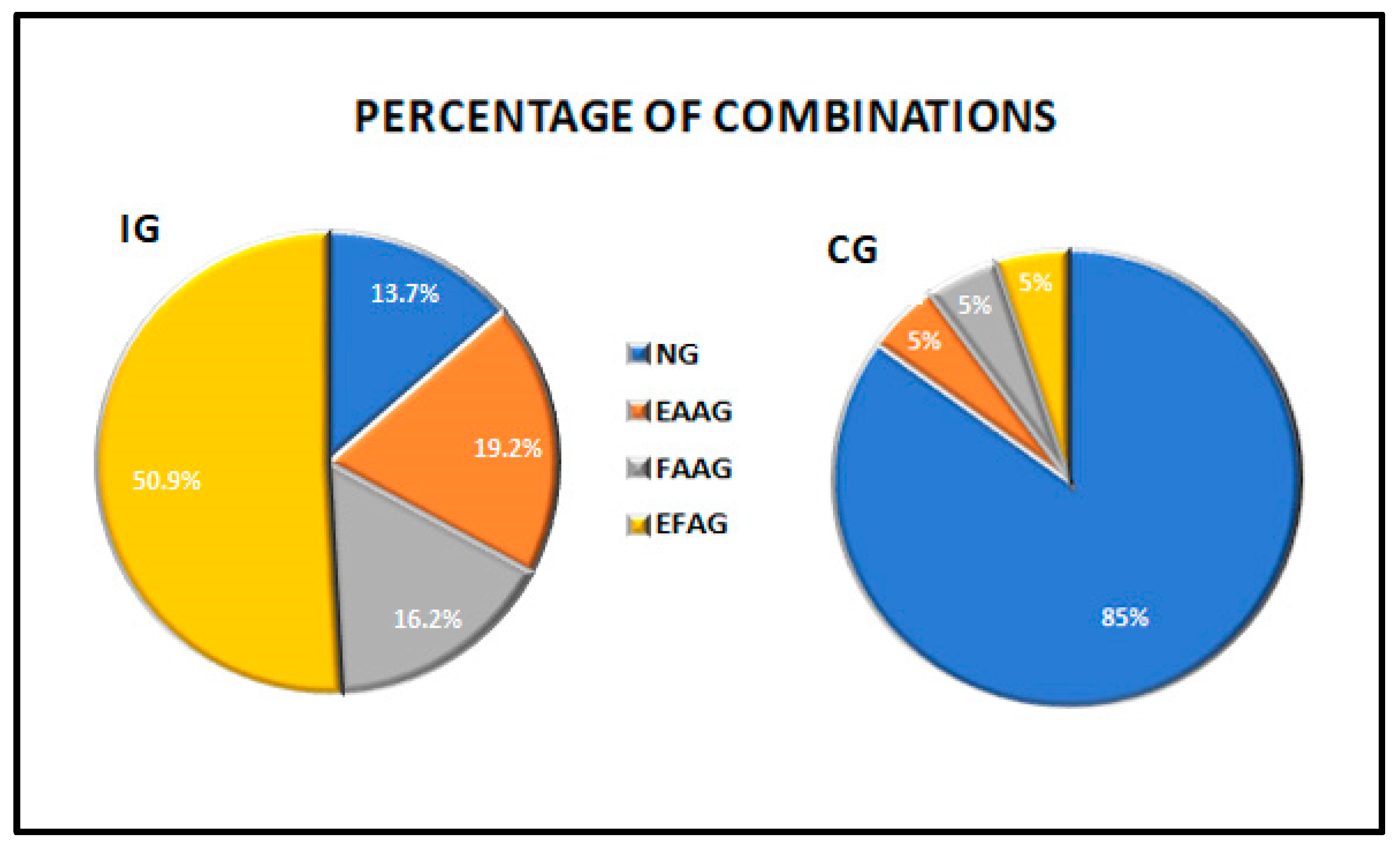
- NG = the normal group that included dogs having a score of 0 in both extension and flexion;
- EFAG = the extension and flexion abnormal group that included dogs having scores 1–4 in both extension and flexion;
- EAAG = the extension angle (reduced) abnormal group that included dogs with stifles having an extension angle with scores 1 to 4 and a flexion angle having a score of 0;
- FAAG = the flexion angle (increased) abnormal group that included dogs with stifles having an extension angle with a score of 0 and a flexion angle with scores 1–4.
2.2. Statistical Analysis
3. Results
3.1. Descriptive Analysis
3.2. Statistical Analysis
- In the IG, the Spearman’s rank correlation coefficient revealed a significant level of correlation (p = 0.0031; rho = 0.192) between the extension and flexion angles.
- In the IG, each injured stifle angle of each dog was investigated. The scores of motion in both extension and flexion were then assessed as a normal angle (NA = score 0) or an abnormal angle (AA = scores 1, 2, 3 or 4). The data are reported in Table 2.
- In the IG, the chi-square test revealed a statistically significant relationship of the NAs and AAs between the two angle motion groups (EA and FA) (p = 0.01). There was a reduced ROM in 86.3% of the cases, and there was a significant prevalence in the alteration of the extension angle. The combinations of the scores were analysed: 13.7% of the dogs had a normal ROM, 50.9% of stifles had a reduced ROM due to an alteration of both movements, 19.2% had abnormal extension with normal flexion and 16.2% had abnormal flexion and normal extension (Figure 1 and Table 3). There was a prevalence of abnormal extension, having a contingency coefficient of 0.16.
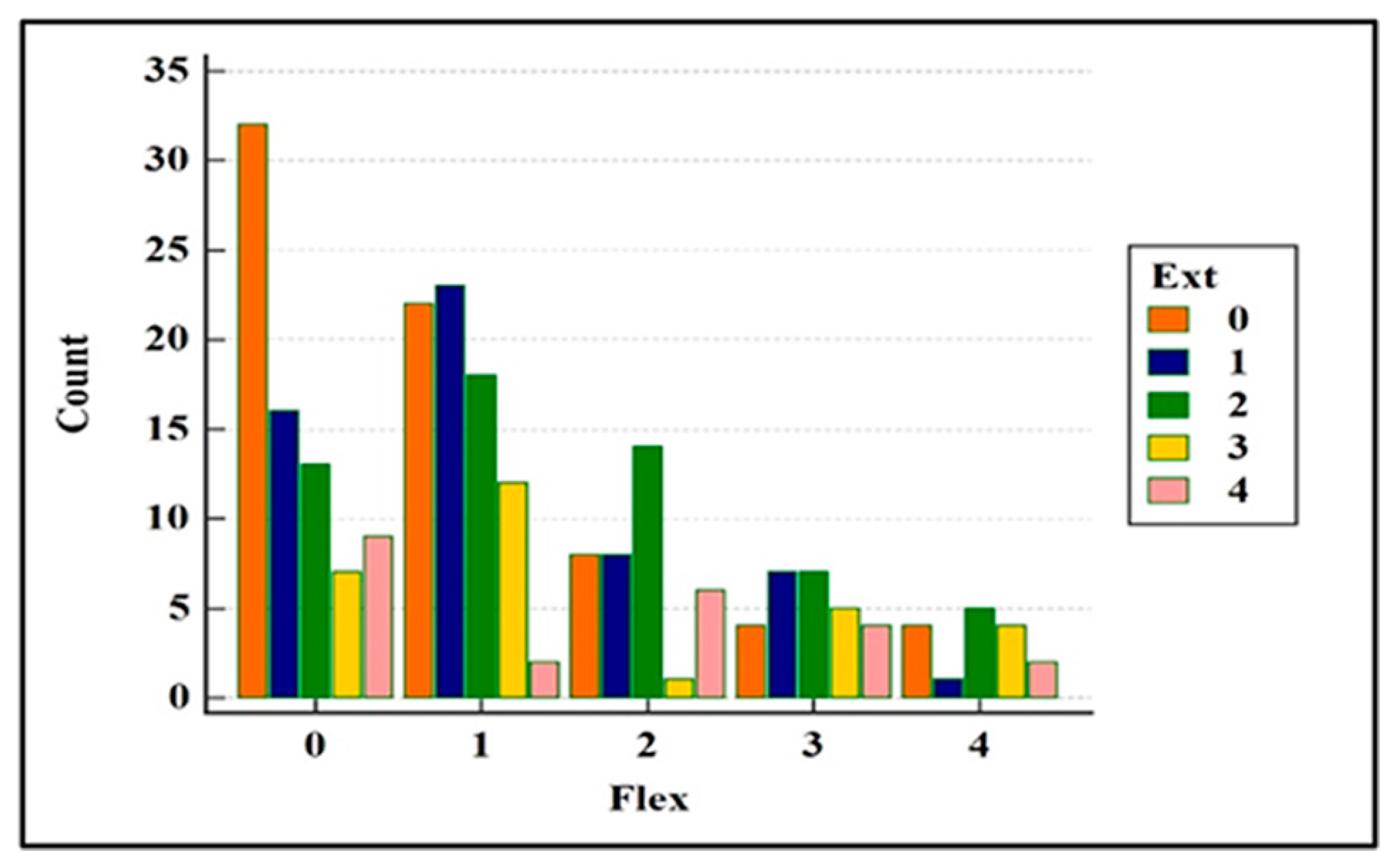
- All five scores (0 to 4) were then evaluated to obtain more details. In the IG, the combinations of the scores of the EAs and FAs were statistically significant, with p = 0.0248 (Figure 2). Only 13.7% (n.33) of the stifles were normal. The stifles with normal EAs (score 0) were associated with abnormal FAs distributed in the slight prevalence score. The stifles with normal FAs (score 0) were associated with abnormal EAs mainly distributed in the slight and the mild scores. Overall, the alterations of the EAs were found in all the scores of severity; instead, the prevalence of the frequency of the alterations of the FA was found in score 1 (slight). The contingency coefficient was 0.33.
- The dogs were grouped based on weight: Group A < 20 kg, Group B from 21 to 30 kg and Group C > 31 kg. The demographic data are listed in Table 1. The chi-square test for the trends revealed a statistically significant difference in the EA among the dogs in Groups B and C, having greater alterations (p = 0.0098 and p = 0.0124, groups B and C, respectively). The alterations in the FA were not significant.
- No significant differences were found between males and females in either the extension or the flexion angles.
- All ruptures were present for more than 2 weeks. Dogs with known lameness and/or trauma occurrence within the past 30 days were classified as the acute group (AG). There were 16 dogs (6.84%) (out of 234) with acute injuries. The chi-square test for the relationship of NAs and AAs was not significant (p = 0.36). There were 218 (93.16%) dogs with chronic disease (Chronic Group: ChG), and the chi-square test revealed a significant difference in the normal and abnormal angles of motion (NAs and AAs), p = 0.027. In the ChG, the alteration of extension was prevalent at flexion, having a percentage of 19.3%, which was the same as the value found in the IG (Table 3).
- The IG was divided into two groups of normal and abnormal muscle mass. The investigation revealed that NMG included 22 dogs, and there was not a significant difference between normal and abnormal angles (p = 0.55). The DMG consisted of 212 dogs, and there was a significative difference between NAs and AAs (p = 0.019). The percentage of the combination of normal and abnormal extension and flexion had the same trend of IG (Table 3).
4. Discussion
5. Conclusions
Author Contributions
Funding
Institutional Review Board Statement
Informed Consent Statement
Conflicts of Interest
References
- Hady, L.L.; Fosgate, G.T.; Weh, J.M. Comparison of range of motion in Labrador Retrievers and Border Collies. J. Vet. Med. Anim. Health 2015, 7, 122–127. [Google Scholar]
- Jaegger, G.; Marcellin-Little, D.J.; Levine, D. Reliability of goniometry in Labrador Retrievers. Am. J. Vet. Res. 2002, 63, 979–986. [Google Scholar] [CrossRef] [PubMed]
- Mitchell, W.S.; Millar, J.; Sturrock, R.D. An evaluation of goniometry as an objective parameter for measuring joint motion. Scott Med. J. 1975, 20, 57–59. [Google Scholar] [CrossRef] [PubMed]
- Boone, D.C.; Azen, S.P.; Lin, C.M.; Spence, C.; Baron, C.; Lee, L. Reliability of goniometric measurements. Phys. Ther. 1978, 58, 1355–1360. [Google Scholar] [CrossRef] [PubMed]
- Goodwin, J.; Clark, C.; Deakes, J.; Burdon, D.; Lawrence, C. Clinical methods of goniometry: A comparative study. J. Phys. Ther Sci. 1992, 14, 10–15. [Google Scholar] [CrossRef]
- Svensson, M.; Lind, V.; Löfgren Harringe, M. Measurement of knee joint range of motion with a digital goniometer: A reliability study. Physiother. Res. Int. 2019, 24, e1765. [Google Scholar] [CrossRef]
- Grohmann, J.E.L. Comparison of two methods of goniometry. Phys. Ther. 1983, 63, 922–925. [Google Scholar] [CrossRef]
- Clapper, M.P.; Wolf, S.L. Comparison of the reliability of the Orthoranger and the standard goniometer for assessing active lower extremity range of motion. Phys. Ther. 1988, 68, 214–218. [Google Scholar] [CrossRef]
- Fraeulin, L.; Holzgreve, F.; Brinkbäumer, M.; Dziuba, A.; Friebe, D.; Klemz, S.; Schmitt, M.; Theis, A.A.L.; Tenberg, S.; Van Mark, A.; et al. Intra-and inter-rater reliability of joint range of motion tests using tape measure, digital inclinometer and inertial motion capturing. PLoS ONE 2020, 15, e0243646. [Google Scholar] [CrossRef]
- Jaeger, G.H.; Marcellin-Little, D.J.; DePuy, V.; Lascelles, B.D.X. Validity of goniometric joint measurements in cats. Am. J. Vet. Res. 2007, 68, 822–826. [Google Scholar] [CrossRef]
- Liljebrink, Y.; Bergh, A. Goniometry: Is it a reliable tool to monitor passive joint range of motion in horses? Equine Vet. J. 2010, 42, 676–682. [Google Scholar] [CrossRef]
- Adair, H.S., III; Marcellin-Little, D.J.; Levine, D. Validity and repeatability of goniometry in normal horses. Vet. Comp. Orthop. Traumatol. 2016, 29, 314–319. [Google Scholar] [CrossRef]
- Bergh, A.; Lauridsen, N.G.; Hesbach, A.L. Concurrent Validity of Equine Joint Range of Motion Measurement: A Novel Digital Goniometer versus Universal Goniometer. Animals 2020, 10, 2436. [Google Scholar] [CrossRef]
- Govoni, V.M.; Rahal, S.C.; Agostinho, F.S.; Conceição, R.T.; Tsunemi, M.H.; El-Warrak, A.O. Goniometric measurements of the forelimb and hindlimb joints in sheep. Vet. Comp. Orthop. Traumatol. 2012, 25, 297–300. [Google Scholar] [CrossRef]
- Thomovsky, S.A.; Chen, A.V.; Kiszonas, A.M.; Lutskas, L.A. Goniometry and limb girth in miniature Dachshunds. J. Vet. Med. 2016, 2016, 5846052. [Google Scholar] [CrossRef] [PubMed] [Green Version]
- Sabanci, S.S.; Ocal, M.K. Comparison of goniometric measurements of the stifle joint in seven breeds of normal dogs. Vet. Comp. Orthop. Traumatol. 2016, 29, 214–219. [Google Scholar] [PubMed]
- Formenton, M.R.; de Lima, L.G.; Vassalo, F.G.; Joaquim, J.G.F.; Rosseto, L.P.; Fantoni, D.T. Goniometric Assessment in French Bulldogs. Front. Vet. Sci. 2019, 6, 424. [Google Scholar] [CrossRef] [PubMed] [Green Version]
- Johnston, S.A. Osteoarthritis: Joint anatomy, physiology, and pathobiology. Vet. Clin. N. Am. Small Anim. Pract. 1997, 27, 699–723. [Google Scholar] [CrossRef]
- Crook, T.; McGowan, C.; Pead, M. Effect of passive stretching on the range of motion of osteoarthritic joints in 10 labrador retrievers. Vet. Rec. 2007, 160, 545–547. [Google Scholar] [CrossRef]
- Marcellin-Little, D.J.; Levine, D. Principles and application of range of motion and stretching in companion animals. Vet. Clin. N. Am. Small Anim. Pract. 2015, 45, 57–72. [Google Scholar] [CrossRef]
- Montasell, X.; Dupuis, J.; Huneault, L.; Ragetly, G.R. Short-and long-term outcomes after shoulder excision arthroplasty in 7 small breed dogs. Can. Vet. J. 2018, 59, 277–283. [Google Scholar]
- Clarke, E.; Aulakh, K.S.; Hudson, C.; Barnes, K.; Gines, J.A.; Liu, C.C.; Aulakh, H.K. Effect of sedation or general anesthesia on elbow goniometry and thoracic limb circumference measurements in dogs with naturally occurring elbow osteoarthritis. Vet. Surg. 2020, 49, 1428–1436. [Google Scholar] [CrossRef] [PubMed]
- Huňáková, K.; Hluchý, M.; Špaková, T.; Matejová, J.; Mudroňová, D.; Kuricová, M.; Rosocha, J.; Ledecký, V. Study of bilateral elbow joint osteoarthritis treatment using conditioned medium from allogeneic adipose tissue-derived MSCs in Labrador retrievers. Res. Vet. Sci. 2020, 132, 513–520. [Google Scholar] [CrossRef]
- Bertocci, G.; Smalley, C.; Brown, N.; Bialczak, K.; Carroll, D. Aquatic treadmill water level influence on pelvic limb kinematics in cranial cruciate ligament-deficient dogs with surgically stabilised stifles. J. Small Anim. Pract. 2018, 59, 121–127. [Google Scholar] [CrossRef]
- Barnes, K.; Faludi, A.; Takawira, C.; Aulakh, K.; Rademacher, N.; Liu, C.C.; Lopez, M.J. Extracorporeal shock wave therapy improves short-term limb use after canine tibial plateau leveling osteotomy. Vet. Surg. 2019, 48, 1382–1390. [Google Scholar] [CrossRef] [PubMed]
- Roh, Y.H.; Jung, J.H.; Lee, J.H.; Jeong, J.M.; Jeong, S.M.; Lee, H. Clinical Results of Distal Femoral Osteotomy for Treatment of Grade 4 Medial Patella Luxation with Concurrent Distal Femoral Varus in Small Breeds Dogs: 13 Cases. J. Vet. Clin. 2020, 37, 135–140. [Google Scholar] [CrossRef]
- Pinna, S.; Lanzi, F.; Tassani, C.; Mian, G. Intra-articular replacement of a ruptured cranial cruciate ligament using the Mini-TightRope in the dog: A preliminary study. J. Vet. Sci. 2020, 21, e53. [Google Scholar] [CrossRef]
- Pinna, S.; Lanzi, F.; Grassato, L. Bologna Healing Stifle Injury Index: A Comparison of Three Surgical Techniques for the Treatment of Cranial Cruciate Ligament Rupture in Dogs. Front. Vet. Sci. 2020, 7, 567473. [Google Scholar] [CrossRef]
- Piras, L.A.; Mancusi, D.; Olimpo, M.; Gastaldi, L.; Rosso, V.; Panero, E.; Staffieri, B.; Peirone, B. Post-operative analgesia following TPLO surgery: A comparison between cimicoxib and tramadol. Res. Vet. Sci. 2021, 136, 351–359. [Google Scholar] [CrossRef]
- Chomsiriwat, P.; Ma, A. Comparison of the Effects of Electro-acupuncture and Laser Acupuncture on Pain Relief and Joint Range of Motion in Dogs with Coxofemoral Degenerative Joint Disease. Am. J. Tradit. Chin. Vet. Med. 2019, 14, 11–20. [Google Scholar]
- Alves, J.C.; Santos, A.; Jorge, P.; Lavrador, C.; Carreira, L.M. Clinical and diagnostic imaging findings in police working dogs referred for hip osteoarthritis. BMC Vet. Res. 2020, 16, 425. [Google Scholar] [CrossRef]
- Moeller, E.M.; Allen, D.A.; Wilson, E.R.; Lineberger, J.A.; Lehenbauer, T. Long-term outcomes of thigh circumference, stifle range-of-motion, and lameness after unilateral tibial plateau levelling osteotomy. Vet. Comp. Orthop. Traumatol. 2010, 23, 37–42. [Google Scholar] [PubMed]
- Mölsä, S.H.; Hyytiäinen, H.K.; Hielm-Björkman, A.K.; Laitinen-Vapaavuori, O.M. Long-term functional outcome after surgical repair of cranial cruciate ligament disease in dogs. BMC Vet. Res. 2014, 10, 266. [Google Scholar] [CrossRef] [PubMed] [Green Version]
- Marsolais, G.S.; Dvorak, G.; Conzemius, M.G. Effects of postoperative rehabilitation on limb function after cranial cruciate ligament repair in dogs. J. Am. Vet. Med. Assoc. 2002, 220, 1325–1330. [Google Scholar] [CrossRef] [PubMed] [Green Version]
- Hoelzler, M.G.; Millis, D.L.; Francis, D.A.; Weigel, J.P. Results of arthroscopic versus open arthrotomy for surgical management of cranial cruciate ligament deficiency in dogs. Vet. Surg. 2004, 33, 146–153. [Google Scholar] [CrossRef]
- Jandi, A.S.; Schulman, A.J. Incidence of motion loss of the stifle joint in dogs with naturally occurring cranial cruciate ligament rupture surgically treated with tibial plateau leveling osteotomy: Longitudinal clinical study of 412 cases. Vet. Surg. 2007, 36, 114–121. [Google Scholar] [CrossRef] [PubMed]
- Au, K.K.; Gordon-Evan, W.J.; Dunning, D.; O’Dell-Anderson, K.J.; Knap, K.E.; Griffon, D.; Johnson, A.L. Comparison of short-and long-term function and radiographic osteoarthrosis in dogs after postoperative physical rehabilitation and tibial plateau leveling osteotomy or lateral fabellar suture stabilization. Vet. Surg. 2010, 39, 173–180. [Google Scholar] [CrossRef]
- Mostafa, A.A.; Griffon, D.J.; Thomas, M.W.; Constable, P.D. Morphometric characteristics of the pelvic limb musculature of Labrador Retrievers with and without cranial cruciate ligament deficiency. Vet. Surg. 2010, 39, 380–389. [Google Scholar] [CrossRef]
- Drygas, K.A.; McClure, S.R.; Goring, R.L.; Pozzi, A.; Robertson, S.A.; Wang, C. Effect of cold compression therapy on postoperative pain, swelling, range of motion, and lameness after tibial plateau leveling osteotomy in dogs. J. Am. Vet. Med. Assoc. 2011, 238, 1284–1291. [Google Scholar] [CrossRef] [Green Version]
- Gordon-Evans, W.J.; Dunning, D.; Johnson, A.L.; Knap, K.E. Effect of the use of carprofen in dogs undergoing intense rehabilitation after lateral fabellar suture stabilization. J. Am. Vet. Med. Assoc. 2011, 239, 75–80. [Google Scholar] [CrossRef]
- MacDonald, T.L.; Allen, D.A.; Monteith, G.J. Clinical assessment following tibial tuberosity advancement in 28 stifles at 6 months and 1 year after surgery. Can. Vet. J. 2013, 54, 249–254. [Google Scholar]
- Monk, M.L.; Preston, C.A.; McGowan, C.M. Effects of early intensive postoperative physiotherapy on limb function after tibial plateau leveling osteotomy in dogs with deficiency of the cranial cruciate ligament. Am. J. Vet. Res. 2006, 67, 529–536. [Google Scholar] [CrossRef] [Green Version]
- Marsolais, G.S.; McLean, S.; Derrick, T.; Conzemius, M.G. Kinematic analysis of the hind limb during swimming and walking in healthy dogs and dogs with surgically corrected cranial cruciate ligament rupture. J. Am. Vet. Med. Assoc. 2003, 222, 739–743. [Google Scholar] [CrossRef] [PubMed] [Green Version]
- Pinna, S.; Lanzi, F.; Grassato, L. Evidence-Based Veterinary Medicine: A Tool for Evaluating the Healing Process After Surgical Treatment for Cranial Cruciate Ligament Rupture in Dogs. Front. Vet. Sci. 2020, 6, 65. [Google Scholar] [CrossRef] [PubMed] [Green Version]
- Hyytiäinen, H.K.; Mölsä, S.H.; Junnila, J.T.; Laitinen-Vapaavuori, O.M.; Hielm-Björkman, A.K. Ranking of physiotherapeutic evaluation methods as outcome measures of stifle functionality in dogs. Acta Vet. Scand. 2013, 55, 29. [Google Scholar] [CrossRef] [Green Version]
- Colborne, G.R.; Innes, J.F.; Comerford, E.J.; Owen, M.R.; Fuller, C.J. Distribution of power across the hind limb joints in Labrador Retrievers and Greyhounds. Am. J. Vet. Res. 2005, 66, 1563–1571. [Google Scholar] [CrossRef]
- Irrgang, J.J.; Harner, C.D. Loss of motion following knee ligament reconstruction. Sports Med. 1995, 19, 150–159. [Google Scholar] [CrossRef]
- Pinna, S.; Lanzi, F.; Cordella, A.; Diana, A. Relationship between the stage of osteoarthritis before and six months after tibial tuberosity advancement procedure in dogs. PLoS ONE 2019, 14, e0219849. [Google Scholar] [CrossRef] [Green Version]
- Pinna, S.; Landucci, F.; Tribuiani, A.M.; Carli, F.; Venturini, A. The effects of pulsed electromagnetic field in the treatment of osteoarthritis in dogs: Clinical study. Pak. Vet. J. 2013, 33, 96–100. [Google Scholar]
- Cook, J.L.; Luther, J.K.; Beetem, J.; Karnes, J.; Cook, C.R. Clinical comparison of a novel extracapsular stabilization procedure and tibial plateau leveling osteotomy for treatment of cranial cruciate ligament deficiency in dogs. Vet. Surg. 2010, 39, 315–323. [Google Scholar] [CrossRef]
- Gordon, W.J.; Conzemius, M.G.; Riedesel, E.; Besancon, M.F.; Evans, R.; Wilke, V.; Ritter, M.J. The relationship between limb function and radiographic osteoarthrosis in dogs with stifle osteoarthrosis. Vet. Surg. 2003, 32, 451–454. [Google Scholar] [CrossRef] [PubMed]
- Johnson, J.M.; Johnson, A.L. Cranial cruciate ligament rupture: Pathogenesis, diagnosis, and postoperative rehabilitation. Vet. Clin. N. Am. Small Anim. Pract. 1993, 23, 717–733. [Google Scholar] [CrossRef]
- Moore, E.V.; Weeren, R.; Paek, M. Extended long-term radiographic and functional comparison of tibial plateau leveling osteotomy vs tibial tuberosity advancement for cranial cruciate ligament rupture in the dog. Vet. Surg. 2020, 49, 146–154. [Google Scholar] [CrossRef] [PubMed]


| Dog/Stifle | n. | % | Age | Weight | ||||
|---|---|---|---|---|---|---|---|---|
| Median | Range | 95% CI | Median | Range | 95% CI | |||
| IG | 234 | 100 | 6 | 1–13 | 6–7 | 30 | 4–75 | 28–32 |
| Male | 99 | 42.3 | 6 | 1–13 | 5–7 | 35 * | 4–75 | 30–38 |
| Female | 135 | 57.7 | 7 | 1–13 | 6–7 | 28 | 4–56 | 26–30 |
| Group A | 64 | 27.4 | 8 | 2–13 | 7–9.3 | 10 | 4–18 | 8–11 |
| Group B | 91 | 38.9 | 6 | 1–12 | 5–6.3 | 29 | 21–35 | 28–30 |
| Group C | 79 | 33.8 | 5 | 1–13 | 5–6 | 42 | 36–75 | 40–44.9 |
| CG | 20 | 100 | 6 | 1–12 | 3–7.8 | 28 | 14–55 | 22–29.8 |
| Angle Motion | Scores | Total AA | |||||
|---|---|---|---|---|---|---|---|
| 0 (normal) | 1 (slight) | 2 (mild) | 3 (moderate) | 4 (severe) | |||
| IG | EA | 70 (30%) | 54 (23%) | 58 (25%) | 29 (12%) | 23(10%) | 164 (70%) |
| FA | 77 (33%) | 76 (32%) | 38 (16%) | 27 (12%) | 16 (7%) | 157 (67%) | |
| CG | EA | 17 (85%) | 2 (10%) | 1 (5%) | - | - | 3 (15%) |
| FA | 17 (85%) | 3 (15%) | - | - | - | 3 (15%) | |
| IG | CG | AG | ChG | NMG | DMG | |
|---|---|---|---|---|---|---|
| NG (0 + 0) | 32 (13.7%) | 17 (85%) | 4 (25%) | 28 (12.8) | 4 (18.2%) | 28 (13.2%) |
| EAAG (1 + 0) | 45 (19.2) | 1 (5%) | 3 (18.8%) | 42 (19.3%) | 5 (22.7%) | 40 (18.9%) |
| FAAG (0 + 1) | 38 (16.2%) | 1 (5%) | 2 (12.5%) | 36 (16.5%) | 3 (13.6%) | 35 (16.5%) |
| EFAG (1 + 1) | 119 (50.9%) | 1 (5%) | 7 (43.8%) | 112 (51.4%) | 10 (45.5%) | 109 (51.4%) |
| Total dog/group | 234 | 20 | 16 | 218 | 22 | 212 |
Publisher’s Note: MDPI stays neutral with regard to jurisdictional claims in published maps and institutional affiliations. |
© 2021 by the authors. Licensee MDPI, Basel, Switzerland. This article is an open access article distributed under the terms and conditions of the Creative Commons Attribution (CC BY) license (https://creativecommons.org/licenses/by/4.0/).
Share and Cite
Pinna, S.; Lanzi, F.; Tassani, C. The Effect of Cranial Cruciate Ligament Rupture on Range of Motion in Dogs. Vet. Sci. 2021, 8, 119. https://doi.org/10.3390/vetsci8070119
Pinna S, Lanzi F, Tassani C. The Effect of Cranial Cruciate Ligament Rupture on Range of Motion in Dogs. Veterinary Sciences. 2021; 8(7):119. https://doi.org/10.3390/vetsci8070119
Chicago/Turabian StylePinna, Stefania, Francesco Lanzi, and Chiara Tassani. 2021. "The Effect of Cranial Cruciate Ligament Rupture on Range of Motion in Dogs" Veterinary Sciences 8, no. 7: 119. https://doi.org/10.3390/vetsci8070119
APA StylePinna, S., Lanzi, F., & Tassani, C. (2021). The Effect of Cranial Cruciate Ligament Rupture on Range of Motion in Dogs. Veterinary Sciences, 8(7), 119. https://doi.org/10.3390/vetsci8070119


