
Excitement-Induced Cutaneous Bleeding (Haematidrosis-like) in a Dog


Abstract
1. Introduction
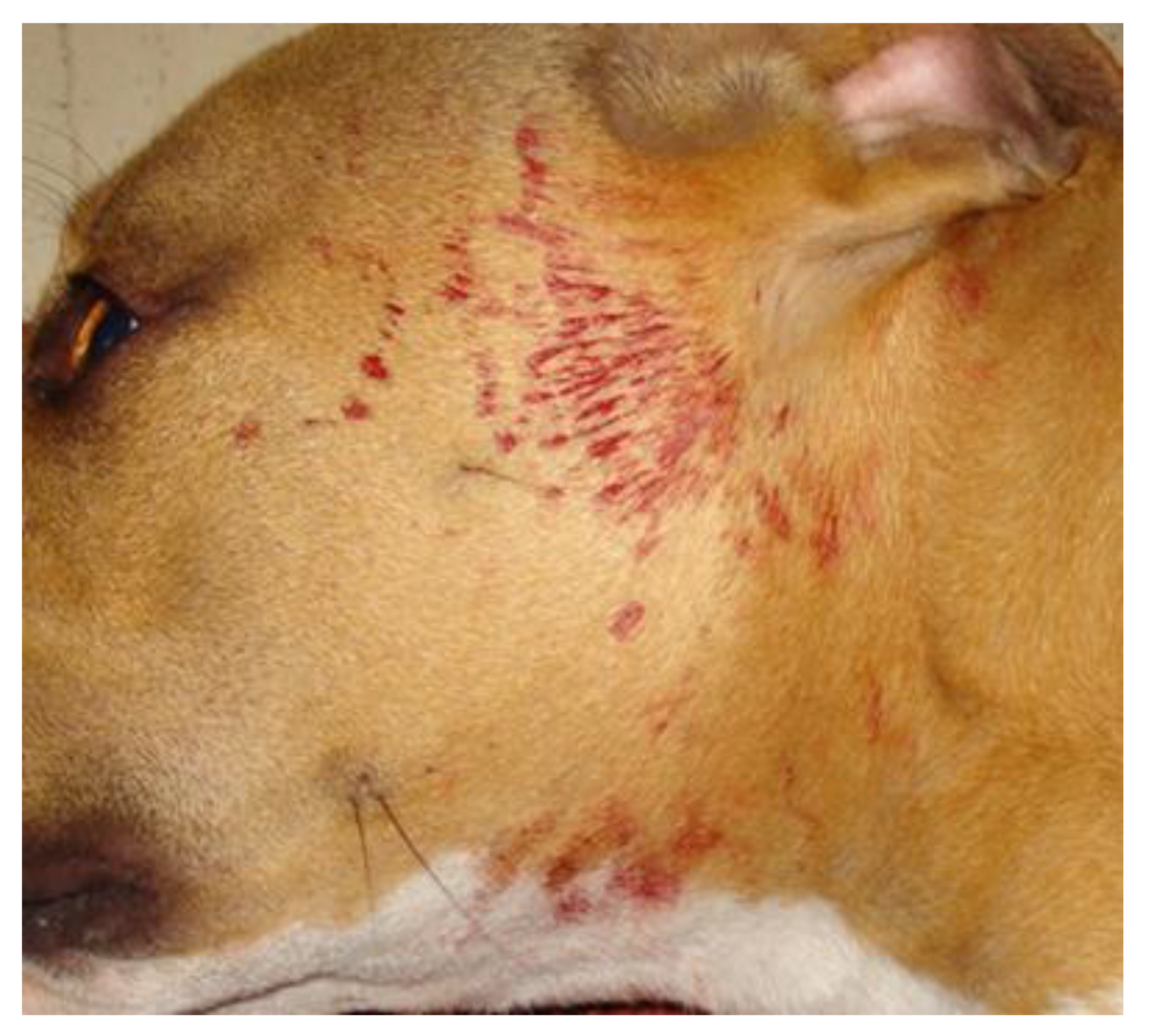
2. Case Presentation
3. Discussion
4. Conclusions
Author Contributions
Funding
Institutional Review Board Statement
Informed Consent Statement
Data Availability Statement
Conflicts of Interest
References
- Kluger, N. Hematidrosis (bloody sweat): A review of the recent literature (1996–2016). Acta Dermatovenerol. Alp. Pannonica Adriat. 2018, 27, 85–90. [Google Scholar] [CrossRef] [PubMed]
- Kechichian, E.; Khoury, E.; Richa, S.; Tomb, R. Religious stigmata: A dermato-psychiatric approach and differential diagnosis. Int. J. Dermatol. 2018, 57, 885–893. [Google Scholar] [CrossRef] [PubMed]
- Tshifularo, M. Blood otorrhea: Blood stained sweaty ear discharges: Hematohidrosis; four case series (2001–2013). Am. J. Otolaryngol. 2014, 35, 271–273. [Google Scholar] [CrossRef] [PubMed][Green Version]
- Murota, H.; Kotobuki, Y.; Yamaga, K.; Yoshioka, Y. Female child with hematidrosis of the palm: Case report and published work review. J. Dermatol. 2020, 47, 166–168. [Google Scholar] [CrossRef] [PubMed]
- Shahgholi, Ε. A case series of hematohidrosis: A puzzling medical phenomenon. Turk. J. Pediatrics 2018, 60, 757–761. [Google Scholar] [CrossRef] [PubMed]
- Scott, D.W.; Miller, W.H.J. Hemathidrosis. In Equine Dermatology, 2nd ed.; Scott, D.W., Miller, W.H.J., Eds.; Elsevier Saunders: St. Louis, MO, USA, 2011; p. 460. [Google Scholar]
- Pardon, B.; Steukers, L.; Dierick, J.; Ducatelle, R.; Saey, V.; Maes, S.; Vercauteren, G.; De Clercq, K.; Callens, J.; De Bleecker, K.; et al. Haemorrhagic diathesis in neonatal calves: An emerging syndrome in Europe. Transbound. Emerg. Dis. 2010, 57, 135–146. [Google Scholar] [CrossRef] [PubMed]
- Miller, W.H.J.; Griffin, C.E.; Campbell, K.L. Flushing. In Muller and Kirk’s Small Animal Dermatology, 7th ed.; Miller, W.H.J., Griffin, C.E., Campbell, K.L., Eds.; Elsevier Mosby: St. Louis, MO, USA, 2013; p. 628. [Google Scholar]
- Miller, W.H.J.; Griffin, C.E.; Campbell, K.L. Diagnostic methods. In Muller and Kirk’s Small Animal Dermatology, 7th ed.; Miller, W.H.J., Griffin, C.E., Campbell, K.L., Eds.; Elsevier Mosby: St. Louis, MO, USA, 2013; pp. 57–107. [Google Scholar]
- Rostaher, A.; Hofer-Inteeworn, N.; Kümmerle-Fraune, C.; Fischer, N.M.; Favrot, C. Triggers, risk factors and clinico-pathological features of urticaria in dogs-a prospective observational study of 24 cases. Vet. Dermatol. 2017, 28, e38–e39. [Google Scholar] [CrossRef] [PubMed]
- Declercq, J. Urticarial vasculitis in a French bulldog. Vet. Dermatol. 2015, 26, 72–73. [Google Scholar] [CrossRef] [PubMed]
- Takahashi, T.; Kadosawa, T.; Nagase, M.; Matsunaga, S.; Mochizuki, M.; Nishimura, R.; Sasaki, N. Visceral mast cell tumors in dogs: 10 cases (1982–1997). J. Am. Vet. Med Assoc. 2000, 216, 222–226. [Google Scholar] [CrossRef] [PubMed]
- Maher, E.R.; McNiel, E.A. Pheochromocytoma in dogs and cats. Vet. Clin. N. Am.-Small Anim. Pract. 1997, 27, 359–380. [Google Scholar] [CrossRef]
- Innerå, M. Cutaneous vasculitis in small animals. Vet. Clin. N. Am.-Small Anim. Pract. 2013, 43, 113–134. [Google Scholar] [CrossRef] [PubMed]
- Jain, N.C.; Switzer, J.W. Autoimmune thrombocytopenia in dogs and cats. Vet. Clin. N. Am.-Small Anim. Pract. 1981, 11, 421–434. [Google Scholar] [CrossRef]
- Burton, A.G.; Jandrey, K.E. Use of thromboelastography in clinical practice. Vet. Clin. N. Am.-Small Anim. Pract. 2020, 50, 1397–1409. [Google Scholar] [CrossRef] [PubMed]
- Brooks, M.B.; Catalfamo, J.L. Current diagnostic trends in coagulation disorders among dogs and cats. Vet. Clin. N. Am.-Small Anim. Pract. 2013, 43, 1349–1372. [Google Scholar] [CrossRef] [PubMed]
- Scott, D.W.; Miller, W.H.; Griffin, C. Structure and function of the skin. In Mueller & Kirk’s Small Animal Dermatology; W.B. Saunders: Philadelphia, PA, USA, 2001; pp. 1–70. [Google Scholar]
- Bonamonte, D.; Vestita, M.; Filoni, A.; Giudice, G.; Angelini, G. Religious stigmata as malingering artifact: Report of a case and review of the literature. Medicine 2016, 95, e5354. [Google Scholar] [CrossRef] [PubMed]
- Shahriari, M.; Bazrafshan, A.; Karimi, M.; Shakibazad, N. Case series of bloody sweating; a scary event for families. Acta Haematol. Pol. 2020, 51, 258–260. [Google Scholar] [CrossRef]
- Carvalho, A.C.S.; Machado-Pinto, J.; Nogueira, G.C.; Almeida, L.M.C.; Nunes, M.B. Hematidrosis: A case report and review of the literature. Int. J. Dermatol. 2008, 47, 1058–1059. [Google Scholar] [CrossRef] [PubMed]
- Mora, E.; Lucas, J. Hematidrosis: Blood sweat. Blood 2013, 121, 1493. [Google Scholar] [CrossRef] [PubMed][Green Version]
- Manonukul, J.; Wisuthsarewong, W.; Chantorn, R.; Vongirad, A.; Omeapinyan, P. Hematidrosis: A pathologic process or stigmata. A case report with comprehensive histopathologic and immunoperoxidase studies. Am. J. Dermatopathol. 2008, 30, 135–139. [Google Scholar] [CrossRef] [PubMed]
- Altamura, A.C.; Moliterno, D.; Paletta, S.; Maffini, M.; Mauri, M.C.; Bareggi, S. Understanding the pharmacokinetics of anxiolytic drugs. Expert Opin. Drug Metab. Toxicol. 2013, 9, 423–440. [Google Scholar] [CrossRef] [PubMed]


Publisher’s Note: MDPI stays neutral with regard to jurisdictional claims in published maps and institutional affiliations. |
© 2021 by the authors. Licensee MDPI, Basel, Switzerland. This article is an open access article distributed under the terms and conditions of the Creative Commons Attribution (CC BY) license (https://creativecommons.org/licenses/by/4.0/).
Share and Cite
Sofou, E.I.; Gavra, A.; Saridomichelakis, M.N. Excitement-Induced Cutaneous Bleeding (Haematidrosis-like) in a Dog. Vet. Sci. 2021, 8, 327. https://doi.org/10.3390/vetsci8120327
Sofou EI, Gavra A, Saridomichelakis MN. Excitement-Induced Cutaneous Bleeding (Haematidrosis-like) in a Dog. Veterinary Sciences. 2021; 8(12):327. https://doi.org/10.3390/vetsci8120327
Chicago/Turabian StyleSofou, Evi I., Anna Gavra, and Manolis N. Saridomichelakis. 2021. "Excitement-Induced Cutaneous Bleeding (Haematidrosis-like) in a Dog" Veterinary Sciences 8, no. 12: 327. https://doi.org/10.3390/vetsci8120327
APA StyleSofou, E. I., Gavra, A., & Saridomichelakis, M. N. (2021). Excitement-Induced Cutaneous Bleeding (Haematidrosis-like) in a Dog. Veterinary Sciences, 8(12), 327. https://doi.org/10.3390/vetsci8120327