Article

Serotyping, Genotyping and Virulence Genes Characterization of Pasteurella multocida and Mannheimia haemolytica Isolates Recovered from Pneumonic Cattle Calves in North Upper Egypt

1
Bacteriology, Mycology and Immunology Department, Faculty of Veterinary Medicine, Beni-Suef University, Beni Suef 62511, Egypt
2
Animal Reproduction Research Institute, Giza 12511, Egypt
3
Microbiology Department, Faculty of Veterinary Medicine, Matrouh University, Matrouh 51511, Egypt
4
Friedrich-Loeffler-Institut, Institute of Bacterial Infections and Zoonoses, Naumburger Str. 96a, 07743 Jena, Germany
5
Faculty of Veterinary Medicine, Benha University, Moshtohor, Toukh 13736, Egypt
6
Veterinary Medicine Department (Infectious Diseases), Faculty of Veterinary Medicine, Beni-Suef University, Beni Suef 62511, Egypt
*
Author to whom correspondence should be addressed.
Vet. Sci. 2020, 7(4), 174; https://doi.org/10.3390/vetsci7040174
Submission received: 26 September 2020 / Revised: 26 October 2020 / Accepted: 7 November 2020 / Published: 10 November 2020

Abstract

:
Pasteurella (P.) multocida and Mannheimia (M.) haemolytica are the most two common pathogenic bacterial agents causing pneumonia in calves. Both bacteria are associated with significant economic losses in the cattle industry due to high morbidity and mortality rates, especially in the case of severe infections. The objectives of the present study were to perform serotyping and genotyping, as well as characterization of the virulence-associated genes in 48 bacterial isolates; 33 P. multocida and 15 M. haemolytica. All strains were isolated from pneumonic cattle calves showing respiratory manifestations such as fever, nasal discharges, and rapid breathing in North Upper Egypt governorates (Beni-Suef and El-Fayoum). PCR was applied as a confirmatory test using a specific universal gene, kmt1, and rpt2 for P. multocida and M. haemolytica, respectively. The results show that 29 (87.9%) P. multocida and 15 (100%) M. haemolytica isolates were positive for the corresponding universal gene. The results of serotyping indicate that 86.2% of P. multocida isolates belonged to serotype B:2, while 13.8% were untyped. Meanwhile, 60% and 40% of M. haemolytica isolates belonged to serotype 2 and serotype 1, respectively. Investigation of virulence-associated genes showed that all the tested P. multocida isolates harbored nanB, omp87, and toxA genes. Four M. haemolytica isolates harbored both gcp and lktC genes and of these, three isolates harbored the ssa gene. Sequencing of toxA gene of P. multocida and lktC gene of M. haemolytica in the current strains indicated a great homology with strains uploaded in gene banks from different hosts and localities worldwide.

1. Introduction

Bovine respiratory disease (BRD) is a significant cause of morbidity and mortality among beef cattle globally. Pasteurella multocida (P. multocida) and Mannheimia haemolytica (M. haemolytica), which are commensal Gram-negative bacteria in the upper respiratory tract of animals, represent the major bacterial causative agents for BRD [1]. Under stresses including environmental, managemental, and/or infectious factors, those agents often produce mild to severe clinical signs. The incubation period varies from 3 to 5 days. In peracute cases, sudden death within 24–36 h with clear clinical signs may be observed. In chronic cases, they may cause permanent lung damage such as fibrosis, adhesions and/or abscesses that are affecting the performance [1]. The clinical manifestations include a rise in temperature, respiratory distress with nasal discharge, and frothing from the mouth, and then recumbency and death may be the result [2]. Disease incidence is most often in 6–24 month-old animals and groups of less than 10 animals. The disease was seasonal, occurring only in rainy seasons of the year, and victims were only cattle and buffaloes [2]. Thus, early recognition and treatment of BRD are so important [3]. Respiratory disorders in animal production sectors in Egypt were reported to cause a considerable economic loss due to lower productivity and death [4]. Pasteurella multocida is a zoonotic bacterium causing hemorrhagic septicaemia (HS), which is a major disease of cattle and buffaloes occurring as catastrophic epizootics in many African and Asian countries characterized by an acute, highly fatal septicemia with high morbidity and mortality [3]. In humans, P. multocida has been tightly associated with animal exposure, usually involving soft-tissue sites within 24 h after animal bites, especially dog or cat, or scratch wounds. Serious respiratory tract infections including pneumonia, empyema, and lung abscesses are typically found in patients with underlying pulmonary disease. In more serious cases, a bacteremia can occur by spreading from a localized bite wound or from another localized source of infection, such as pneumonia, meningitis, or arthritis. Varieties of other serious invasive infections such as meningitis, endocarditis, and peritonitis, have also been reported, but are rare [5].
HS is caused by certain serotypes of P. multocida including B:2 in Asia and E:2 in Africa based on the Carter and Heddleston system, which is corresponding to the newer B:6 and E:6 serotypes in Namioka–Carter classification [6]. OIE added that A:1 and A:3 serotypes have been incriminated in an HS-like syndrome in bovines in India with mainly characterized by pneumonia ended with death. On the other hand, M. haemolytica (formerly Pasteurella haemolytica) is considered one of the most important pathogens in ruminants of all ages and it is the principal cause of bovine and ovine pneumonic pasteurellosis or shipping fever. It is responsible for considerable economic losses to the livestock industries all over the world [3,7]. Two biotypes have been recognized for the taxon Pasteurella haemolytica: biotype A, an isolate that ferments L-arabinose, and biotype T, an isolate that ferments trehalose [8]. Based on capsular polysaccharide antigen typing using the indirect haemagglutination test, P. haemolytica complex has been identified as 17 serotypes; including 13 A serotypes (A:1, A:2, A:5, A:6, A:7, A:8, A:9, A:11, A:12, A:13, A:14, A:16, and A:17) and 4 T serotypes (serotypes 3, 4, 10 and 15) [8]. P. haemolytica biotype A was later allocated to a new genus Mannheimia and renamed as M. haemolytica, while the four T serotypes were named Bibersteinia trehalosi. Later, serotype A:11 was classified into a new taxon as M. glucosida, due to its different biochemical profile, leaving 12 serotypes of M. haemolytica [9].
Many virulence genes are important in the pathogenesis of P. multocida, such as fimbriae and adhesins (nanB and nanH), a variety of outer membrane proteins (OMPs) such as protectins (ompA, ompH, omp87 and plpB), and toxins such as dermonecrotoxin (toxA) [1,10,11]. These virulence factors facilitate the colonization and invasion of P. multocida through impairing the host defense mechanisms, destruction of host tissues, and/or stimulation of a noxious host inflammatory response [12]. The toxA and the OMPs-encoding genes have been suggested as epidemiological markers as they are found in high prevalence in pneumonic P. multocida isolates [13]. M. haemolytica can colonize and establish infection in the lungs due to various virulence factors, including capsule, adhesins, lipopolysaccharide (LPS), OMPs, and various proteases [14]. The virulence of M. haemolytica is linked to different virulence genes, such as leukotoxin (lkt), leukotoxin C (lktC), putative adhesin (ahs), O-sialoglycoprotease (gcp), outer membrane lipoprotein (gs60), transferring-binding protein B (tbpB) and UDP-N-acetyl-D-glucosamine-2-epimerase (nmaA). Characterization of these genes provides important information about the pathogenicity of M. haemolytica [14,15]
The present study was designed to perform serotyping and genotyping of P. multocida and M. haemolytica isolates obtained from pneumonic calves in North Upper Egypt Governorates, as well as characterization of virulence-associated genes in both bacteria.

2. Materials and Methods

2.1. Sampling and Bacterial Isolation and Identification

A total of 189 pneumonic cattle calves of both sexes aged from 3 to 18 months were sampled. All calves showed respiratory manifestations including fever, nasal discharges, and rapid breathing during veterinary convoys in different villages and towns of North Upper Egypt Governorates (EL-Fayoum and Beni-Suef) during the period from January 2017 to December 2017. Forty-eight bacterial isolates (33 P. multocida and 15 M. haemolytica) were recovered from deep nasal swaps. Isolates were identified by traditional methods including morphology using Gram’s and Leishman’s stains, as well as colonial and biochemical characteristics. The suspected isolates of P. multocida and M. haemolytica were tested for hemolysis on blood agar and growth on MacConkey’s agar in addition to biochemical tests, i.e., oxidase, catalase, indole, triple sugar iron agar medium, citrate utilization and sugar fermentation (glucose, lactose, sucrose, and mannitol) tests [7]. The distribution of samples and bacterial isolates is shown in Table 1.
Before sampling, approval was obtained from the Ethical Committee at the Office of the Dean at faculty of veterinary medicine, Beni-Suef university (code, BSU/0139/24122016), as well as obtaining permission from the farm owners.

2.2. Molecular Confirmation and Serotyping of P. multocida and M. haemolytica Isolates by PCR

A PCR assay was applied on P. multocida and M. haemolytica isolates as a confirmatory test using specific universal genes for P. multocida (kmt1 gene) and M. haemolytica (rpt2 gene). Genomic DNA was extracted by QIAamp® DNA extraction Mini Kit (Cat. No. 51304 supplied from QIAGEN, Valencia, CA, USA) according to manufacturer’s instructions. Extracted DNA was kept at −80 °C until being used in PCR amplification. The sets of the primer pairs were designed in Metabion Company (Planegg, Germany). Primers sequences and amplified products for the targeted genes for P. multocida and M. haemolytica isolates are illustrated in Table 2. Temperature and time conditions of the primers during PCR are shown in Table 3 according to specific authors and Emerald Amp GT PCR master-mix (Takara) kit.
All PCR-positive P. multocida and M. haemolytica isolates for kmt1 and rpt2 universal genes, respectively, were serotyped. P. multocida isolate serotyping was conducted using capsular type B antisera by rapid slide agglutination test according to Rimler and Rhoades [16]. Meanwhile, M. haemolytica isolates were serotyped using rapid plate agglutination procedure as described by Frank and Wessman [17]. Serotyping of P. multocida and M. haemolytica isolates was performed in the Department of Clinical Microbiology, Central Health Laboratories, Ministry of Health, Cairo, Egypt.

2.3. Detection of Virulence-Associated Genes in P. multocida and M. haemolytica Isolates by PCR

Detection of virulence-associated genes was done by PCR on ten multidrug-resistant (MDR) isolates (5 P. multocida and 5 M. haemolytica) which were selected according to the results of El-Seedy et al. [7]. The selected isolates were previously tested against 12 antimicrobial agents and showed resistance to oxytetracycline, ampicillin-sulbactam, amoxicillin-clavulanic acid, kanamycin, cefquinome, amikacin, ceftriaxone, ciprofloxacin, and enrofloxacin [7]. PCR was applied to determine the nanB, omp87 and toxA virulence-associated genes in P. multocida isolates and ssa, gcp and lktC virulence-associated genes in M. haemolytica isolates. Primers sequences and amplified products for the targeted genes for P. multocida and M. haemolytica isolates are illustrated in Table 2. The temperature and time conditions of the primers during PCR are shown in Table 3.

2.4. Gene Sequencing and Sequence Analysis

Due to limited resources and funding, sequencing of toxA gene of one strain of P. multocida serotype B:2 as well as lktC gene of one strain of M. haemolytica serotype 2 was applied. The amplified products of the toxA gene of P. multocida and lktC gene of M. haemolytica isolates were purified from the gel using QIAquick Gel Extraction Kits (Qiagen Inc., Valencia, CA, USA). All steps of purification were run in accordance with the manufacturer’s instructions using reagents provided in the kits. For gene sequencing, the Bigdye Terminator V3.1 cycle sequencing kit (Perkin-Elmer/Applied Biosystems, Foster City, CA, USA, Cat. No.4336817) was used. The purified PCR products were sequenced along with sequencing primers (forward and reverse) on an applied biosystem 3130 automated DNA sequencer (ABI, 3130, USA). ABLAST® analysis (basic local alignment search tool) was initially performed to establish sequence identity to gene bank accessions according to Altschul et al. [24]. The sequence reaction was done according to the instruction of the manufacture. A comparative analysis of sequence was performed using the CLUSTAL W multiple sequence alignment program, version 1.83 of the MegAlign module of laser gene DNA star software pairwise, which was designed by Thompson et al. [25]. Sequence alignments and phylogenetic analyses were done using maximum likelihood, neighbor-joining, and maximum parsimony in MEGA6 [26]. The sequenced genes of P. multocida and M. haemolytica Egyptian strains were compared with 14 and 17 strains, respectively, which were uploaded in gene bank representing different clinical lesions, hosts, and localities worldwide.

3. Results

3.1. Molecular Confirmation and Serotyping of P. multocida and M. haemolytica Isolates by PCR

The results of the PCR assay reveal that twenty-nine (87.9%) P. multocida isolates and fifteen (100%) M. haemolytica isolates were positive for the kmt1 and rpt2 universal genes, respectively (Table 4). Serotyping of kmt1 PCR-positive P. multocida isolates (n = 29) indicated that twenty-five isolates (86.2%) were P. multocida serotype B:2, while four isolates (13.8%) were untyped. Meanwhile, serotyping of rpt2 PCR-positive M. haemolytica isolates (n = 15) revealed that nine isolates (60%) were M. haemolytica serotype 2 (A:2) and six isolates (40%) were serotype 1 (A:1).

3.2. Detection of Selected Virulence-Associated Genes of P. multocida and M. haemolytica Isolates

Among five tested MDR P. multocida isolates, all harbored nanB, omp87, and toxA genes. Meanwhile, out of the five tested MDR M. haemolytica isolates, four isolates (80%) harbored both gcp and lktC, of which three isolates (60%) harbored the ssa gene (Table 5).

3.3. Sequencing Analysis of the Selected Genes

Sequencing analysis of P. multocida serotype B:2 toxA gene: Amino acid and nucleotide alignment reports of the sequenced 276 amino acids and 828 bp nucleotides of P. multocida serotype B2 toxA showed great homology between the Egyptian strain and the different P. multocida strains uploaded in the genebank. The sequence distance of P. multocida serotype B:2 toxA was created by the MegAlign module of Lasergene DNAStar. Sequence identities between the isolated Egyptian strain and fourteen P. multocida strains uploaded in gene bank showed 99.4–100% homology (Table 6). The phylogenetic tree for P. multocida serotype B:2 toxA partial sequences was generated using maximum likelihood, neighbor-joining, and maximum parsimony in MEGA6. The phylogenetic tree showed clear clustering of the Egyptian strain with different P. multocida strains uploaded in the gene bank (Figure 1).
Sequencing analysis of M. haemolytica serotype 2 lktC gene: Amino acid and nucleotide alignment reports of the sequenced 149 amino acids and 447 bp nucleotides of M. haemolytica A2 lktC revealed great homology between the Egyptian strain and the different M. haemolytica strains uploaded in the gene bank. The sequence distance of M. haemolytica A2 lktC was created by the MegAlign module of Lasergene DNAStar. Sequence identities between the isolated Egyptian strain and seventeen M. haemolytica strains uploaded in genebank showed 99.6–100% homology (Table 7). The phylogenetic tree for M. haemolytica serotype A2 lktC gene partial sequences was generated using maximum likelihood, neighbor-joining, and maximum parsimony in MEGA6. The phylogenetic tree revealed clear clustering of the Egyptian strain with different M. haemolytica strains uploaded in genebank (Figure 2).

4. Discussion

Bacterial infections causing pneumonia in calves can be fatal. The pathogens P. multocida and M. haemolytica are the two most common bacterial agents causing calves’ pneumonia in Egypt and worldwide [7]. Respiratory disorders in animal production units in Egypt were reported to cause a considerable loss due to lower productivity and death [4]. One of the challenges of bovine respiratory medicine is the early recognition and treatment of clinical cases of BRD. As P. multocida and M. haemolytica are commensals in the upper respiratory tract of animals, they represent the main bacterial etiology for BRD. Successful treatment occurs if antibiotics were given at the initial stages of the disease [27]. Pasteurella multocida-polymerase chain reaction (PM-PCR) and capsular PCR assays are highly specific rapid efficient tools for the diagnosis of Pasteurella species, especially in epidemiological studies [28,29]. Capsular PCR assay was found to be a very convenient and reliable method for serogrouping of P. multocida, in contrast to the conventional serogrouping method, which is very slow and requires the production and maintenance of a battery of hyperimmune sera, which is difficult to produce [29,30]. Primers for P. multocida were designed to detect a fragment of the kmt1 gene encoding the outer membrane protein, producing an amplification product unique to all strains of P. multocida [31]. The present PCR method was designed to identify P. multocida serogroup B strains by amplification of the kmt1 gene with genotyping of it depending on the cap loci at bcbD that is highly specific for serogroup B.
The present study focused on molecular identification as well as serotyping and genotyping of P. multocida and M. haemolytica isolates from pneumonic calves in North Upper Egypt governorates. The PCR assay was applied as a confirmatory identification of P. multocida and M. haemolytica isolates using the kmt1 and rpt2 universal genes, respectively. The results of PCR reveal that 87.9% and 100% of P. multocida isolates and M. haemolytica isolates, respectively, were positive for the corresponding universal gene. The PCR results of P. multocida using the kmt1 gene are in agreement with the results obtained by Balakrishnan and Roy [32], who identified P. multocida strains recovered from sheep using the primers kmt1. In addition, Abbas et al. [31] identified the P. multocida isolates from different hosts using the primers kmt1 and they also considered these primers unique to all strains of P. multocida. Meanwhile, the obtained results for the detection of the rpt2 gene in M. haemolytica isolates are similar to those previously studied by Ryan and Lo [33]. Serotyping of P. multocida and M. haemolytica was conducted only on the PCR-positive isolates for universal genes. The results of the serotyping of P. multocida isolates (n = 29) using capsular type B antisera indicate that 86.2% isolates were serotype B:2, while 13.8% of isolates were untyped. These results run hand in hand with reports by Elshemey and Abd-Elrahman [2], who serotyped fifty P. multocida isolates from HS outbreak in cattle and buffalo in Alexandria province and found P. multocida type B:2 in 100% of strains. In addition, Abbas et al. [31] reported that 100% of P. multocida isolates from HS cases of cattle and buffalo in different governorates of Egypt belonged to serotype B:2. Regarding the untyped P. multocida isolates, after capsular and somatic antigen detection, some of the isolates cannot be differentiated because they may react similarly in both the antigens [34]. Furthermore, the agglutination of homologous antiserum may fail [31]. Passive hemagglutination has a substantial concern, as that test can be rendered ineffective by the loss of P. multocida capsule after repeated subcultures in vitro [35]. Moreover, the agglutination failure of serogroups A, D, and F with homologous antisera is one of the main causes of reduced sensitivity in this phenotypic test [36]. We speculated that the untyped isolates might be related to serogroups other than B, especially capsular group E, which is the most common in Africa. This point is supported by what was reported by OIE [6] and Farooq et al. [27], in that serotypes B:2 and E:2 were the most common serotypes of P. multocida associated with HS in animals in Asia and Africa, respectively.
The serotyping of M. haemolytica isolates (n = 15) showed that 60% and 40% of isolates belonged to serotype 2 and serotype 1, respectively. These results were supported by other studies [15,37,38], in which M. haemolytica serotypes (A:1, A:2, and A:6) were the most prevalent isolates recovered from cattle with BRD. Singh et al. [14] and Kabeta et al. [39] reported that HS is mainly caused by M. haemolytica serotype 1, and the disease is most commonly found in calves. Apart from outer membrane proteins and capsular antigens, the virulence-associated genes (toxA, nanB, oma87, and others) are playing important roles in the pathogenesis of P. multocida [40]. These virulence factors facilitate the colonization and invasion of P. multocida through impairing the host defense mechanisms, destruction of host tissues, and/or stimulation of a noxious host inflammatory response [12]. The toxA and the OMPs-encoding genes have been suggested as epidemiological markers and are found in high prevalence in pneumonic P. multocida isolates [13]. PCR-based methods have been used to ascertain their distribution in strains recovered from a wide range of sources and disease conditions [40]. The virulence of M. haemolytica is linked to different virulence genes including lkt, especially lktC, gcp, and other genes, and characterization of these genes provides important information about the pathogenicity of M. haemolytica [14,15].
In the current study, PCR assay was applied on five MDR P. multocida isolates to determine nanB, omp87, and toxA virulence genes and on five M. haemolytica isolates to determine ssa, gcp, and lktC virulence genes. The results indicate that all the tested P. multocida isolates harbored all the tested genes (100%); meanwhile, four of the tested M. haemolytica isolates (80%) harbored both gcp and lktC, of which three isolates only (60%) harbored ssa gene. The obtained PCR results of P. multocida using the toxA gene agreed with data obtained by Devi et al. [41], who concluded that the toxA gene is an important marker gene for defining the pathogenic potential of P. multocida strains. Meanwhile, Ewers et al. [40] recorded that the toxA was found only in 12.5% of all isolates from small ruminants in Germany. In addition, Vougidou et al. [13] reported that some of the genes including the toxA and the OMPs-encoding genes have been suggested as epidemiological markers. On the contrary, Sarangi et al. [19] reported that all the virulence-associated genes, except the toxA gene, were found to be regularly distributed among P. multocida isolates. The results of PCR using fimbriae and adhesins encoding gene (nanB) and outer membrane proteins encoding gene (omp87) are supported by those reported by Katsuda et al. [11], who analyzed 378 P. multocida isolates using PCR and detected the presence of nanB and omp87 in most of the isolates. In addition, Jamali et al. [10] examined 141 P. multocida isolates for the detection of different virulence genes and found that omp87 and nanB genes were present in all isolates. On the other hand, the obtained PCR results of M. haemolytica are similar to those recorded by Singh et al. [14], who recorded lkt as species-specific for ruminants. In addition, Klima et al. [15] detected lktC and gcp in all M. haemolytica isolates. Meanwhile, Klima et al. [42] recorded OMPs serine protease encoding (ssa) gene as one of the top ten antigens detected among 240 M. haemolytica. Moreover, Ayalew et al. [43] previously identified M. haemolytica OMPs that may be an important immunogen, including serotype 1-specific antigen (ssa1) by using immunoproteomic analyses.
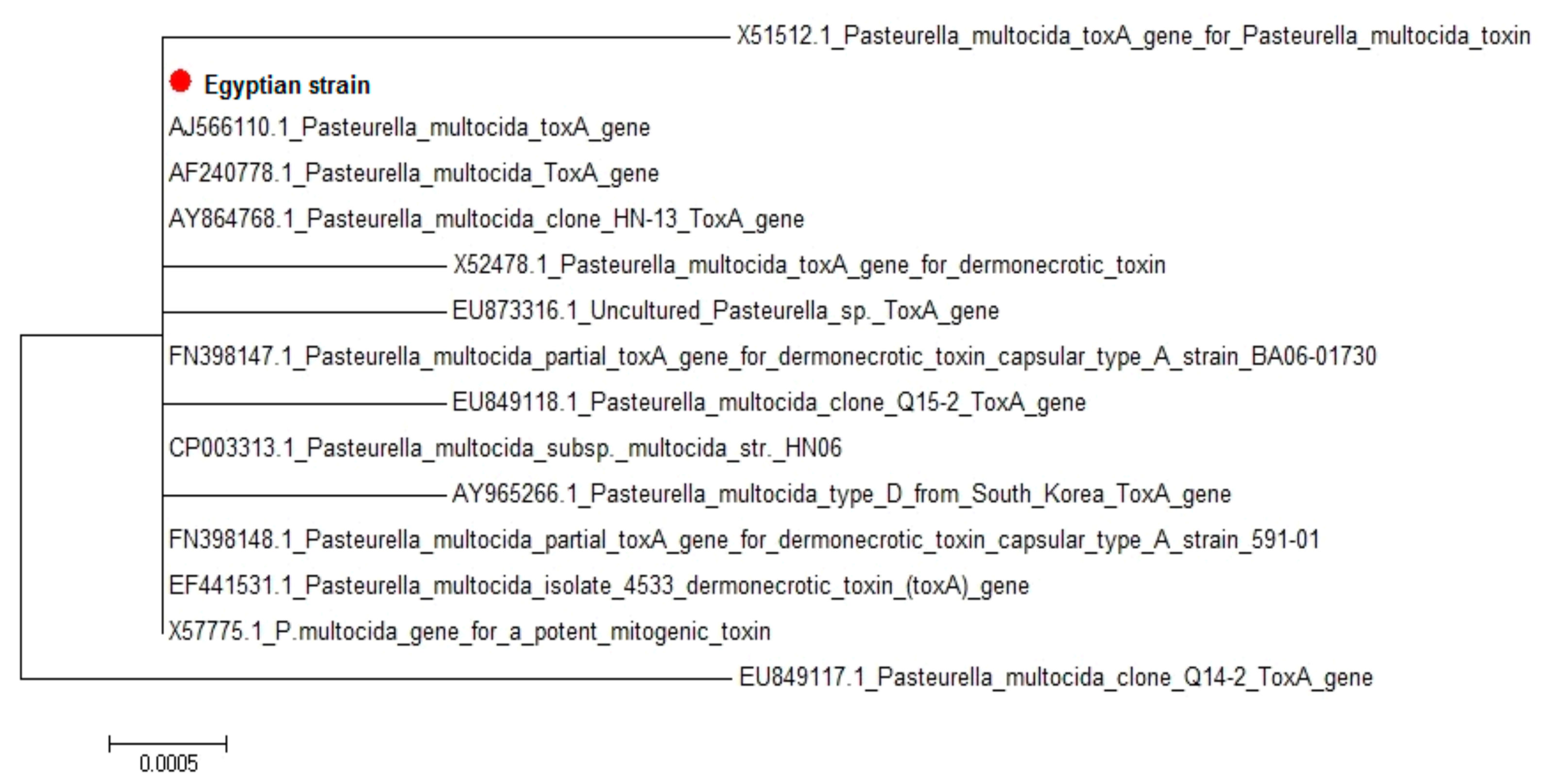
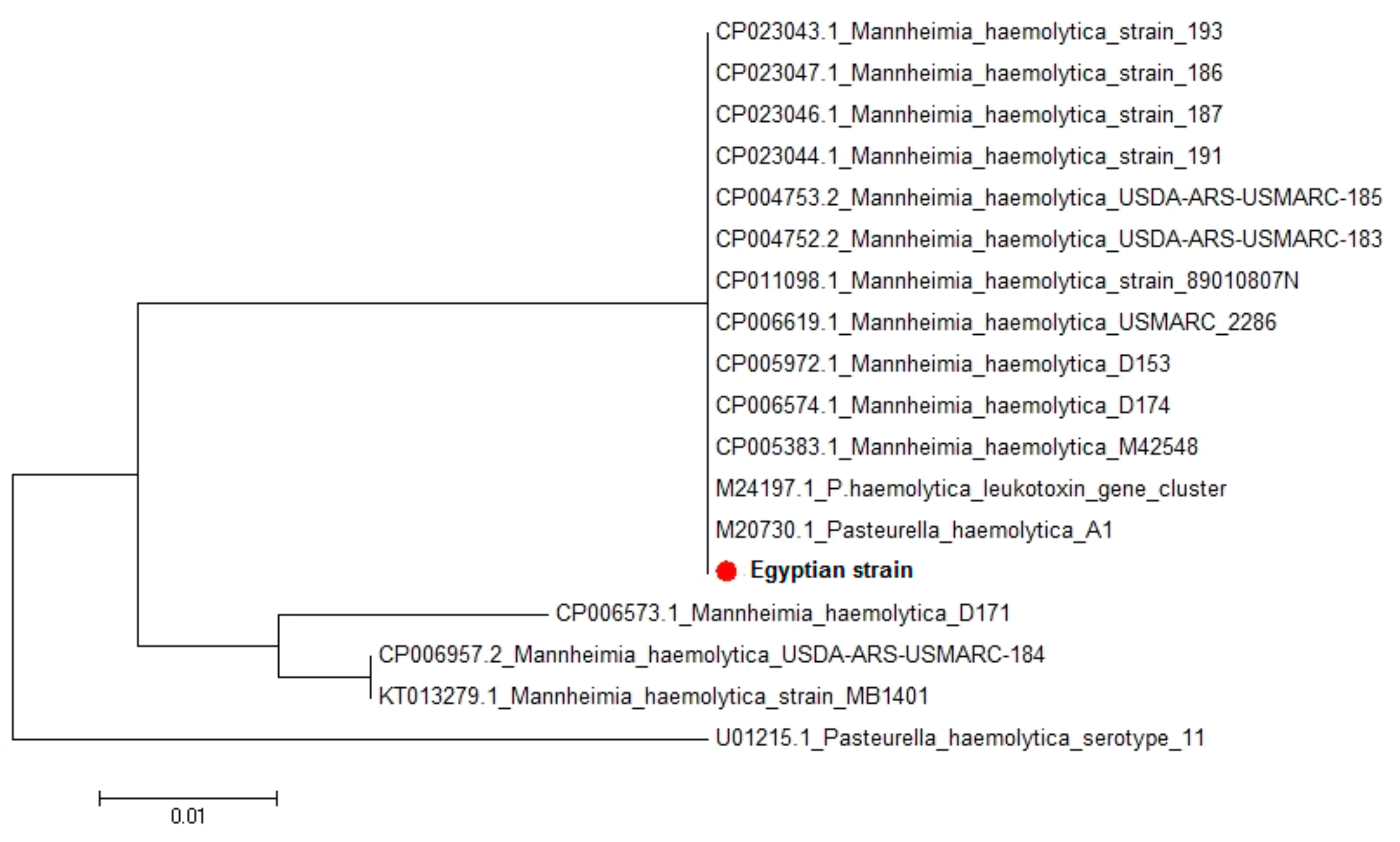
In the present study, the toxA gene of P. multocida serotype B:2 and lktC gene of M. haemolytica serotype 2 (the most prevalent serotype) were sequenced. Regarding P. multocida toxA gene, amino acid and nucleotide sequence analysis showed great homology between the Egyptian strain and the fourteen P. multocida strains uploaded in gene bank representing different clinical lesions, hosts, and localities worldwide. Generally, the sequence identities between the isolated Egyptian strain and different P. multocida strains uploaded from gene bank revealed 99.4–100% homology. This could suggest the high pathogenicity of the isolated strain and its high affinity to cause respiratory problems in infected animals [44], where toxA plays an important in destruction of lung tissues, and/or stimulation of a noxious host inflammatory response. Therefore, the toxA gene is considered an epidemiological marker found mostly in pneumonic P. multocida isolates [12]. Pullinger et al. [45] reported that the P. multocida toxin (PMT) acts as a potent mitogen. Sequence analysis of the structural gene for PMT, toxA, suggested that it was horizontally acquired because it had a low G+C content relative to the P. multocida genome [45]. Concerning M. haemolytica lktC gene, amino acid and nucleotide sequence analysis also indicated great homology between the Egyptian strain and the seventeen M. haemolytica strains uploaded in genebank representing different clinical lesions, hosts, and localities worldwide. Sequence identities between the isolated Egyptian strain and different M. haemolytica strains uploaded in genebank revealed 92.6–100% homology. This leukotoxin has been implicated as a major virulence factor in the pathogenesis of M. haemolytica, helping in the colonization and invasion of the lung tissues by impairing the primary lung defense mechanism and subsequent immune response or by the induction of inflammation as a consequence of leukocyte lysis [46]. Therefore, characterization of such a gene provides important information about the pathogenicity of M. haemolytica [14]. Moreover, Highlander et al. [47] reported that M. haemolytica secreted a 102-kilodalton leukotoxin that was believed to be involved in the pathogenesis of severe bovine pneumonia and considered lktC as the activator for leukotoxin (lktA).

5. Conclusions

The situation of P. multocida and M. haemolytica infections in North Upper Egypt is complicated due to the different stresses facing the young calves, including managemental, environmental, and infectious factors. To control such infections, firstly, stress factors should be avoided in addition to rapid diagnosis and efficient treatment of disease calves using suitable antimicrobial agents after application of the sensitivity test. Detection of Pasteurella species in the clinical material was greatly accelerated by the use of molecular techniques such as PCR using universal genes. Serotyping of isolates indicated that P. multocida serotype B:2 and M. haemolytica serotype 2 were the most prevalent in Egypt. Detection of some virulence-associated genes indicated that all the tested P. multocida isolates harbored nanB, omp87, and toxA virulence genes, while ssa, gcp, and lktC virulence genes were determined in most of tested M. haemolytica isolates. Sequencing of P. multocida toxA gene and M. haemolytica lktC gene from the isolated strains indicated a great homology between the isolated strains and the other strains uploaded in gene bank of different clinical lesions, hosts and localities worldwide. More scientific and field benefits could be gained by further investigations to know more about the current epidemiological situation of P. multocida and M. haemolytica infections in Egypt, and this step is the cornerstone for the prevention and control of these diseases through designing autogenous vaccines or minimizing the importation of animals from the endemic countries.

Author Contributions

Sample collection, S.E.S., A.H.A., E.K., methodology and data analysis F.R.E.-S., A.H.A., H.M.H., A.M.N., S.E.S., A.M.S.M., and G.W., writing and reviewing the original draft, A.H.A., G.W., A.M.S.M., and F.R.E.-S. All authors have read and agreed to the published version of the manuscript.

Funding

This research has been funded by the internal funding of Bacteriology, Mycology and Immunology Department, Faculty of Veterinary Medicine, Beni-Suef University, Egypt as well as project No. 115-2016 of the ministry of agriculture, GOVS sector for the Animal Reproduction Research Institute, Giza, Egypt.

Acknowledgments

The authors would like to thank the Bacteriology, Mycology, and Immunology Department staff at the Faculty of Veterinary Medicine, Beni-Suef University, and staff in Animal Reproduction Research Institute, Giza, Egypt, for providing technical help.

Conflicts of Interest

The authors declare no conflict of interest.

References

  1. Khamesipour, F.; Momtaz, H.; Mamoreh, M.A. Occurrence of virulence factors and antimicrobial resistance in Pasteurella multocida strains isolated from slaughter cattle in Iran. Front. Microbiol. 2014, 5, 1–10. [Google Scholar] [CrossRef] [Green Version]
  2. Elshemey, T.M.; Abd-Elrahman, A.H. Hemorrhagic Septicemia Outbreak as a Consequence to SAT2 FMD Infection in Buffalo and Cattle in Alexandria Province, Egypt. Life Sci. J. 2013, 10, 816–822. [Google Scholar]
  3. Zaher, K.S.; Syame, S.M.; Elhewairy, H.M.; Marie, H.S.H. Investigation of Bovine Respiratory Disease Complex in Egypt with Emphasis on Some Viral and Bacterial Pathogens. Life Sci. J. 2014, 11, 56–62. [Google Scholar]
  4. Kaoud, H.; El-Dahshan, A.R.; Zaki, M.M.; Nasr, S.A. Occurrence of Mannheimia haemolytica and Pasteurella trehalose among ruminants in Egypt. N. Y. Sci. J. 2010, 3, 135–141. [Google Scholar]
  5. Giordano, A.; Dincman, T.; Clyburn, B.E.; Steed, L.L.; Rockey, D.C. Clinical features and outcomes of Pasteurella multocida infection. Medicine 2015, 94, e1285. [Google Scholar] [CrossRef]
  6. Office International des Épizooties. Terrestrial Manual. Haemorrhagic Septicaemia; OIE: London, UK, 2008; pp. 739–751. [Google Scholar]
  7. El-Seedy, F.R.; Hassan, H.M.; Nabih, A.M.; Salem, S.E.; Khalifa, E.; Menshawy, A.M.S.; Abed, A.H. Respiratory affections in calves in upper and middle Egypt: Bacteriologic, immunologic and epidemiologic studies. Adv. Anim. Vet. Sci. 2020, 8, 558–569. [Google Scholar] [CrossRef]
  8. Haig, S.G. Adherence of Mannheimia haemolytica to ovine bronchial epithelial cells. Biosci. Horiz. Int. J. Stud. Res. 2011, 4, 50–60. [Google Scholar] [CrossRef] [Green Version]
  9. Legesse, A.; Abayneh, T.; Mamo, G.; Gelaye, E.; Tesfaw, L.; Yami, M.; Belay, A. Molecular characterization of Mannheimia haemolytica isolates associated with pneumonic cases of sheep in selected areas of Central Ethiopia. BMC Microbiol. 2018, 18, 205. [Google Scholar] [CrossRef] [PubMed]
  10. Jamali, H.; Rezagholipour, M.; Fallah, S.; Dadrasnia, A.; Chelliah, S.; Velappan, R.D.; Wei, K.S.C.; Ismail, S. Prevalence, characterization and antibiotic resistance of Pasteurella multocida isolated from bovine respiratory infection. Vet. J. 2014, 202, 381–383. [Google Scholar] [CrossRef] [PubMed]
  11. Katsuda, K.; Hoshinoo, K.; Ueno, Y.; Kohmoto, M.; Mikami, O. Virulence genes and antimicrobial susceptibility in Pasteurella multocida isolates from calves. Vet. Microbiol. 2013, 167, 737–741. [Google Scholar] [CrossRef] [PubMed]
  12. Harper, M.; Boyce, J.D.; Adler, B. Pasteurella multocida pathogenesis: 125 years after Pasteur. FEMS Microbioliol. Lett. 2006, 265, 1–10. [Google Scholar] [CrossRef] [PubMed]
  13. Vougidou, C.; Sandalakis, V.; Psaroulaki, A.; Siarkou, V.; Petridou, E.; Ekateriniadou, L. Distribution of the ompA-types among ruminant and swine pneumonic strains of Pasteurella multocida exhibiting various cap-locus and toxA patterns. Microbiol. Res. 2015, 174, 1–8. [Google Scholar] [CrossRef] [PubMed]
  14. Singh, K.; Ritchey, J.W.; Confer, A.W. Mannheimia haemolytica: Bacterial-Host interactions in bovine pneumonia. Vet. Pathol. 2011, 48, 338–348. [Google Scholar] [CrossRef] [Green Version]
  15. Klima, C.L.; Alexander, T.W.; Hendrick, S.; McAllister, T.A. Characterization of Mannheimia haemolytica isolated from feedlot cattle that were healthy or treated for bovine respiratory disease. Canad. J. Vet. Res. 2014, 78, 38–45. [Google Scholar]
  16. Rimler, R.B.; Rhoades, K.R. Hyaluronidase and chondroitinase activity of Pasteurella multocida serotype B:2 involved in hemorrhagic septicaemia. Vet. Rec. 1994, 134, 67–68. [Google Scholar] [CrossRef]
  17. Frank, G.H.; Wessman, G.E. Rapid plate agglutination procedure for serotyping Pasteurella haemolytica. J. Clin. Microbiol. 1978, 7, 142–145. [Google Scholar]
  18. Office International des Épizooties (OIE). Terrestrial Manual. Manual of Diagnostic Tests and Vaccines for Terrestrial Animals, Haemorrhagic Septicaemia; OIE: Paris, France, 2012; Chapter 2.4.12. [Google Scholar]
  19. Sarangi, L.N.; Priyadarshini, A.; Kumar, S.; Thomas, P.; Gupta, S.K.; Nagaleekar, V.K.; Singh, V.P. Virulence Genotyping of Pasteurella multocida Isolated from Multiple Hosts from India. Sci. World J. 2014, 2014. [Google Scholar] [CrossRef] [Green Version]
  20. Sabiel, Y.A.; Musa, M.T.; Zyl, A.V. Identification of Mannheimia haemolytica and Pasteurella multocida by Polymerase Chain Reaction and Random Amplification of Polymorphic DNA. Sud. J. Vet. Res. 2012, 27, 55–59. [Google Scholar]
  21. Tang, X.; Zhao, Z.; Hu, J.; Wu, B.; Cai, X.; He, Q.; Chen, H. Isolation, antimicrobial resistance and virulence genes of Pasteurella multocida strains from swine in China. J. Clin. Microbiol. 2009, 47, 951–958. [Google Scholar] [CrossRef] [Green Version]
  22. Deressa, A.; Asfaw, Y.; Lubke, B.; Kyule, M.W.; Tefera, G.; Zessin, K.H. Molecular detection of Pasteurella multocida and Mannheimia haemolytica in sheep respiratory infections in Ethiopia. Inter. J. Appl. Res. Vet. Med. 2010, 8, 101–108. [Google Scholar]
  23. Hawari, D.; Hassawi, D.S.; Sweiss, M. Isolation and identification of Mannheimia haemolytica and Pasteurella multocida in sheep and goats using biochemical tests and random amplified polymorphic DNA (RAPD) analysis. J. Biol. Sci. 2008, 8, 1251–1254. [Google Scholar] [CrossRef] [Green Version]
  24. Altschul, S.F.; Gish, W.; Miller, W.; Myers, E.W.; Lipmanl, D.J. Basic Local Alignment Search Tool. J. Mol. Biol. 1990, 215, 403–410. [Google Scholar] [CrossRef]
  25. Thompson, J.D.; Higgins, D.G.; Gibson, T.J.; Clustal, W. Improving the sensitivity of progressive multiple sequence alignment through sequence weighting, position-specific gap penalties and weight matrix choice. Nucleic Acids Res. 1994, 22, 4673–4680. [Google Scholar] [CrossRef] [PubMed] [Green Version]
  26. Tamura, K.; Stecher, G.; Peterson, D.; Filipski, A.; Kumar, S. MEGA6: Molecular evolutionary genetics analysis version 6.0. Mol. Biol. Evol. 2013, 30, 2725–2729. [Google Scholar] [CrossRef] [Green Version]
  27. Farooq, U.; Saeed, Z.; Khan, M.A.; Ali, I.; Qamar, M.F. Sero-surveillance of hemorrhagic septicemia in buffaloes and cattle in Southern Punjab, Pakistan. Pak. Vet. J. 2011, 31, 254–256. [Google Scholar]
  28. Güler, L.; Gündüz, K.; Sarisahin, A.S. Capsular typing and antimicrobial susceptibility of Pasteurella multocida isolated from different hosts. Kafkas Üniversitesi Vet. Fakültesi Derg. 2013, 19, 843–849. [Google Scholar] [CrossRef] [Green Version]
  29. Kumar, P.; Singh, V.P.; Agrawal, R.K.; Singh, S. Identification of Pasteurella multocida isolates of ruminant origin using polymerase chain reaction and their antibiogram study. Trop. Anim. Health Prod. 2009, 41, 573–578. [Google Scholar] [CrossRef]
  30. Boyce, J.D.; Harper, M.; Wilkie, I.W.; Adler, B. Pasteurella. Pathogenesis of Bacterial Infections in Animals, 4th ed.; Gyles, C.L., Prescott, J.F., Songer, J.G., Thoen, C.O., Eds.; Blackwell Publishing: Hoboken, NJ, USA, 2010; pp. 325–346. [Google Scholar]
  31. Abbas, A.M.; Abd El-Moaty, D.A.M.; Zaki, E.S.A.; El-Sergany, E.F.; El-Sebay, N.A.; Fadl, H.A.; Samy, A.A. Use of molecular biology tools for rapid identification and characterization of Pasteurella spp. Vet. World 2018, 11, 100–1014. [Google Scholar] [CrossRef] [Green Version]
  32. Balakrishnan, G.; Roy, P. Isolation, identification and antibiogram of Pasteurella multocida isolation of avian origin. Tamilnadu J. Vet. Anim. Sci. 2012, 8, 199–200. [Google Scholar]
  33. Ryan, K.A.; Lo, R.Y. Characterization of a CACAG pentanucleotide repeat in Pasteurella haemolytica and its possible role in modulation of a novel type III restriction-modification system. Nucleic. Acids Res. 1999, 27, 1505–1511. [Google Scholar] [CrossRef] [Green Version]
  34. Ranjan, R.; Panda, S.K.; Acharya, A.P.; Singh, A.P.; Gupta, M.K. Molecular diagnosis of haemorrhagic septicaemia- A review. Vet. World 2011, 4, 189–192. [Google Scholar]
  35. Dziva, F.; Muhairwa, A.P.; Bisgaard, M.; Christensen, H. Diagnostic and typing options for investigating diseases associated with Pasteurella Multocida. Vet. Microbiol. 2008, 128, 1–22. [Google Scholar] [CrossRef] [PubMed]
  36. Jabbari, A.R.; Esmaelzadeh, M.; Moazeni, J.G.R. Polymerase chain reaction of Pasteurella multocida capsules isolated in Iran. Iran J. Vet. Res. 2006, 7, 50–55. [Google Scholar]
  37. Katsuda, K.; Kohmoto, M.; Mikami, O. Relationship between serotype and the antimicrobial susceptibility of Mannheimia haemolytica isolates collected between 1991 and 2010. Vet. Sci. 2013, 94, 205–208. [Google Scholar] [CrossRef] [PubMed]
  38. Klima, C.L.; Cook, S.R.; Zaheer, R.; Laing, C.; Gannon, V.P.; Xu, Y.; Rasmussen, J.; Potter, A.; Hendrick, S.; Alexander, T.W.; et al. Comparative genomic analysis of Mannheimia haemolytica from bovine sources. PLoS ONE 2016, 11, e0149520. [Google Scholar] [CrossRef]
  39. Kabeta, T.; Fikadu, T.; Zenebe, T.; Kebede, G. Review on the pneumonic pasteurellosis of cattle. Acad. J. Anim. Dis. 2015, 4, 177–184. [Google Scholar]
  40. Ewers, C.; Lubke-Becker, A.; Bethe, A.; Kiebling, S.; Filter, M.; Wieler, L.H. Virulence genotype of Pasteurella multocida strains isolated from different hosts with various disease status. Vet. Microbiol. 2006, 114, 304–317. [Google Scholar] [CrossRef]
  41. Devi, L.; Durlav, P.; Das, S.K.; Sharma, R.K.; Mukherjee, S.; Hazarika, R.A. Virulence gene profiling of porcine Pasteurella multocida isolates of Assam. Vet. World 2018, 11, 348–354. [Google Scholar] [CrossRef]
  42. Klima, C.L.; Zaheer, R.; Cook, S.R.; Rasmussen, J.; Alexander, T.W.; Potter, A.; Hendrick, S.; McAllister, T.A. In silico identification and high throughput screening of antigenic proteins as candidates for a Mannheimia haemolytica vaccine. Vet. Immunol. Immunopathol. 2018, 195, 19–24. [Google Scholar] [CrossRef]
  43. Ayalew, S.; Shrestha, B.; Montelongo, M.; Wilson, A.E.; Confer, A.W. Proteomic analysis and immunogenicity of Mannheimia haemolytica vesicles. Clin. Vaccine Immunol. 2013, 20, 191–196. [Google Scholar] [CrossRef] [Green Version]
  44. Liu, W.; Yang, M.; Xu, Z.; Zheng, H.; Liang, W.; Zhou, R.; Wu, B.; Chen, H. Complete genome sequence of Pasteurella multocida HN06, a toxigenic strain of serogroup D. J. Bacteriol. 2012, 194, 3292–3303. [Google Scholar] [CrossRef] [PubMed] [Green Version]
  45. Pullinger, G.D.; Bevir, T.; Lax, A.J. The Pasteurella multocida toxin is encoded within a lysogenic bacteriophage. Mol. Microbiol. 2004, 51, 255–269. [Google Scholar] [CrossRef] [PubMed] [Green Version]
  46. Lo, R.Y.C.; Strathdee, C.A.; Shewen, P.E. Nucleotide Sequence of the Leukotoxin Genes of Pasteurella haemolytica Al. Infect. Immun. 1987, 55, 1987–1996. [Google Scholar] [CrossRef] [PubMed] [Green Version]
  47. Highlander, S.K.; Engler, M.J.; Weinstock, G.M. Secretion and expression of the Pasteurella haemolytica Leukotoxin. J. Bacteriol. 1990, 172, 2343–2350. [Google Scholar] [CrossRef] [Green Version]
Figure 1. Phylogenetic tree for P. multocida serotype B:2 toxA showing clear clustering of the Egyptian strain with different strains uploaded in the gene bank.
Vetsci 07 00174 g001
Figure 2. Phylogenetic tree for M. haemolytica A2 lktC partial sequences showing clear clustering of the Egyptian strain with different strains uploaded in the genebank.
Vetsci 07 00174 g002
Table 1. Samples and bacterial isolates in different Governorates.
GovernoratesNo. of SamplesNo. of IsolatesTotal
P. multocidaM. haemolytica
El-Fayoum106221032
Beni-Suef8311516
Total189331548
Table 2. Primers sequences and amplified products for the targeted genes for P. multocida and M. haemolytica isolates.
BacteriaTarget GenePrimer Sequence 5′-3′Amplified ProductReference
P. multocidakmt1FATCCGCTATTTACCCAGTGG460 bp[18]
RGCTGTAAACGAACTCGCCAC
nanBFGTCCTATAAAGTGACGCCGA554 bp[19]
RACAGCAAAGGAAGACTGTCC
omp87FAGGTGAAAGAGGTT ATG200 bp [20]
RTACCTAA CTCAACCAAC
toxAFCTTAGATGAGCGACAAGG864 bp[21]
RGAATGCCACACCTCTATAG
M. haemolyticarpt2FGTTTGTAAGATATCCCATTT1022 bp[22]
RCGTTTTCCACTTGCGTGA
ssaFTTCACATCTTCATCCTC325 bp[23]
RTTTTCATCCTCTTCGTC
gcpFCGCCCCTTTTGGTTTTCTAA420 bp[15]
RGTAAATGCCCTTCCATATGG
lktCFGGAAACATTACTTGGCTATGG440 bp
RTGTTGCCAGCTCTTCTTGATA
Table 3. Cycling conditions of the different primers during PCR.
BacteriaTarget GenePrimary DenaturationSecondary DenaturationAmplification (35 Cycles)
AnnealingExtensionFinal Extension
P. multocidakmt194 C/5 min94 C/30 s55 C/1 min72 C/1 min72 C/10 min
nanB94 C/5 min94 C/30 s50 C/40 s72 C/40 s72 C/10 min
omp8794 C/5 min94 C/30 s48 C/30 s72 C/30 s72 C/7 min
toxA94 C/5 min94 C/30 s48 C/40 s72 C/45 s72 C/10 min
M. haemolyticarpt295 C/3 min95 C/1 min48 C/1 min72 C/30 s72 C/10 min
ssa94 C/5 min94 C/30 s45 C/40 s72 C/40 s72 C/10 min
gcp94 C/5 min94 C/30 s58 C/40 s72 C/40 s72 C/10 min
lktC94 C/5 min94 C/30 s58 C/40 s72 C/40 s72 C/10 min
Table 4. Results of PCR as a confirmatory identification of P. multocida and M. haemolytica isolates.
Tested BacteriaNo. of the Tested IsolatesPositive PCR Result
No.%
P. multocida332987.9
M. haemolytica1515100
%: Percentages were calculated according to the corresponding number of the tested isolates.
Table 5. Prevalence of virulence-associated genes among the examined P. multocida and M. haemolytica isolates.
Tested BacteriaTested GeneNo. of the Tested IsolatesPositiveNegative
No.%No.%
P. multocidananB5510000
omp87510000
toxA510000
M. haemolyticassa5360240
gcp480120
lktC 480120
%: Percentages were calculated according to the corresponding number of the tested isolates.
Table 6. Nucleotides and amino acid identity and sequence distance of P. multocida serotype B2 toxA gene between the isolated Egyptian strain and different strains uploaded in the gene bank.
Percent Identity
Divergence 123456789101112131415
1 99.899.899.899.999.999.999.999.999.899.999.899.699.599.91AF240778.1
20.1 99.899.899.999.999.999.999.999.899.999.899.699.599.92EU873316.1
30.10.2 99.899.999.999.999.999.999.899.999.899.699.599.93EU849118.1
40.10.20.2 99.999.999.999.999.999.899.999.899.699.599.94AY965266.1
50.00.10.10.1 10010010010099.910099.999.899.61005CP003313.1
60.00.10.10.10 10010010099.910099.999.899.61006FN398148.1
70.00.10.10.100 10010099.910099.999.899.61007FN398147.1
80.00.10.10.1000 10099.910099.999.899.61008EF441531.1
90.00.10.10.10000 99.910099.999.899.61009AY854768.1
100.00.10.10.100000 99.999.899.699.599.910AJ566110.1
110.00.10.10.1000000 99.999.899.610011X57775.1
120.10.20.20.20.10.10.10.10.10.10.1 99.699.599.912X52478.1
130.20.40.40.40.20.20.20.20.20.20.20.4 99.499.813X51512.1
140.40.50.50.50.40.40.40.40.40.40.40.50.6 99.614EU849117.1
150.00.10.10.100000000.10.20.4 15Egyptian Strain
123456789101112131415
Table 7. Nucleotides and amino acid identity and sequence distance of M. haemolytica serotype A2 lktC gene between the Egyptian strain and different strains uploaded in the genebank.
DivergencePercent Identity
123456789101112131415161718
1 10010010010010010010010010010010010095.795.794.692.61001CP023043.1
20.0 10010010010010010010010010010010095.795.794.692.61002CP023047.1
30.00.0 10010010010010010010010010010095.795.794.692.61003CP023046.1
40.00.00.0 10010010010010010010010010095.795.794.692.61004CP023044.1
50.00.00.00.0 10010010010010010010010095.795.794.692.61005CP004753.2
60.00.00.00.00.0 10010010010010010010095.795.794.692.61006CP004752.2
70.00.00.00.00.00.0 10010010010010010095.795.794.692.61007CP011098.1
80.00.00.00.00.00.00.0 10010010010010095.795.794.692.61008CP005619.1
90.00.00.00.00.00.00.00.0 10010010010095.795.794.692.61009CP005972.1
100.00.00.00.00.00.00.00.00.0 10010010095.795.794.692.610010CP005574.1
110.00.00.00.00.00.00.00.00.00.0 10010095.795.794.692.610011CP005383.1
120.00.00.00.00.00.00.00.00.00.00.0 10095.795.794.692.610012M24197.1
130.00.00.00.00.00.00.00.00.00.00.00.0 95.795.794.692.610013M20730.1
144.44.44.44.44.44.44.44.44.44.44.44.44.4 1009893.195.714CP006957.2
154.44.44.44.44.44.44.44.44.44.44.44.44.40.0 9893.195.715KT013279.1
165.65.65.65.65.65.65.65.65.65.65.65.65.62.12.1 94.694.616CP006573.1
177.97.97.97.97.97.97.97.97.97.97.97.97.97.47.45.6 92.617U01215.1
180.00.00.00.00.00.00.00.00.00.00.00.00.04.44.45.67.9 18Egyptian strain
123456789101112131415161718
Publisher’s Note: MDPI stays neutral with regard to jurisdictional claims in published maps and institutional affiliations.

