1. Introduction
PRV, also known as porcine herpesvirus type 1 (PHV1), is classified as an α-herpesvirus, specifically within the Varicellovirus genus [
1,
2]. Swine serve as the natural hosts, and PRV can also infect a broad spectrum of other animals, including ruminants, carnivores, rodents, and lagomorphs [
3]. Infection with PRV results in considerable economic losses for the global livestock industry, particularly in swine farming. Pseudorabies has been shown to cause nearly 100% mortality in piglets, reproductive failure in sows, growth retardation in finishing pigs, and increased susceptibility to secondary infections. Although eradication programs can enhance farm profitability, they entail significant costs [
4].
The mainstay of PRV control is vaccination; however, currently available vaccines are licensed exclusively for use in pigs and face dual challenges related to efficacy and safety. Since 2011, outbreaks in Bartha-K61-vaccinated herds in China have persisted due to emerging variant strains [
5,
6]. These variants exhibit significant genetic and antigenic divergence from classical strains, with vaccine efficacy declining from 100% against the classical SC strain to just 50% [
7]. The 2022 isolate CH/JLHF/2022 largely evaded maternal antibodies, causing 100% mortality in piglets [
8]. Adding to these efficacy concerns, a 2025 case report raised a potential safety issue: Chen et al. [
9] reported an outbreak in PRV-negative Danish purebred pigs imported to Taiwan, China. Both groups exhibited 100% morbidity, with mortality rates of 67.6% and 13.4%, accompanied by severe immunopathology. These observations suggest a possible association with Bucharest strain live vaccine (Zoetis PR-VAC PLUS) immunization, constituting a potential safety signal. As a potential zoonotic pathogen, PRV has caused markedly rising human infections, manifesting as viral encephalitis [
10,
11,
12] and endophthalmitis [
13], the latter often leading to permanent blindness [
2,
14]. This highlights its growing threat to public health, particularly to swine industry workers [
15].
Type I interferons (IFN-α/β) are key mediators of the innate antiviral response. Their production is initiated upon host recognition of viral nucleic acids by pattern recognition receptors (PRRs) such as Toll-like receptors (TLRs) and RIG-I-like receptors (RLRs), as well as the DNA sensor cGAS. This recognition activates the cGAS/STING pathway, which triggers downstream cascades involving TBK1 kinase and leads to phosphorylation of the transcription factors IRF3 and IRF7. Phosphorylated IRF3/7 then dimerize and translocate to the nucleus to drive type I interferon gene expression [
16]. Once secreted, type I IFNs bind to the IFN-α/β receptor (IFNAR), activating the JAK-STAT pathway and leading to the formation of the ISGF3 complex (STAT1-STAT2-IRF9). ISGF3 translocates to the nucleus to induce the expression of numerous interferon-stimulated genes (ISGs). The resulting ISG products inhibit viral replication through diverse mechanisms, such as blocking protein translation, degrading viral nucleic acids, or modulating cellular metabolism, thereby establishing an antiviral state at the site of infection [
17].
Flavonoids, a class of naturally occurring polyphenolic compounds, have been demonstrated to exert regulatory effects on specific phases of the viral life cycle, thereby impeding viral replication in diverse viruses, including herpesviruses [
18]. Wogonin has been shown to modulate NF-κB and JNK/p38 MAPK pathways to exert anti-HSV activity [
19]. Similarly, naringenin has been shown to exert antiviral activity, with effects including inhibition of virion assembly and modulation of viral protein translation [
20]. Among these bioactive flavonoids, isoliquiritigenin, a hydroxychalcone derived from licorice root, has attracted considerable interest due to its pleiotropic pharmacological effects, including antioxidant, anti-inflammatory, antidiabetic, cardioprotective, hepatoprotective, neuroprotective, and anticancer properties [
21]. To date, studies on the antiviral activity of isoliquiritigenin remain limited. A preliminary in vitro study has reported that isoliquiritigenin can reduce the viral titers of vesicular stomatitis virus, influenza A virus, encephalomyocarditis virus, and herpes simplex virus type 1 [
22]. Additionally, isoliquiritigenin exhibits potent antiviral activity against spring viremia of carp virus (SVCV) in aquaculture models, an effect attributed to the dual modulation of host antioxidant defenses and immune responses [
23]. However, its efficacy against PRV and the underlying mechanisms remain unexplored. Given the critical role of IFN-I in controlling PRV infection and the virus’s ability to subvert this pathway, this study aimed to evaluate the regulatory effects of isoliquiritigenin against PRV both in vitro and in vivo, focusing on the cGAS/STING and JAK/STAT signaling cascades, to clarify its antiviral mechanism and facilitate the development of novel anti-PRV agents.
2. Materials and Methods
2.1. Cell, Virus, and Isoliquiritigenin
The porcine kidney cell line (PK-15) and African green monkey kidney cells (Vero) were obtained from the China Center for Type Culture Collection (China Center for Type Culture Collection, Wuhan, China). Both cell lines were cultured in Eagle‘s Minimum Essential Medium (EMEM) supplemented with 10% (v/v) calf serum (Gibco, Grand Island, NY, USA), 100 U/mL penicillin, and 100 μg/mL streptomycin. For the maintenance medium (MM), the serum concentration was reduced to 2%.
The pseudorabies virus (Ra strain), a virulent field isolate, was purchased from the China Veterinary Culture Collection Center (Beijing, China) and propagated in PK-15 cells. After five passages, the virus was maintained at the Natural Medicine Research Center, Sichuan Agricultural University (Chengdu, China). Prior to use in this study, the virus was passaged for an additional 10 passages in PK-15 cells. The 50% tissue culture infectious dose (TCID50) of the virus stock was determined to be 105.7TCID50/mL according to the Reed–Muench method.
Isoliquiritigenin (catalog No. Y91617A, Adamas Life, manufacturer direct, Shanghai, China) with a purity of ≥97% (determined by actual test; Bio-Active Grade) was used in this study. The compound was dissolved in dimethyl sulfoxide (DMSO; Beijing Solarbio Science & Technology Co., Ltd., Beijing, China) to prepare a 200× stock solution, which was then diluted with maintenance medium to the desired working concentrations immediately before use. The final DMSO concentration in all experimental groups was ≤0.5%.
2.2. Cytotoxic Concentration Assay
The cytotoxicity of isoliquiritigenin against PK-15 cells was evaluated using the CCK-8 assay. PK-15 cells were seeded in 96-well plates and treated with two-fold serial dilutions of isoliquiritigenin ranging from 1000 to 15.63 µM, with six replicates per concentration. After incubation for 48 h, 10 µL of CCK-8 solution was added to each well, and the plates were further incubated at 37 °C for 30 min. Absorbance was then measured at 450 nm using a microplate reader. Cell viability was calculated as: (ODISL − ODblank)/(ODcontrol − ODblank) × 100%, where ODISL represents cells treated with isoliquiritigenin, ODcontrol represents untreated cells, and ODblank represents medium-only wells. The half-maximal cytotoxic concentration (CC50) was determined by fitting the dose–response data to a nonlinear regression model (log(inhibitor) vs. normalized response, variable slope) using GraphPad Prism 9.0 (Dotmatics, Boston, MA, USA).
2.3. Antiviral Activity Assay
PK-15 cell monolayers in 96-well plates were infected with PRV at 100 TCID50 per well. For this, the virus stock with a titer of 105·7 TCID50/100 µL was diluted 10−3.7 (approximately 5000-fold) in maintenance medium to obtain 100 TCID50 per 100 µL, and 100 µL of this dilution was added to each well. One hour post-infection, the virus-containing medium was removed and replaced with medium containing various concentrations of isoliquiritigenin (two-fold serial dilutions from 1000 to 15.63 µM). When virus-infected, untreated cells exhibited approximately 80% cytopathic effect (CPE); 10 µL of CCK-8 solution was added to each well. After incubation at 37 °C for 30 min, absorbance was measured at 450 nm using a microplate reader (Bio-Rad, USA). Viral inhibition rate (%) was calculated using the following formula: (OD PRV+ ISL − ODPRV)/(ODMock − ODPRV) × 100%, where ODPRV+ISL represents the OD value of cells treated with isoliquiritigenin and infected with PRV, ODPRV represents the OD value of cells infected with PRV without drug treatment (virus control), and ODMock represents the OD value of uninfected and untreated cells. The 50% inhibitory concentration (IC50) was then calculated by fitting the inhibition rates against drug concentrations using nonlinear regression analysis in GraphPad Prism 9.0 (Dotmatics, Boston, MA, USA). The selectivity index (SI) was calculated as CC50/IC50.
To evaluate the antiviral activity of isoliquiritigenin at different multiplicities of infection (MOI), PK-15 cells in 96-well plates were infected with PRV at MOIs of 0.01, 0.1, and 1. Following a 1-h adsorption period, the inoculum was removed and replaced with maintenance medium containing 62.5 µM isoliquiritigenin. At 24 h post-infection, total DNA was extracted using DNAiso Reagent (D305; TaKaRa, Dalian, China) according to the manufacturer’s instructions to detect viral DNA copies. For determination of viral gene copies, fluorescent quantitative PCR (FQ-PCR) was performed using a Bio-Rad CFX96 Connect™ Real-Time PCR Detection System (Bio-Rad, Hercules, CA, USA) [
24].
2.4. Mode of Action Assay
To determine which stage of viral infection was inhibited by isoliquiritigenin, a mode-of-action analysis was conducted using the following five experimental setups [
25]. (1) Prevention: The cells were exposed to isoliquiritigenin (62.5 μM) at 37 °C for 1 h, after which they were infected with PRV at a concentration of 100 TCID
50. (2) Inactivation: The PRV (10,000 TCID
50) was subjected to an incubation with isoliquiritigenin (62.5 μM) at 37 °C for 1 h. Thereafter, the PRV was diluted to a concentration of 100-fold, and was subsequently introduced to the cells for the purpose of infection. (3) Adsorption: The PRV (100 TCID
50) was incubated with isoliquiritigenin (62.5 μM) at 4 °C for 1 h prior to infection of the cells. The cells then underwent a thorough washing process to ensure the removal of any unadsorbed virus. Following this, the cells were subjected to an incubation at 37 °C. (4) Penetration: PK-15 cells grown in 6-well plates were pre-chilled at 4 °C for 30 min. The cells were then incubated with PRV (100 TCID
50) at 4 °C for 1 h to allow viral adsorption. After removal of unadsorbed virus, the cells were gently washed three times with pre-chilled PBS. Pre-chilled maintenance medium containing isoliquiritigenin (62.5 μM) was then added, and the cells were incubated at 37 °C for 1 h to allow viral penetration. Following this incubation, the virus-containing medium was removed, and the cells were washed three times with citrate-citrate sodium buffer (pH 3.0) to inactivate and remove any remaining surface-bound virus. Finally, 2 mL of fresh maintenance medium was added to each well, and the cells were further incubated at 37 °C in a 5% CO
2 incubator for 48 h. Viral DNA copies were quantified by FQ-PCR as described above. (5) Post-infection: The cells were infected with PRV (100 TCID
50) at 37 °C for 1 h, then treated with isoliquiritigenin (62.5 μM) after removing unbound virus. After further incubation for 48 h, viral DNA copies were detected by FQ-PCR method described above.
2.5. Effect of Isoliquiritigenin on the PRV Growth Curve
PK-15 cells, which had been seeded in 6-well plates, were infected with PRV (MOI = 0.1). After incubation at 37 °C for 1 h, the inoculum was replaced with medium containing isoliquiritigenin (62.5 μM). Cells were harvested at 2, 4, 6, 8, 12, and 18 hpi, and viral DNA was extracted. Viral DNA copies were quantified by fluorescent quantitative PCR (FQ-PCR) as described previously.
2.6. Real-Time PCR Assay
Total RNA was extracted from cells cultured in 6-well plates using TRIzol reagent (RA101-01, Biomed, Beijing, China). Reverse transcription was performed using the M-MLV First-Strand cDNA Synthesis Kit (MT403-01, Biomed, Beijing, China). Quantitative real-time PCR was then carried out using the Hieff UNICON Universal Blue qPCR SYBR Master Mix (11184ES08, Yeasen, Shanghai, China) with the primers listed in
Table 1. The PCR cycling was performed at 95 °C for 3 min, followed by 40 cycles of 95 °C for 10 s, 59.8 °C for 30 s, and 55 °C for 5 s. The Ct value of the target gene was normalized in relation to the expression of the internal reference gene, β-actin. The relative mRNA expression level of each target gene was calculated according to the 2
−ΔΔCt method.
2.7. cGAMP Activity Assay
To assess cyclic guanosine monophosphate-adenosine monophosphate (cGAMP) activity, PK-15 cell monolayers infected with PRV (MOI = 1) were treated with 62.5 µM isoliquiritigenin for 1 h. The cells were washed with PBS and then lysed on ice for 30 min. The lysates were centrifuged and the supernatant was collected. Super Nuclease (1000 U/mL; Sino Biological, Beijing, China; Cat. No. SSNP01) was added and incubated at 37 °C for 30 min. The mixture was then heated at 95 °C for 5 min and centrifuged at 12,000 rpm at 4 °C for 5 min. The supernatant was retained and added to PK-15 cells for 6 h. Total RNA was then extracted, and RT-qPCR was performed to measure IFN-β mRNA levels in the recipient cells, serving as an indirect readout of cGAMP activity in the original cell lysates [
26].
2.8. Western Blotting Assay
PK-15 cells were divided into four groups: Mock, uninfected-untreated group; ISL, uninfected group treated with isoliquiritigenin; PRV, infected-untreated group; PRV + ISL, infected group treated with isoliquiritigenin. Following a 1 h adsorption period, the virus-containing inoculum was removed and replaced with fresh culture medium supplemented with or without 62.5 µM isoliquiritigenin as appropriate. Total proteins were extracted using a commercial kit (BOSTER, Wuhan, China, Cat. No. AR0103-100) at 2, 6, and 12 hpi, respectively. Then, the proteins were separated by 10% sodium dodecyl sulfate-polyacrylamide gel electrophoresis (SDS-PAGE) alongside a pre-stained protein ladder (Chengdu Rongwei Gene Technology Co., Ltd., Cat. No. M201-01, Chengdu, China) with a molecular weight range of 10–180 kDa, and transferred to polyvinylidene difluoride (PVDF) membranes (Millipore, Burlington, MA, USA). The membranes were blocked in 5% (w/v) skim milk diluted with Tris-buffered saline containing 0.1% (v/v) Tween 20 (TBST) for 90 min at room temperature, followed by incubation with primary antibodies against IRF3 (1:500, #11312–1-AP; Proteintech, Wuhan, China), p-IRF3 (1:500, #29528–1-AP; Bioss, Beijing, China), STAT1 (1:500, #66545–1-IG; Proteintech, Wuhan, China), p-STAT1 (1:500, #orb7016; Biorobyt, Durham, NC, USA), and β-actin (1:5000, #bs-0061R; Boster, Wuhan, China) at 4 °C overnight. The membranes were washed with TBST and incubated with horseradish peroxidase-conjugated secondary antibody (BA1056; Boster, Wuhan, China, 1:5000) at room temperature for 1.5 h. The proteins were visualized using an enhanced chemiluminescence (ECL) substrate (Bio-Rad, Hercules, CA, USA). The protein expression levels were normalized to β-actin, and the relative band intensities were quantified using ImageJ software (Version 1.47; NIH, Bethesda, MD, USA).
Western blot quantification and normalization: The band intensities were quantified using ImageJ software (Version 1.47; NIH, USA). To correct for variations in total protein loading, the raw integrated density (gray value) values of total IRF3, phosphorylated IRF3 (p-IRF3), STAT1, and p-STAT1 were first normalized to the corresponding β-actin band intensity within the same sample. To assess the activation levels of IRF3 and STAT1, the ratio of phosphorylated to total protein was calculated by dividing the normalized phosphorylated protein value (p-IRF3 or p-STAT1) by the normalized total protein value (IRF3 or STAT1) for each individual sample. The data are presented as the relative protein levels (p-IRF3/IRF3 and p-STAT1/STAT1 ratios). All primary antibodies were purchased from commercial suppliers and have been validated by the manufacturers for cross-reactivity against the relevant species (mouse, pig and monkey) as indicated in the product datasheets. Western blotting was performed, and densitometric quantification was done by investigators blinded to group allocation.
2.9. Immunofluorescence Staining in Cultured Cells
PK-15 cells seeded onto glass slides were fixed with 4% paraformaldehyde (PFA) in PBS at room temperature for 15 min. After fixation, the cells were washed three times with PBS for 5 min each. Subsequently, the cells were permeabilized with 0.1% Triton X-100 (Solarbio, Beijing, China) in PBS at room temperature for 10 min. After washing, the cells were blocked with 5% bovine serum albumin (BSA, Beijing Solarbio Science & Technology Co., Ltd., Beijing, China) in PBS at room temperature for 30 min to reduce non-specific binding. The cells were then incubated with anti-p-IRF3 primary antibody (1:200, #29528–1-AP; Bioss, Beijing, China) overnight at 4 °C. After washing with PBS, the cells were incubated with CoraLite594-conjugated secondary antibody (SA00013–4; Proteintech, Wuhan, China, 1:100) at room temperature for 1 h in the dark. After washing, the nuclei were counterstained with DAPI (1 μg/mL in PBS; Beijing Solarbio Science & Technology Co., Ltd., Beijing, China) for 5 min, followed by a final wash with PBS. The slides were mounted with an anti-fade mounting medium and coverslipped. Images were acquired using a fluorescence microscope (Olympus VS200, Evident Scientific, Shanghai, China). IF staining analysis was performed and quantified by operators blinded to the experimental groups. Relative fluorescence intensities were quantified using Fiji (an enhanced distribution of ImageJ, Version 1.47; NIH, Bethesda, MD, USA).
2.10. STAT1 Gene Silencing Method
STAT1 siRNA was designed and synthesized by Youkang Biotechnology Co., Ltd. (Chengdu, China). The targeting sequence for transient silencing was 5′-GCACGGUGAUGUUAGACAATT-3′ (sense) and 3′-UUGUCUAACAUCACCGUGCTT-5′ (antisense), which was specifically designed and validated for targeting Vero cell STAT1 mRNA. The non-targeting siRNA sequence was 5′-UUCUCCGAACGUGUCACGUTT-3′ (sense) and 5′-ACGUGACACGUUCGGAGAATT-3′ (antisense). Both siRNAs were specifically designed and validated by Youkang Biotechnology Co., Ltd., Chengdu, China. (The non-targeting siRNA served as the negative control.) In addition, a transfection reagent-only control group (cells treated with Lipofectamine 2000 alone, without any siRNA) was included. The siRNAs were transfected into subconfluent (approximately 50%) Vero cell monolayers using Lipofectamine 2000 (Cat. No. 11668030; Thermo Fisher Scientific, Waltham, MA, USA). Cells were incubated at 37 °C with 5% CO2 for 24 h. The transfection medium was then replaced with maintenance medium containing PRV (MOI = 1), with or without isoliquiritigenin. Following a 12 h incubation, the total cellular protein was extracted in accordance with the manufacturer’s protocol (Wuhan Boster, AR0103-100, Wuhan, China). Subsequently, a Western blot analysis was conducted as described above.
2.11. Animals, Experimental Design and Sample Collection
Forty specific pathogen-free (SPF) male KM mice weighing 20 ± 2 g were purchased from Chengdu Dossy Experimental Animals Co., Ltd. The mice were acclimated to the experimental environment for 7 days prior to the commencement of the study. Mice were housed in an SPF facility, 5 mice per cage, under a 12 h light/12 h dark cycle at 22 ± 2 °C and 50 ± 10% humidity, with food and water ad libitum. All procedures involving animals and their care in this study were approved by the Ethics Committee of Sichuan Agricultural University (Approval No. 20251109) and were conducted in strict accordance with the Regulations on the Management of Laboratory Animals (State Science and Technology Commission of China, No. 2, 1988) and the Interim Measures for the Management of Laboratory Animals in Sichuan Province (Sichuan Provincial Department of Science and Technology, China, No. 25, 2013). Mice were randomly divided into four groups (
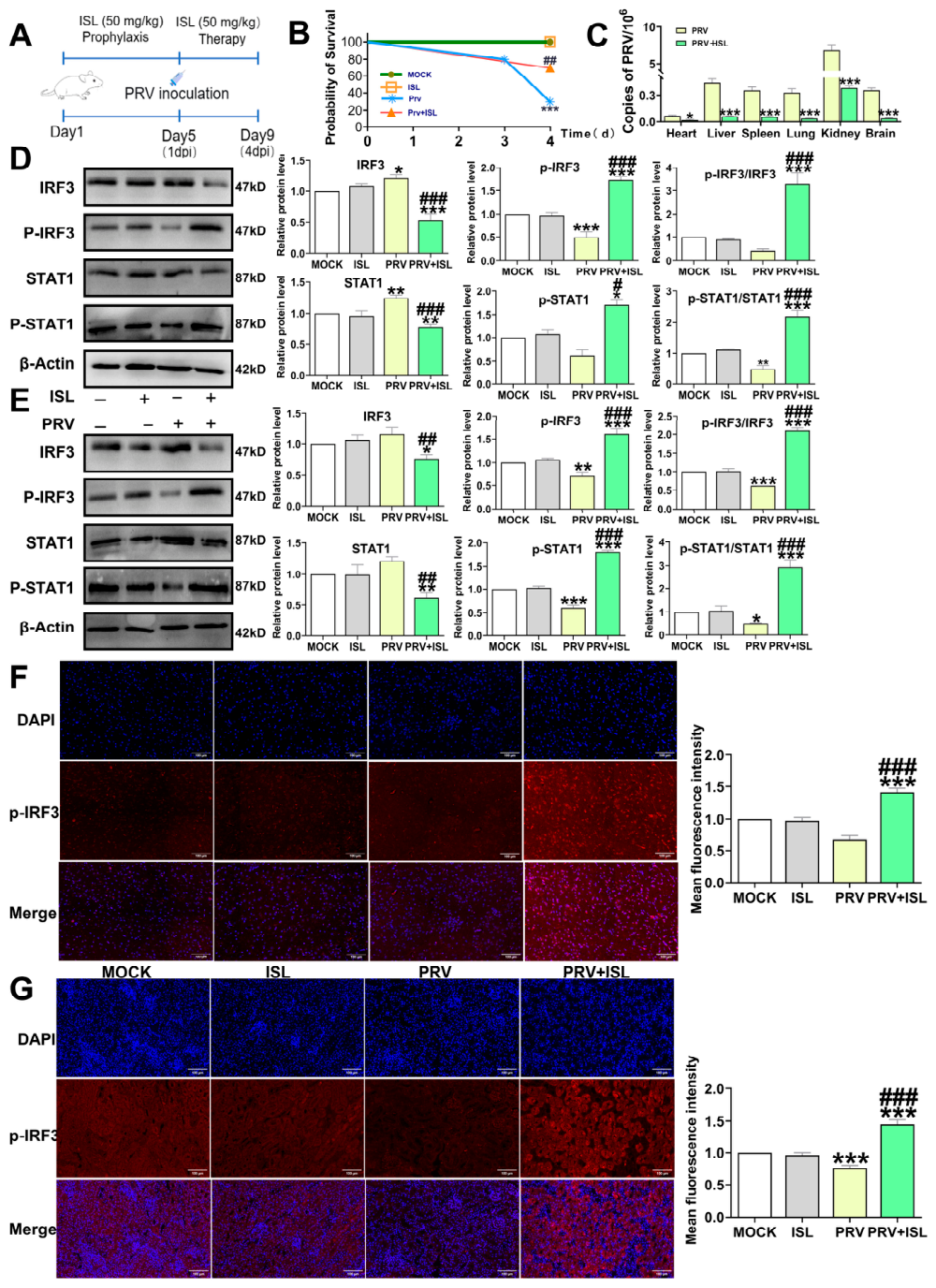
n = 10): an uninfected-untreated group (Mock), an uninfected group treated with isoliquiritigenin (ISL), an infected-untreated group (PRV), and an infected group treated with isoliquiritigenin (PRV + ISL). The Mock group received the vehicle (0.5% CMC-Na) by oral gavage at the same volume and according to the same schedule (once daily for five consecutive days) as the ISL-treated groups. The dose of 50 mg/kg was selected based on preliminary dose-finding experiments, in which 50, 100, and 200 mg/kg were tested; the 50 mg/kg dose exhibited the most favorable balance between antiviral efficacy and safety in mice and was therefore chosen for all subsequent in vivo experiments. All treatments were administered via intragastric gavage. The mice in the isoliquiritigenin-treated groups received isoliquiritigenin via oral gavage for five consecutive days. The treatment protocol consisted of two phases: a preventive phase and a therapeutic phase. During the preventive phase (days 1–5), mice in the ISL and PRV+ISL groups received 50 mg/kg isoliquiritigenin once daily by oral gavage for five consecutive days, while those in the Mock and PRV groups received an equal volume of vehicle (0.5% CMC-Na). On day 5, mice in the PRV and PRV+ISL groups were intraperitoneally challenged with 0.1 mL of PRV at 2 × 10
4 TCID
50, following the protocol established in our previous study [
27]. At this dose, the mice exhibited stable infection; the Mock and ISL groups received an equal volume of vehicle via intraperitoneal injection. After viral challenge, the therapeutic phase was initiated: the ISL and PRV+ISL groups continued to receive the same dose of isoliquiritigenin (50 mg/kg) daily by gavage for an additional 4 days (days 6–9), whereas the Mock and PRV groups received an equal volume of 0.5% CMC-Na, until the endpoint of the experiment. Humane endpoints were strictly defined for early euthanasia of moribund animals. Mice were monitored at least twice daily for signs of morbidity, including severe lethargy, hunched posture, labored breathing, paralysis, and inability to reach food or water. Animals meeting any of these criteria were humanely euthanized by cervical dislocation prior to the endpoint to minimize suffering. Mortality was recorded daily by investigators blinded to the group allocation. At 4 days post-infection (dpi), the mortality rate in the PRV group reached 70%, which exceeded the predefined humane endpoint threshold (≥50%) and prompted early termination of the experiment. All surviving mice in each group were then euthanized by cervical dislocation at this unified endpoint (4 dpi) to ensure consistency in sample collection timing. Tissues from the brain, kidneys, heart, liver, lungs, and spleen were collected, and then subjected to cryogenic homogenization in liquid nitrogen, and total RNA was extracted using the TRIzol method as described above. The total proteins of brain and kidney specimens (approximately 100 mg) were processed according to the protocol stipulated by the manufacturer (Wuhan Boster, AR0101-30, Wuhan, China) for the purpose of Western blotting analysis, which was performed and quantified by operators blinded to the experimental groups.
2.12. Immunofluorescence Staining of Tissue Sections
Brain and kidney specimens were fixed in 4% paraformaldehyde for 24 h. Targeted sections of the brain and kidneys were then excised, dehydrated, embedded in paraffin, and sectioned. After deparaffinization and rehydration, the sections were treated with 0.2% Triton X-100. Antigen retrieval was performed by heating the sections in sodium citrate buffer using a microwave. The sections were then blocked with 3% BSA for 40 min. For immunofluorescence, sections were incubated with anti-p-IRF3 primary antibody (1:200, #29528-1-AP; Bioss, Beijing, China) overnight at 4 °C, followed by incubation with CoraLite594-conjugated secondary antibody (SA00013-4; Proteintech, Wuhan, China; 1:100) for 1 h at room temperature in the dark. Nuclei were counterstained with DAPI. Images were acquired using a fluorescence microscope. IF staining analysis was performed and quantified by operators blinded to the experimental groups.
2.13. Statistical Analysis
All data are expressed as mean ± SD from at least three independent biological replicates. Statistical analyses were performed using GraphPad Prism 9.0. One-way or two-way analysis of variance (ANOVA) was applied as appropriate, followed by Tukey’s post hoc test for multiple comparisons. Differences were considered statistically significant at p < 0.05, p < 0.01, and p < 0.001. For the animal survival experiment, the Kaplan–Meier method with the log-rank (Mantel–Cox) test was used to compare survival curves between groups.
4. Discussion
In the present study, we report that isoliquiritigenin exhibits antiviral activity against PRV both in vitro and in vivo. Mechanistically, we found that PRV infection significantly reduced intracellular cGAMP levels and suppressed the transcriptional expression of key genes including STING, TBK1, IRF3, and IRF7, while isoliquiritigenin treatment effectively reversed these effects and restored cGAMP content. Additionally, isoliquiritigenin treatment enhanced IRF3 and STAT1 phosphorylation and upregulated the expression of multiple ISGs. These findings suggest that isoliquiritigenin may exert its antiviral effects against PRV in a manner associated with modulation of the cGAS/STING and JAK/STAT signaling cascades. Beyond its direct impact on these innate immune cascades, isoliquiritigenin has been reported to exert cytoprotective and immunomodulatory effects via activation of the Nrf2-dependent antioxidant pathway and suppression of the NF-κB-mediated inflammatory response, thereby mitigating BDE-47(2,2′,4,4′-tetrabromodiphenyl ether)-induced oxidative stress, apoptosis, and immune dysfunction [
30]. This suggests that the anti-PRV activity of isoliquiritigenin may arise from the synergistic coordination of multiple signaling pathways, which, together with its intrinsic anti-inflammatory and antioxidant properties, constitutes the molecular basis for its potent antiviral efficacy both in vitro and in vivo.
The antiviral activity of a compound is commonly evaluated by two key parameters: the IC
50 and SI. The IC
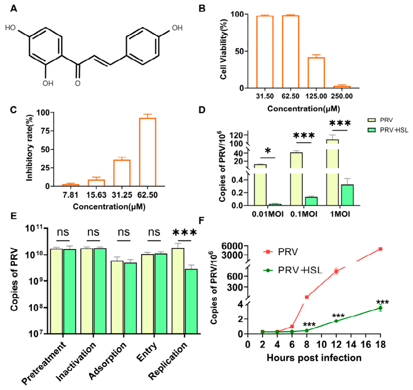
50 of isoliquiritigenin against PRV was determined to be 35.47 μM, with a SI of 3.34. By comparison, a flavonoid mixture derived from
Ocotea notata leaves was reported to exhibit SI values of 5.5 against HSV-1 and 8.5 against HSV-2 [
31], whereas quercetin demonstrated an SI exceeding 20 against HSV-1 [
32]. In this context, the SI of isoliquiritigenin is relatively modest, suggesting a moderate selectivity and a somewhat narrow therapeutic window. In antiviral drug discovery, an SI below 10 generally indicates a narrow therapeutic window, meaning that the effective concentration is close to the cytotoxic concentration. Importantly, the SI is highly dependent on cell type, virus strain, and assay conditions; direct comparisons across studies must be made with caution. In addition, a moderate SI does not preclude further development—natural product leads with SI values in the single digits have been successfully optimized through medicinal chemistry, formulation, or combination strategies. This principle is exemplified by the optimization of betulinic acid, a natural pentacyclic triterpenoid that initially exhibited limited potency and weak selectivity. Through systematic amidation modifications, researchers successfully transformed betulinic acid into a lead candidate with improved selective toxicity (compound 9; HeLa: IC
50 = 5.4 μM, SI > 9.3; MCF-7: IC
50 = 7.0 μM, SI > 7.2). This case provides a compelling precedent for the notion that natural product leads with modest SI values can be advanced through rational structural optimization, supporting the feasibility of similar strategies for isoliquiritigenin [
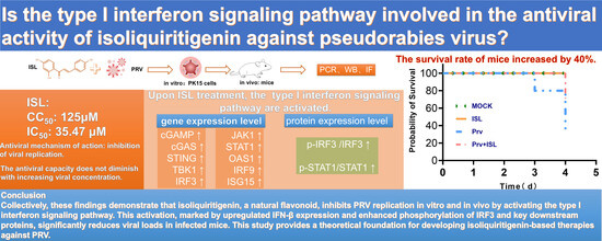
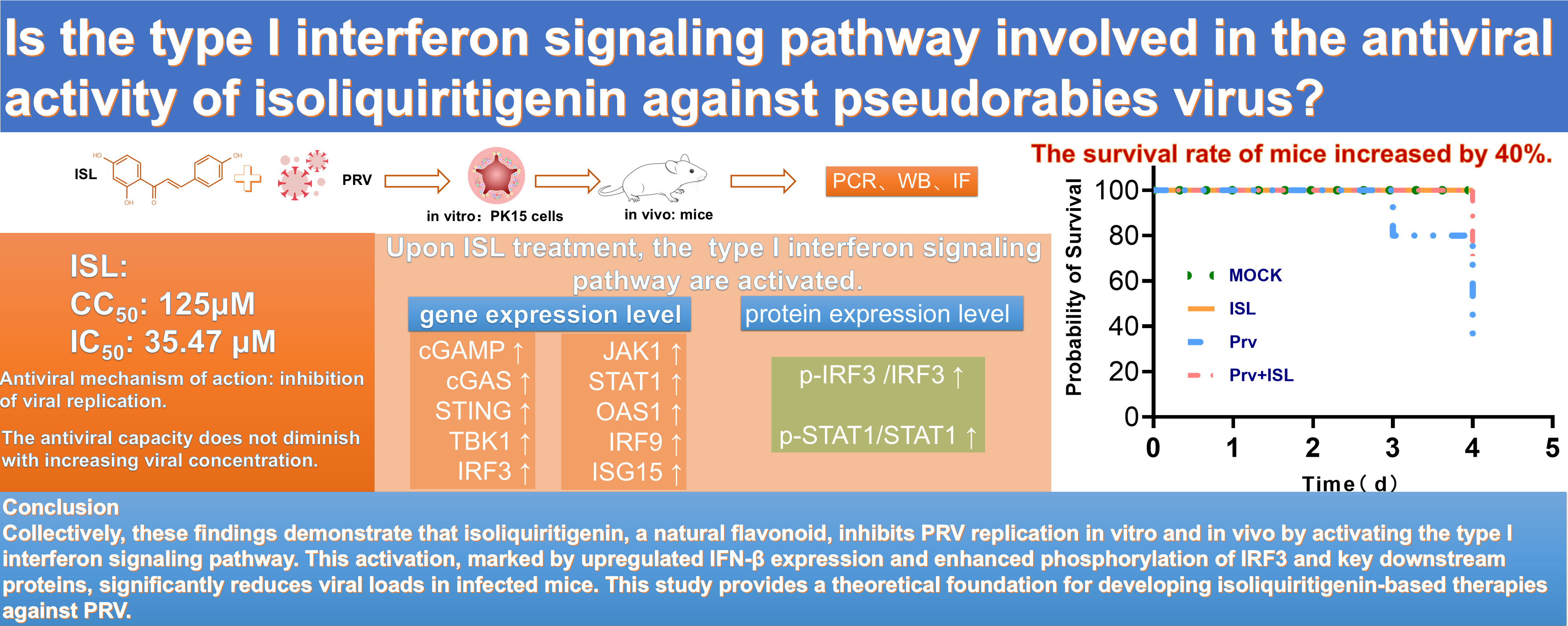
33]. Notably, the compound demonstrated significant in vivo efficacy in our mouse model (a 40-percentage-point reduction in mortality), suggesting that despite the narrow in vitro window, sufficient exposure can be achieved to exert antiviral effects. Since no PRV-specific drug is currently available on the market, a direct head-to-head comparison with an approved antiviral agent is not feasible. However, isoliquiritigenin can be compared with other natural products that possess anti-herpesvirus activity. For instance, resveratrol has been reported to inhibit PRV with an IC
50 of 17.17 μM (SI > 15.31) [], and kaempferol with an IC
50 of 25.57 μM (SI 9.97) [
25], both exhibiting anti-PRV activity in PK-15 cells. In comparison, isoliquiritigenin (IC
50 = 62.5 µM, SI 3.34) shows relatively weaker antiviral activity. This may be due to differences in chemical structure, which could affect its binding affinity to target proteins. Nonetheless, this does not diminish its value as a promising lead compound for anti-PRV drug development. Rather than positioning it as an immediate clinical candidate, isoliquiritigenin should be considered a viable starting point for structural optimization to improve its potency and selectivity. Future efforts in rational derivatization, formulation development, or combination therapy with other antiviral agents are expected to further enhance its therapeutic utility against PRV infection.
The importance of the cGAS/STING pathway in antiviral defense is well documented. Kombu polysaccharide enhances cGAS/STING signaling during HSV-1 infection by stabilizing STING protein through inhibition of autophagic degradation [
34], and cGAS knockout mice exhibit significantly increased susceptibility to HSV-1 infection [
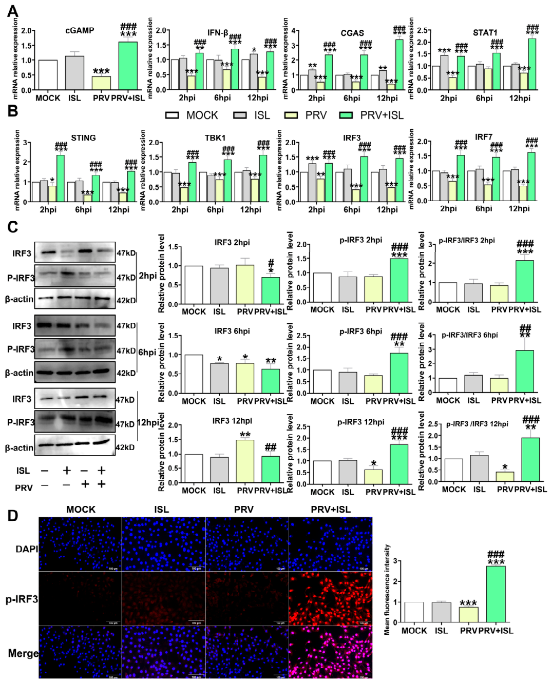
12]. Although these studies were conducted in the context of HSV-1, they underscore the general importance of the cGAS/STING pathway in DNA virus defense. Our results indicate that cGAS/STING signaling activity, as reflected by the cGAMP-stimulated IFN-β response, is significantly reduced following PRV infection, suggesting that PRV suppresses type I interferon pathway activation. However, isoliquiritigenin treatment not only enhanced this cGAMP-mediated IFN-β response but also elevated the transcriptional levels of STING, TBK1, and IRF3. It is worth noting that some of the observed pathway effects may be indirect, resulting from reduced PRV replication. However, in the PRV+ISL group, the expression levels of cGAS and STAT1 were even higher than those in the Mock group (
Figure 2A). This suggests that ISL may exert dual effects: inhibiting viral replication and positively modulating the cGAS/STING and JAK/STAT pathways. Nevertheless, our current data only support a correlation between ISL treatment and pathway activation. The direct molecular targets of ISL remain unclear. Future studies using binding assays and gene knockout experiments are needed to verify causality. In addition, more direct evidence is required to determine whether ISL directly interferes with PRV immune evasion.
PRV has evolved multiple strategies to antagonize this pathway. The envelope protein UL13 recruits the E3 ubiquitin ligase RNF5, promoting STING degradation through non-canonical K27/K29 ubiquitination [
35]. Another viral protein, US2, facilitates K48-linked ubiquitination-dependent degradation of STING by recruiting the E3 ubiquitin ligase TRIM21 [
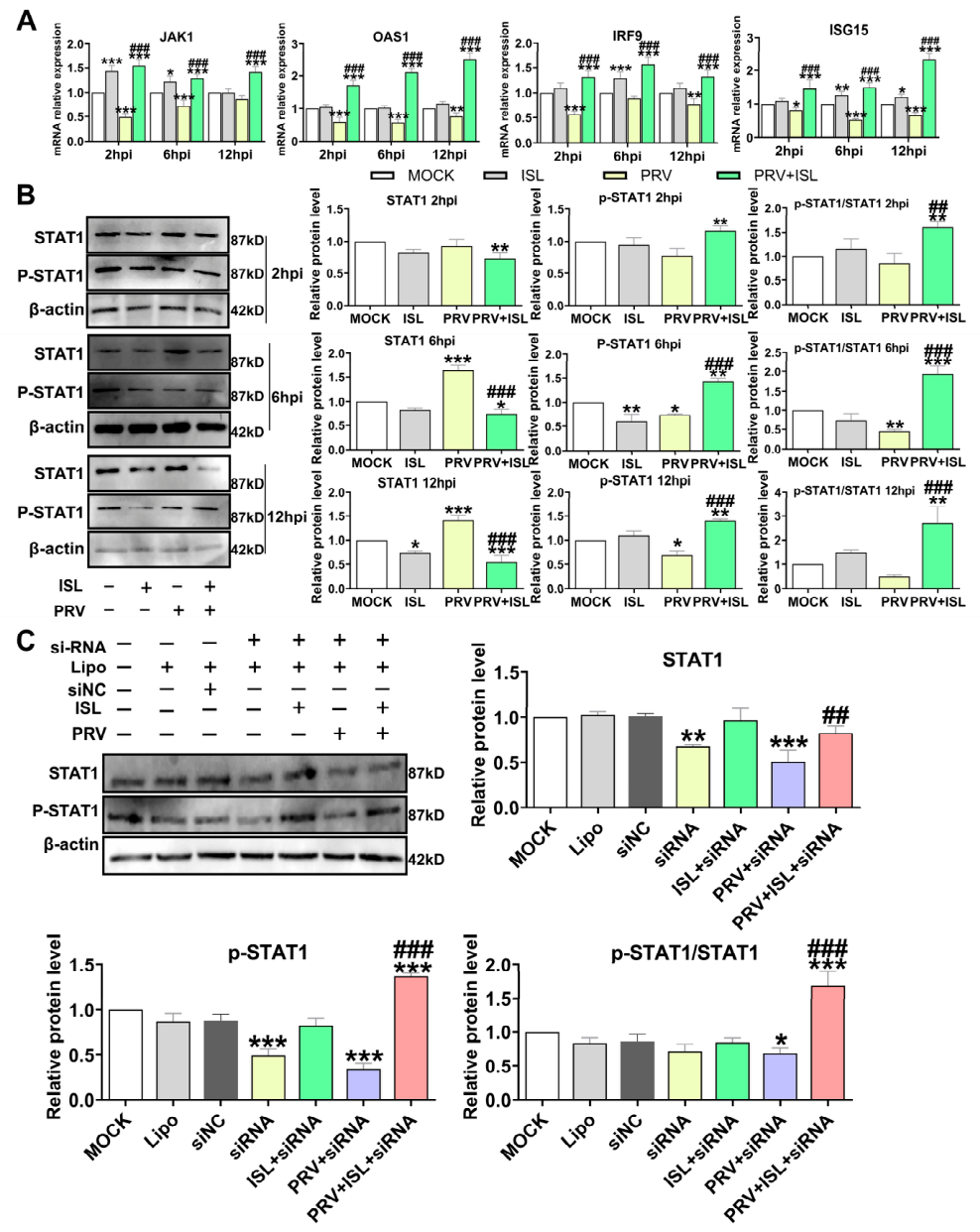
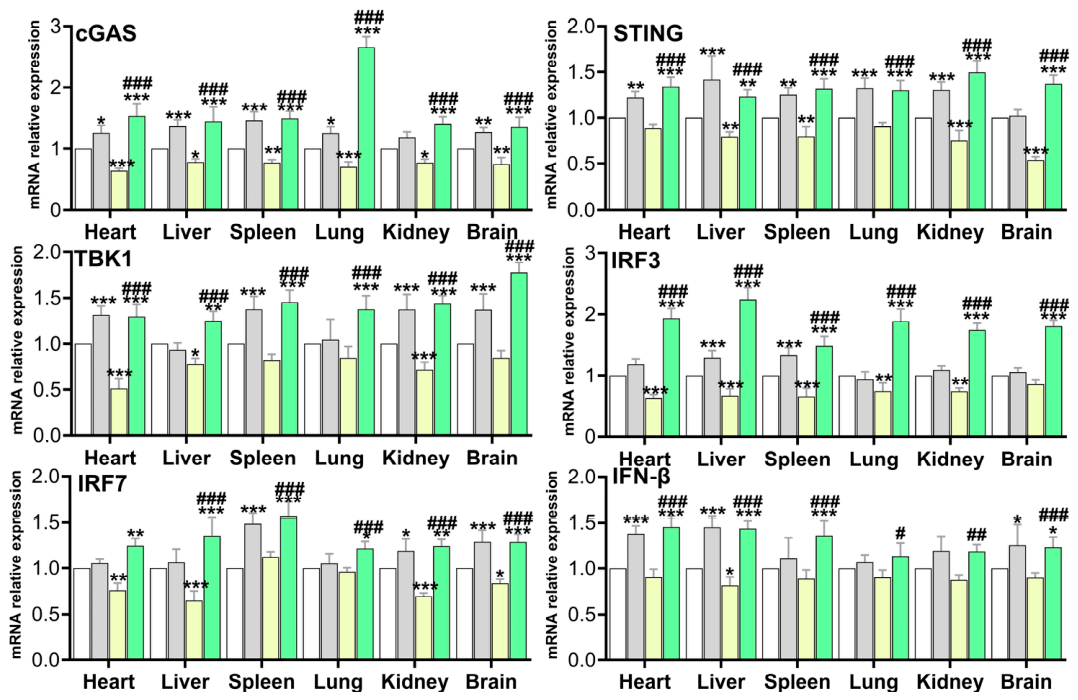
36]. Our results show that isoliquiritigenin treatment reverses PRV-induced transcriptional suppression of STING, suggesting that it may counteract PRV immune evasion by maintaining STING expression. Furthermore, PRV infection significantly suppressed the mRNA expression of downstream signaling genes, including TBK1, IRF3, and IRF7, while isoliquiritigenin treatment effectively reversed this suppression, restoring their transcript levels to values significantly higher than those of the virus-only control. At the protein level, isoliquiritigenin also promoted IRF3 phosphorylation, with levels markedly rebounding and even becoming significantly enhanced following treatment. Collectively, these results suggest that isoliquiritigenin modulates the PRV-suppressed cGAS/STING pathway by upregulating STING, TBK1, and IRF3 mRNA expression, while also promoting IRF3 phosphorylation at the protein level, together contributing to counteracting PRV-mediated suppression of this pathway.
Beyond the cGAS/STING pathway, we also examined whether isoliquiritigenin affects downstream JAK/STAT signaling. Notably, exogenous expression of IFN-α or IFN-β in PK-15 cells has been shown to significantly inhibit PRV replication while maintaining cellular morphology and strongly inducing key effector molecules such as ISG15 [
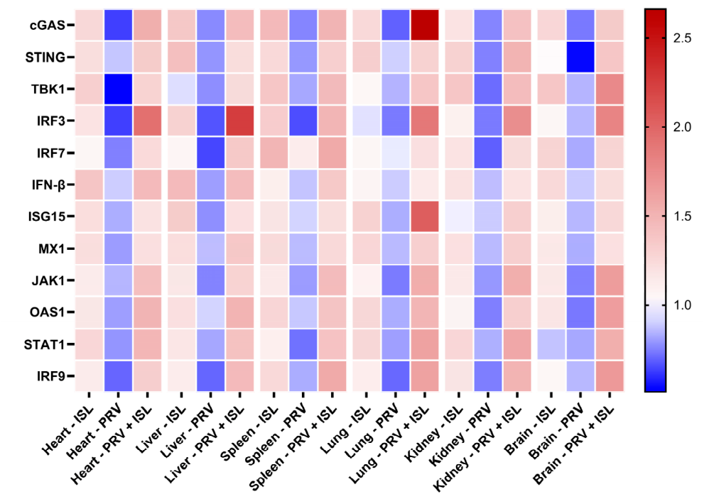
37]. In the present study, through dual validation in cellular and animal models, isoliquiritigenin treatment significantly enhanced ISG15 expression under PRV infection. This finding was consistent across both in vitro and in vivo settings, further supporting the role of isoliquiritigenin in activating the type I interferon pathway.
It has been established that PRV directly blocks downstream signaling cascades triggered by secreted type I interferons. The viral protein UL50, a deoxyuridine triphosphate hydrolase (dUTPase), targets interferon receptors by inducing IFNAR1 degradation via the lysosomal pathway, thereby directly blocking exogenous interferon signaling. This results in inhibition of STAT1 phosphorylation and subsequent ISG expression [
38]. Furthermore, PRV potently inhibits JAK/STAT signaling by specifically interfering with JAK kinase or STAT protein functions through its encoded viral proteins [
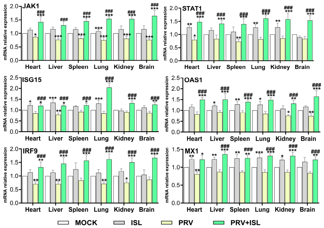
39]. Our findings are consistent with and extend these observations, demonstrating that isoliquiritigenin treatment significantly restores PRV-inhibited STAT1 phosphorylation, upregulates JAK1 and STAT1 mRNA expression, and promotes transcription of multiple key genes. To further investigate the potential role of STAT1 in the antiviral mechanism of isoliquiritigenin, we performed siRNA-mediated knockdown of STAT1 in Vero cells prior to isoliquiritigenin treatment and PRV infection. Vero cells are naturally deficient in type I interferon production, allowing us to effectively exclude the influence of upstream interferon signaling. As expected, STAT1 siRNA significantly reduced STAT1 protein expression compared with the blank control. Notably, even under STAT1-deficient conditions, isoliquiritigenin treatment still partially restored STAT1 phosphorylation in PRV-infected cells. This indicates that isoliquiritigenin can activate the JAK1/STAT pathway, thereby contributing to its antiviral efficacy against PRV. Collectively, these findings suggest that isoliquiritigenin may counteract PRV immune evasion through stimulation of the JAK/STAT signaling pathway.
To further verify whether isoliquiritigenin inhibits PRV proliferation by modulating the type I interferon response, a PRV-infected mouse model was established. Treatment with isoliquiritigenin improved clinical signs in infected mice, as evidenced by reduced incidence of morbidity (including lethargy, hunched posture, and labored breathing) compared to the PRV-only group, and effectively increased the survival rate. This mechanism is similar to that of rosmarinic acid, a natural compound that activates the cGAS/STING pathway, reverses PRV-induced inhibition, induces IFN-β expression, and thereby suppresses viral replication, alleviates inflammation, and reduces mortality both in vitro and in vivo [
40]. Similarly, myricetin has been shown to impede PRV replication through the EGFR/PI3K/Akt and type I interferon signaling pathways [
41,
42,
43]. These findings are consistent with our observations that isoliquiritigenin activates type I interferon signaling. The antiviral effects of both compounds are attributable to their capacity to enhance the host innate immune response and support the concept that “targeting host immune pathways” represents an effective strategy against PRV infection.
The present study further supports this notion, as isoliquiritigenin effectively reduces viral load in the heart, liver, spleen, lung, kidney, and brain tissues of PRV-infected mice while concurrently upregulating the transcriptional levels of genes associated with the cGAS/STING and JAK/STAT signaling pathways in these tissues. Furthermore, isoliquiritigenin enhanced p-IRF3 protein expression in brain and kidney tissues, indicating systemic activation of the cGAS/STING signaling pathway in mice. These findings suggest that isoliquiritigenin treatment may enhance host resistance to PRV at the tissue level, although the contribution of this pathway activation to tissue protection requires further investigation.
Several limitations of this study should be acknowledged. (1) An exogenous cGAMP positive control was not included in the cGAMP assay, so the IFN-β signal should be interpreted as an indirect functional readout. (2) STAT1 knockdown was performed in Vero cells rather than PK-15 cells, which are the natural host for PRV; Vero cells reduce endogenous interferon interference but do not fully mimic the natural infection microenvironment. (3) The evidence for cGAS/STING involvement remains correlative, and additional loss-of-function experiments such as STING or IRF3 knockdown/knockout are needed to provide definitive proof. (4) The 50 mg/kg dose was selected based on preliminary experiments, but pharmacokinetic and toxicity data for this dose are lacking. (5) Tissue molecular data were obtained from both moribund and surviving animals using random sampling; however, usable tissues were not available from all deceased animals, particularly in the PRV group, which had the highest mortality. Surviving animals in this group may represent a less severely affected subpopulation, but results from moribund animals were consistent with those from surviving groups, which partially mitigates the concern that the findings are solely driven by survivorship bias. (6) Although viral loads in brain tissues were reduced after ISL treatment, drug concentration in the brain was not measured, and it remains unclear whether ISL crosses the blood–brain barrier or acts indirectly through peripheral immunomodulation. (7) No obvious toxicity was observed in the ISL-only group, but histopathological analysis was not performed. (8) All in vivo efficacy data were obtained from mice; the PRV infection model in mice has been validated in multiple studies [
44] and is generally accepted in the field, but validation in the natural host (pigs) is essential before translation to clinical or swine industry applications.




 ,
,