Article

Prevalence and Risk Factors of Mycoplasma Hyopneumoniae in Swine Farms, Mainland China, 2003–2024: A Meta-Analysis

1
College of Veterinary Medicine (Institute of Comparative Medicine), Yangzhou University, Yangzhou 225009, China
2
Jiangsu Co-Innovation Center for Prevention and Control of Important Animal Infectious Diseases and Zoonoses, Yangzhou 225009, China
3
Independent Researcher, New York, NY 11355, USA
*
Author to whom correspondence should be addressed.
These authors contributed equally to this work and should be considered co-first authors.
Vet. Sci. 2025, 12(9), 863; https://doi.org/10.3390/vetsci12090863
Submission received: 26 July 2025 / Revised: 19 August 2025 / Accepted: 29 August 2025 / Published: 5 September 2025
(This article belongs to the Section Veterinary Microbiology, Parasitology and Immunology)

Simple Summary

Mycoplasma hyopneumoniae (M. hyopneumoniae) is a major respiratory pathogen in pigs, causing significant economic losses to swine production. This study systematically evaluated its prevalence and risk factors in swine farms across mainland China from 2003 to 2024. The pooled prevalence was 33.4%, with higher rates in clinically affected farms (52.9%) than subclinical ones (11.5%). Infection rates varied by region, age, farm scale, diagnostic method, and sample type. This research highlights the need for region-specific biosecurity, vaccination strategies, and diagnostic improvements to better control the disease and reduce its impact on the swine industry.

Abstract

This is the first systematic review and meta-analysis on the prevalence and risk factors of M. hyopneumoniae infection in swine farms across mainland China from 2003 to 2024. A total of 54 eligible cross-sectional studies were analyzed by stratifying farms as subclinically or clinically infected. The overall pooled prevalence of M. hyopneumoniae was estimated as 33.4%, with clinical infection farms showing a significantly higher prevalence (52.9%) than subclinical farms (11.5%). Subgroup analyses revealed significant variations in infection rates based on age, sampling year, geographic region, farming scale, season, sampling type, and diagnostic method. Small-scale farms, farms with breeding swine, and farms in the Northwest region showed the highest infection rates. Diagnostic methods and sampling types also significantly influenced detection rates. Sensitivity analyses confirmed the robustness of the results, while publication bias was addressed using the Trim-and-Fill method. To effectively reduce the burden of M. hyopneumoniae in the swine industry in mainland China, future efforts should prioritize enhanced biosecurity, improved diagnostic accuracy, and region-specific vaccination and management strategies.

1. Introduction

M. hyopneumoniae is a key respiratory pathogen in swine and the causative agent of a common, widely distributed disease that occurs year-round. It is restricted to swine and is present in all major swine-raising countries, causing tremendous global financial losses to the swine industry due to its role in porcine respiratory disease complex (PRDC) [1,2]. The primary losses stem from the costs associated with treatment and vaccination, as well as reduced performance and higher mortality rates resulting from secondary infections [3].
M. hyopneumoniae, a species within the genus Mycoplasma, lacks a cell wall, which enhances its tolerance to external environmental conditions and enables its survival under various conditions, particularly in dry and warm environments. Additionally, it exhibits intrinsic resistance to many commonly used antibiotics, such as penicillin [4,5]. M. hyopneumoniae is primarily transmitted via airborne routes, with an increased risk of spread in high-density farming environments. Prolonged exposure to environmental stressors, including overcrowding and poor ventilation, can exacerbate disease severity. Additionally, direct contact between swine serves as a significant mode of transmission [6]. As M. hyopneumoniae infection progresses, it stimulates a sustained immune response that contributes to chronic respiratory disease, characterized by persistent inflammation of the airways. In parallel, the pathogen can interfere with innate defense mechanisms such as mucociliary clearance, which facilitates long-term colonization. This dual effect leads to a significant decline in production performance and immune function, accompanied by clinical symptoms such as coughing, tachypnea, and elevated body temperature [7]. Moreover, the chronic and recurrent nature of Mycoplasma infections presents significant challenges to disease prevention and control in swine farms and has led to substantial economic losses in the global swine industry [8,9,10]. M. hyopneumoniae infection is highly prevalent in domestic swine populations and has been reported in nearly all swine-producing regions, with infection rates ranging from 38% to 100% [11].
A peculiar feature of pig farming in mainland China is the coexistence of modern intensive production systems with a large number of smallholder and backyard farms. Currently, Mycoplasmal Pneumonia of Swine (MPS) is widely prevalent in swine farms across mainland China, typically occurring in a sporadic manner. In certain regions with suboptimal farming conditions and unfavorable climates, MPS may exhibit an endemic trend [12,13]. Due to characteristics such as the prolonged infection period, variable incubation time, potential for long-distance transmission, and antimicrobial resistance, complete eradication of the disease from swine farms remains challenging. The current control measures for M. hyopneumoniae infections in swine include vaccination and antibiotic treatment. Vaccination is a key strategy in managing the disease; however, its effectiveness can vary due to factors such as the alignment between vaccine strains and field strains, as well as the immunization protocols used [14]. Although antibiotic treatment can help control the infection to some extent, it also raises concerns about antimicrobial resistance [15,16]. Additional control measures involve optimizing management practices and strengthening biosecurity [1,8].
Given the significant differences in the clinical manifestations and epidemiological characteristics of M. hyopneumoniae infection in swine farms across mainland China, this study categorizes farms into two groups: subclinical infection farms and clinical infection farms. Clinical infection farms refer to herds in which M. hyopneumoniae infection is present but without evident clinical respiratory signs; infection is usually detected through laboratory tests or by characteristic lung lesions at slaughter. By contrast, subclinical infection farms are those in which pigs show no clinical signs and no evidence of infection was detected during sampling. This stratification aims to more accurately capture variations in prevalence estimates across different farm types.
The objectives of this study are to determine the prevalence of M. hyopneumoniae across different regions and farm types in mainland China, identify associated risk factors, and highlight high-risk areas to inform targeted prevention and control measures. Additionally, the study aims to provide evidence-based guidance for optimizing farm management and biosecurity practices, considering farm scale, pig age, and regional context, thereby reducing infection risk and supporting effective, differentiated control strategies.

2. Materials and Methods

2.1. Search Strategy

This study adhered to the PRISMA (Preferred Reporting Items for Systematic Reviews and Meta-Analyses) guidelines [17]. To ensure the inclusion of all relevant information in the analysis, the PRISMA checklist was applied (Supplementary Data S1).
All the literature conducted on the prevalence of M. hyopneumoniae in swine from January 2003 to January 2025 was searched using four databases, including English databases (PubMed and Web of Science) and Chinese databases (CNKI and Wan Fang databases). The Chinese search terms were “Mycoplasma pneumonia” and “Pig”. The English search terms were “Mycoplasma pneumonia”, “Pig” or “Swine”, and “China”. Additionally, the references of the published papers were checked to retrieve papers that were not found via database searching.

2.2. Inclusion and Exclusion Criteria

The full-text articles were retrieved, and studies were included in the review based on the following criteria: 1. Study location: Mainland China. 2. Study subjects: Swine. 3. Study design: Cross-sectional studies. 4. Detection methods: Clearly stated with a detailed description of procedures. 5. Sample size: Greater than 30 with accurate data. 6. Sample background: Information must be fully described and documented. 7. Publication status: No duplicate publications.
The literature review was conducted independently by two reviewers. In cases where the results were inconsistent, discrepancies were resolved either by consulting a third reviewer or through discussion and negotiation. Articles that did not meet the inclusion criteria were excluded.
The screened literature for M. hyopneumoniae prevalence was carefully read, and the following information was collected: the first author, year of publication, sampling year, geographic region, age, diagnostic method, sampling type, farming scale, total sample size, and number of positive samples. Although studies were identified from most provinces, certain regions such as Tibet, Ningxia, and Hainan were scarcely represented, reflecting the limited availability of published data.

2.3. Quality Assessment and Data Extraction

After removing duplicate records and review articles, the titles and abstracts of all remaining studies were screened to preliminarily exclude those unrelated to the prevalence of M. hyopneumoniae in swine. The full texts were then reviewed to determine eligibility based on the following criteria: (1) the study was conducted in mainland China; (2) the study population consisted of swine; (3) the article reported a cross-sectional study; (4) the diagnostic method and procedures were clearly described; (5) the number of clinical samples was greater than 10, and accurate data were available; (6) the background information of the samples was complete. Literature screening was independently performed by two reviewers. Studies not meeting the inclusion criteria were excluded. Disagreements or uncertainties regarding eligibility were resolved by a third reviewer or through discussion.
Cross-sectional studies in animals differ from randomized clinical trials. Their systematic evaluation methods are not yet fully established, and these studies typically lack control groups. Therefore, the systematic evaluation methods for animal cross-sectional studies were adapted from the Cochrane quality assessment framework. The quality of the studies was assessed by evaluating the following six criteria using RevMan 5.4. A score of 2 was assigned if a study met a given criterion, 1 if the criterion was unclear or not explicitly described, and 0 if it was not met. The quality assessment criteria included the following: 1. whether all relevant details were reported; 2. whether the study provided a detailed description of experimental procedures; 3. whether the sampling process was conducted randomly; 4. whether the study spanned multiple time points; 5. whether statistical analyses were performed; 6. whether missing data were reported and addressed; 7. whether the study evaluated potential risk factors. Each criterion was assigned equal weight in the overall quality assessment.
Studies with a total quality evaluation score below 7 points were excluded from the statistical analysis.

2.4. Statistical Analysis

Data processing was performed using Stata software (version 16), and the double-arcsine transformation (PFT) was applied to normalize the data distribution [18,19]. The heterogeneity of M. hyopneumoniae infection rates across the included studies was assessed using the I2 and Q tests. If p ≥ 0.10 and I2 < 50%, it suggests low or no statistical heterogeneity among the study effect sizes, and a fixed-effects model should be applied for the analysis. Conversely, if p < 0.10 and I2 ≥ 50%, it indicates significant statistical heterogeneity, warranting the use of a random-effects model to combine the estimated infection rates and their 95% confidence intervals (CIs) [20]. To determine whether publication bias exists in this study, a funnel plot was drawn, and publication bias is assessed using the Begg test or Egger test. If publication bias is detected, the Trim-and-Fill method can be applied for correction, followed by recalculating the overall effect size [21,22]. Furthermore, sensitivity analysis is performed to verify its robustness.
Subgroup analyses were performed to explore the sources of heterogeneity, followed by meta-regression analyses to investigate the risk factors for M. hyopneumoniae infection. The subgroups considered in the analyses included year of sample collection, sampling season, diagnostic methods, age group of swine, farming scale (large-scale, medium-scale, and small-scale), and farm type (subclinical infection farms and clinical infection farms).

3. Results

3.1. Literature Search Results

A total of 1676 articles were retrieved from four Chinese and English databases based on the keywords, including 54 from the China National Knowledge Infrastructure (CNKI), 1527 from Wanfang Data, 58 from Web of Science, and 37 from PubMed. After eliminating duplicate studies and following the screening process outlined in the Cochrane Handbook, articles were assessed based on their titles, abstracts, full texts, and predefined exclusion criteria. Ultimately, 54 articles were included for data analysis. The screening process and results are presented in Figure 1.
The data screened from the 54 studies on M. hyopneumoniae prevalence are shown in Table 1.

3.2. Data Extraction and Quality Analysis

The quality of each study was assessed based on a checklist of five items, and the corresponding scores are illustrated in Supplementary Data S2. Among the 54 included studies, 42 were rated as high quality, 12 as medium quality, and none as low quality.

3.3. Heterogeneity Analysis

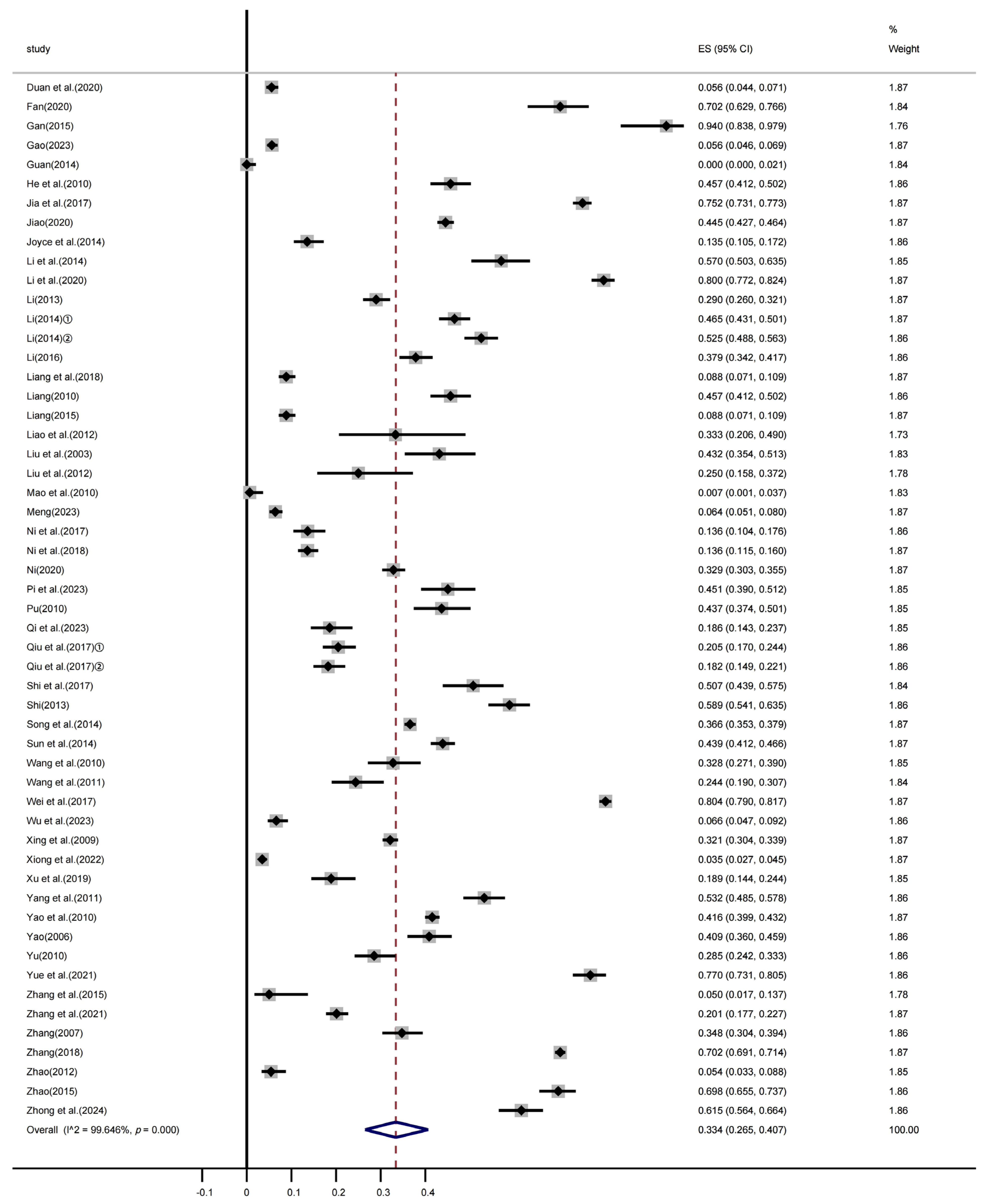
A meta-analysis was conducted based on data extracted from the final 54 included studies, revealing significant heterogeneity among the studies (χ2 = 14984.484, p < 0.001; I2 = 99.646%). A random-effects model was used to calculate the pooled prevalence, resulting in an estimated prevalence of 33.4% (95% CI: 26.5–40.7%). Figure 2 presents the forest plot of M. hyopneumoniae prevalence in China from 2002 to 2024.
The points represent the prevalence rates, and the horizontal lines represent the 95% confidence intervals corresponding to the effect sizes (ESs). The diamond shape at the bottom indicates the pooled effect size.
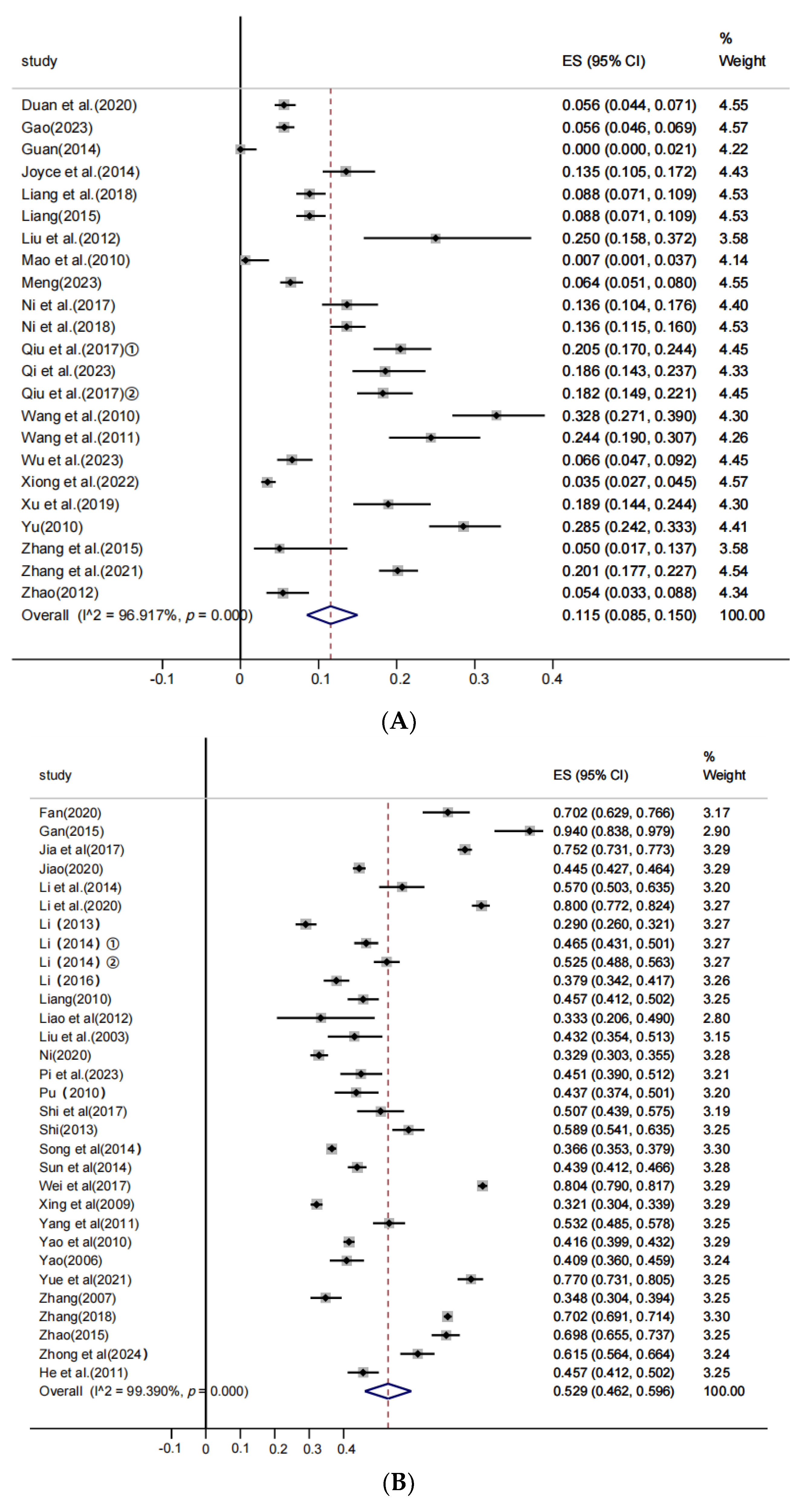
The included studies were further stratified into subclinical infection farms and clinical infection farms. Based on 23 studies of subclinical infection farms, significant heterogeneity was observed (χ2 = 713.517, p < 0.001; I2 = 96.917%), and the estimated pooled prevalence calculated by the random-effects model was 11.5% (95% CI: 8.5–15.0%). Figure 3A shows the forest plot for M. hyopneumoniae prevalence in subclinical infection farms. Data from 31 studies on clinical infection farms also demonstrated significant heterogeneity (χ2 = 4917.036, p < 0.001; I2 = 99.390%). The pooled prevalence estimated using the random-effects model was 52.9% (95% CI: 46.2–59.6%). Figure 3B depicts the forest plot of M. hyopneumoniae prevalence in clinical infection farms.

3.4. Subgroup Analysis

Subgroup analysis was conducted to explore potential sources of heterogeneity, with results stratified by age, sampling year, geographic region, farming scale, season, sampling type, and diagnostic method, as presented in Table 2.
The possible causes of M. hyopneumoniae infection heterogeneity were analyzed. The studies were divided into subgroups on the basis of study age, sampling year, geographic region, farming scale, season, sampling type, and diagnostic method.
In clinically infected swine farms, significant differences in M. hyopneumoniae prevalence were observed among different age groups (p = 0.005). The infection rates, from highest to lowest, were as follows: breeding swine (61.7%), fattening swine (49.1%), suckling swine (38.0%), nursery swine (36.1%), and growing swine (32.7%). In contrast, no significant age-related differences were found in subclinically infected farms (p = 0.606), with prevalence rates of 13.7% in suckling swine, 20.7% in nursery swine, 11.7% in growing swine, 16.9% in fattening swine, and 17.2% in breeding swine. The overall pooled prevalence was estimated at 39.80% (95% CI: 34.1–45.6%).
In the subgroup analysis by sampling year, the prevalence of M. hyopneumoniae in clinically infected swine farms showed an increasing trend over time, with infection rates of 49.7% before 2013, 54.6% during 2014–2018, and 56.2% during 2019–2024. However, these differences were not statistically significant. In subclinically infected swine farms, the prevalence remained relatively stable across the three periods, ranging from 10% to 12%, with no significant differences observed.
In the regional subgroup analysis, significant differences in M. hyopneumoniae prevalence were observed among clinically infected swine farms (p = 0.005). The highest prevalence was reported in the Northwest region (64.2%), followed by the Qinghai–Tibet region (58.9%), the Northern region (55.7%), and the Southern region (47.4%). In subclinically infected swine farms, no significant regional differences were found (p = 0.272), with prevalence rates of 9.4% in the Northern region, 9.6% in the Southern region, and 11.8% in the Qinghai–Tibet region. Due to the availability of only one study from the Northwest region, it was excluded from the subclinical analysis. Overall, the prevalence in subclinically infected farms was significantly lower than that in clinically infected farms across all regions.
In the subgroup analysis based on farming scale, significant differences in M. hyopneumoniae prevalence were observed among farms of different scales (p = 0.012). The highest infection rate was observed in small-scale farms (≤100 swine) at 35.7%, followed by medium-scale farms (100–500 swine) at 27.9%, while large-scale farms (>500 swine) had the lowest prevalence at 2.3%.
Seasonal subgroup analysis showed no statistically significant differences in M. hyopneumoniae prevalence across seasons in either subclinically infected farms (p = 0.127) or clinically infected farms (p = 0.105). In subclinically infected farms, the prevalence was 16.6% in spring, 4.4% in summer, 5.0% in autumn, and 9.4% in winter. In clinically infected farms, the corresponding rates were 54.7% in spring, 36.0% in summer, 65.1% in autumn, and 47.1% in winter.
The subgroup analysis based on sampling type revealed significant differences in clinically infected swine farms (p = 0.033). The highest prevalence was observed in tissue samples (56.8%), followed by serum samples (44.6%), with the lowest in various swab samples (35.4%). In contrast, no significant differences were found among sampling types in subclinically infected farms (p = 0.513), where serum samples showed the highest prevalence (13.7%), followed by swab samples (10.8%), with the lowest in tissue samples (8.3%).
The subgroup analysis based on diagnostic methods revealed substantial differences in M. hyopneumoniae prevalence (p = 0.000). The infection rate detected by antibody-based methods was 40.2%, while antigen-based methods yielded a lower prevalence of 20.2%. The highest prevalence was observed with histopathological examination, primarily based on pathological tissue samples, at 63.1%. Diagnostic heterogeneity should therefore be considered when interpreting prevalence estimates, and future studies would benefit from standardized, validated protocols. In addition, slaughter lesion scoring data from Chinese studies indicate that using the Combined Lung and Pleural (CLP) scoring method on 12,630 lungs across 247 batches reported median bronchopneumonia lesions in approximately 59.7% of lungs, with an average affected lung surface area of 3.66%, a bronchopneumonia lesion area of 5.8%, a scar tissue proportion of 19.8%, pleuritis on apical and cardiac lobes at 0.62%, diaphragmatic pleuritis at 4.07%, and an APPI of 0.12 [77].

3.5. Publication Bias and Sensitivity Analysis

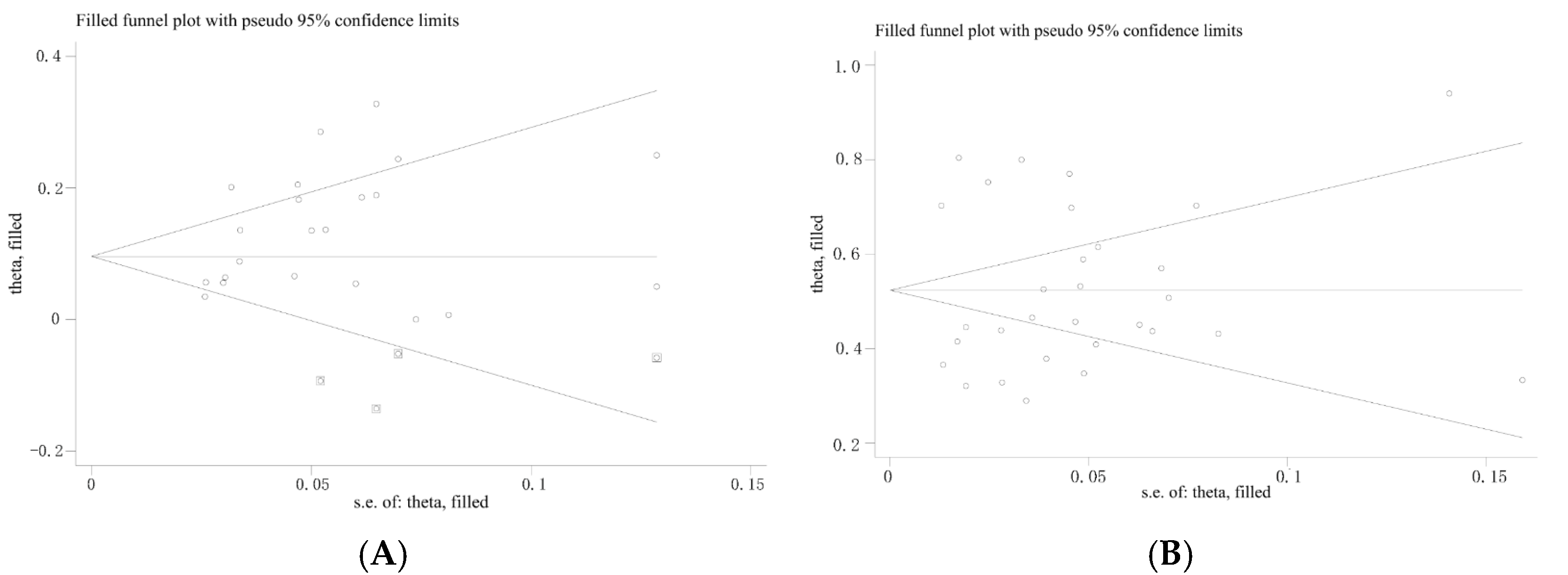
Funnel plots were used to assess potential publication bias among the included studies. Both the funnel plot for subclinically infected swine farms (Figure 4A) and that for clinically infected swine farms (Figure 4B) exhibited noticeable asymmetry (represented by dots), indicating a potential presence of publication bias in the included literature.
Egger’s test was further applied to evaluate publication bias among the included studies. The results indicated a significant publication bias (p = 0.050, Figure 5A; p = 0.000, Figure 5B) [78].
Subsequently, the Trim-and-Fill method was employed to further assess the impact of publication bias on the results [21,22]. After adding six imputed studies for subclinically infected swine farms and none for clinically infected swine farms, the meta-analysis was re-conducted, yielding an adjusted p-value of 0.000 (Figure 6). The heterogeneity remained significant, indicating that the corrected results are robust.
In this study, the sensitivity analysis (leave-one-out sensitivity analysis) revealed that omitting any single study did not substantially affect the pooled effect size or its 95% confidence interval (Figure 7). This indicates that the meta-analysis results are not overly influenced by any individual study. The overall consistency of the estimates across all iterations supports the robustness and reliability of the findings.

4. Discussion

M. hyopneumoniae prevalence in domestic swine herds varies widely across countries that employ modern swine production systems, ranging from 30% to 80%, depending on the region and diagnostic methods used. In Spain and Portugal, 31% of slaughtered swine exhibited lung lesions compatible with the pathogen, and 24.2% tested positive by PCR [79]. In Argentina, molecular detection revealed a prevalence of 48% in fattening swine [80]. In Belgium and the Netherlands, infection rates among young swine ranged from 7.1% to 10.9% [81]. Recent surveillance in the United States has shown that although the infection rate in breeding herds decreased from 21.75% in 2021 to 13.5% in 2022 due to enhanced vaccination and biosecurity measures, a resurgence was observed in growing swine during the wean-to-finish phase in 2024 [82]. As of 2024, M. hyopneumoniae remains a major concern in China’s swine industry. A study analyzing 989 samples from 27 provinces between 2018 and 2020 reported an increasing positivity rate from 7.2% in 2018 to 18.4% in 2019, and 43.8% in 2020 [72]. Nevertheless, as the distribution of samples across provinces was uneven and the sample size in several provinces was relatively limited, these data should be interpreted with caution, since they may not fully reflect the true prevalence at the provincial level. Compared with these countries, mainland China shows a relatively higher overall burden of M. hyopneumoniae, particularly in small-scale farms and provinces with harsher climates such as the Northwest and Qinghai–Tibet Plateau. These international comparisons indicate that while the general epidemiological patterns of M. hyopneumoniae are broadly similar worldwide, the scale, persistence, and regional heterogeneity of infection in mainland China reflect unique challenges that require region-specific control strategies.
Subclinical infections, often asymptomatic, may evade detection while still contributing to disease transmission. In contrast, clinically affected farms exhibit overt respiratory symptoms, resulting in higher detection rates. Given the marked differences in clinical manifestations and epidemiological characteristics of M. hyopneumoniae infections across swine farms in mainland China, this study stratified farms into subclinical and clinical infection categories to better reflect infection dynamics and improve the accuracy of pooled prevalence estimates. This stratification not only enhances epidemiological insight but also informs the development of targeted control strategies. As highlighted by [83,84], classification based on infection status is essential for effective disease surveillance and intervention planning. By adopting this approach, our study provides a more nuanced understanding of M. hyopneumoniae prevalence in both subclinical and clinical settings.
To the best of our knowledge, this is the first systematic review and meta-analysis to evaluate the pooled prevalence of M. hyopneumoniae in mainland China. A comprehensive analysis of studies published between 2003 and 2024 was conducted, and 54 eligible articles were included. The aggregated data suggest that the estimated overall prevalence of M. hyopneumoniae infection in swine farms in mainland China is 33.4%. Stratified analysis indicates prevalence rates of 11.5% in subclinically infected farms and 52.9% in clinically infected farms.
The infection rates of M. hyopneumoniae in clinically infected swine farms were significantly higher than those in subclinically infected farms across all age groups. In clinical farms, the highest prevalence was observed in breeding and fattening swine (>49%), which was significantly greater than in the other three age groups. This suggests an age-related increase in M. hyopneumoniae infection, with breeding and fattening swine identified as high-risk groups. Previous studies have demonstrated a similar trend. A large-scale study conducted in Belgium and the Netherlands reported that M. hyopneumoniae prevalence increased from 8.5% in 3–5-week-old piglets to 16.2% in 6–11-week-old swine, and up to 53.4% in 12–25-week-old fattening swine [5], indicating a clear age-related rise in infection. In contrast, the prevalence across all age groups in subclinical farms remained below 21%. The high I2 values observed indicate substantial heterogeneity among studies, potentially attributable to differences in diagnostic methods, geographic regions, and sample sources. Therefore, these results should be interpreted with caution.
The infection rate of M. hyopneumoniae in clinically infected swine farms varies significantly across different regions and is markedly higher than that in subclinical farms. Among clinical infection farms, regional differences are significant, with the highest infection rates observed in the Northwest region, followed by the Qinghai–Tibet Plateau and northern regions, while southern regions exhibit relatively lower infection rates. However, in contrast to clinical farms, subclinical farms exhibited minimal regional variation, with rates fluctuating between 9% and 12% [72].
The higher prevalence of M. hyopneumoniae in the Northwest and Qinghai–Tibet Plateau regions may be attributed to the predominance of free-range and small-scale farming practices and relatively weak biosecurity measures, and scarce veterinary support facilitates transmission. By contrast, southern regions are dominated by large-scale commercial farms with stricter management and vaccination, resulting in lower prevalence. These findings underscore how the dual structure of pig farming in mainland China drives regional disparities and highlight the need for tailored control strategies that consider both production systems and local conditions. Climatic factors also play a crucial role in the epidemiology of M. hyopneumoniae. Harsh environmental conditions characterized by aridity, low temperatures, and large diurnal temperature variations can impair the immune function of swine and enhance the pathogenicity, thus increasing infection rates. Conversely, in some southern regions, despite limited management, higher humidity and warmer climates may influence pathogen survival and transmission differently, resulting in complex regional infection patterns [69]. In addition, provinces in southern China, where industrialized farming is more advanced and infrastructure is better developed, generally reported lower prevalence. Northern provinces, which combine both smallholder and intensive farms, showed intermediate values. Therefore, control strategies for M. hyopneumoniae should be tailored to specific geographical areas and inter-provincial disparities by integrating local farming practices, climatic conditions, and management levels to develop scientifically sound and effective comprehensive prevention and control measures. Future research should emphasize longitudinal multi-regional and multi-seasonal studies to more thoroughly elucidate key factors influencing M. hyopneumoniae infection rates, thereby providing a theoretical basis for precision control.
Subgroup analysis based on farm scale revealed significant differences in M. hyopneumoniae infection rates among swine farms in mainland China. The highest prevalence was observed in small-scale farms (35.7%), followed by medium-scale farms (27.9%), while large-scale farms had the lowest infection rate (2.3%). Large-scale intensive farms often implement strict biosecurity protocols—such as all-in/all-out systems, disinfection routines, and vaccination programs—reducing pathogen transmission [16]. They also benefit from better infrastructure and greater investment in disease prevention [85]. In contrast, smallholder farms typically lack proper facilities, have inadequate hygiene and low vaccination coverage, and maintain frequent contact with external animals, people, or equipment, all of which increase the risk of infection [86,87]. These conditions increase the risk of M. hyopneumoniae transmission not only within farms but also across regions, since biosecurity barriers are insufficient to prevent the circulation of respiratory pathogens. The limited implementation of all-in/all-out management and the mixing of animals of different origins further exacerbate the persistence of infection in small-scale systems. In mainland China, this dual structure presents unique epidemiological challenges, highlighting the need for differentiated strategies that account for farm scale and management level.
Inadequate disease monitoring further hampers timely control. However, a larger farm scale does not necessarily eliminate the risk of infection. The critical factor lies in implementing scientific husbandry practices and stringent biosecurity measures.
Subgroup analysis by sample type revealed significant differences among clinically infected swine farms (p = 0.033). The highest infection rate was detected in tissue samples (56.8%), followed by serum samples (44.6%), with the lowest in various swab samples (35.4%). In contrast, no significant differences were found among subclinically infected farms (p = 0.513), with serum, swabs, and tissue showing infection rates of 13.7%, 10.8%, and 8.3%, respectively. The reported prevalence of M. hyopneumoniae infection is significantly influenced by sample type. Common specimens include lung tissue, bronchoalveolar lavage fluid (BALF), nasal swabs, tonsillar swabs, and oral fluids. Lung tissue and BALF, which reflect infection at the primary colonization sites, often yield higher detection rates than upper respiratory tract samples due to higher bacterial load [86,88]. In contrast, nasal or tonsillar swabs may result in false negatives, particularly in subclinical or chronic infections [89]. Oral fluids are useful for herd-level surveillance but have limited sensitivity for detecting individual infections [90]. These differences in detection rates highlight the need for careful sample selection based on diagnostic goals. Studies show that relying solely on non-invasive sampling may underestimate true prevalence, affecting epidemiological assessments and control strategies [91,92]. Therefore, incorporating more sensitive sample types, such as BALF or postmortem lung tissue, is recommended for accurate prevalence estimation.
Subgroup analysis by diagnostic method revealed significant differences in the detection rates of M. hyopneumoniae among testing approaches. In this study, three categories of diagnostic methods were used to detect M. hyopneumoniae: antigen detection methods (PCR and AGPT), antibody detection methods (ELISA and agglutination tests), and histopathological examination. PCR and AGPT directly detect the pathogen and are highly sensitive during the acute infection phase but may yield false negatives due to low pathogen load or poor sampling technique [86]. In contrast, ELISA and agglutination tests detect antibodies and are useful for assessing previous exposure or vaccination response, although they are limited by the window period before seroconversion [93]. Histopathology, while valuable for confirming chronic or late-stage lesions, requires postmortem samples and may not differentiate M. hyopneumoniae from other respiratory pathogens without specific staining [94]. These discrepancies reflect differing stages of infection. Such patterns help interpret diagnostic results relative to infection stage: PCR-positive but ELISA-negative results indicate early infection, whereas ELISA-positive but PCR-negative results suggest past infection or vaccination [95]. Although CLP-based lesion scoring provides valuable confirmation of subclinical infections in Chinese pig herds, such data remain scarce. More systematic, abattoir-based surveillance is needed to complement laboratory-based prevalence estimates. In general, differences among sample features strongly influence prevalence estimates of M. hyopneumoniae. Tissue samples usually yield higher detection rates as they represent the primary site of infection, while serum mainly reflects past exposure, and swabs tend to underestimate prevalence due to low and variable bacterial load. Moreover, the diagnostic method also plays a role, with PCR and histopathology detecting active infection, whereas serology reflects prior infection or vaccination. Therefore, combining molecular, serological, and pathological approaches provides a more accurate diagnosis and comprehensive understanding of infection dynamics in a herd.
The outcomes of this meta-analysis carry several important implications. First, the identification of higher prevalence in clinically affected herds, small-scale farms, and specific regions highlights priority targets for intervention. Region-specific vaccination programs, enhanced biosecurity, and improved diagnostic strategies are essential to reduce transmission. Second, the persistence of infection in smallholder systems indicates that national policies should support these farms through education, subsidies for vaccination, and better access to veterinary services. Finally, the study provides an evidence-based foundation for policymakers and industry stakeholders to optimize resource allocation, improve herd health management, and mitigate the substantial economic losses caused by M. hyopneumoniae in mainland China.
There are several limitations to this study related to the quality, consistency, and geographic coverage of the included literature. Uncontrolled confounders (e.g., vaccination status, animal health, and sampling protocols) limit causal inferences. Limited studies from certain regions (e.g., Tibet, Ningxia, and Hainan) reduce geographic representativeness, and therefore the results should be interpreted with caution for these areas. High variability in diagnostic methods, including differences in sensitivity and specificity among PCR, ELISA, AGPT, and histopathology, together with the unclear distinction between vaccine- and infection-induced antibodies, may affect the accuracy and comparability of prevalence estimates.

5. Conclusions

This meta-analysis reveals a high and variable prevalence of M. hyopneumoniae in swine farms in mainland China, particularly in clinically infected herds, small-scale operations, and certain regions. These findings call for targeted control strategies considering farm type, swine age, climate, and diagnostic methods. Enhancing biosecurity, improving diagnostics, and tailoring regional vaccination and management programs are essential. Future research should prioritize longitudinal monitoring and integrated control efforts to address the complex nature of M. hyopneumoniae transmission.

Supplementary Materials

The following supporting information can be downloaded at: https://www.mdpi.com/article/10.3390/vetsci12090863/s1.

Author Contributions

Conceptualization: H.Z. (Hongyu Zhou) and H.Z. (Huiling Zhang); methodology: H.Z. (Hongyu Zhou), X.L. and T.Z.; software: H.Z. (Hongyu Zhou) and X.Z.; validation: X.Z. and T.Z.; formal analysis: L.Y., H.Z. (Huiling Zhang), X.Z. and X.L.; investigation: H.Z. (Hongyu Zhou) and H.Z. (Huiling Zhang); resources: L.Y.; data curation: X.Z., X.L. and T.Z.; writing—original draft preparation: H.Z. (Hongyu Zhou) and X.Z.; writing—review and editing: H.Z. (Huiling Zhang) and T.Z.; visualization: T.Z. and H.Z. (Hongyu Zhou); supervision: T.Z.; project administration: H.Z. (Huiling Zhang) and T.Z.; funding acquisition: T.Z. All authors have read and agreed to the published version of the manuscript.

Funding

Supported by the 111 Project D18007, the Priority Academic Program Development of Jiangsu Higher Education Institutions (PAPD), and the Yangzhou University Student Science and Technology Innovation Program XCX20240808.

Institutional Review Board Statement

Not applicable.

Informed Consent Statement

Not applicable.

Data Availability Statement

The original contributions presented in this study are included in the article/Supplementary Material. Further inquiries can be directed to the corresponding author(s).

Conflicts of Interest

The authors declare no conflicts of interest.

References

  1. Thacker, E.; Minion, F. Mycoplasmosis. In Diseases of Swine, 10th ed.; Zimmerman, J.J., Ramirez, A., Schwartz, K.J., Stevenson, G.W., Eds.; Wiley-Blackwell Publishing: Ames, IA, USA, 2012; pp. 779–798. [Google Scholar]
  2. Ferraz, M.E.S.; Almeida, H.M.S.; Storino, G.Y.; Sonálio, K.; Souza, M.R.; Moura, C.A.A.; Oliveira, S.; Pieters, M. Lung consolidation caused by Mycoplasma hyopneumoniae has a negative effect on productive performance and economic revenue in finishing pigs. Prev. Vet. Med. 2020, 182, 105091. [Google Scholar] [CrossRef]
  3. Holst, S.; Yeske, P.; Pieters, M. Elimination of Mycoplasma hyopneumoniae from breed-to-wean farms: A review of current protocols with emphasis on herd closure and medication. J. Swine Health Prod. 2015, 23, 321–330. [Google Scholar] [CrossRef]
  4. Jafari Jozani, R.; Khallawi, M.F.H.A.; Trott, D.; Petrovski, K.; Low, W.Y.; Hemmatzadeh, F. Unravelling Antimicrobial Resistance in Mycoplasma hyopneumoniae: Genetic Mechanisms and Future Directions. Vet. Sci. 2024, 11, 542. [Google Scholar] [CrossRef]
  5. Maes, D.; Sibila, M.; Kuhnert, P.; Segalés, J.; Haesebrouck, F.; Pieters, M. Update on Mycoplasma hyopneumoniae infections in pigs: Knowledge gaps for improved disease control. Transbound. Emerg. Dis. 2018, 65 (Suppl. S1), 110–124. [Google Scholar] [CrossRef]
  6. Garcia-Morante, B.; De Abreu, C.; Underwood, G.; Lara Puente, J.H.; Pieters, M. Characterization of a Mycoplasma hyopneumoniae aerosol infection model in pigs. Vet. Microbiol. 2024, 299, 110296. [Google Scholar] [CrossRef] [PubMed]
  7. Shi, S.; Zhang, X.; Zhou, Y.; Tang, H.; Zhao, D.; Liu, F. Immunosuppression Reduces Lung Injury Caused by Mycoplasma pneumoniae Infection. Sci. Rep. 2019, 9, 7147. [Google Scholar] [CrossRef] [PubMed]
  8. Shi, S.; Zhang, X.; Zhou, Y.; Tang, H.; Zhao, D.; Liu, F. Control of Mycoplasma hyopneumoniae infections in pigs. Vet. Microbiol. 2018, 225, 104–109. [Google Scholar]
  9. Poeta Silva, A.P.S.; Marostica, T.P.; McDaniel, A.; Minion, F.C.; Oliveira, S.; Pieters, M. Comparison of Mycoplasma hyopneumoniae response to infection by route of exposure. Vet. Microbiol. 2021, 258, 109118. [Google Scholar] [CrossRef]
  10. Liu, T.; Zhang, Y.; Zhao, H.; Wu, Q.; Xin, J.; Pan, Q. Mycoplasma hyopneumoniae inhibits the unfolded protein response to prevent host macrophage apoptosis and M2 polarization. Infect. Immun. 2024, 92, e00051-24. [Google Scholar] [CrossRef]
  11. Maes, D.; Boyen, F.; Haesebrouck, F.; Gautier-Bouchardon, A.V. Antimicrobial treatment of Mycoplasma hyopneumoniae infections. Vet. J. 2020, 259–260, 105474. [Google Scholar] [CrossRef]
  12. Yu, M.; Wang, H.L.; Wang, X.F.; Wang, T.; Wang, K.W.; Yu, J.; Zhang, Y.Y.; Han, H.; Wu, J.Q. Investigation on Mycoplasma hyopneumoniae, Mycoplasma hyorhinis and PRRSV infections in large-scale swine farms. China Anim. Health Inspect. 2015, 32, 8–10, 18. [Google Scholar]
  13. Gao, Y.M.; Chen, G.S.; Ni, S.T.; Tong, Z.; Wang, H.; Yang, F.; Yang, L.J.; Mo, Y.P.; Tan, C. Survey and analysis of Mycoplasma hyopneumoniae infection in some areas of southern China in 2022. Acta Vet. Zootech. Sin. 2024, 55, 3064–3074. [Google Scholar]
  14. Maes, D.; Boyen, F.; Devriendt, B.; Verheyen, A.; Haesebrouck, F. Perspectives for improvement of Mycoplasma hyopneumoniae vaccines in pigs. Vet. Res. 2021, 52, 67. [Google Scholar] [CrossRef] [PubMed]
  15. Khan, S.; Patel, A.; Shah, P. Investigating antimicrobial resistance mechanisms in Mycoplasma hyopneumoniae and implications for treatment. Antimicrob. Agents Chemother. 2023, 67, 301–309. [Google Scholar]
  16. Maes, D.; Segalés, J.; Meyns, T.; Sibila, M.; Pieters, M.; Haesebrouck, F. Control of Mycoplasma hyopneumoniae infections in pigs. Vet. Microbiol. 2008, 126, 297–309. [Google Scholar] [CrossRef] [PubMed]
  17. Page, M.J.; McKenzie, J.E.; Bossuyt, P.M.; Boutron, I.; Hoffmann, T.C.; Mulrow, C.D.; Shamseer, L.; Tetzlaff, J.M.; Akl, E.A.; Brennan, S.E.; et al. The PRISMA 2020 statement: An updated guideline for reporting systematic reviews. BMJ 2021, 372, n71. [Google Scholar] [CrossRef]
  18. Doi, S.A.; Xu, C. The Freeman-Tukey double arcsine transformation for the meta-analysis of proportions: Recent criticisms were seriously misleading. J. Evid.-Based Med. 2021, 14, 259–261. [Google Scholar] [CrossRef]
  19. Zhang, X.; Zhou, H.; Ye, L.; Shi, J.; Zhang, H.; Zhang, T. The Occurrence and Meta-Analysis of Investigations on Intestinal Parasitic Infections Among Captive Wild Mammals in Mainland China. Vet. Sci. 2025, 12, 182. [Google Scholar] [CrossRef]
  20. Higgins, J.P.T.; Thomas, J.; Chandler, J.; Cumpston, M.; Li, T.; Page, M.J.; Welch, V.A. (Eds.) Cochrane Handbook for Systematic Reviews of Interventions; Version 6.2; Wiley: Hoboken, NJ, USA, 2021. [Google Scholar]
  21. Duval, S.; Tweedie, R. Trim and fill: A simple funnel-plot-based method of testing and adjusting for publication bias in meta-analysis. Biometrics 2000, 56, 455–463. [Google Scholar] [CrossRef]
  22. Shi, L.Y.; Lin, L.F. The trim-and-fill method for publication bias: Practical guidelines and recommendations based on a large database of meta-analyses. Medicine 2019, 98, e15987. [Google Scholar] [CrossRef]
  23. Duan, Q.P.; Zhang, Y.J.; Zhao, S. Epidemiological investigation of major respiratory diseases in some large-scale pig farms in Guangxi from 2015 to 2019. J. Anim. Vet. Sci. 2020, 52, 120–127. [Google Scholar]
  24. Fan, C.M. Epidemiological investigation and analysis of respiratory diseases in some household pig farms in Hunan Province. China Swine Ind. 2020, 15, 80–83. [Google Scholar] [CrossRef]
  25. Gan, Y. The Diagnosis of Mycoplasmal Pneumonia and the Establishment of Negative Pigs. Master’s Thesis, Nanjing Agricultural University, Nanjing, China, 2015. [Google Scholar]
  26. Gao, Y.M. Epidemiological Investigation and Immunization Efficacy Evaluation of Mycoplasma hyopneumoniae in a Breeding Pig Farm of a Company in Guangxi. Master’s Thesis, Huazhong Agricultural University, Wuhan, China, 2023. [Google Scholar] [CrossRef]
  27. Guan, Z. Epidemiological Investigation of Mixed Respiratory Infections in Pigs in Some Large-Scale Farms in Eastern Sichuan. Master’s Thesis, Sichuan Agricultural University, Ya’an, China, 2014. [Google Scholar]
  28. He, Y.; Xu, M.J.; Zhou, D.H.; Zou, F.C.; Lin, R.Q.; Yin, C.C.; He, X.H.; Liang, R.; Liang, M.; Zhu, X.Q. Seroprevalence of Mycoplasma hyopneumoniae in pigs in subtropical southern China. Trop. Anim. Health Prod. 2011, 43, 695–698. [Google Scholar] [CrossRef] [PubMed]
  29. Jia, W.Y.; Wei, Y.M.; Qian, Z.B. Epidemiological and serological investigation of major swine diseases in Zhangye City. J. Anim. Husb. Vet. Med. 2017, 36, 73–75. [Google Scholar]
  30. Jiao, Q.L. Epidemiological Investigation and Immunoprevention Research on Mycoplasmal Pneumonia and Infectious Atrophic Rhinitis in Large-Scale Pig Farms in Shandong Province. Master’s Thesis, Shandong Agricultural University, Tai’an, China, 2020. [Google Scholar] [CrossRef]
  31. Maingi, W.J.; Xiong, Q.Y.; Wei, Y.N. Detection of Mycoplasma hyorhinis and Mycoplasma hyopneumoniae infection in pig farms in Jiangsu Province, China, by nested PCR. Chin. J. Zoonoses 2014, 30, 800–805. [Google Scholar]
  32. Li, Z.C. Epidemiological Investigation of Mycoplasma hyopneumoniae (MPS) and Clinical Immunization Efficacy Experiment in Ningbo Region. Master’s Thesis, Nanjing Agricultural University, Nanjing, China, 2016. [Google Scholar]
  33. Li, P.F. Epidemiological Investigation and Immunization Comparison Test of Mycoplasma hyopneumoniae in Some Areas of Shanxi Province. Master’s Thesis, Shanxi Agricultural University, Taiyuan, China, 2013. [Google Scholar]
  34. Li, X.P.; Bian, S.S.; Cao, J. Detection and analysis of Mycoplasma hyopneumoniae infection in pigs from large-scale slaughterhouses in Xinjiang. J. Anim. Vet. Sci. 2020, 52, 121–125. [Google Scholar]
  35. Li, Y.Y. Epidemiological Investigation of Respiratory Disease Syndrome in Luchuan Pigs in Guangxi and Isolation and Identification of Porcine Circovirus Type 2. Master’s Thesis, Guangxi University, Nanning, China, 2014. [Google Scholar]
  36. Li, S. Epidemiological Investigation of Mycoplasma hyopneumoniae in Northern Xinjiang. Master’s Thesis, Shihezi University, Shihezi, China, 2014. [Google Scholar]
  37. Li, S.; Li, J.; Zhou, X. Molecular epidemiological investigation of Mycoplasma hyopneumoniae in parts of Northern Xinjiang. J. Anim. Vet. Sci. 2014, 46, 107–109. [Google Scholar]
  38. Liang, Q.L. Serological Epidemiological Investigation of Influenza A, Hepatitis E, Mycoplasma hyopneumoniae, and Haemophilus parasuis in Wild Boars. Master’s Thesis, Jilin Agricultural University, Changchun, China, 2019. [Google Scholar] [CrossRef]
  39. Liang, R. Toxoplasmosis, Chlamydiosis, and Mycoplasmosis in Some Pig Farms in Guangdong. Master’s Thesis, South China Agricultural University, Guangzhou, China, 2010. [Google Scholar] [CrossRef]
  40. Liang, Q.L.; Zou, Y.; Gao, Y.H.; Nie, L.B.; Zhang, X.X.; Hu, G.X.; Du, R.; Zhu, X.Q. First report of Mycoplasma hyopneumoniae seroprevalence in farmed wild boars in China. Acta Trop. 2018, 182, 212–214. [Google Scholar] [CrossRef]
  41. Liao, Q.S.; Li, Z.X.; Ding, L.C.; Chai, J.; Xu, J. Epidemiological Research of Major Infectious Disease from Partial Pig Farm from 2009 to 2010. China Anim. Husb. Vet. Med. 2012, 39, 194–199. [Google Scholar]
  42. Liu, M.M.; Jia, L.J.; Xue, S.J.; Liang, W.F.; Zhang, S.F. Comparison of PCR methods to different target genes of Mycoplasma suis. Anim. Husb. Vet. Med. 2012, 44, 1–4. [Google Scholar]
  43. Liu, B.S.; Li, Y.B.; Wang, Q.Q.; Kong, F.Y.; Shi, Z.Z.; Pu, Y.H.; Wang, J.Y.; Chang, H.R. Epidemiological investigation and diagnosis of swine diseases in Honghe Prefecture in the summer of 2002. Yunnan J. Anim. Husb. Vet. Med. 2003, 4, 16–17. [Google Scholar]
  44. Mao, A.M.; Wang, Y.J.; Bi, Z.S. Detection and analysis of common pathogens in cases of unknown high fever disease in pigs in Wuxi from 2007 to 2008. China Anim. Health Inspect. 2010, 27, 50–52. [Google Scholar]
  45. Meng, R. Epidemiological investigation of major diseases in Changbai pigs in Dushan County, Guizhou Province. China Livest. Poultry Breed. 2023, 19, 155–159. [Google Scholar]
  46. Ni, L.G.; Tao, Y.; Xu, P. Epidemiological investigation report on Mycoplasma hyopneumoniae in Sujiang pigs. Mod. Anim. Husb. 2017, 41–42. [Google Scholar]
  47. Ni, L.G.; Zhao, X.T.; Tao, Y.; Chen, Z. Epidemiological investigation of Mycoplasma hyopneumoniae in different pig breeds. Heilongjiang J. Anim. Sci. Vet. Med. 2018, 91–95. [Google Scholar] [CrossRef]
  48. Ni, L.G. Molecular Genetic Mechanisms Underlying Differences in Susceptibility to Mycoplasma hyopneumoniae Between Jiangquhai and Duroc Pigs. Master’s Thesis, Yangzhou University, Yangzhou, China, 2020. [Google Scholar] [CrossRef]
  49. Pi, Z.Y.; Wang, C.F.; Wang, C.X. Epidemiological investigation of Mycoplasma hyopneumoniae in pigs around Yining City, Xinjiang. China Swine Ind. 2023, 18, 87–89. [Google Scholar] [CrossRef]
  50. Pu, T.C. Serological Investigation and Pathological Analysis of Mycoplasma hyopneumoniae (MPS) in a Pig farm. Master’s Thesis, Guizhou University, Guiyang, China, 2010. [Google Scholar] [CrossRef]
  51. Qi, J.; Zhang, J.; Zhao, C.P.; Lu, Y.; Tan, Y.; Zhang, X.; Wang, J.; Zhou, S.; Shi, K. Epidemiological investigation of common swine diseases in Songtao County, Guizhou. Mod. Anim. Husb. Sci. Technol. 2023, 35–39. [Google Scholar] [CrossRef]
  52. Qiu, G.; Rui, Y.; Li, K.; Huang, S.; Han, Z.; Wang, X.; Jiang, W.; Luo, H.; Lan, Y.; Li, J. Detection and phylogenetic analysis of Mycoplasma hyopneumoniae from Tibetan pigs in western China. Trop. Anim. Health Prod. 2017, 49, 1545–1551. [Google Scholar] [CrossRef]
  53. Qiu, G.; Rui, Y.; Zhang, J.; Zhang, L.; Huang, S.; Wu, Q.; Li, K.; Han, Z.; Liu, S.; Li, J. Macrolide-Resistance Selection in Tibetan Pigs with a High Load of Mycoplasma hyopneumoniae. Microb Drug Resist. 2018, 24, 1043–1049. [Google Scholar] [CrossRef]
  54. Shi, X.C. Serological Epidemiological Investigation of Six Infectious Diseases in Tibetan Pig Populations in Nyingchi, Tibet. Master’s Thesis, Northwest A&F University, Xianyang, China, 2013. [Google Scholar]
  55. Shi, L.M.; Feng, B.K.; Du, X.Y.; Sun, Q.; Zhang, Y. Epidemiological investigation and serological detection of Mycoplasma hyopneumoniae in pigs. Hubei J. Anim. Husb. Vet. Med. 2017, 38, 13–15. [Google Scholar] [CrossRef]
  56. Song, Q.; Zhang, W.; Song, W.; Liu, Z.; Khan, M.K.; He, L.; Fang, R.; Li, P.; Zhou, Y.; Hu, M.; et al. Seroprevalence and risk factors of Mycoplasma suis infection in pig farms in central China. Prev. Vet. Med. 2014, 117, 215–221. [Google Scholar] [CrossRef]
  57. Sun, Q.; Wei, J.; Gan, Y.Q.; Xia, L.M.; Lu, J. Investigation on the infection of swine asthma in large-scale pig farms in Shanghai. Livest. Poult. Ind. 2014, 7, 68–69. [Google Scholar]
  58. Wang, G.P.; Dai, W.; Jia, X.Y. Detection of Mycoplasma hyopneumoniae and Mycoplasma hyorhinis infection in pigs in Changsha by PCR. Swine Prod. 2011, 03, 81–82. [Google Scholar] [CrossRef]
  59. Wang, G.P.; Hu, S.F.; Jiang, F.F.; Li, X.Y.; Yu, X.L.; Yang, X.D. Detection of Mycoplasma hyopneumoniae and Mycoplasma hyorhinis in subclinically infected pigs. In Proceedings of the 11th National Symposium on Surveillance and Eradication of Major Diseases in Large-Scale Pig Farms; Huazhong Agricultural University Animal Medical College: Wuhan, China, 2010. [Google Scholar]
  60. Wei, Y.M.; Qian, Z.B. Epidemiological and serological investigation report on major diseases in large-scale pig farms. J. Anim. Husb. Vet. Med. 2017, 36, 97–101. [Google Scholar]
  61. Wu, Y.; Li, N.M.; Zhang, Y.X.; Zhang, W.; Chen, S.; Huang, Y.; Tian, Y.; Zeng, Z.; Wu, X.; Wang, B. Establishment and application of a multiplex PCR detection method for porcine respiratory bacterial pathogens. Prog. Vet. Med. 2023, 44, 82–87. [Google Scholar] [CrossRef]
  62. Xing, F.S.; Wang, W.H.; Zhang, Y.M.; Cheng, X.Y. Epidemiological investigation and pathological diagnosis of Mycoplasma hyopneumoniae in pigs. J. Northwest A&F Univ. (Agric. Sci.) 2009, 18, 67–70. [Google Scholar]
  63. Xiong, J.P.; Liang, S.H. Epidemiological investigation and analysis of major swine diseases in Yongchang County. J. Anim. Husb. Vet. Med. 2022, 41, 180–186. [Google Scholar]
  64. Xu, L.; Lan, S.; Li, J.; Yu, B.; Zhang, P.; Li, B.; Yuan, X. Epidemiological investigation of three Mycoplasma species in pigs in Zhejiang Province from 2014 to 2018. Zhejiang Agric. Sci. 2019, 60, 2368–2370. [Google Scholar] [CrossRef]
  65. Yang, J.J.; Li, S.; Li, J.; Lu, X.G. Epidemiological investigation of Mycoplasma hyopneumoniae in large-scale pig farms in northern Xinjiang. China Anim. Health Inspect. 2011, 28, 44–46. [Google Scholar]
  66. Yao, R.Y. Study on ELISA Antibody Detection Method for Mycoplasma hyopneumoniae and Cloning of Nucleotide Reductase Gene. Master’s Thesis, Huazhong Agricultural University, Wuhan, China, 2006. [Google Scholar]
  67. Yao, J.; Zhang, L.; Wu, X.; Meng, F.; Wang, J.; Han, Y.; Fan, Z.; Lei, Y. Epidemiological survey of Mycoplasma hyopneumoniae in breeding pig farms. Swine Prod. 2010, 2, 57–58. [Google Scholar] [CrossRef]
  68. Yu, J. Serological Epidemiological Investigation and Comprehensive Prevention and Control Strategies for Mycoplasma hyopneumoniae Infection in a Certain District. Master’s Thesis, Shanghai Jiao Tong University, Shanghai, China, 2010. [Google Scholar]
  69. Yue, W.; Liu, Y.; Meng, Y.; Ma, H.; He, J. Prevalence of porcine respiratory pathogens in slaughterhouses in Shanxi Province, China. Vet. Med. Sci. 2021, 7, 1339–1346. [Google Scholar] [CrossRef] [PubMed]
  70. Zhang, W.Y. Epidemiological survey of Mycoplasma hyopneumoniae in breeding pig farms. Today’s Anim. Husb. Vet. Med. 2018, 34, 65. [Google Scholar]
  71. Zhang, S.H. Serological Epidemiological Investigation of Major Respiratory Pathogens in Large-Scale Pig Farms in Hainan Province and Prevention and Control of Porcine Respiratory Disease Syndrome. Master’s Thesis, Huazhong Agricultural University, Wuhan, China, 2007. [Google Scholar]
  72. Zhang, H.; Wang, Y.; Gao, L.; Wang, Y.; Wei, R. Genotype diversity of Mycoplasma hyopneumoniae in Chinese swine herds based on multilocus sequence typing. BMC Vet. Res. 2021, 17, 347. [Google Scholar] [CrossRef] [PubMed]
  73. Zhang, X.; Wu, P.; Li, S.Z.; Fu, Y.F.; Xu, Z.; Liang, T.; Yang, S.F.; Chen, X.H.; Qi, Y.V.; Sheng, J.L. Establishment and Preliminary Application of the Multiplex PCR Method for Detection of 4 Kinds of Swine Diseases. China Anim. Husb. Vet. Med. 2015, 42, 2612–2618. [Google Scholar]
  74. Zhao, H.W. Establishment and Application of a Multiplex PCR Method for Four Pathogens of Porcine Respiratory Disease Syndrome. Master’s Thesis, Anhui Agricultural University, Hefei, China, 2012. [Google Scholar]
  75. Zhao, X.P. Diagnosis and treatment of Mycoplasma hyopneumoniae pneumonia in pigs. Hubei Anim. Husb. Vet. Med. 2015, 36, 24–25. [Google Scholar] [CrossRef]
  76. Zhong, D.; Zheng, J.; Guan, Z.; Wahaab, A.; Zhang, J.; Anwar, M.N.; Wei, J. Prevalence and phylogenetic analysis of Mycoplasma hyopneumoniae in native pigs from Shanghai, China. Pak. Vet. J. 2024, 44, 954–956. [Google Scholar]
  77. Shen, S.; Zhang, J. Scientific Scoring of Pig Lungs Effectively Monitors Respiratory Diseases in Pig Farms. Swine Ind. Sci. 2023, 40, 94–96. [Google Scholar]
  78. Barendregt, J.J.; Doi, S.A.; Lee, Y.Y.; Norman, R.E.; Vos, T. Meta-analysis of prevalence. J. Epidemiol. Commun. Health 2013, 67, 974–978. [Google Scholar] [CrossRef]
  79. Pallarés, F.J.; Añón, J.A.; Rodríguez-Gómez, I.M.; Gómez-Laguna, J.; Fabré, R.; Sánchez-Carvajal, J.M.; Ruedas-Torres, I.; Carrasco, L. Prevalence of mycoplasma-like lung lesions in pigs from commercial farms from Spain and Portugal. Porc. Health Manag. 2021, 7, 26. [Google Scholar] [CrossRef]
  80. Coppola, F.; D’Addio, E.; Casini, L.; Sagona, S.; Aloisi, M.; Felicioli, A. Hematological and serum biochemistry values in free-ranging crested porcupine. Vet. Sci. 2020, 7, 171. [Google Scholar] [CrossRef]
  81. Vangroenweghe, F.A.; Labarque, G.G.; Piepers, S.; Strutzberg-Minder, K.; Maes, D. Mycoplasma hyopneumoniae infections in peri-weaned and post-weaned pigs in Belgium and The Netherlands: Prevalence and associations with climatic conditions. Vet. J. 2015, 205, 93–97. [Google Scholar] [CrossRef] [PubMed]
  82. Cezar, G.; Magalhães, E.; Rupasinghe, K.; Chandra, S.; Silva, G.; Almeida, M.; Crim, B.; Burrough, E.; Gauger, P.; Siepker, C.; et al. Using diagnostic data from veterinary diagnostic laboratories to unravel macroepidemiological aspects of porcine circoviruses 2 and 3 in the United States from 2002–2023. PLoS ONE 2024, 19, e0311807. [Google Scholar] [CrossRef]
  83. Pieters, M.; Maes, D. Mycoplasmosis. In Diseases of Swine, 11th ed.; Zimmermann, J.J., Karriker, L.A., Ramirez, A., Schwartz, K.J., Stevenson, G.W., Zhang, J., Eds.; Wiley: New York, NY, USA, 2019; pp. 863–883. [Google Scholar]
  84. Clavijo, M.J.; Pantoja, L.G.; Holtkamp, D.I.; Yeske, P.; Johnson, C.; Sprague, M.; Fano, E.; Main, R.; McDowell, E.; Painter, T.; et al. Herd classification system for Mycoplasma hyopneumoniae infection status in breeding herds. J. Swine Health Prod. 2021, 29, 319–326. [Google Scholar] [CrossRef] [PubMed]
  85. Nathues, H.; Alarcon, P.; Rushton, J.; Jolie, R.; Fiebig, K.; Jimenez, M.; Geurts, V.; Nathues, C. Cost of porcine reproductive and respiratory syndrome virus at individual farm level—An economic disease model. Prev. Vet. Med. 2017, 142, 16–29. [Google Scholar] [CrossRef] [PubMed]
  86. Fablet, C.; Marois-Créhan, C.; Grasland, B.; Simon, G.; Jestin, A.; Kobisch, M.; Madec, F. Infectious agents associated with respiratory diseases in 125 farrow-to-finish pig herds: A cross-sectional study. Vet. Microbiol. 2012, 157, 152–163. [Google Scholar] [CrossRef] [PubMed]
  87. Calderón Díaz, J.A.; Fitzgerald, R.M.; Shalloo, L.; Rodrigues da Costa, M.; Niemi, J.; Leonard, F.C.; Kyriazakis, I.; García Manzanilla, E. Financial Analysis of Herd Status and Vaccination Practices for Porcine Reproductive and Respiratory Syndrome Virus, Swine Influenza Virus, and Mycoplasma hyopneumoniae in Farrow-to-Finish Pig Farms Using a Bio-Economic Simulation Model. Front. Vet. Sci. 2020, 7, 556674. [Google Scholar] [CrossRef]
  88. Sibila, M.; Pieters, M.; Molitor, T.; Maes, D.; Haesebrouck, F.; Segalés, J. Current perspectives on the diagnosis and epidemiology of Mycoplasma hyopneumoniae infection. Vet. J. 2009, 181, 221–231. [Google Scholar] [CrossRef]
  89. Pieters, M.; Daniels, J.; Rovira, A. Comparison of sample types and diagnostic methods for in vivo detection of Mycoplasma hyopneumoniae during early stages of infection. Vet. Microbiol. 2017, 203, 103–109. [Google Scholar] [CrossRef]
  90. Tang, Y.W. Promoting translational research in human and veterinary medical virology. Vet. Microbiol. 2013, 165, 2–6. [Google Scholar] [CrossRef]
  91. Palzer, A.; Ritzmann, M.; Wolf, G.; Heinritzi, K. Associations between pathogens in healthy pigs and pigs with pneumonia. Vet. Rec. 2008, 162, 267–271. [Google Scholar] [CrossRef]
  92. Villarreal, I.; Meyns, T.; Dewulf, J.; Vranckx, K.; Calus, D.; Pasmans, F.; Haesebrouck, F.; Maes, D. The effect of vaccination on the transmission of Mycoplasma hyopneumoniae in pigs under field conditions. Vet. J. 2011, 188, 48–52. [Google Scholar] [CrossRef]
  93. Pieters, M.; Daniels, J.; Rovira, A.; Murtaugh, M.P.; Morrison, R. Comparison of real-time PCR and ELISA for detection of Mycoplasma hyopneumoniae. J. Vet. Diagn. Investig. 2017, 29, 593–597. [Google Scholar]
  94. Villarreal, I.; Maes, D.; Vranckx, K.; Pasmans, F.; Haesebrouck, F. Serological and pathological evaluation of Mycoplasma hyopneumoniae infection in pigs. Vet. J. 2011, 188, 48–52. [Google Scholar] [CrossRef]
  95. Fablet, C.; Marois-Créhan, C.; Simon, G.; Grasland, B.; Jestin, A.; Kobisch, M.; Madec, F. Infectious agents in respiratory diseases of pigs: Comparison of detection methods. Vet. Microbiol. 2012, 157, 152–163. [Google Scholar] [CrossRef]
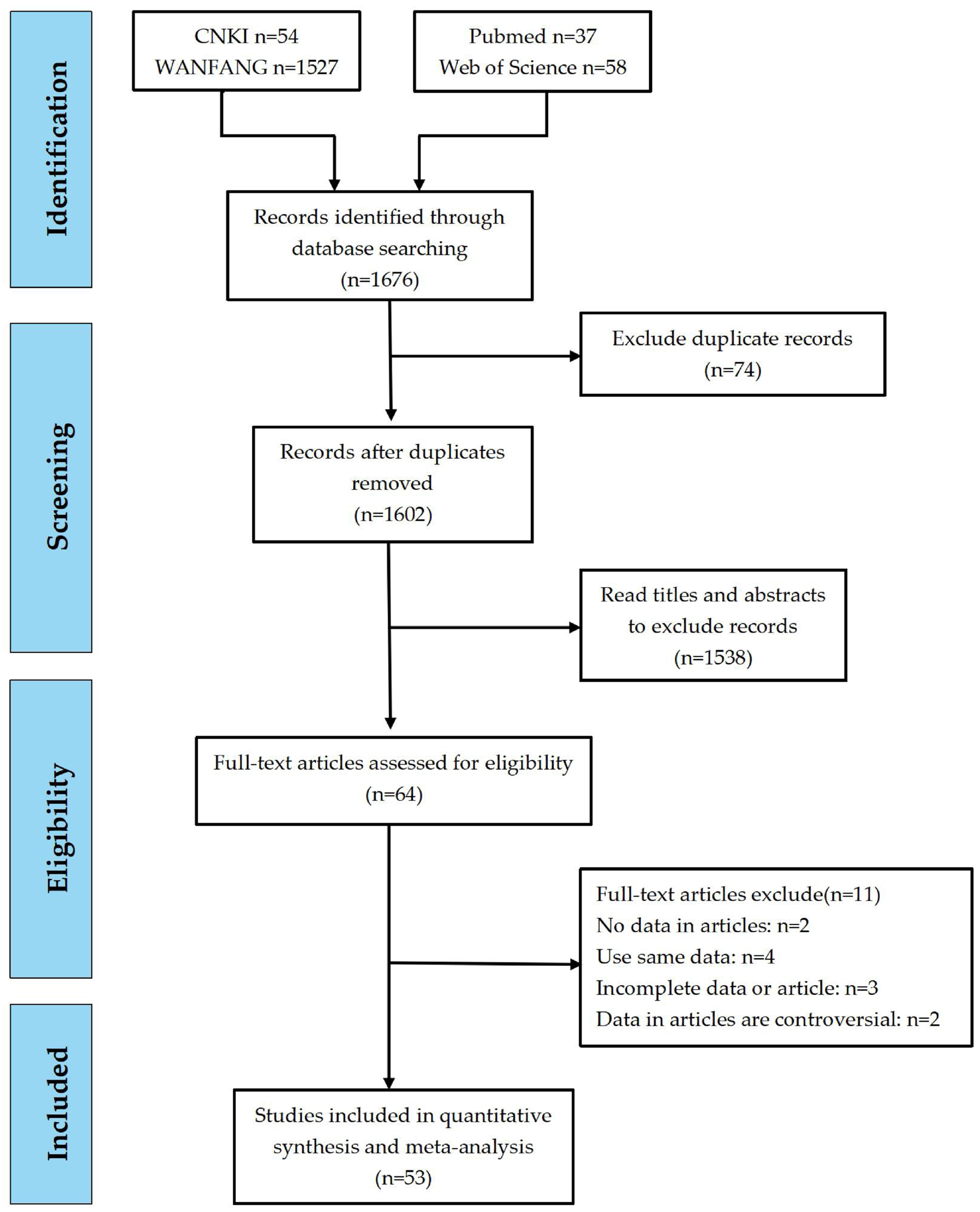
Figure 1. Flow diagram of the selection of eligible studies.
Vetsci 12 00863 g001
Figure 2. Summary forest plot of the estimated M. hyopneumoniae prevalence with random-effects analyses.
Vetsci 12 00863 g002
Figure 3. Forest plot of the prevalence of M. hyopneumoniae in swine with random-effects analysis in subclinical infection swine farms (A) and clinical infection swine farms (B).
Vetsci 12 00863 g003
Figure 4. Funnel plot showing publication bias in studies reporting the prevalence of M. hyopneumoniae in subclinical infection swine farms (A) and clinical infection swine farms (B).
Vetsci 12 00863 g004
Figure 5. Egger test for publication bias in epidemiological studies of M. hyopneumoniae in subclinical infection swine farms (A) and clinical infection swine farms (B). SND: standard normal deviate; precision: inverse of the variance.
Vetsci 12 00863 g005
Figure 6. Filled funnel plots for subclinical infection swine farms (A) and clinical infection swine farms (B) with the Trim-and-Fill Method. ◯ indicates observed studies; ⌼ indicates missing studies.
Vetsci 12 00863 g006
Figure 7. Leave-one-out sensitivity analysis of epidemiological studies on M. hyopneumoniae in subclinical infection swine farms (A) and clinical infection swine farms (B) (the circles indicate the pooled relative risk estimate when each individual trial is omitted).
Vetsci 12 00863 g007
Table 1. Baseline data of the studies included in the meta-analysis.
No.AuthorsPublication YearDiagnostic TechniqueSample TypeNo. of Positive SamplesNo. of Samples
1Duan et al. [23]2020PCRPathological material621114
2Fan [24]2020AGPT, histopathological examinationPathological material118168
3Gan [25]2015PCRNasal swabs4750
4Gao [26]2023PCRThroat swabs831478
5Guan [27]2014PCRLung0183
6He et al. [28]2010ELISASerum210460
7Jia et al. [29]2017Agglutination testSerum12331639
8Jiao [30]2020Histopathological examination, PCRLung, throat swabs11191375
9Joyce et al. [31]2014PCRNasal swabs54399
10Li [32]2016PCR, ELISALung, nasal swabs245647
11Li [33]2013PCR, ELISAPathological material,
serum, nasopharyngeal swabs
247852
12Li et al. [34] 2020Histopathological examinationLung730917
13Li, Y.Y. [35]2014PCRLung364782
14Li, S. [36]2014ELISA, PCRSerum, lung352670
15Li et al. [37]2014PCRLung122214
16Liang [38]2015ELISASerum78882
17Liang [39]2010ELISASerum210460
18Liang et al. [40]2018ELISASerum49619
19Liao et al. [41]2012ELISASerum1339
20Liu et al. [42]2012PCRSerum1560
21Liu et al. [43]2003ELISASerum63146
22Mao et al. [44]2010PCRPathological material1151
23Meng [45]2023PCRSerum691080
24Ni et al. [46]2017PCRNasal swabs48352
25Ni et al. [47]2018PCR, ELISANasal swabs, serum119876
26Ni [48]2020PCR, ELISANasal swabs, serum, lung, tracheal swab4141260
27Pi et al. [49]2023PCRNasal swabs, lung114253
28Pu [50]2010ELISASerum100229
29Qi et al. [51]2023PCRLung, nasal swabs49264
30Qiu et al. [52]2017ELISASerum93454
31Qiu et al. [53]2017PCRNasal swabs, tracheal swab, alveolar lavage fluid82450
32Shi [54]2013ELISASerum249423
33Shi et al. [55]2017ELISASerum103203
34Song et al. [56]2014ELISASerum20565619
35Sun et al. [57]2014ELISASerum5621280
36Wang et al. [58]2011PCRLung50205
37Wang et al. [59]2010PCRLung, nasal swabs, tracheal swab, alveolar lavage fluid78238
38Wei et al. [60]2017Agglutination testSerum26873343
39Wu et al. [61]2023PCRLung, nasal swabs31469
40Xing et al. [62]2009AGPTPathological material8862757
41Xiong et al. [63]2022PCRSerum521500
42Xu et al. [64]2019PCRPathological material45238
43Yang et al. [65]2011ELISA, PCRSerum, lung232436
44Yao [66]2006ELISASerum152372
45Yao et al. [67]2010Histopathological examination, PCR, ELISALung, serum, nasopharyngeal swabs14513492
46Yu [68]2010ELISASerum105368
47Yue et al. [69]2021PCRLung378491
48Zhang [70]2018Histopathological examination, PCRSerum, lung42146000
49Zhang [71]2007ELISASerum146420
50Zhang et al. [72]2021PCRPathological material199989
51Zhang et al. [73]2015PCRPathological material360
52Zhao [74]2012PCRLung, nasal swabs16276
53Zhao [75]2015Histopathological examinationLung335480
54Zhong et al. [76]2024ELISASerum224364
Antibody tests: ELISA and agglutination test; antigen tests: PCR and AGPT (agar gel precipitation test). Swabs: nasal, throat, nasopharyngeal, and tracheal. Lung: lung tissue samples.
Table 2. Results of subgroup analysis of data on the prevalence of M. hyopneumoniae infection.
SubgroupNo. of StudiesNo. of Positive SamplesNo. of SamplesInfection RateHeterogeneityP
Estimates95% CIX2p-ValueI2/%
Age 39.80%[0.341; 0.456]
Subclinical infection swine farmsSuckling swine3251440.137[0.000; 0.480]///0.606
Nursery swine219800.207[0.132; 0.305]///
Growing swine3453870.117[0.052; 0.201]///
Fattening swine5613340.169[0.080; 0.282]16.2720.00181.563
Breeding swine3362200.172[0.085; 0.277]6.9430.07456.790
Clinical infection swine farmsSuckling swine18187838120.380[0.246; 0.523]1246.4620.00098.6360.005
Nursery swine11158536250.361[0.161; 0.589]1753.0590.00099.430
Growing swine16114639960.327[0.235; 0.426]570.1020.00097.369
Fattening swine21345976830.491[0.364; 0.619]2363.7280.00099.154
Breeding swine18237847190.617[0.495; 0.734]994.1290.00098.189
Period 29.80%[0.232; 0.367]
Subclinical infection swine farmsBefore 2013724016420.107[0.031; 0.218]223.6400.00097.3170.944
2014–20181063357120.110[0.084; 0.139]158.2010.00089.254
2019–2024745853760.124[0.066; 0.197]342.9610.00097.959
Clinical infection swine farmsBefore 201316998220,7370.497[0.394; 0.600]3046.1620.00099.5080.803
2014–201810783012,9110.546[0.430; 0.659]1329.9970.00099.323
2019–20246166835600.562[0.234; 0.862]1946.3020.00099.743
Region 33.40%[0.256; 0.416]
Subclinical infection swine farmsNorthern region2939420.094[0.076; 0.114]///0.272
South region1368257160.096[0.056; 0.145]434.1940.00096.776
Qinghai–Tibet Plateau region213110690.118[0.099; 0.138]///
Clinical infection swine farmsNorthern region6441694470.557[0.375; 0.731]1465.310.00099.6590.005
South region13259561440.474[0.418; 0.530]208.6390.00094.248
Qinghai–Tibet Plateau region12494230.589[0.541; 0.635]///
Northwest region7547074680.642[0.538; 0.739]434.4080.00098.619
Farming scale 25.60%[0.160; 0.365] 0.000
Small-scale swine farms836219740.357[0.192; 0.541]531.7990.00098.3080.012
Medium-scale swine farms324315600.279[0.062; 0.573]297.6660.00098.992
Large-scale swine farms311910760.023[0.000; 0.169]///
Season 31.30%[0.209; 0.428]
Subclinical infection swine farmsSpring31709020.166[0.054; 0.321]///0.127
Summer2337200.044[0.030; 0.061]///
Autumn2295700.050[0.034; 0.070]///
Winter312310240.094[0.020; 0.215]///
Clinical infection swine farmsSpring665517190.547[0.259; 0.819]///0.105
Summer477511660.360[0.107; 0.664]765.0550.00099.346
Autumn348812150.651[0.564; 0.73323.0810.00087.002
Winter32215270.471[0.309; 0.636]///
Sampling type 31.60%[0.260; 0.374]
Subclinical infection swine farmsSerum854454860.137[0.082; 0.203]283.0420.00097.5270.513
Swab729527280.108[0.069; 0.155]77.430.00090.96
Tissue1038632100.083[0.030; 0.156]315.7420.00097.15
Clinical infection swine farmsSerum21910921,0680.446[0.369; 0.525]2415.5300.00099.1720.033
Swab8157040010.354[0.252; 0.462]246.880 0.00096.760
Tissue14625711,1990.568[0.449; 0.683]1791.307 0.00099.274
Diagnostic method 24.20%[0.192; 0.296]
Antibody testing26961920,9730.402[0.131; 0.494]4408.8210.00099.410%0.000
Antigen testing33553719,2560.202[0.137; 0.275]8490.0690.00099.282%
Histopathological testing5463771110.631[0.463; 0.783]811.3490.00099.384%
Disclaimer/Publisher’s Note: The statements, opinions and data contained in all publications are solely those of the individual author(s) and contributor(s) and not of MDPI and/or the editor(s). MDPI and/or the editor(s) disclaim responsibility for any injury to people or property resulting from any ideas, methods, instructions or products referred to in the content.