Review

Advancing In Vitro Tools for Oncologic Research in Cats and Dogs

1
Institute of Animal Pathology, Vetsuisse Faculty, University of Bern, 3012 Bern, Switzerland
2
Graduate School for Cellular and Biomedical Sciences, University of Bern, 3012 Bern, Switzerland
3
Bern Center for Precision Medicine and Cancer Therapy Resistance Cluster, Department for BioMedical Research, University of Bern, 3008 Bern, Switzerland
*
Author to whom correspondence should be addressed.
Vet. Sci. 2025, 12(9), 815; https://doi.org/10.3390/vetsci12090815
Submission received: 24 July 2025 / Revised: 20 August 2025 / Accepted: 22 August 2025 / Published: 26 August 2025
(This article belongs to the Special Issue Focus on Tumours in Pet Animals: 2nd Edition)

Simple Summary

Technological advances now allow scientists to grow cells in a dish outside the body, which has become a fundamental tool in life sciences and especially in cancer research. In recent years, there has been growing interest in using these technologies for pets such as cats and dogs. Both cats and dogs develop cancer similar to humans, such as mammary tumors (breast cancer), which can lead to devastating outcomes and make this disease highly important to study. Growing tumors in a dish makes it possible to study cancer cells without harming the patient, and, in the best case, artificially grown cells can help predict whether a drug will work. While these technologies are constantly improving, such that cells can even form three-dimensional mini tumors, we still face challenges in fully reproducing disease behavior in the body. This review summarizes the current state of tumor culture, with a focus on mammary tumors in cats and dogs.

Abstract

In vitro culture systems have advanced cancer biology, particularly through 2D and 3D tumor cultures. These have answered numerous scientific inquiries and propelled human oncologic research, with growing recognition of their potential to improve cancer treatment in companion animals, specifically cats and dogs. These species develop cancer spontaneously, closely resembling specific human cancer subtypes. For example, canine and feline mammary tumors are especially valuable for studying tumor biology. In vitro models from these tumors therefore offer a unique opportunity for veterinary cancer research. Recent 3D cell culture advancements provide promising platforms for predicting therapeutic responses in human cancer and may be applied to mammary tumors in animals. However, while limitations in fully recapitulating in vivo conditions and predicting chemotherapy response have been observed in colorectal tumoroids, similar challenges are emerging in mammary and breast tumors. In particular, canine mammary tumors and human breast cancers share critical heterogeneity and microenvironmental factors usually inadequately modeled in vitro. This review critically examines the predictivity of 3D mammary tumoroids from humans and companion animals, highlighting challenges related to stromal and immune cell preservation, reproducibility, and the translational gap between in vitro findings and clinical outcomes. We propose future directions to optimize these models for both comparative oncology and veterinary-specific applications.

1. Introduction

A cornerstone of both veterinary and human cancer research has been the development of in vitro culture systems, making the study of cancer cells outside of the patient possible. The first attempts at cell culture date back to the late 19th century, when a wide range of cell types were extracted from various organisms and kept viable outside of the donor for a limited amount of time. A pivotal discovery marked the establishment of the first immortalized human cancer cell line, HeLa cells [1,2]. The ability of cells to virtually proliferate indefinitely in a dish ex vivo has opened numerous avenues of biological research, particularly in cancer research. Over time, the repertoire of culturable mammalian cell lines has expanded substantially, and the culturing techniques have dramatically evolved with the addition of new technologies and deeper understanding of cellular biology. Nowadays, in vitro systems have become progressively more elaborate, attempting to emulate in vivo settings, including complex three-dimensional (3D) systems, from organoid culture to 3D bioprinting [3,4,5,6,7,8]. However, despite the advancement of in vitro culture systems, the complexity of in vivo animal models such as mice remains unrivaled, and their use still holds place in most preclinical studies [7,8,9].
Human and veterinary oncology are traditionally viewed as entirely different fields because of the differences between species. However, accumulating evidence has revealed striking parallels across species, making them increasingly difficult to distinguish from each other when inspected in detail. Similarly to humans, neoplastic diseases account for a significant proportion of morbidity and mortality in cats and dogs, according to records throughout America and Europe, with mammary tumors, cutaneous epithelial tumors, and lymphomas being frequently reported among the malignant cases [10,11,12,13,14,15,16,17,18,19]. In this context, both canine and feline mammary tumors (CMT and FMT) display striking similarities to human breast cancer (HBC), with both closely recapitulating key morphologic and clinicopathologic characteristics of their human counterparts [20,21,22,23]. CMT is a heterogeneous disease encompassing a variety of histological subtypes, and approximately half of all cases are malignant [24,25,26]. In contrast, FMT is typically highly aggressive and presents a high metastatic rate at diagnosis. Notably, FMT closely resembles the triple-negative subtype in human breast cancer (HBC)—defined by the absence of estrogen receptors (ERs), progesterone receptors (PRs), and human epidermal growth factor 2 (HER2) [21,25].
Furthermore, companion animals share a similar living environment with their owners and are therefore exposed to similar environmental risk factors. These parallels have led to the introduction of therapeutic strategies originally developed for humans into veterinary medicine. At the same time, cancer in pets offers significant potential to model human disease and serves as a valuable model to study tumor biology and treatment strategies. This reciprocal relationship between companion animals and humans is underscored by the One Health approach, which promotes the advancement of cancer research for both pets and owners [27,28,29,30,31,32]. For instance, the receptor tyrosine kinase inhibitor toceranib (Palladia TM), initially developed for treating mast cell tumors in dogs, provided essential preclinical data for the development of its human analog, sunitinib (Sutent TM), used for a variety of human cancer types [33,34]. Clinical trials involving toceranib not only improved treatment outcomes for dogs but also offered valuable insights into human cancer therapies. This example illustrates the potential in bridging the gaps in oncologic research between species and underscores the necessity for collaborative research efforts.
This review presents an overview of the application of in vitro culture systems, with a focus on canine and feline mammary tumors. We will explore both classical and advanced systems used to study cancer in cats and dogs. In addition, we will discuss the advantages and limitations of the currently available systems, highlighting the potential of comparative oncology bridging cancer research across species.

2. Conventional Two-Dimensional (2D) Culture

2.1. Overview of 2D Culture

To understand the advancements in in vitro systems, it is essential to examine the conventional two-dimensional (2D) culture first. The basic but still canonically used in vitro culture system involves propagating adherent cells in flasks as a monolayer supplemented with various formulations of growth media. This popular system is characterized by its low cost, user-friendliness, and scalability compared to the more complex three-dimensional (3D) systems discussed later. Cells are usually seeded, allowed to grow until reaching confluency, and passaged for continuous growth.

2.2. Immortalization

Before focusing on cancer-derived in vitro models, it is important to clarify that the classification of culturable cells as either “normal” or “cancerous” is an oversimplification. In most research settings, the ability to proliferate virtually indefinitely is essential, which poses a major limitation in culturing normal, non-transformed cells. In an ideal world, autologous normal cells—e.g., normal mammary cells derived from the same individual as the mammary tumor—would serve as corresponding controls.
However, even under optimal conditions, culturing primary normal somatic cells—those derived from normal donor tissues—will result in replicative senescence. This is primarily due to the activation of tumor suppressor genes, such as TP53 and RB1, in response to the programmed shortening of telomeric DNA upon reaching a critical length. This occurs because DNA polymerase is unable to fully copy the telomeric ends during replication [35,36]. Typically, genetic modifications are required to counteract the telomeric shortening and/or bypass the tumor suppressor pathways. This can be achieved by expression of telomerase reverse transcriptase (TERT), a ribonucleoprotein which adds hexameric repeats during DNA replication. In somatic cells, TERT is physiologically repressed and is typically restricted to specific cell types such as germ cells, stem cells and activated lymphocytes, and the deactivation of tumor suppressor genes can result in an immortalized phenotype [37].
Most cancer cells typically bypass senescence by acquiring mutations affecting telomere regulation, and such alterations also need to be introduced in normal 2D cells to avoid senescence. A systematic analysis of telomere length in over 6800 human cancer samples revealed that 72% harbored TERT-expressing mutations, followed by samples showing alterations in ATRX or DAXX, key genes in the alternative lengthening of telomeres (ALT) pathway. In the remaining samples, telomere length was positively correlated with alterations in tumor suppressor genes such as TP53 and RB1 [36]. Hence, the typical immortalization protocols involve the introduction of a TERT-expressing vector and/or deactivation of TP53 and RB1, usually by using viral oncogenes such as Simian virus 40 (SV40) or Human papilloma virus 16 (HPV16) [38].
Although extensive characterization of telomeric biology in companion animals remains elusive, various early studies identified frequent mutations in similar genes across tumors, including TP53, RB1, ATRX, and TERT [13,39,40,41,42]. These findings suggest that telomeric biology is highly conserved across species, extending beyond humans. Further evidence is provided by the use of human TERT-expressing vectors and/or human oncoviruses to successfully immortalize various normal canine and feline cells, suggestive of a shared biological foundation across species [42,43,44,45,46,47,48,49,50]. Despite these parallels, telomerase activity is rarely measured in companion animal tumor studies, which may lead to the underestimation of its prevalence. However, Uyama et al. [51] detected telomerase activity in all eight FMT cell lines established in their laboratory showing continuous growth—further supporting the cross-species homology of these mechanisms.
Notably, in mice, low levels of TERT are also expressed in adult somatic cells [52,53]. This special trait explains why mouse cells have significantly longer telomers (40–80 kb) [54] compared to humans (5–15 kb) [54], cats (5–26 kb) [55,56], and dogs (11–27 kb) [57]. Despite somatic mouse cells expressing TERT, the stricter regulation prevents indefinite telomere lengthening, contrary to immortalized cell lines [58,59]. Still, the fact that immortalization is easier achieved in mouse cells simplifies the process of establishing stable and long-term 2D cultures from mouse tissues.

2.3. Oncogenic Transformation

Given the overlapping features observed in vitro, it is crucial not to confuse immortalized cells with cancer cells. Immortalization only involves bypassing of replicative senescence, allowing cells to divide indefinitely. However, immortalized non-cancerous cell lines still lack certain properties exclusive to cancer cells, such as evasion of growth suppression or resistance to programmed cell death. For instance, normal cells enter a proliferative quiescence when they reach confluency as a monolayer known as contact inhibition or initiate apoptosis upon loss of anchorage through a process called anoikis. In contrast, cancer cells would continue to divide uncontrollably under these conditions [60,61]. In some cases, spontaneous immortalization in vitro may occur, a phenomenon frequently observed in rodent cell lines, likely due to the inherent TERT expression in these species (Figure 1) [58,62,63]. This phenotype was also shown in canine embryonic fibroblasts [42]. However, to the authors’ knowledge, this instance has not been reported in normal adult somatic cell lines from both dogs and cats, further underscoring the importance of telomerase activity in this process.
The soft agar assay is widely used to assess oncogenic transformation, where the ability to proliferate independently of anchorage is demonstrated, adherent cells being suspended in low-concentration agar, allowing the assessment of resistance to anoikis. This technique can also be adapted to culture some cancer lines in 3D, as will be discussed later. Another well-established method examines the tumorigenic potential of transformed cells in vivo using mice as xenotransplantation models. These models are considered more reliable as they reflect the interaction between the inoculated cells with the complex in vivo environment. Additionally, xenotransplantation models are useful to study long-term tumor progression and metastasis, with tumors often passaged in these mice to maintain tumor lineage and assess metastatic potential [64]. However, these models also have limitations, including the low engraftment rates, the foreign species origin of the microenvironment, the use of immunocompromised animals, the high cost, and ethical concerns [65].
Given this context, for immortalized cell lines to acquire typical hallmarks of cancer, additional pathway perturbations are required. Interestingly, species vary in the number of mutational hits required for successful oncogenic transformation. For instance, while human fibroblasts require perturbations in five different pathways—specifically p53, pRb, PP2A, telomerase, and Ras pathways—to achieve oncogenic transformation, mouse fibroblasts only require perturbations in p53 and Ras [66,67,68]. Such a disparity may explain the phenomenon called Peto’s paradox, where larger animal species do not show higher cancer incidences than smaller species, despite the significantly higher absolute number of cells [68,69]. This observation suggests that larger species harbor additional mechanisms that prevent an increase in malignant cell transformation, possibly correlating with the number of mutational hits required to transform cells across species.
Even more intriguing, studies on canine cell lines suggest that dogs resemble mice in their susceptibility to oncogenic transformation, despite the significant difference in body mass and life span. This was supported by the successful oncogenic transformation of embryonic immortalized canine fibroblasts using h-Ras in addition to the introduction of a catalytic telomerase subunit [42]. In another study [70], TP53 knockout in canine fibroblasts with a presumptive mutation in TFE3 was found to induce a tumorigenic phenotype. However, these results contrast with findings from a study [71] that used human cytomegalovirus (HCMV), known to induce oncogenic transformation in human cell lines [72]. While canine kidney cells were immortalized upon infection with SV40 or HCMV and anchorage-independent growth was promoted, only 1 out of 40 mice developed tumors when inoculated with these cells.
In contrast, the literature is significantly sparser regarding oncogenic transformation in feline cell lines. While successful immortalization was demonstrated in feline endothelial cells [50], respiratory epithelia [43], and intestinal epithelia [49] using combinations of SV40, HPV16, and hTERT, no investigations assessing their tumorigenicity have been conducted. Notably, the two studies involving epithelial cells [43,49] both observed that transformed cells acquired double positivity for cytokeratin and vimentin, suggestive of epithelial–mesenchymal transition (EMT). While EMT is a physiological process during embryologic transdifferentiation, EMT-inducing transcription factors are commonly upregulated in tumors and promote malignant transformation in mouse mammary epithelia [73]. Thus, the EMT phenotype in these feline epithelial cell lines may reflect potential malignant transformation.
Together, these findings highlight the complexity of tumorigenesis across species and the challenges associated with 2D cultures. They emphasize the need for future investigations in companion animals, possibly also by improving current cell culture systems. Elucidating the underlying mechanisms of the differences and similarities may result in more relevant translation across veterinary and human oncology.

3. Three-Dimensional (3D) Culture

3.1. Overview of 3D Culture

As technology advances, in vitro culture systems have become increasingly more sophisticated to bridge the gaps between in vitro models and their primary counterparts. While conventional 2D culture systems provide powerful ex vivo tools, the loss of native multicellular architecture and change in cell shape through the growth on hard plastic can substantially impact gene expression [74], resulting in altered cell differentiation [75] and metabolic activity [76]. The limitations of 2D cultures drove the development of more physiologically relevant in vitro three-dimensional (3D) models, such as organoids, tumoroids, and spheroids, using various systems aiming to reflect the in vivo tissue environment. These 3D systems can be broadly divided into scaffold-based or scaffold-free approaches, both aiming to recapitulate in vivo complexities.
In this context, scaffolds refer to supportive materials that mimic the native extracellular matrix (ECM) for cells to grow on or within. Alternatively, scaffold-free approaches, including static suspension cultures, dynamic microfluidic devices, and bioreactors, rely on meticulously controlled biochemical cues to promote 3D growth [77]. With the wide range of available 3D culture systems, determining the most suitable one depends on the experimenters’ specific needs. Scaffold-based systems excel in mimicking in vivo microenvironments but require cell-type-specific optimization. While they are limited in nutrient access and waste removal due to restricted diffusion, scaffold-free systems offer high-density expansion and superior control over the culture environment. On the other hand, scaffold-free systems lack the mechanical support and are often more costly to implement in term of consumables and equipment [78]. Both approaches rely on specific culture medium formulations that vary dramatically across studies, significantly impacting cell behavior and experimental outcomes, making the selection of an appropriate medium formulation critical [77,79,80]. Given the vastness of the topic of 3D culture technologies, we will narrow our discussion to the application of 3D models to highlight their significance in advancing veterinary oncology.

3.2. Organoids vs. Spheroids vs. Tumoroids: Definition Matters

To better understand the distinct advantages of 3D models, it is important to clarify key terminologies first. Across studies, authors classify their in vitro models as organoids, spheroids, and, in the context of the mammary gland, mammospheres. Etymologically speaking, a spheroid (“sphere-like”) describes a geometric feature—likewise, a mammosphere refers to a spherical structure derived from the mammary gland—whereas an organoid (“organ-like”) implies organotypic complexity. This interchangeable use of these terms to describe various types of 3D models has been a source of confusion. Essentially, the core feature of organoids is their ability to arise from pluripotent or multipotent stem cells and to self-renew into multicellular 3D structures, mimicking key morphological and functional characteristics of an organ [4,80,81]. This process often depends on Wnt signaling, a critical pathway promoting the expansion of stem cells and organoid formation [77,80]. Notably, Wnt signaling activation also induces TERT expression, contributing to the long-term culture capability of organoids [82].
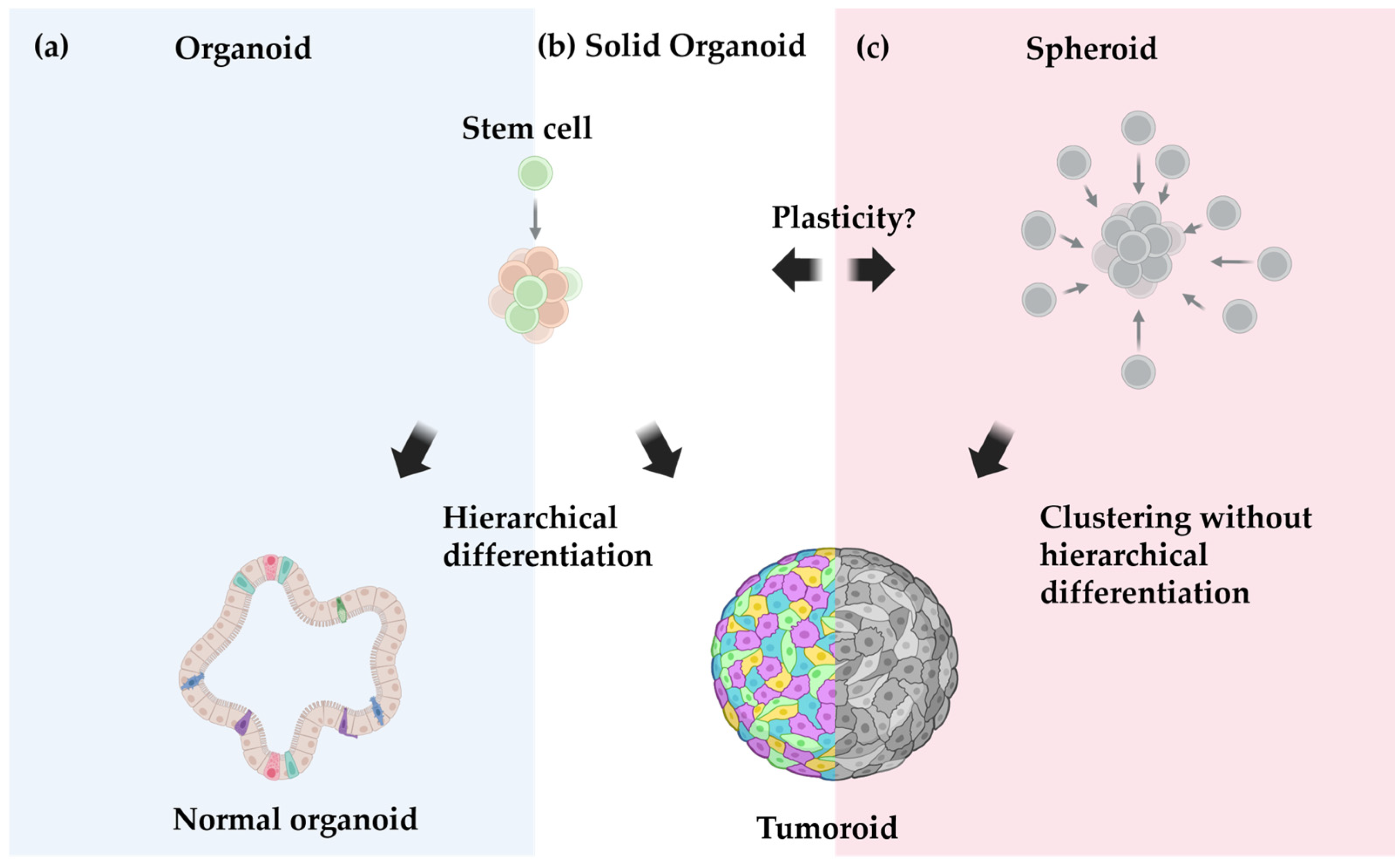
Given this context, organoid culture relies on the natural differentiation pathways of these cells—meaning that the composition and architecture of the final organoid depend on both the commitment state of the starting stem cells and the culture conditions (Figure 2). For instance, naïve adult stem cells from the intestinal crypt will differentiate in various epithelial portions of the intestine. This applies similarly to the mammary gland. On the other hand, genetic modification can reintroduce the required stemness in differentiated somatic cells, allowing for the formation of complex brain organoids from a single induced pluripotent stem cell (iPSC) [77,80,81,83].
While the definition of an organoid is generally straightforward, classifying tumor-derived 3D structures may pose a challenge, particularly regarding solid tumors that lack clear morphological heterogeneity and architecture. Traditionally, “spheroids” refers to 3D structures generated from any cells forming simple, miniature spheres. One of the earliest models, developed in the 1970s, used hamster cancer cell lines cultured in spinning flasks to keep cells in suspension [84]. Although spheroids lack a tissue-specific architecture, they exhibit a characteristic layering due to the centripetally decreasing nourishment gradient, reflecting the behavior of poorly vascularized solid tumors and provide physiological relevance to drug response studies similar to organoids. Building on this model, researchers typically refer to spherical 3D structures derived from tumor cell lines as spheroids [85,86], implying that these structures have low heterogeneity and complexity due to the observed phenotypes in 2D cultures. While convenient, this nomenclature may overlook the potential organotypic complexity of these 3D tumor models. For instance, Petersen et al. [87] showed that 2D cell lines of non-cancerous mammary epithelium cells formed an organotypic glandular architecture with various forms of cellular differentiation when 3D culture conditions were introduced—illustrating the potential for cells to generate organotypic complexities, even after being adapted to 2D growth.
In this context, 3D culture studies of CMT exhibit organoid characteristics, displaying glandular histomorphological features while maintaining the mutational landscape and immunophenotypic signature of the original tumor tissue [88]. In contrast, such organoid complexities may be less evident in 3D models derived from tumors with rather compact solid growth, such as canine urothelial tumors [89].
The distinction between tumor organoids and tumor spheroids is crucial, as the way we conceptualize tumor-derived 3D structures depends on the tumor growth model we subscribe to. Therefore, we propose the term “tumoroids” (“tumor-like”) as a more appropriate nomenclature for tumor-derived 3D models. According to the cancer stem cell (CSC) theory, only a subset of specialized cells within a tumor are responsible for disease initiation and driving disease progression. These CSCs can additionally differentiate into a variety of non-tumorigenic cells with a limited capacity for self-renewal. Given this context, in vitro conditions suitable for organoid culture should logically produce tumor organoids from these CSCs. However, some researchers may argue that the non-tumorigenic population in 3D HBC cultures may result from contamination by surrounding untransformed cells [90]. It is an interesting paradox that organoid culture conditions favor the growth of normal tissues over cancerous ones [91].
Although the CSC model is still debated, there are cancer cell subpopulations expressing stem cell markers such as CD133 or CD44 [92,93]. Both markers have also been detected in 3D CMT culture via Western blotting, although the bands appeared faint [94]. Along these lines, the authors performed lineage tracing experiments to track individual cell fate over time, combined with ALDH activity measurements providing evidence for a hierarchical structure within these CMT tumor organoids [79,94].
In contrast, the stochastic model of carcinogenesis posits that any oncogenetically transformed cells can expand clonally and equally contribute to tumor growth [95]. This model suggests a tumor architecture, where all transformed cells drive tumor progression without the need for hierarchical differentiation. There may still be intra-tumoral heterogeneity, but which subpopulation thrives is stochastic and not the result of a pre-defined program. In case the tumor cells are fairly homogeneous, tumor spheroids may better align with the idea of a homogenous cell aggregation of limited complexity.
At the same time, the line between both models starts to blur given the concept of cancer cell plasticity, positing a continuous transition of cancer cells into—or their reversion to—progenitor-like states [61]. And if there is plasticity, can a spheroid become a tumoroid and vice versa? Does this mean that the distinction is ultimately not as rigid as we thought and that one should rather focus on their cellular origin—whether derived from cancerous cells (i.e., tumoroids) or non-transformed cells.
Ultimately, without clear evidence of differentiation or organization, the distinction between a tumor organoid and a tumor spheroid may require additional investigation such as lineage tracing experiments. While organoid formation typically relies heavily on the culture medium supplements, cancer cells may lose these dependencies to activate oncogenic pathways [96], allowing the formation of 3D organoids even under suboptimal conditions. This further complicates the classification, suggesting that a more encompassing term, such as tumoroids (“tumor-like”), might be appropriate, with further subclassification based on whether a structure exhibits an organoid- or spheroid-like phenotype.

4. Mammary Tumor Models

4.1. Advancements and Challenges in Establishing In Vitro Models for Mammary Tumors

With the growing attention to alternative HBC models, there has been a substantial increase in the number of mammary tumor models in companion animals (Table 1). Significant progress has been made with 2D CMT and FMT models. However, the development of 3D tumoroids remains scarce yet potentially more impactful. This scarcity of 3D models may stem from challenges in optimizing growth medium formulations and financial constraints associated with the necessary reagents.
Efforts to develop 3D CMT models have seen some success. Cocola et al. [79,94] were among the first to create CMT tumoroids, using tissue samples of eight CMT patients. However, the actual success rate of producing the primary organoids remains unclear, as failed attempts may not have been reported. In addition, only short-term cultures lasting up to five passages were achieved. In contrast, Inglebert et al. [88] established the first large-scale organoid biobank, generating 23 tumoroid lines derived from 16 dogs (some dogs had multiple tumors), yielding a success rate of around 75%. These tumoroid lines demonstrated long-term potential, sustaining growth for up to 20 passages. Notably, these numbers are remarkably higher than those reported in HBC tumoroid cultures with success rates as low as 3.33% [90]. That said, the authors observed a reduction in estrogen receptor expression at the protein level with extended passages, suggesting some degree of cellular adaptation to the in vitro conditions.
Despite advancements in CMT models, the development of 3D FMT models remains elusive. Success rates for establishing cell lines have ranged from 2.5% to 40% [51,97,98]. These relatively low numbers could indicate potentially inherent challenges in establishing primary feline tumoroids, possibly attributed to suboptimal culture conditions. Additionally, the actual number of failed attempts often remains unclear due to the tendency for negative results to go unpublished, further complicating progress in this field. Despite these apparent challenges, the existence of feline intestinal and liver organoids [99,100,101] established using similar protocols to those for canine and human organoids suggests that the development of a 3D FMT culture may be feasible. Refining protocols from CMT research could help address the obstacles to establishing FMT tumoroid models.
Table 1. Summary of studies on establishing in vitro canine and feline mammary tumor models.
SpeciesModel TypeTumor SourceNumber of AnimalsReferenceHighlights
Canine2D
  • Primary carcinoma and metastases
2Van der Burg, 1989 [102]
  • 4 tumor cell lines established
  • Primary mixed adenoma
1Priosoeryanto et al., 1995 [103]
  • 1 tumor cell line established
  • Carcinoma and adenoma
5Hellmén, 1992 [104]
  • 5 tumor cell lines established
  • Using 3 different modes of sampling: 1) tumor fragment DMSO frozen vs. 2) fine-needle aspirate vs. 3) tumor fragment freshly processed
  • Primary carcinoma and metastases
4Uyama et al., 2006 [105]
  • 4 pairs of tumor cell lines from 4 dogs (primary and metastatic) established
  • Primary carcinoma
1Caceres et al., 2015 [106]
  • 1 tumor cell line established
  • Primary carcinoma
1Mei et al., 2021 [107]
  • 1 tumor cell line established
  • Primary mixed adenocarcinoma
1Li et al., 2021 [108]
  • 1 tumor cell line established
  • Included tumor types: complex carcinoma, complex adenoma, and mixed adenoma
10 out of 12Yeom et al., 2023 [109]
  • 10 tumor cell lines established
  • Targeted sequencing for two variants of PIK3CA
  • Validated for in vitro drug assays
  • Included tumor types: complex carcinoma, mixed tumor, in situ carcinoma, and simple carcinoma
8Park et al., 2024 [110]
  • 8 tumor cell lines established
  • Targeted sequencing for a variant of PIK3CA
  • Validated for in vitro drug assays
  • Identified over 200 highly expressed genes and enriched EMT signatures
Canine3D
  • Not specified
8Cocola et al., 2009 [94]
  • 8 tumoroid lines and 8 normal mammary glands established
  • Could be maintained for up to 5 passages
  • Included tumor types: complex carcinoma, simple carcinoma, mixed tumor, mixed carcinoma, and malignant myoepithelioma
16Inglebert et al., 2022 [88]
  • 23 different tumoroids and normal mammary organoids out of 32 samples established
  • Suitable for long-term culture
  • Validated for gene editing and in vitro drug assays
  • Verified conserved mutational landscape in in vitro models using whole-genome sequencing (WGS)
Feline2D
  • Primary carcinoma
4 out of 30Norval et al., 1985 [97]
  • 4 tumor cell lines established
  • 1× primary carcinoma
  • 3× pulmonary metastases
4 out of 135Minke et al., 1991 [96]
  • 4 tumor cell lines established
  • Primary carcinoma and metastases
  • Including 1 thoracocentesis sample
5 out of 13Uyama et al., 2005 [51]
  • 5 cell lines established
  • Telomerase activity detected in all cell lines
  • Primary carcinoma
1Borges et al., 2016 [111]
  • 1 cell line established
  • Primary carcinoma
1Granados-Soler et al., 2018 [112]
  • 1 cell line established
  • Immunophenotypic characterization
  • Copy-number variation analysis of cell lines and tumors
  • Validated for in vitro drug assays
  • Investigated EMT-related gene expression levels

4.2. Predictive Drug Response and Biomarkers in Mammary Tumor Models

Building on the development of these models, one of the arguably most clinically relevant aspects of primary in vitro models is their potential to predict drug response without the ethical concerns of in vivo testing. Several studies have investigated drug response involving CMT and FMT models to identify clinically translatable vulnerabilities [88,109,110,112,113]. For example, as early as 1981, von Hoff et al. [114] demonstrated that human tumoroids grown in soft agar had an impressive 88% sensitivity and 94% specificity when subjected to a panel of cytotoxic drugs. This study highlights the remarkable potential of primary in vitro models to serve as functional tools for predicting therapy response. However, a limitation of this application was the low success rate of 25% in growing the samples in vitro, likely due to the suboptimal growth medium formulation. More recent attempts, for example, in colorectal cancer, the role model for developing tumoroids, the response of patients to oxaliplatin was predicted with a sensitivity of 70% and a specificity of 71% using patient-derived tumoroids [115]. This shows that we still need to substantially improve the current tumoroid growth conditions to achieve reliable predictions.
With the recent revelation of PIK3CA mutations being frequently observed in CMTs, researchers are focusing on exploiting this mutation for targeted therapy, similar to approaches in HBC [108,109,110,116]. Both 2D [109,110,116] and 3D [88] CMT lines have conserved their matching mutations in vitro. However, results across studies have been variable. While most studies [88,109,110] showed increased drug sensitivities associated with PIK3CA mutations, Maeda et al. [116] reported mixed results between PIK3CA-mutated lines and wild-type lines. Still, most of these studies used a targeted approach to identify mutated CMT lines, in which additional genomic alterations may not have been accounted for, which could potentially have impacted drug response. Although all authors used CMT models harboring the same mutations, the tumors may differ in their biological behavior. For instance, Yeom et al. [109] found no evident correlations between the mutation and tumor malignancy. They further observed cells reminiscent of chondrocytes in two of the CMT cell lines, further underscoring the heterogeneity between CMT lines. These findings highlight how even well-characterized mutations, such as PIK3CA, warrant further investigation and more standardized methods to better understand their impact on tumor behavior and drug response in CMT models.
In contrast, investigations into drug response in FMT models remain limited. Recent copy-number variation (CNV) analysis identified several altered cancer-related genomic regions as prognostic markers, supported by retrospective clinical survival data [117]. While there are several targetable genes within these altered genomic regions, including JAK2, PD-L1, and PD-L2, further validation experiments are needed to establish their suitability as appropriate candidates for targeted therapy approaches. Despite the potential application of immune checkpoint inhibitors targeting PD-L1 and PD-L2, the current lack of established FMT in vitro co-culture systems with immune cells presents an additional obstacle. Limited studies have tested drug response in FMT models so far. One study [113] subjected an FMT cell line to 5-fluorouracil (5-FU), an antimetabolite for disrupting DNA synthesis, in combination with a panel of drugs known for their synergistic effects in HBC patients. While 5-FU significantly decreased cell viability, none of the combinations proved to have additional effect, contrary to HBC cell lines. The authors concluded that these results may reflect the aggressive biological behavior of the FMT cells used. The precise reasons for the lack of drug response in FMT remains unclear, as several factors are capable of influencing experimental outcomes, ranging from growth medium formulation to even passage number. For instance, it was shown that an FMT cell line’s sensitivity to doxorubicin doubled with successive passages, similar to findings in human cell lines [112,118]. One possible explanation could be the enrichment of certain cells through selective pressure, a phenomenon observed in HBC studies, where genomic aberrations shift with increased passaging [90]. In another study, Gameiro et al. [119] demonstrated a synergistic inhibitory effect of receptor tyrosine kinase inhibitors (lapatinib and neratinib) and rapamycin in FMT lines, including the same line used in the 5-FU study. However, passage numbers were not reported, which is common in many human and veterinary studies, making it difficult to assess the potentially confounding effects of passage stage on drug responses. These examples emphasize the need for a more stringent standardization across studies to improve experimental reproducibility and to derive meaningful results.

4.3. Exploring Novel Biomarkers with CRISPR/Cas9 Screening

While significant attention has been given to drug testing and mutation-specific drug response prediction, genetic screenings using CRISPR/Cas9 technologies have recently emerged as a powerful tool for discovering novel predictive biomarkers, as well as investigating tumor-specific vulnerabilities and resistance mechanisms. In contrast to traditional genomic screening that identifies mutations in wild-type genomes, CRISPR/Cas9 screening provides functional context for genomic alterations. For example, CRISPR/Cas9 screening enabled the identification of LRP6 in a CMT cell line as a critical tumor-specific host factor that promotes infections by the oncolytic virus, the Ondersteepoort strain of Canine Distemper Virus (CDV-OP) [120]. These findings lay the mechanistic framework for refining oncolytic viral therapies across different tumor types and species, potentially enhancing their precision and therapeutic effectiveness.
Moreover, CRISPR/Cas9 screening was successfully applied to both CMT tumoroids and normal canine mammary organoids using a custom-designed CRISPR/Cas9 sub-library targeting over 800 druggable genes. This approach opens new opportunities for uncovering cancer-specific vulnerabilities and mechanisms [88,121]. A key advantage of this approach is the ability to directly compare normal and tumor cells from the same animal, deepening our understanding of tumor-specific genetic dependencies to further improve cancer treatment and the development of novel therapies. Although these approaches have not yet been implemented in FMT models, their application to feline models appears promising.
Despite the pressing need for effective therapies in FMT, such advanced methodologies remain significantly underrepresented in FMT research. FMT is often proposed as a model for triple-negative breast cancer (TNBC) in women. While TNBC is recognized as the most aggressive subtype of HBC, it is frequently susceptible to poly (ADP-ribose) polymerase inhibitors (PARPi) due to the homologous recombination deficiencies (HRDs) that arise from mutations in BRCA1/2 genes [122]. Genetic studies investigating BRCA1/2 mutations in cats have produced mixed results. One study [123] reported no mutations in a cohort of 24 patients, whereas a different study [124] claimed that 3 out of 9 cats carried variants with moderate impact. It is important to note that although BRCA1/2 mutations lead to HRDs, there are also BRCA-independent genetic alterations that can result in HRDs. Considering the remarkable clinicopathologic resemblance between FMT and TBNC, it is plausible that FMT may exhibit HRDs as well. In vitro FMT models present promising ex vivo investigations to explore this hypothesis by establishing functional testing for HRDs in FMT [125].

4.4. Lost in Translation

Despite progress in modeling mammary tumors in vitro, translating some findings into clinical applications remains challenging, even using 3D tumoroid models. Current scaffold-based CMT tumoroids can mimic certain aspects of the in vivo environment, but they are still largely limited to epithelial monocultures, excluding stromal and immune cells that are crucial in shaping tumor behavior. This limitation likely contributes to the observed discrepancies in drug sensitivities between in vitro and in vivo mouse mammary tumor models, due to the protective effects of the tumor microenvironment [126,127]. For instance, studies in canine mast cell tumors have demonstrated that co-culturing with stromal cells significantly improves cell viability [128]. Similarly, in a study with human cell lines [129], cancer-associated fibroblasts (CAFs) were found to secrete a specific profile of growth factors and chemokines, promoting proliferation as well as inducing resistance to targeted antibody therapy.
These observations raise the important question of how accurately in vitro systems can predict clinically relevant drug efficacies in cancer therapy. In this context, the use of patient-derived tumoroid cultures to guide individualized treatment decisions conceptually aligns with bacteriologic culture-based diagnostics. Interestingly, both in vitro tumoroid culture and antimicrobial susceptibility testing (AST) share similar limitations regarding their predictive value in vivo. In both cases, the systems tend to be selected for specific subpopulations and are susceptible to pre-analytical errors, including variability in clinical sampling and processing. While AST generally correlates well with clinical outcomes, therapy failure still occurs due to factors such as biofilm formation or drug-limiting conditions at the infection site (e.g., pH or perfusion), similar to tumoroid models without a tumor microenvironment [130,131].
The profound influence of the tumor microenvironment on in vitro models emphasizes the need for comprehensive characterization and optimization of co-culture systems in the future. Accurately replicating the tumor microenvironment requires detailed profiling of the various cellular components involved. Advanced transcriptomic approaches, such as single-cell RNA sequencing (scRNAseq), provide powerful tools for extensive mapping of the cellular composition and gene expression profiles within both the tumor and its microenvironment [132]. Moving forward, integrating these approaches will be pivotal for improving the fidelity and translational relevance of in vitro models. Nonetheless, financial constraints pose a persistent barrier. The high costs associated with essential culture reagents and advanced technologies hinder the development and optimization of these complex in vitro systems, ultimately dictating the pace of progress in veterinary oncology.
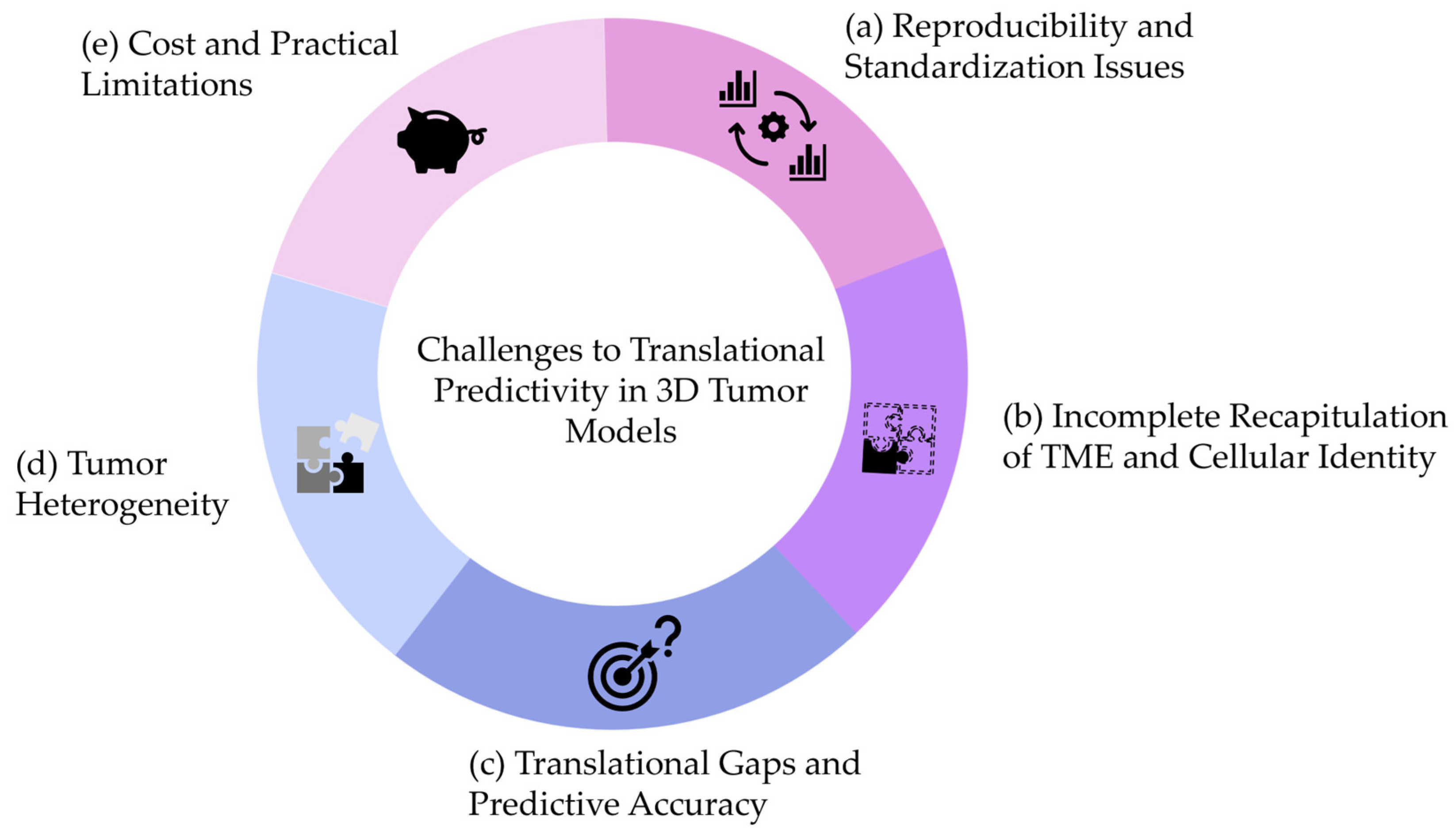
In summary, we think that the challenges summarized in Figure 3 need to be addressed to improve the predictivity of 3D tumoroids in mammary tumors.
(a)
Reproducibility and Standardization Issues
Variability in culture conditions remains a major obstacle. Mammary tumoroid protocols, whether derived from human or canine tissues, are frequently based on “homebrew” media compositions that vary between laboratories. Even when commercial kits like those adapted for veterinary oncology are used, slight differences in growth factors, extracellular matrix components, and culture duration may result in significant variability in drug sensitivity readouts. This variability complicates the predictive accuracy when translating in vitro findings to clinical or veterinary settings.
(b)
Incomplete Recapitulation of the Native TME
Although 3D tumoroids have provided an improved platform over conventional 2D cultures, current models for breast and mammary tumors often suffer from a reduction or loss of non-epithelial components. For instance, while human breast cancer tumoroids may retain certain epithelial characteristics, they often lack a fully representative TME—missing stromal support cells, resident immune cells, and the complex extracellular matrix—which are essential for accurate drug response prediction. Moreover, hormone receptor-positive tumor cells appear to lose these during the tumoroid culture. These components may lead to underestimation of therapeutic resistance mechanisms typically observed in vivo.
(c)
Translational Gaps and Predictive Accuracy
Similar to findings in CRC, the sensitivity and specificity of 3D tumoroid models in predicting drug responses in breast and mammary tumors remain moderate. This highlights the discrepancies between in vitro drug responses and the complexity of tumor behavior in a living organism. These discrepancies are partly attributed to the heterogeneous nature of both human and canine mammary tumors, where distinct subtypes (e.g., triple-negative breast cancer in humans and complex carcinoma in dogs) present unique challenges for in vitro modeling.
(d)
Addressing Tumor Heterogeneity
Intrinsic heterogeneity within breast tumors is a well-documented phenomenon, affecting prognosis and therapeutic responsiveness. Despite maintaining some level of histological and molecular fidelity, current mammary tumoroids do not fully capture inter- and intra-tumoral diversity. Incorporating methods such as multi-regional sampling and single-cell sequencing into tumoroid platforms may improve their predictive utility, allowing for a more tailored approach—both in human oncology and in veterinary practice.
(e)
Cost and Practical Limitations
Generating and maintaining mammary tumoroids is often more laborious and costly compared to traditional models. This is especially true when optimizing conditions to preserve aspects of the TME. While some studies on CRC have demonstrated cost-effective methods, the financial constraints in veterinary research may require further innovations, such as automation and high-throughput screening methods, to achieve more consistent and economically feasible results in mammary tumor models.
To enhance the translational applicability of mammary tumoroids, future research should focus on several key directions. First, integrating multi-cellular co-culture systems that incorporate fibroblasts, immune cells, and endothelial cells will better recapitulate the complexity of human and animal tumor microenvironments. Second, establishing standardized culture guidelines addressing growth media composition, ECM substrates and culture duration will be crucial to reduce inter-laboratory variability. Additionally, advances in automation and AI-assisted monitoring should be leveraged to generate more refined and predictive readouts. Finally, rigorous in vivo validation is critical to correlate tumoroid drug response profiles with clinical outcomes in both canine and feline mammary tumor cases.

5. Final Remarks

Overall, the effort to replicate in vivo conditions in vitro has led to substantial progress in the field of veterinary oncology for cats and dogs. While 2D models have established foundational knowledge, the development of 3D tumoroid models presents an exciting opportunity for more accurate experimental outcomes. However, critical challenges remain, especially in establishing robust FMT models, addressing experimental variation across studies, and integrating the tumor microenvironment in vitro. Ultimately, overcoming these challenges will be crucial for advancing the field of veterinary oncology, ensuring that cancer research yields meaningful experimental outcomes for companion animals.

Author Contributions

Conceptualization, S.R. and C.H.; Writing—Original Draft Preparation, S.R. and C.H.; Writing—Review and Editing, S.R. and C.H.; Supervision, S.R.; Figure Preparation, C.H. All authors have read and agreed to the published version of the manuscript.

Funding

The research of the Rottenberg laboratory on cat mammary tumors is supported by the EveryCat Health Foundation (EC24F-229). The contents of this publication are solely the responsibility of the authors and do not necessarily represent the views of the EveryCat Health Foundation, its officers, directors, affiliates, or agents. Moreover, the current research in the Rottenberg laboratory is supported by the Swiss National Science Foundation (320030M_219453), the European Union (ERC-2019-AdG-883877), the Swiss Cancer Research Foundation (KFS-5519-02-2022), the Department of Defense (W81XWH-22-1-0557 to S.R.), and the ISREC foundation.

Institutional Review Board Statement

Not applicable.

Informed Consent Statement

Not applicable.

Data Availability Statement

Not applicable.

Acknowledgments

We thank Leonore Aeschlimann, Simone de Brot, Llorenç Grau Roma, and Demeter Túrós for reading the manuscript and providing their valuable feedback.

Conflicts of Interest

The authors declare no conflicts of interest.

References

  1. Höxtermann, E. Cellular ‘Elementary Organisms’ In Vitro. The Early Vision of Gottlieb Haberlandt and Its Realization. Physiol. Plant. 1997, 100, 716–728. [Google Scholar] [CrossRef]
  2. Zhao, C. Cell Culture: In Vitro Model System and a Promising Path to In Vivo Applications. J. Histotechnol. 2023, 46, 1–4. [Google Scholar] [CrossRef]
  3. Lorvellec, M.; Pellegata, A.F.; Maestri, A.; Turchetta, C.; Alvarez Mediavilla, E.; Shibuya, S.; Jones, B.; Scottoni, F.; Perocheau, D.P.; Cozmescu, A.C.; et al. An In Vitro Whole-Organ Liver Engineering for Testing of Genetic Therapies. iScience 2020, 23, 101808. [Google Scholar] [CrossRef]
  4. Clevers, H. Modeling Development and Disease with Organoids. Cell 2016, 165, 1586–1597. [Google Scholar] [CrossRef]
  5. Brassard, J.A.; Nikolaev, M.; Hübscher, T.; Hofer, M.; Lutolf, M.P. Recapitulating Macro-Scale Tissue Self-Organization through Organoid Bioprinting. Nat. Mater. 2021, 20, 22–29. [Google Scholar] [CrossRef]
  6. Shin, W.; Kim, H.J. 3D In Vitro Morphogenesis of Human Intestinal Epithelium in a Gut-on-a-Chip or a Hybrid Chip with a Cell Culture Insert. Nat. Protoc. 2022, 17, 910–939. [Google Scholar] [CrossRef]
  7. Neufeld, L.; Yeini, E.; Reisman, N.; Shtilerman, Y.; Ben-Shushan, D.; Pozzi, S.; Madi, A.; Tiram, G.; Eldar-Boock, A.; Ferber, S.; et al. Microengineered Perfusable 3D-Bioprinted Glioblastoma Model for in Vivo Mimicry of Tumor Microenvironment. Sci. Adv. 2021, 7, eabi9119. [Google Scholar] [CrossRef] [PubMed]
  8. Bhatia, S.N.; Ingber, D.E. Microfluidic Organs-on-Chips. Nat. Biotechnol. 2014, 32, 760–772. [Google Scholar] [CrossRef]
  9. Ben-David, U.; Siranosian, B.; Ha, G.; Tang, H.; Oren, Y.; Hinohara, K.; Strathdee, C.A.; Dempster, J.; Lyons, N.J.; Burns, R.; et al. Genetic and Transcriptional Evolution Alters Cancer Cell Line Drug Response. Nature 2018, 560, 325–330. [Google Scholar] [CrossRef]
  10. Pinello, K.; Amorim, I.; Pires, I.; Canadas-Sousa, A.; Catarino, J.; Faísca, P.; Branco, S.; Peleteiro, M.C.; Silva, D.; Severo, M.; et al. Vet-OncoNet: Malignancy Analysis of Neoplasms in Dogs and Cats. Vet. Sci. 2022, 9, 535. [Google Scholar] [CrossRef]
  11. Graf, R.; Grüntzig, K.; Hässig, M.; Axhausen, K.W.; Fabrikant, S.; Welle, M.; Meier, D.; Guscetti, F.; Folkers, G.; Otto, V.; et al. Swiss Feline Cancer Registry: A Retrospective Study of the Occurrence of Tumours in Cats in Switzerland from 1965 to 2008. J. Comp. Pathol. 2015, 153, 266–277. [Google Scholar] [CrossRef]
  12. MacVean, D.W.; Monlux, A.W.; Anderson, P.S.; Silber Jr, S.L.; Roszel, J.F. Frequency of Canine and Feline Tumors in a Defined Population. Vet. Pathol. 1978, 15, 700–715. [Google Scholar] [CrossRef] [PubMed]
  13. Ludwig, L.; Dobromylskyj, M.; Wood, G.A.; van der Weyden, L. Feline Oncogenomics: What Do We Know about the Genetics of Cancer in Domestic Cats? Vet. Sci. 2022, 9, 547. [Google Scholar] [CrossRef] [PubMed]
  14. Graf, R.; Pospischil, A.; Guscetti, F.; Meier, D.; Welle, M.; Dettwiler, M. Cutaneous Tumors in Swiss Dogs: Retrospective Data From the Swiss Canine Cancer Registry, 2008–2013. Vet. Pathol. 2018, 55, 809–820. [Google Scholar] [CrossRef]
  15. Merlo, D.F.; Rossi, L.; Pellegrino, C.; Ceppi, M.; Cardellino, U.; Capurro, C.; Ratto, A.; Sambucco, P.L.; Sestito, V.; Tanara, G.; et al. Cancer Incidence in Pet Dogs: Findings of the Animal Tumor Registry of Genoa, Italy. J. Vet. Intern. Med. 2008, 22, 976–984. [Google Scholar] [CrossRef]
  16. Dorn, C.R.; Taylor, D.O.N.; Schneider, R.; Hibbard, H.H.; Klauber, M.R. Survey of Animal Neoplasms In Alameda and Contra Costa Counties, California. II. Cancer Morbidity in DOls and Cats From Alameda Countyl,2. J. Natl. Cancer Inst. 1968, 40, 307–318. [Google Scholar]
  17. Grüntzig, K.; Graf, R.; Hässig, M.; Welle, M.; Meier, D.; Lott, G.; Erni, D.; Schenker, N.S.; Guscetti, F.; Boo, G.; et al. The Swiss Canine Cancer Registry: A Retrospective Study on the Occurrence of Tumours in Dogs in Switzerland from 1955 to 2008. J. Comp. Pathol. 2015, 152, 161–171. [Google Scholar] [CrossRef]
  18. Egenvall, A.; Bonnett, B.N.; Hedhammar, Å.; Olson, P. Mortality in over 350,000 Insured Swedish Dogs from 1995-2000: II. Breed-Specific Age and Survival Patterns and Relative Risk for Causes of Death. Acta Vet. Scand. 2005, 46, 121. [Google Scholar] [CrossRef]
  19. Dobson, J.M.; Samuel, S.; Milstein, H.; Rogers, K.; Wood, J.L.N. Canine Neoplasia in the UK: Estimates of Incidence Rates from a Population of Insured Dogs. J. Small Anim. Pract. 2002, 43, 240–246. [Google Scholar] [CrossRef] [PubMed]
  20. Hayes, A.A.; Mooney, S. Feline Mammary Tumors. Vet. Clin. N. Am.—Small Anim. Pract. 1985, 15, 513–520. [Google Scholar] [CrossRef]
  21. Zappulli, V.; Rasotto, R.; Caliari, D.; Mainenti, M.; Peña, L.; Goldschmidt, M.H.; Kiupel, M. Prognostic Evaluation of Feline Mammary Carcinomas: A Review of the Literature. Vet. Pathol. 2015, 52, 46–60. [Google Scholar] [CrossRef]
  22. Sorenmo, K.U.; Rasotto, R.; Zappulli, V.; Goldschmidt, M.H. Development, Anatomy, Histology, Lymphatic Drainage, Clinical Features, and Cell Differentiation Markers of Canine Mammary Gland Neoplasms. Vet. Pathol. 2011, 48, 85–97. [Google Scholar] [CrossRef]
  23. Soares, M.; Correia, J.; Peleteiro, M.C.; Ferreira, F. St Gallen Molecular Subtypes in Feline Mammary Carcinoma and Paired Metastases-Disease Progression and Clinical Implications from a 3-Year Follow-up Study. Tumor Biol. 2016, 37, 4053–4064. [Google Scholar] [CrossRef]
  24. Goldschmidt, M.H.; Peña, L.; Rasotto, R.; Zappulli, V. Classification and Grading of Canine Mammary Tumors. Vet. Pathol. 2011, 48, 117–131. [Google Scholar] [CrossRef]
  25. Goldschmidt, M.H.; Peña, L.; Zappulli, V. Tumors of the Mammary Gland. In Tumors in Domestic Animals; Wiley: Hoboken, NJ, USA, 2016; pp. 723–765. [Google Scholar]
  26. Salas, Y.; Márquez, A.; Diaz, D.; Romero, L. Epidemiological Study of Mammary Tumors in Female Dogs Diagnosed during the Period 2002–2012: A Growing Animal Health Problem. PLoS ONE 2015, 10, e0127381. [Google Scholar] [CrossRef]
  27. Hambly, J.N.; Ruby, C.E.; Mourich, D.V.; Bracha, S.; Dolan, B.P. Potential Promises and Perils of Human Biological Treatments for Immunotherapy in Veterinary Oncology. Vet. Sci. 2023, 10, 336. [Google Scholar] [CrossRef]
  28. Mestrinho, L.A.; Santos, R.R. Translational Oncotargets for Immunotherapy: From Pet Dogs to Humans. Adv. Drug Deliv. Rev. 2021, 172, 296–313. [Google Scholar] [CrossRef] [PubMed]
  29. LeBlanc, A.K.; Mazcko, C.N. Improving Human Cancer Therapy through the Evaluation of Pet Dogs. Nat. Rev. Cancer 2020, 20, 727–742. [Google Scholar] [CrossRef]
  30. Oh, J.H.; Cho, J.Y. Comparative Oncology: Overcoming Human Cancer through Companion Animal Studies. Exp. Mol. Med. 2023, 55, 725–734. [Google Scholar] [CrossRef]
  31. Garden, O.A.; Volk, S.W.; Mason, N.J.; Perry, J.A. Companion Animals in Comparative Oncology: One Medicine in Action. Vet. J. 2018, 240, 6–13. [Google Scholar] [CrossRef]
  32. Cannon, C.M. Cats, Cancer and Comparative Oncology. Vet. Sci. 2015, 2, 111–126. [Google Scholar] [CrossRef]
  33. London, C.A.; Hannah, A.L.; Zadovoskaya, R.; Chien, M.B.; Kollias-Baker, C.; Rosenberg, M.; Downing, S.; Post, G.; Boucher, J.; Shenoy, N.; et al. Phase I Dose-Escalating Study of SU11654, a Small Molecule Receptor Tyrosine Kinase Inhibitor, in Dogs with Spontaneous Malignancies12. Clin. Cancer Res. 2003, 9, 2755–2768. [Google Scholar] [PubMed]
  34. Liao, A.T.; Chien, M.B.; Shenoy, N.; Mendel, D.B.; McMahon, G.; Cherrington, J.M.; London, C.A. Inhibition of Constitutively Active Forms of Mutant Kit by Multitargeted Indolinone Tyrosine Kinase Inhibitors. Blood 2002, 100, 585–593. [Google Scholar] [CrossRef] [PubMed]
  35. Karlseder, J.; Smogorzewska, A.; de Lange, T. Senescence Induced by Altered Telomere State, Not Telomere Loss. Science (1979) 2002, 295, 2446–2449. [Google Scholar] [CrossRef]
  36. Barthel, F.P.; Wei, W.; Tang, M.; Martinez-Ledesma, E.; Hu, X.; Amin, S.B.; Akdemir, K.C.; Seth, S.; Song, X.; Wang, Q.; et al. Systematic Analysis of Telomere Length and Somatic Alterations in 31 Cancer Types. Nat. Genet. 2017, 49, 349–357. [Google Scholar] [CrossRef]
  37. Yuan, X.; Xu, D. Telomerase Reverse Transcriptase (TERT) in Action: Cross-Talking with Epigenetics. Int. J. Mol. Sci. 2019, 20, 3338. [Google Scholar] [CrossRef]
  38. Hubbard, K.; Ozer, H.L. Mechanism of Immortalization. Age 1999, 22, 65–69. [Google Scholar] [CrossRef]
  39. Wong, K.; Ludwig, L.; Krijgsman, O.; Adams, D.J.; Wood, G.A.; Van Der Weyden, L. Comparison of the Oncogenomic Landscape of Canine and Feline Hemangiosarcoma Shows Novel Parallels with Human Angiosarcoma. DMM Dis. Models Mech. 2021, 14, dmm049044. [Google Scholar] [CrossRef]
  40. Wong, K.; Abascal, F.; Ludwig, L.; Aupperle-Lellbach, H.; Grassinger, J.; Wright, C.W.; Allison, S.J.; Pinder, E.; Phillips, R.M.; Romero, L.P.; et al. Cross-Species Oncogenomics Offers Insight into Human Muscle-Invasive Bladder Cancer. Genome Biol. 2023, 24, 191. [Google Scholar] [CrossRef]
  41. Van Leeuwen, I.S.; Hellmèn, E.; Cornelisse, C.J.; Van den Burgh, B.; Rutteman, G.R. P53 Mutations in Mammary Tumor Cell Lines and Corresponding Tumor Tissues in the Dog. Anticancer Res. 1996, 16, 3737–3744. [Google Scholar]
  42. You, S.; Moon, J.-H.; Kim, T.-K.; Kim, S.-C.; Kim, J.-W.; Yoon, D.-H.; Kwak, S.; Hong, K.-C.; Choi, Y.-J.; Kim, H. Cellular Characteristics of Primary and Immortal Canine Embryonic Fibroblast Cells. Exp. Mol. Med. 2004, 36, 325–335. [Google Scholar] [CrossRef]
  43. Lee, Y.; Berríos-Vázquez, G.; Maes, R.K.; Kiupel, M.; Desmarets, L.M.B.; Nauwynck, H.J.; Soboll Hussey, G. Development of Immortalized Feline Respiratory Epithelial Cells in an Air-Liquid-Interface Culture System for Feline Herpesvirus-1 Study. Virus Res. 2023, 326, 199063. [Google Scholar] [CrossRef]
  44. Pelst, M.; Höbart, C.; de Rooster, H.; Devriendt, B.; Cox, E. Immortalised Canine Buccal Epithelial Cells’ CXCL8 Secretion Is Affected by Allergen Extracts, Toll-like Receptor Ligands, IL-17A and Calcitriol. Vet. Res. 2022, 53, 72. [Google Scholar] [CrossRef] [PubMed]
  45. Guo, L.; Wang, Z.; Li, J.; Li, J.; Cui, L.; Dong, J.; Meng, X.; Qian, C.; Wang, H. Immortalization Effect of SV40T Lentiviral Vectors on Canine Corneal Epithelial Cells. BMC Vet. Res. 2022, 18, 181. [Google Scholar] [CrossRef] [PubMed]
  46. Matsumura, T.; Takesue, M.; Westerman, K.A.; Okitsu, T.; Sakaguchi, M.; Fukazawa, T.; Totsugawa, T.; Noguchi, H.; Yamamoto, S.; Stolz, D.B.; et al. Establishment of an Immortalized Human-Liver Endothelial Cell Line with SV40T and HTERT. Transplantation 2004, 77, 1357–1365. [Google Scholar] [CrossRef]
  47. Yasumura, Y.; Teshima, T.; Nagashima, T.; Takano, T.; Michishita, M.; Taira, Y.; Suzuki, R.; Matsumoto, H. Immortalized Canine Adipose-Derived Mesenchymal Stem Cells as a Novel Candidate Cell Source for Mesenchymal Stem Cell Therapy. Int. J. Mol. Sci. 2023, 24, 2250. [Google Scholar] [CrossRef] [PubMed]
  48. López, S.M.; Balog-Alvarez, C.; Canessa, E.H.; Hathout, Y.; Brown, K.J.; Vitha, S.; Bettis, A.K.; Boehler, J.; Kornegay, J.N.; Nghiem, P.P. Creation and Characterization of an Immortalized Canine Myoblast Cell Line: Myok9. Mamm. Genome 2020, 31, 95–109. [Google Scholar] [CrossRef]
  49. Desmarets, L.M.; Theuns, S.; Olyslaegers, D.A.; Dedeurwaerder, A.; Vermeulen, B.L.; Roukaerts, I.D.; Nauwynck, H.J. Establishment of Feline Intestinal Epithelial Cell Cultures for the Propagation and Study of Feline Enteric Coronaviruses. Vet. Res. 2013, 44, 71. [Google Scholar] [CrossRef]
  50. Olyslaegers, D.A.J.; Desmarets, L.M.B.; Dedeurwaerder, A.; Dewerchin, H.L.; Nauwynck, H.J. Generation and Characterization of Feline Arterial and Venous Endothelial Cell Lines for the Study of the Vascular Endothelium. BMC Vet. Res. 2013, 9, 170. [Google Scholar] [CrossRef]
  51. Uyama, R.; Hong, S.-H.; Nakagawa, T.; Yazawa, M.; Kadosawa, T.; Mochizuki, M.; Tsujimoto, H.; Nishimura, R.; Sasaki, N. Establishment and Characterization of Eight Feline Mammary Adenocarcinoma Cell Lines. J. Veter-Med. Sci. 2005, 67, 1273–1276. [Google Scholar] [CrossRef]
  52. Gorbunova, V.; Seluanov, A. Coevolution of Telomerase Activity and Body Mass in Mammals: From Mice to Beavers. Mech. Ageing Dev. 2009, 130, 3–9. [Google Scholar] [CrossRef]
  53. Prowse, K.R.; Greider, C.W. Developmental and Tissue-Specific Regulation of Mouse Telomerase and Telomere Length. Proc. Natl. Acad. Sci. USA 1995, 92, 4818–4822. [Google Scholar] [CrossRef]
  54. Pfeiffer, V.; Lingner, J. Replication of Telomeres and the Regulation of Telomerase. Cold Spring Harb Perspect. Biol. 2013, 5, a010405. [Google Scholar] [CrossRef]
  55. McKevitt, T.; Nasir, L.; Wallis, C.; Argyle, D. A Cohort Study of Telomere and Telomerase Biology in Cats. Am. J. Vet. Res. 2003, 12, 1496–1499. [Google Scholar] [CrossRef]
  56. Brümmendorf, T.H.; Mak, J.; Sabo, K.M.; Baerlocher, G.M.; Dietz, K.; Abkowitz, J.L.; Lansdorp, P.M. Longitudinal Studies of Telomere Length in Feline Blood Cells: Implications for Hematopoietic Stem Cell Turnover in Vivo. Exp. Hematol. 2002, 30, 1147–1152. [Google Scholar] [CrossRef] [PubMed]
  57. Fick, L.J.; Fick, G.H.; Li, Z.; Cao, E.; Bao, B.; Heffelfinger, D.; Parker, H.G.; Ostrander, E.A.; Riabowol, K. Telomere Length Correlates with Life Span of Dog Breeds. Cell Rep. 2012, 2, 1530–1536. [Google Scholar] [CrossRef] [PubMed]
  58. Greenberg, R.A.; Allsopp, R.C.; Chin, L.; Morin, G.B.; DePinho, R.A. Expression of Mouse Telomerase Reverse Transcriptase during Development, Differentiation and Proliferation. Oncogene 1998, 16, 1723–1730. [Google Scholar] [CrossRef] [PubMed]
  59. Wu, K.-J.; Grandori, C.; Amacker, M.; Simon-Vermot, N.; Polack, A.; Lingner, J.; Dalla-Favera, R. Direct Activation of TERT Transcription by C-MYC. Nat. Genet. 1999, 21, 220–224. [Google Scholar] [CrossRef]
  60. Pavel, M.; Renna, M.; Park, S.J.; Menzies, F.M.; Ricketts, T.; Füllgrabe, J.; Ashkenazi, A.; Frake, R.A.; Lombarte, A.C.; Bento, C.F.; et al. Contact Inhibition Controls Cell Survival and Proliferation via YAP/TAZ-Autophagy Axis. Nat. Commun. 2018, 9, 2961. [Google Scholar] [CrossRef]
  61. Hanahan, D. Hallmarks of Cancer: New Dimensions. Cancer Discov. 2022, 12, 31–46. [Google Scholar] [CrossRef]
  62. Heimann, R.; Rice, R.H. Rat Esophageal and Epidermal Keratinocytes: Intrinsic Differences in Culture and Derivation of Continuous Lines. J. Cell Physiol. 1983, 117, 362–367. [Google Scholar] [CrossRef]
  63. Zhao, X.; Zhao, Q.; Luo, Z.; Yu, Y.; Xiao, N.A.; Sun, X.; Cheng, L. Spontaneous Immortalization of Mouse Liver Sinusoidal Endothelial Cells. Int. J. Mol. Med. 2015, 35, 617–624. [Google Scholar] [CrossRef] [PubMed]
  64. Hassan, B.B.; Elshafae, S.M.; Supsavhad, W.; Simmons, J.K.; Dirksen, W.P.; Sokkar, S.M.; Rosol, T.J. Feline Mammary Cancer: Novel Nude Mouse Model and Molecular Characterization of Invasion and Metastasis Genes. Vet. Pathol. 2017, 54, 32–43. [Google Scholar] [CrossRef]
  65. Chen, C.; Lin, W.; Huang, Y.; Chen, X.; Wang, H.; Teng, L. The Essential Factors of Establishing Patient-Derived Tumor Model. J. Cancer 2021, 12, 28. [Google Scholar] [CrossRef] [PubMed]
  66. Rangarajan, A.; Hong, S.J.; Gifford, A.; Weinberg, R.A. Species-and Cell Type-Specific Requirements for Cellular Transformation. Cancer Cell 2013, 3, 171–183. [Google Scholar] [CrossRef]
  67. Hahn, W.C.; Dessain, S.K.; Brooks, M.W.; King, J.E.; Elenbaas, B.; Sabatini, D.M.; DeCaprio, J.A.; Weinberg, R.A. Enumeration of the Simian Virus 40 Early Region Elements Necessary for Human Cell Transformation. Mol. Cell Biol. 2002, 22, 2111–2123. [Google Scholar] [CrossRef]
  68. Firsanov, D.; Zacher, M.; Tian, X.; Zhao, Y.; George, J.C.; Sformo, T.L.; Ali Biashad, S.; Gilman, A.; Hamilton, N.; Patel, A.; et al. DNA Repair and Anti-Cancer Mechanisms in the Longest-Living Mammal: The 1 Bowhead Whale 2 3. BioRxiv 2023. [Google Scholar] [CrossRef]
  69. Vincze, O.; Colchero, F.; Lemaître, J.F.; Conde, D.A.; Pavard, S.; Bieuville, M.; Urrutia, A.O.; Ujvari, B.; Boddy, A.M.; Maley, C.C.; et al. Cancer Risk across Mammals. Nature 2022, 601, 263–267. [Google Scholar] [CrossRef]
  70. Eun, K.; Park, M.G.; Jeong, Y.W.; Jeong, Y.I.; Hyun, S.H.; Hwang, W.S.; Kim, S.H.; Kim, H. Establishment of TP53-Knockout Canine Cells Using Optimized CRIPSR/Cas9 Vector System for Canine Cancer Research. BMC Biotechnol. 2019, 19, 1. [Google Scholar] [CrossRef]
  71. Yelle, J.; Lussier, G.; Pramatarova, A.; Hamelin, C. Low Tumorigenicity of Canine Cells Transformed by the Human Cytomegalovirus. Biol. Cell 1990, 70, 9–18. [Google Scholar] [CrossRef]
  72. Geder, L.; Lausch, R.; O’Neill, F.; Rapp, F. Oncogenic Transformation of Human Embryo Lung Cells by Human Cytomegalovirus. Science (1979) 1976, 192, 1134–1137. [Google Scholar] [CrossRef] [PubMed]
  73. Morel, A.P.; Hinkal, G.W.; Thomas, C.; Fauvet, F.; Courtois-Cox, S.; Wierinckx, A.; Devouassoux-Shisheboran, M.; Treilleux, I.; Tissier, A.; Gras, B.; et al. EMT Inducers Catalyze Malignant Transformation of Mammary Epithelial Cells and Drive Tumorigenesis towards Claudin-Low Tumors in Transgenic Mice. PLoS Genet. 2012, 8, e1002723. [Google Scholar] [CrossRef] [PubMed]
  74. Meyers, J.; Craig, J.; Odde, D.J.J. Potential for Control of Signaling Pathways via Cell Size and Shape. Curr. Biol. 2006, 16, 1685–1693. [Google Scholar] [CrossRef] [PubMed]
  75. De Belly, H.; Stubb, A.; Yanagida, A.; Labouesse, C.; Jones, P.H.; Paluch, E.K.; Chalut, K.J. Membrane Tension Gates ERK-Mediated Regulation of Pluripotent Cell Fate. Cell Stem Cell 2021, 28, 273–284.e6. [Google Scholar] [CrossRef]
  76. Wrzesinski, K.; Rogowska-Wrzesinska, A.; Kanlaya, R.; Borkowski, K.; Schwämmle, V.; Dai, J.; Joensen, K.E.; Wojdyla, K.; Carvalho, V.B.; Fey, S.J. The Cultural Divide: Exponential Growth in Classical 2D and Metabolic Equilibrium in 3D Environments. PLoS ONE 2014, 9, e106973. [Google Scholar] [CrossRef]
  77. Zhao, Z.; Chen, X.; Dowbaj, A.M.; Sljukic, A.; Bratlie, K.; Lin, L.; Fong, E.L.S.; Balachander, G.M.; Chen, Z.; Soragni, A.; et al. Organoids. Nat. Rev. Methods Primers 2022, 2, 94. [Google Scholar] [CrossRef]
  78. McKee, C.; Chaudhry, G.R. Advances and Challenges in Stem Cell Culture. Colloids Surf. B Biointerfaces 2017, 159, 62–77. [Google Scholar] [CrossRef]
  79. Cocola, C.; Molgora, S.; Piscitelli, E.; Veronesi, M.C.; Greco, M.; Bragato, C.; Moro, M.; Crosti, M.; Gray, B.; Milanesi, L.; et al. FGF2 and EGF Are Required for Self-Renewal and Organoid Formation of Canine Normal and Tumor Breast Stem Cells. J. Cell Biochem. 2017, 118, 570–584. [Google Scholar] [CrossRef]
  80. Sato, T.; Vries, R.G.; Snippert, H.J.; Van De Wetering, M.; Barker, N.; Stange, D.E.; Van Es, J.H.; Abo, A.; Kujala, P.; Peters, P.J.; et al. Single Lgr5 Stem Cells Build Crypt-Villus Structures in Vitro without a Mesenchymal Niche. Nature 2009, 459, 262–265. [Google Scholar] [CrossRef]
  81. Sato, T.; Clevers, H. Growing Self-Organizing Mini-Guts from a Single Intestinal Stem Cell: Mechanism and Applications. Science 2013, 340, 1190–1194. [Google Scholar] [CrossRef] [PubMed]
  82. Kessler, M.; Hoffmann, K.; Brinkmann, V.; Thieck, O.; Jackisch, S.; Toelle, B.; Berger, H.; Mollenkopf, H.J.; Mangler, M.; Sehouli, J.; et al. The Notch and Wnt Pathways Regulate Stemness and Differentiation in Human Fallopian Tube Organoids. Nat. Commun. 2015, 6, 8989. [Google Scholar] [CrossRef] [PubMed]
  83. Lancaster, M.A.; Renner, M.; Martin, C.A.; Wenzel, D.; Bicknell, L.S.; Hurles, M.E.; Homfray, T.; Penninger, J.M.; Jackson, A.P.; Knoblich, J.A. Cerebral Organoids Model Human Brain Development and Microcephaly. Nature 2013, 501, 373–379. [Google Scholar] [CrossRef]
  84. Sutherland, R.M.; McCredie, J.A.; Inch, W.R. Growth of Multicell Spheroids in Tissue Culture as a Model of Nodular Carcinomas2. J. Natl. Cancer Inst. 1971, 46, 113–120. [Google Scholar] [CrossRef] [PubMed]
  85. Hainline, K.M.; Gu, F.; Handley, J.F.; Tian, Y.F.; Wu, Y.; de Wet, L.; Vander Griend, D.J.; Collier, J.H. Self-Assembling Peptide Gels for 3D Prostate Cancer Spheroid Culture. Macromol. Biosci. 2019, 19, e1800249. [Google Scholar] [CrossRef]
  86. Dorrigiv, D.; Goyette, P.A.; St-Georges-Robillard, A.; Mes-Masson, A.M.; Gervais, T. Pixelated Microfluidics for Drug Screening on Tumour Spheroids and Ex Vivo Microdissected Tumour Explants. Cancers 2023, 15, 1060. [Google Scholar] [CrossRef]
  87. Petersen, O.W.; R0nnov-Jessen, L.; Howlettt, A.R.; Bissellt, M.J. Interaction with Basement Membrane Serves to Rapidly Distinguish Growth and Differentiation Pattern of Normal and Malignant Human Breast Epithelial Cells (Extracelular Matrix/Rapid Transformation Assay/Breast Cancer/Tissue Structure and Function). Proc. Natl. Acad. Sci. USA 1992, 89, 9064–9068. [Google Scholar] [CrossRef]
  88. Inglebert, M.; Dettwiler, M.; Hahn, K.; Letko, A.; Drogemuller, C.; Doench, J.; Brown, A.; Memari, Y.; Davies, H.R.; Degasperi, A.; et al. A Living Biobank of Canine Mammary Tumor Organoids as a Comparative Model for Human Breast Cancer. Sci. Rep. 2022, 12, 18051. [Google Scholar] [CrossRef]
  89. Elbadawy, M.; Fujisaka, K.; Yamamoto, H.; Tsunedomi, R.; Nagano, H.; Ayame, H.; Ishihara, Y.; Mori, T.; Azakami, D.; Uchide, T.; et al. Establishment of an Experimental Model of Normal Dog Bladder Organoid Using a Three-Dimensional Culture Method. Biomed. Pharmacother. 2022, 151, 113105. [Google Scholar] [CrossRef]
  90. Goldhammer, N.; Kim, J.; Timmermans-Wielenga, V.; Petersen, O.W. Characterization of Organoid Cultured Human Breast Cancer. Breast Cancer Res. 2019, 21, 141. [Google Scholar] [CrossRef] [PubMed]
  91. Schwerd-Kleine, P.; Würth, R.; Cheytan, T.; Michel, L.; Thewes, V.; Gutjahr, E.; Seker-Cin, H.; Kazdal, D.; Neuberth, S.-J.; Thiel, V.; et al. Biopsy-Derived Organoids in Personalised Early Breast Cancer Care: Challenges of Tumour Purity and Normal Cell Overgrowth Cap Their Practical Utility. Int. J. Cancer 2025, 156, 2200–2209. [Google Scholar] [CrossRef]
  92. Al-Hajj, M.; Wicha, M.S.; Benito-Hernandez, A.; Morrison, S.J.; Clarke, M.F. Prospective Identification of Tumorigenic Breast Cancer Cells. Proc. Natl. Acad. Sci. USA 2003, 100, 3983–3988. [Google Scholar] [CrossRef]
  93. Singh, S.K.; Hawkins, C.; Clarke, I.D.; Squire, J.A.; Bayani, J.; Hide, T.; Henkelman, R.M.; Cusimano, M.D.; Dirks, P.B. Identification of Human Brain Tumour Initiating Cells. Nature 2004, 432, 396–401. [Google Scholar] [CrossRef]
  94. Cocola, C.; Anastasi, P.; Astigiano, S.; Piscitelli, E.; Pelucchi, P.; Vilardo, L.; Bertoli, G.; Beccaglia, M.; Veronesi, M.C.; Sanzone, S.; et al. Isolation of Canine Mammary Cells with Stem Cell Properties and Tumour-Initiating Potential. Reprod. Domest. Anim. 2009, 44, 214–217. [Google Scholar] [CrossRef]
  95. Ward, R.J.; Dirks, P.B. Cancer Stem Cells: At the Headwaters of Tumor Development. Annu. Rev. Pathol. 2007, 2, 175–189. [Google Scholar] [CrossRef]
  96. Goldschneider, D.; Mehlen, P. Dependence Receptors: A New Paradigm in Cell Signaling and Cancer Therapy. Oncogene 2010, 29, 1865–1882. [Google Scholar] [CrossRef]
  97. Norval, M.; Maingay, J.; Else, R.W. Characteristics of a Feline Mammary Carcinoma Cell Line. Res. Vet. Sci. 1985, 2, 157–164. [Google Scholar] [CrossRef]
  98. Minke, J.M.; Schuuring, E.; van den Berghe, R.; Stolwijk, J.A.; Boonstra, J.; Cornelisse, C.; Hilkens, J.; Misdorp, W. Isolation of Two Distinct Epithelial Cell Lines from a Single Feline Mammary Carcinoma with Different Tumorigenic Potential in Nude Mice and Expressing Different Levels of Epidermal Growth Factor Receptors. Cancer Res. 1991, 51, 4028–4037. [Google Scholar]
  99. Kruitwagen, H.S.; Oosterhoff, L.A.; Vernooij, I.G.W.H.; Schrall, I.M.; van Wolferen, M.E.; Bannink, F.; Roesch, C.; van Uden, L.; Molenaar, M.R.; Helms, J.B.; et al. Long-Term Adult Feline Liver Organoid Cultures for Disease Modeling of Hepatic Steatosis. Stem Cell Rep. 2017, 8, 822–830. [Google Scholar] [CrossRef]
  100. Haaker, M.W.; Kruitwagen, H.S.; Vaandrager, A.B.; Houweling, M.; Penning, L.C.; Molenaar, M.R.; van Wolferen, M.E.; Oosterhoff, L.A.; Spee, B.; Helms, J.B. Identification of Potential Drugs for Treatment of Hepatic Lipidosis in Cats Using an in Vitro Feline Liver Organoid System. J. Vet. Intern. Med. 2020, 34, 132–138. [Google Scholar] [CrossRef]
  101. Tekes, G.; Ehmann, R.; Boulant, S.; Stanifer, M.L. Development of Feline Ileum-and Colon-Derived Organoids and Their Potential Use to Support Feline Coronavirus Infection. Cells 2020, 9, 2085. [Google Scholar] [CrossRef]
  102. van der Burg, B.; van Selm-Miltenburg, A.J.; van Maurik, P.; Rutteman, G.R.; Misdorp, W.; de Laat, S.W.; van Zoelen, E.J. Isolation of Autonomously Growing Dog Mammary Tumor Cell Lines Cultured in Medium Supplemented with Serum Treated to Inactivate Growth Factors. J. Natl. Cancer Inst. 1989, 81, 1545–1551. [Google Scholar] [CrossRef]
  103. Priosoeryanto, B.P.; Tateyama, S.; Yamaguchi, R.; Uchida, K. Establishment of a Cell Line (MCM-B2) from a Benign Mixed Tumour of Canine Mammary Gland. Res. Vet. Sci. 1995, 58, 272–276. [Google Scholar] [CrossRef]
  104. Hellmén, E. Characterization of Four in Vitro Established Canine Mammary Carcinoma and One Atypical Benign Mixed Tumor Cell Lines. Vitr. Cell Dev. Biol. 1992, 28, 309–319. [Google Scholar] [CrossRef]
  105. Uyama, R.; Nakagawa, T.; Hong, S.-H.; Mochizuki, M.; Nishimura, R.; Sasaki, N. Establishment of Four Pairs of Canine Mammary Tumour Cell Lines Derived from Primary and Metastatic Origin and Their E-Cadherin Expression. Vet. Comp. Oncol. 2006, 4, 104–113. [Google Scholar] [CrossRef]
  106. Caceres, S.; Peña, L.; DeAndres, P.J.; Illera, M.J.; Lopez, M.S.; Woodward, W.A.; Reuben, J.M.; Illera, J.C. Establishment and Characterization of a New Cell Line of Canine Inflammatory Mammary Cancer: IPC-366. PLoS ONE 2015, 10, e0122277. [Google Scholar] [CrossRef]
  107. Mei, C.; Xin, L.; Liu, Y.; Lin, J.; Xian, H.; Zhang, X.; Hu, W.; Xia, Z.; Wang, H.; Lyu, Y. Establishment of a New Cell Line of Canine Mammary Tumor CMT-1026. Front. Vet. Sci. 2021, 8, 744032. [Google Scholar] [CrossRef]
  108. Li, R.; Wu, H.; Sun, Y.; Zhu, J.; Tang, J.; Kuang, Y.; Li, G. A Novel Canine Mammary Cancer Cell Line: Preliminary Identification and Utilization for Drug Screening Studies. Front. Vet. Sci. 2021, 8, 665906. [Google Scholar] [CrossRef]
  109. Yeom, J.; Cho, Y.; Ahn, S.; Jeung, S. Anticancer Effects of Alpelisib on PIK3CA-Mutated Canine Mammary Tumor Cell Lines. Front. Vet. Sci. 2023, 10, 1279535. [Google Scholar] [CrossRef]
  110. Park, S.Y.; Baek, Y.B.; Lee, C.H.; Kim, H.J.; Kim, H.P.; Jeon, Y.J.; Song, J.E.; Jung, S.B.; Kim, H.J.; Moon, K.S.; et al. Establishment of Canine Mammary Gland Tumor Cell Lines Harboring PI3K/Akt Activation as a Therapeutic Target. BMC Vet. Res. 2024, 20, 233. [Google Scholar] [CrossRef]
  111. Borges, A.; Adega, F.; Chaves, R. Establishment and Characterization of a New Feline Mammary Cancer Cell Line, FkMTp. Cytotechnology 2016, 68, 1529–1543. [Google Scholar] [CrossRef]
  112. Granados-Soler, J.L.; Junginger, J.; Hewicker-Trautwein, M.; Bornemann-Kolatzki, K.; Beck, J.; Brenig, B.; Betz, D.; Schille, J.T.; Murua Escobar, H.; Nolte, I. TiHo-0906: A New Feline Mammary Cancer Cell Line with Molecular, Morphological, and Immunocytological Characteristics of Epithelial to Mesenchymal Transition. Sci. Rep. 2018, 8, 13231. [Google Scholar] [CrossRef]
  113. Correia, A.S.; Matos, R.; Gärtner, F.; Amorim, I.; Vale, N. High Drug Resistance in Feline Mammary Carcinoma Cell Line (FMCm) and Comparison with Human Breast Cancer Cell Line (MCF-7). Animals 2021, 11, 2321. [Google Scholar] [CrossRef]
  114. Von Hoff, D.D.; Casper, J.; Bradley, E.; Sandbach, J.; Jones, D.; Makuch, R. Association between Human Tumor Colony-Forming Assay Results and Response of an Individual Patient’s Tumor to Chemotherapy. Am. J. Med. 1981, 70, 1027–1032. [Google Scholar] [CrossRef]
  115. Geevimaan, K.; Guo, J.-Y.; Shen, C.-N.; Jiang, J.-K.; Fann, C.S.J.; Hwang, M.-J.; Shui, J.-W.; Lin, H.-T.; Wang, M.-J.; Shih, H.-C.; et al. Patient-Derived Organoid Serves as a Platform for Personalized Chemotherapy in Advanced Colorectal Cancer Patients. Front. Oncol. 2022, 12, 883437. [Google Scholar] [CrossRef]
  116. Maeda, M.; Ochiai, K.; Michishita, M.; Morimatsu, M.; Sakai, H.; Kinoshita, N.; Sakaue, M.; Onozawa, E.; Azakami, D.; Yamamoto, M.; et al. In Vitro Anticancer Effects of Alpelisib against PIK3CA-mutated Canine Hemangiosarcoma Cell Lines. Oncol. Rep. 2022, 47, 84. [Google Scholar] [CrossRef] [PubMed]
  117. Granados-Soler, J.L.; Bornemann-Kolatzki, K.; Beck, J.; Brenig, B.; Schütz, E.; Betz, D.; Junginger, J.; Hewicker-Trautwein, M.; Escobar, H.M.; Nolte, I. Analysis of Copy-Number Variations and Feline Mammary Carcinoma Survival. Sci. Rep. 2020, 10, 1003. [Google Scholar] [CrossRef]
  118. Kruczynski, A.; Kiss, R. Evidence of a Direct Relationship between the Increase in the in Vitro Passage Number of Human Non-Small-Cell-Lung Cancer Primocultures and Their Chemosensitivity. Lung Cancer 1994, 10, 418. [Google Scholar] [CrossRef]
  119. Gameiro, A.; Nascimento, C.; Correia, J.; Ferreira, F. HER2-Targeted Immunotherapy and Combined Protocols Showed Promising Antiproliferative Effects in Feline Mammary Carcinoma Cell-Based Models. Cancers 2021, 13, 2007. [Google Scholar] [CrossRef] [PubMed]
  120. Gradauskaite, V.; Inglebert, M.; Doench, J.; Scherer, M.; Dettwiler, M.; Wyss, M.; Shrestha, N.; Rottenberg, S.; Plattet, P. LRP6 Is a Functional Receptor for Attenuated Canine Distemper Virus. mBio 2023, 14, e0311422. [Google Scholar] [CrossRef] [PubMed]
  121. Inglebert, M.; Dettwiler, M.; He, C.; Markkanen, E.; Opitz, L.; Naguleswaran, A.; Rottenberg, S. Individualized Pooled CRISPR/Cas9 Screenings Identify CDK2 as a Druggable Vulnerability in a Canine Mammary Carcinoma Patient. Vet. Sci. 2025, 12, 183. [Google Scholar] [CrossRef]
  122. Cocco, S.; Piezzo, M.; Calabrese, A.; Cianniello, D.; Caputo, R.; Di Lauro, V.; Fusco, G.; di Gioia, G.; Licenziato, M.; de Laurentiis, M. Biomarkers in Triple-Negative Breast Cancer: State-of-the-Art and Future Perspectives. Int. J. Mol. Sci. 2020, 21, 4579. [Google Scholar] [CrossRef]
  123. Wiese, D.A.; Thaiwong, T.; Yuzbasiyan-Gurkan, V.; Kiupel, M. Feline Mammary Basal-like Adenocarcinomas: A Potential Model for Human Triple-Negative Breast Cancer (TNBC) with Basal-like Subtype. BMC Cancer 2013, 13, 403. [Google Scholar] [CrossRef]
  124. Govoni, V.M.; Da Silva, T.C.; Guerra, J.M.; Pereira, I.V.A.; Queiroga, F.L.; Cogliati, B. Genetic Variants of BRCA1 and BRCA2 Genes in Cats with Mammary Gland Carcinoma. Vet. Comp. Oncol. 2021, 19, 404–408. [Google Scholar] [CrossRef]
  125. Meijer, T.G.; Nguyen, L.; Van Hoeck, A.; Sieuwerts, A.M.; Verkaik, N.S.; Ladan, M.M.; Ruigrok-Ritstier, K.; van Deurzen, C.H.M.; van de Werken, H.J.G.; Lips, E.H.; et al. Functional RECAP (REpair CAPacity) Assay Identifies Homologous Recombination Deficiency Undetected by DNA-Based BRCAness Tests. Oncogene 2022, 41, 3498–3506. [Google Scholar] [CrossRef] [PubMed]
  126. Duarte, A.A.; Gogola, E.; Sachs, N.; Barazas, M.; Annunziato, S.; De Ruiter, J.R.; Velds, A.; Blatter, S.; Houthuijzen, J.M.; Van De Ven, M.; et al. BRCA-Deficient Mouse Mammary Tumor Organoids to Study Cancer-Drug Resistance. Nat. Methods 2018, 15, 134–140. [Google Scholar] [CrossRef]
  127. Mcmillin, D.W.; Negri, J.M.; Mitsiades, C.S. The Role of Tumour-Stromal Interactions in Modifying Drug Response: Challenges and Opportunities. Nat. Rev. Drug Discov. 2013, 12, 217–228. [Google Scholar] [CrossRef]
  128. Pulz, L.H.; Cordeiro, Y.G.; Huete, G.C.; Cadrobbi, K.G.; Rochetti, A.L.; Xavier, P.L.P.; Nishiya, A.T.; de Freitas, S.H.; Fukumasu, H.; Strefezzi, R.F. Intercellular Interactions between Mast Cells and Stromal Fibroblasts Obtained from Canine Cutaneous Mast Cell Tumours. Sci. Rep. 2021, 11, 23881. [Google Scholar] [CrossRef]
  129. Majety, M.; Pradel, L.P.; Gies, M.; Ries, C.H. Fibroblasts Influence Survival and Therapeutic Response in a 3D Co-Culture Model. PLoS ONE 2015, 10, e0127948. [Google Scholar] [CrossRef]
  130. Richter, A.; Feßler, A.T.; Böttner, A.; Köper, L.M.; Wallmann, J.; Schwarz, S. Reasons for Antimicrobial Treatment Failures and Predictive Value of In-Vitro Susceptibility Testing in Veterinary Practice: An Overview. Vet. Microbiol. 2020, 245, 108694. [Google Scholar] [CrossRef] [PubMed]
  131. Lorian, V.; Burns, L. Predictive Value of Susceptibility Tests for the Outcome of Antibacterial Therapy. J. Antimicrob. Chemother. 1990, 25, 175–181. [Google Scholar] [CrossRef] [PubMed]
  132. Jia, Q.; Chu, H.; Jin, Z.; Long, H.; Zhu, B. High-Throughput Single-Cell Sequencing in Cancer Research. Signal Transduct. Target. Ther. 2022, 7, 145. [Google Scholar] [CrossRef] [PubMed]
Figure 1. Comparison of cell behavior in conventional adherent 2D culture. (a) Normal primary cells undergo a limited number of cell divisions and enter replicative senescence due to telomere shortening. They are susceptible to contact inhibition and anoikis. Xenotransplantation does not result in tumor formation. (b) Non-tumorigenic immortalized cells bypass telomere shortening and replicative senescence but still respond to contact inhibition and anoikis. Xenotransplantation does not result in tumor formation. (c) Traditional cancer cell lines exhibit uncontrolled proliferation and anchorage-independent growth. Xenotransplantation results in tumor formation.
Vetsci 12 00815 g001
Figure 2. Conceptual comparison of organoid and spheroid models. (a) Normal organoids derived from stem cells exhibit hierarchical differentiation, organotypic architectures, and the capacity for self-renewal. (b) Tumor organoids derived from cancer cells with stem-like properties can also exhibit hierarchical organization, although this may be obscured in tumors with solid growth patterns. (c) Tumor spheroids consist of clonally expanding cancer cells that lack hierarchical organization. Cancer cell plasticity may enable transitions between tumor organoids (b) and tumor spheroids (c).
Vetsci 12 00815 g002
Figure 3. Summary of the challenges that need to be addressed to improve the predictivity of 3D tumoroids in mammary tumors.
Vetsci 12 00815 g003
Disclaimer/Publisher’s Note: The statements, opinions and data contained in all publications are solely those of the individual author(s) and contributor(s) and not of MDPI and/or the editor(s). MDPI and/or the editor(s) disclaim responsibility for any injury to people or property resulting from any ideas, methods, instructions or products referred to in the content.