Article

The Role of clbF in the Pathogenicity of Avian Pathogenic Escherichia coli

1
School of Animal Pharmaceutical, Jiangsu Agri-Animal Husbandry Vocational College, Taizhou 225300, China
2
School of Pet Technology, Jiangsu Agri-Animal Husbandry Vocational College, Taizhou 225300, China
3
School of Animal Husbandry and Veterinary Medicine, Jiangsu Vocational College of Agriculture and Forestry, Jurong 212400, China
4
The Third Clinical Medical College, Nanjing University of Chinese Medicine, 138 Xianlin Rd, Nanjing 210023, China
5
Jiangsu Province Academy of Traditional Chinese Medicine, Nanjing 210028, China
*
Author to whom correspondence should be addressed.
Vet. Sci. 2025, 12(8), 727; https://doi.org/10.3390/vetsci12080727 (registering DOI)
Submission received: 17 June 2025 / Revised: 23 July 2025 / Accepted: 28 July 2025 / Published: 1 August 2025
(This article belongs to the Section Veterinary Microbiology, Parasitology and Immunology)

Simple Summary

Avian colibacillosis is an infection caused by Avian pathogenic Escherichia coli (APEC) resulting in high morbidity, high mortality, and significant economic losses. The genotoxin colibactin has been identified as an important virulence factor for Escherichia coli, contributing to pathogenicity in various extraintestinal infections. In this study, deletion of clbF abolished colibactin production in APEC XM without impairing bacterial growth. Crucially, clbF deficiency significantly attenuated the strain’s virulence and altered expression of key virulence-associated genes, establishing its essential role in APEC pathogenicity. Despite current limitations, such as the lack of in-depth mechanistic studies on subsequent in vivo infection, this study provides a crucial first step in understanding clbF’s impact on Escherichia coli pathogenicity. Future research should focus on competition between colibactin-producing and non-producing strains, as well as infection dynamics in clbF-deficient mutants during in vivo host colonization, to elucidate the precise role of clbF in systemic infection.

Abstract

The genotoxin colibactin, a complex secondary metabolite, targets eukaryotic cell cycle machinery and contributes to neonatal sepsis and meningitis. Avian pathogenic Escherichia coli (APEC) XM, which produces this genotoxin, is an agent of poultry diseases with zoonotic potential. In this study, we confirmed that clbF was necessary for the APEC XM strain to produce colibactin, but it did not affect the growth, adhesion, or invasion of cells. Deletion of clbF substantially diminished both virulence and systemic dissemination, but it also changed the gene expression of the antiserum survival factor, adherence and invasion, iron acquisition genes, and the secretion system. In conclusion, clbF is necessary for the synthesis of the genotoxin colibactin and affects the development of APEC meningitis in mice.

1. Introduction

Avian colibacillosis is an infection caused by Avian pathogenic Escherichia coli (APEC) resulting in high morbidity, high mortality, and significant economic losses. APEC belongs to extraintestinal pathogenic Escherichia coli (ExPEC) [1], which shares similarities with neonatal meningitis Escherichia coli (NMEC) [2]. APEC and ExPEC share common virulence genes, such as iucC, hlyA, iss, and ompT [3]. APEC isolates obtained from poultry meningitis cases demonstrate the ability to cause urinary tract infection and meningitis in mouse and rat models [1,4]. Several studies suggest APEC may be a potential foodborne zoonotic pathogen as well as a reservoir for urinary or nervous system infections in humans [1,3].
Colibactin was initially identified in a Finnish neonatal meningitis-sepsis isolate (IHE3034) [5]. This genotoxic compound is produced by bacteria harboring the polyketide synthase (pks) genomic island, such as Escherichia coli, Citrobacter koseri, and Klebsiella pneumoniae. Colibactin induces DNA double-strand breaks and genomic damage in eukaryotic and prokaryotic cells [5]. These genotoxic effects trigger cell cycle arrest and, through activation of the bacterial response of DNA damages, prophage lytic development [6]. Colibactin is a suspected pro-carcinogenic factor for colorectal cancer [7,8] and has been identified as an important virulence factor for ExPEC, contributing to pathogenicity in various extraintestinal infections [9,10]. Furthermore, pks+ strains can be categorized into three ecological types: pathogenic, commensal (fecal), and probiotic [11].
Nineteen genes (clbA to clbS) comprise the 54 kb genomic island known as pks island. In colibactin biosynthesis, the polyketide synthase (PKS) and nonribosomal peptide synthetase (NRPS) assembly line uniquely generates a rare PKS extender unit (AM-ACP), which is critical for constructing genotoxic metabolites [12]. In 2015, Piel and colleagues demonstrated that ClbD and ClbF exhibit homology to ZmaG and ZmaI in zwittermicin biosynthesis and are responsible for producing the AM-ACP extender unit [13]. However, whether ClbF possesses additional roles in colibactin biosynthesis remains unclear.
In this study, we used APEC XM (O2:K1), which was isolated from a meningitis and sepsis duck. This strain induces severe meningitis and sepsis in ducklings, chicks, young mice, and newborn rats [14,15,16]. Previous studies have examined the roles of clbA, clbG, and clbH in disease pathogenesis [17,18,19]. However, whether clbF is one of the key virulence factors contributing to meningitis caused by APEC XM has not yet been reported. Here, we deleted the clbF gene in APEC XM and elucidated how clbF-mediated colibactin production contributes to APEC XM pathogenicity. We assessed invasion and adhesion capacity, LD50 infection experiment, the bacterial loads of various organs, and the expression of virulence factors associated with survival, adhesion/invasion, iron uptake, and secretion system genes. These findings will elucidate the role of clbF in APEC pathogenicity.

2. Materials and Methods

2.1. Ethics Statement

The animal experiments followed the National Institute of Health guidelines for the ethical use of animals in China. All procedures were approved by the Animal Care and Ethics Committee of Jiangsu Agri-animal Husbandry Vocational College (JSAHVC-2025-78). Four-week-old male ICR mice were housed under standard conditions (12 h light/12 h dark). All procedures used isoflurane anesthesia to ensure the animals’ welfare.

2.2. Bacterial Strains, Plasmids, and Culture Conditions

The bacterial strains and plasmids used in this study are listed in Table S1. The APEC XM strain (O2:K1) and the plasmids (pKD3, pKD46, pCP20, and pBR322) were kindly donated by Prof. Guoqiang Zhu at Yangzhou University. The APEC XM strain was isolated from the brain of a duck with septicemia and meningitis. The deletion mutant and complemented mutant of clbF were generated from the APEC XM. All bacterial strains were cultured aerobically in Luria–Bertani (LB) broth at 37 °C with 180 rpm shaking. Strains carrying the temperature-sensitive plasmid pCP20 or pKD46 were grown at 30 °C. The LB broth or LB agar plates (Beyotime, Shanghai, China, ST156, ST158) contained ampicillin (Amp+, 100 μg/mL) (Sangon Biotech, Shanghai, China, B541011) and/or chloramphenicol (Cm+, 34 μg/mL) (Sangon Biotech, Shanghai, China, B541015) as required.

2.3. Construction of the clbF Deletion Mutant and the Complemented Mutant

All primers used for mutant generation are provided in Table S1. The clbF deletion mutant was generated using the λ Red recombination system, as previously described [20]. The full-length clbF sequence was cloned into plasmid pBR322 using the primer pBRclbF-F and pBRclbF-R. Subsequently, the recombinant plasmid pBR322-clbF was transformed into the clbF deletion mutant to construct the complemented strain. Mutant verification was performed by PCR and DNA sequencing for both the deletion mutant (APEC ΔclbF) and complemented strain (APEC ΔclbF/pclbF). The detail method of electroporate and PCR are provided in the Supplementary Materials.

2.4. Growth Test

To determine the growth rates of the APEC XM (WT), APEC ΔclbF, and APEC ΔclbF pclbF strains, all the bacteria were grown at 37 °C in 150 mL LB under continuous agitation (180 rpm). The optical density at 600 nm of the bacterial cultures was measured hourly for 20 h by a microplate reader (BioTek ELx800, Winooski, VT, USA). The growth curve experiment was performed with three biological replicates.

2.5. Mouse Brain Microvascular Endothelial Cells (bEnd.3) Adhesion and Invasion Assay

Cell adhesion assays were carried out as described previously [17]. The bEnd.3 cell line was purchased from the National Collection of Authenticated Cell Cultures (SCSP-5267, Shanghai, China). Cells were cultured in 6-well plates. Bacterial strains (log-phase cultures) underwent two phosphate-buffered saline (PBS, Beyotime, Shanghai, China, ST448) washes and were resuspended in Dulbecco’s modified eagle medium (DMEM, Gibco, Carlsbad, CA, USA, 12634010) prior to assays. Monolayers at 90% confluency were infected at multiplicity of infection (MOI) 100 for 3 h. Unbound bacteria were removed by three sterile PBS washes. For invasion assay, infected cells were incubated in DMEM with 10% fetal bovine serum (FBS, Gibco, Carlsbad, CA, USA, A5256701) containing gentamicin (100 μg/mL, Sangon Biotech, Shanghai, China, A428430) for 1 h to eliminate extracellular bacteria. Cells were then lysed with 0.5% Triton X-100 (37 °C, 30 min, Beyotime, Shanghai, China, ST1723). Lysates underwent 10-fold serial dilution, and viable bacteria were enumerated on LB agar plates.

2.6. Colibactin Cytotoxicity Assays

The bEnd.3 cells (75% confluent) were infected (MOI = 100) with the respective strains. At 4 h post-infection (hpi), the monolayers were PBS-washed and maintained in DMEM/10% FBS with gentamicin (100 μg/mL). To quantify colibactin-induced DNA double-strand breaks (DSBs), γH2AX expression was analyzed via immunofluorescence at 72 hpi. Infected cells were fixed, permeabilized (0.1% Triton X-100, 20 min), and processed using immunofluorescence following an established protocol [17]. For colibactin-induced megalocytosis, infected cells were fixed (4% paraformaldehyde fix solution, Beyotime, Shanghai, China, P0099) at 72 hpi and stained (0.1% methylene blue, 20 min). Cytotoxicity was quantified by the absorbance values of each group at 630 nm by a microplate reader (BioTek ELx800, Winooski, VT, USA).

2.7. Mouse Infection Model Experiment

The 50% lethal dose (LD50) was performed as described previously [16] to assess virulence in mouse models. Groups of five 4-week-old ICR mice were injected intramuscularly with bacterial suspensions containing 104–107 CFU of bacteria in sterile PBS, while control groups were administered PBS alone. For bacterial dissemination analysis, groups of six 4-week-old ICR mice were intraperitoneally injected with 107 CFU each strain in 100 μL saline or an equal volume of sterile saline. The brain, spleen, and blood were collected from each euthanized mouse after 12 h post-infection. Bacteria were isolated from the brains, spleens, and blood and plated on MacConkey agar using 10-fold serial dilutions.

2.8. RNA Extraction and Real-Time Quantitative Polymerase Chain Reaction (RT-qPCR)

The total RNA was extracted from log-phase bacterial cultures using TRIzol reagent (Invitrogen, Carlsbad, CA, USA, 15596026CN) as per the manufacturer’s instructions. The high-quality RNA was converted into cDNA using the PrimeScript RT reagent Kit with gDNA Eraser (Takara, Tokyo, Japan, RR047A). RT-qPCR was performed using the CFX CONNECT Real-time PCR system (Bio-Rad, Hercules, CA, USA,) with ChamQ SYBR Master Mix (Vazyme Biotech, Nanjing, China, Q711-02/03), following this protocol: 95 °C for 10 min; 40 cycles of 95 °C for 30 s, 60 °C for 30 s, and 72 °C for 30 s; final extension at 72 °C for 10 min. Based on transcriptome data from APEC XM infection of bEnd.3 cells [21], 14 virulence-related genes across four categories were selected for detection. There were antiserum survival factor genes (ompA, ompW, and iss), adherence and invasion genes (ibea, fimH, tsh, and fimA), iron acquisition genes (ireA, fyuA, and ChuA), and secretion system genes (Sec ABGY). Primers for target genes are listed in Table S1. Gene expression was normalized to gapA and calculated via the 2−ΔΔCt method.

2.9. Statistical Analysis

Data were analyzed with GraphPad Prism 9.0 software (San Diego, CA, USA) using one-way ANOVA for multiple comparisons. Two different levels of significance were defined: p < 0.05 (indicated by *) and p < 0.01 (indicated by **).

3. Results

3.1. Deletion of clbF Did Not Affect Growth Kinetics, Adhesion, or Invasion

The successful construction of the single gene deletion mutant APEC ΔclbF and the complemented strain APEC ΔclbF/pclbF were confirmed by PCR (Figure 1A). DNA sequencing verified genetic stability in both strains through 30 passages without mutations. APEC ΔclbF and APEC ΔclbF/pclbF grew at a very similar rate to the APEC XM strain in the broth media. (Figure 1B). Multiple studies have supported that cell–cell contact is necessary for colibactin genotoxicity [22]. WT, ΔclbF, and complemented strains exhibited similar adherence to and invasion of bEnd.3 cells after 3 h infection (Figure 1C,D). These results suggest that clbF does not significantly contribute to growth, adhesion, or invasion of cell infection.

3.2. clbF Affects the Colibactin Production

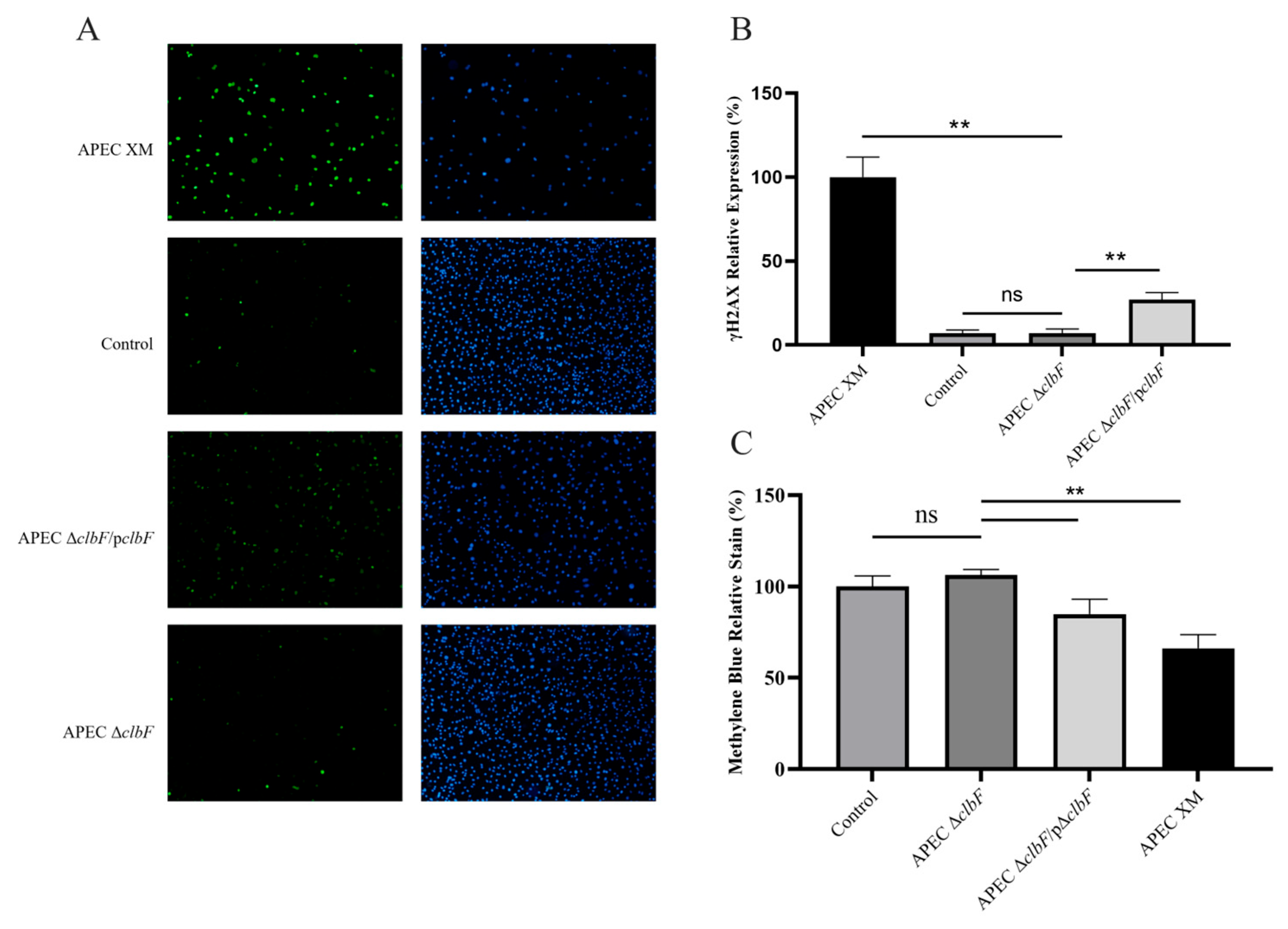
Colibactin production was assessed by quantifying γH2AX expression (a DSBs-sensitive marker) and megalocytosis in infected bEnd.3 cells. Significantly elevated γH2AX cell percentages were observed at 72 hpi in the WT and APEC ΔclbF/pclbF groups compared with the control group, whereas the APEC ΔclbF group showed no increase (see Figure 2A,B). Consistent with colibactin-induced cytopathic effects, the WT and APEC ΔclbF/pclbF strains caused progressive cellular enlargement (megalocytosis). Cytotoxicity quantification via methylene blue absorbance (600 nm) revealed significant reduction in these groups (Figure 2C), while APEC ΔclbF-infected cells maintained control-level absorbance.

3.3. clbF-Attenuated Virulence in In Vivo Infection

A mouse model was used to assess the virulence of wild-type, mutant, and complementation strains. The LD50 values of WT, APEC ΔclbF, and APEC ΔclbF/pclbF were 1.79 × 10⁵, 5.66 × 10⁶, and 3.16 × 10⁵ CFU/mouse, respectively (Table 1). There was a 3.16-fold decrease in virulence for mutant strain compared with the wild strain. Then, we tested the systemic infection effect of clbF in vivo. Bacterial loads in brain and blood at 24 hpi revealed significantly reduced colonization by APEC ΔclbF versus WT (Figure 3A,B). Complementation restored colonization capacity to WT levels across all organs. Thus, clbF deletion substantially diminished both virulence and systemic dissemination.

3.4. Expression of Virulence Factors

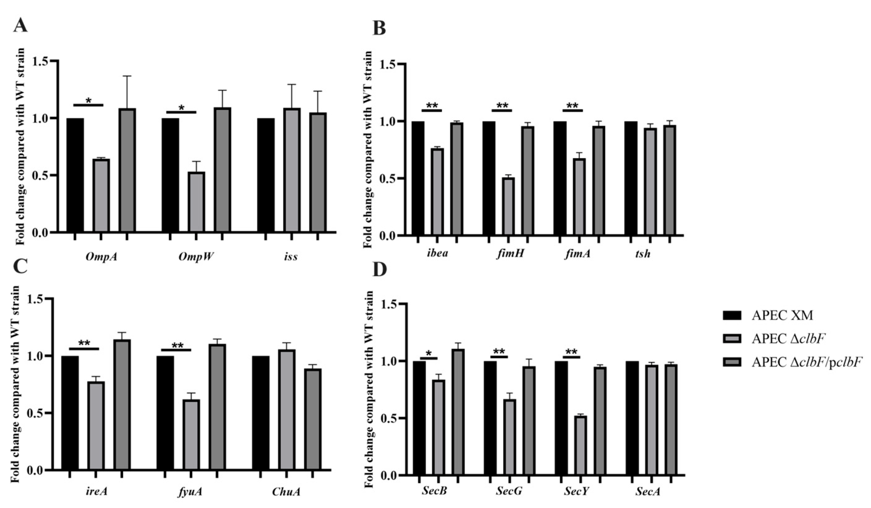
RT-qPCR was used to quantify the expression of the antiserum survival factor genes (ompA, ompW, and iss), adherence and invasion genes (ibea, fimH, tsh, and fimA), iron acquisition genes (ireA, fyuA, and ChuA), and secretion system genes (Sec ABGY). The expressions of ompA, ompW, iss, ibea, fimH, fimA, ireA, fyuA, Sec B, Sec G, and Sec Y genes were down-regulated by 35.5%, 46.6%, 23.5%, 48.8%, 19.5%, 32.2%, 22.2%, 38.1%, 16.6%, 33.2%, and 47.6%, respectively, in the APEC ΔclbF when compared with their expression in the WT strain (Figure 4). There were no differences among all strains in the expression of tsh, ChuA, and Sec A. (Figure 4)

4. Discussion

Colibactin is a structurally uncharacterized genotoxin structure created by human gut commensal Escherichia coli strains containing a 54-kilobase nonribosomal peptide–polyketide biosynthetic gene cluster [5]. The pks+ genotype predominates in E. coli phylogroups A, B1, B2, and D with significantly higher prevalence in B2 [23]. As most ExPEC strains belong to the highly pathogenic phylogroups B2 and D [24], this suggests that the presence of the pks island may be closely linked to the high pathogenicity of Escherichia coli. Notably, PEC and NMEC utilize similar pathogenic strategies for meningitis induction, positioning APEC as an emerging foodborne zoonotic pathogen and a potential reservoir for human extraintestinal infections. Consequently, APEC’s meningitis-causing mechanisms warrant urgent investigation. This study used a pks+ E. coli strain (isolated from a Muscovy duck with meningitis and sepsis), which has previously been used to induce meningitis in newborn or young avian and rodents [25,26]. Mechanistic studies indicate that clbF synthesizes the aminomalonyl (AM) extender unit essential for colibactin production [20,23]. However, clbF’s role in APEC pathogenicity remains undefined. Here, we constructed a clbF-deletion mutant to assess its impact on bacterial biology and virulence.
The γH2AX expression and methylene blue staining definitively demonstrated that clbF deletion severely impaired colibactin production, substantially reducing DNA damage caused by this genotoxin. Rapidly proliferating bacteria exhibit greater competitive advantage in host ecological niche colonization than their slower-growing counterparts. Growth test in vitro showed that clbF does not affect the growth ability of APEC XM. Both LD50 assays and the quantification of bacterial CFUs in 4-week infected mouse organs demonstrated significantly attenuated virulence in the clbF-deletion mutant. These findings establish colibactin as a critical virulence factor in APEC that mediates severe host pathology during infection.
E. coli-induced meningitis requires three essential steps: colonization/invasion, bacteremia, and blood–brain barrier (BBB) traversal. Intraperitoneal infection with clbF-deficient E. coli resulted in significantly reduced bacterial loads in the brains and blood of mice. We hypothesized that clbF deficiency may compromise bloodstream survival and expression of the BBB-crossing virulence factor. Based on prior dual RNA-seq findings [21], we therefore quantified the transcriptional expression of virulence genes, which were related to serum resistance, adhesion and invasion, the secretion system, and iron acquisition/metabolism.
Successful BBB traversal by pathogenic E. coli requires achieving sufficient bacteremia density in the bloodstream. In this study, we have tested three serum resistance-associated genes (ompA, ompW and iss). OmpA, encoding a 35 kDa protein essential for serum survival and blood–brain barrier invasion [27], additionally inhibits dendritic cell maturation by reducing the surface expression of costimulatory markers (such as CD40, HLA-DR, and CD86) [28]. neuC was predominantly associated with neonatal meningitis causing E. coli pathotype [29], which is involved in the biosynthesis of cell surface capsule [30]. In this study, we observed reduced bacterial loads in the blood of mice infected with the clbF deletion mutant, potentially linked to the downregulation of ompA and neuC expression.
Adhesive capacity is fundamental to APEC infectivity and dictates strain-specific pathogenicity variances. The ibeA locus orchestrates NMEC virulence factor expression and promotes early-onset neonatal meningitis pathogenesis through direct bacterial invasion and neutrophil transmigration [31,32,33]. As key virulence determinants, type 1 fimbriae mediate surface attachment and biofilm formation. Their biogenesis requires both FimH (receptor-specific adhesin) [34] and FimA (structural subunit) [35], which facilitate initial phagocytosis while conferring protection against heterophil killing [36]. Separately, tsh gene encodes a proteolytic autotransporter protein that adheres to red blood cells, hemoglobin, and extracellular matrix components [37]. In this study, we observed significant downregulation of major pilus components and ibeA but no detectable change in tsh expression. This likely accounts for the discrepancy between the unchanged adhesion/invasion capacity in vitro and the impaired BBB traversal in the mouse infection model.
Iron is essential for E. coli growth and pathogenesis, driving diverse acquisition strategies [38,39]. The ferric yersiniabactin transporter fyuA enables bloodstream survival [40] and correlates with B2 phylogroup prevalence [41]. In human ExPEC, the virulence factor ireA mediates iron uptake while enhancing adhesion/stress resistance across hosts [38,42]. Although ChuA (a heme-iron receptor) is widely distributed [43], it is non-essential for bacteremia or adhesion/invasion [44]. In this study, we found that the expression of fuyA and ireA was downregulated in the clbF-deletion mutant, potentially explaining the significantly reduced infection rate of the ICR mice. However, the specific underlying mechanisms require further investigation.
While ClbM (a MATE transporter of colibactin) is essential for pre-colibactin translocation to the periplasm [45], its precise mechanism remains elusive. Our dual RNA-seq of TW infecting host cells revealed upregulation of general secretion (Sec) system genes [21]. The Sec system comprises SecA, SecB, and two complexes, SecYEG and SecDFYajC [46], which transports virulence factors across membranes and supports pathogenesis [47]. In this study, we found that the deletion of clbF results in decreased transcription levels of SecB, ecG, and SecY but does not affect SecA transcription. The general system is closely linked to the transport of multiple virulence factors of E. coli. A significant reduction in mortality was observed in the APEC ΔclbF group, which is likely also attributable to the diminished of colibactin production. However, the precise mechanisms involved need further investigation.

5. Conclusions

Our experiments demonstrated that the deletion of clbF blocks colibactin production. Despite having no in vitro effect on adhesion or invasion, the loss of clbF reduced bacterial pathogenicity, an effect potentially associated with genes involved in survival, adhesion/invasion, iron uptake, and secretion systems. These results establish clbF as a key virulence factor essential for APEC XM to induce meningitis.

Supplementary Materials

The following supporting information can be downloaded at https://www.mdpi.com/article/10.3390/vetsci12080727/s1, Table S1: Summary of bacterial strains, plasmids, and primers used in this study; Figure S1: the original image of Figure 1A.

Author Contributions

Conceptualization, P.W.; methodology, P.W. and M.W., formal analysis, M.W., H.W., and L.L.; investigation, M.W., H.W., and L.L.; data curation, L.L.; writing—original draft preparation, M.W. and P.W.; writing—review and editing, P.H. and P.W.; supervision, P.W.; funding acquisition, P.H. and P.W. All authors have read and agreed to the published version of the manuscript.

Funding

This research was funded by the Natural Science Foundation of the Jiangsu Higher Education Institutions of China, grant number No. 22KJB230006, and the Natural Science Fund Project of Jiangsu Vocational College of Agriculture and Forestry (Nos. 2022kj31).

Institutional Review Board Statement

The animal study protocol was approved by the Animal Care and Ethics Committee of Jiangsu Agri-animal Husbandry Vocational College (protocol code JSAHVC-2025-78 and date of approval: 15 January 2025.

Informed Consent Statement

Not applicable.

Data Availability Statement

Dataset available on request from the authors.

Acknowledgments

The authors thank Guoqiang Zhu (Yangzhou University) for generously providing the E. coli strains and plasmids used in this study.

Conflicts of Interest

The authors declare no conflicts of interest.

References

  1. Mellata, M. Human and avian extraintestinal pathogenic Escherichia coli: Infections, zoonotic risks, and antibiotic resistance trends. Foodborne Pathog. Dis. 2013, 10, 916–932. [Google Scholar] [CrossRef]
  2. Tivendale, K.A.; Logue, C.M.; Kariyawasam, S.; Jordan, D.; Hussein, A.; Li, G.; Wannemuehler, Y.; Nolan, L.K. Avian-pathogenic Escherichia coli strains are similar to neonatal meningitis E. coli strains and are able to cause meningitis in the rat model of human disease. Infect. Immun. 2010, 78, 3412–3419. [Google Scholar] [CrossRef] [PubMed]
  3. Ewers, C.; Li, G.; Wilking, H.; Kiessling, S.; Alt, K.; Antáo, E.M.; Laturnus, C.; Diehl, I.; Glodde, S.; Homeier, T.; et al. Avian pathogenic, uropathogenic, and newborn meningitis-causing Escherichia coli: How closely related are they? Int. J. Med. Microbiol. 2007, 297, 163–176. [Google Scholar] [CrossRef] [PubMed]
  4. Skyberg, J.A.; Johnson, T.J.; Johnson, J.R.; Clabots, C.; Logue, C.M.; Nolan, L.K. Acquisition of avian pathogenic Escherichia coli plasmids by a commensal E. coli isolate enhances its abilities to kill chicken embryos, grow in human urine, and colonize the murine kidney. Infect. Immun. 2006, 74, 6287–6292. [Google Scholar] [CrossRef]
  5. Nougayrède, J.P.; Homburg, S.; Taieb, F.; Boury, M.; Brzuszkiewicz, E.; Gottschalk, G.; Buchrieser, C.; Hacker, J.; Dobrindt, U.; Oswald, E. Escherichia coli induces DNA double-strand breaks in eukaryotic cells. Science 2006, 313, 848–851. [Google Scholar] [CrossRef]
  6. Silpe, J.E.; Wong, J.W.H.; Owen, S.V.; Baym, M.; Balskus, E.P. The bacterial toxin colibactin triggers prophage induction. Nature 2022, 603, 315–320. [Google Scholar] [CrossRef]
  7. Lucas, C.; Salesse, L.; Hoang, M.H.T.; Bonnet, M.; Sauvanet, P.; Larabi, A.; Godfraind, C.; Gagnière, J.; Pezet, D.; Rosenstiel, P.; et al. Autophagy of Intestinal Epithelial Cells Inhibits Colorectal Carcinogenesis Induced by Colibactin-Producing Escherichia coli in Apc(Min/+) Mice. Gastroenterology 2020, 158, 1373–1388. [Google Scholar] [CrossRef]
  8. Jans, M.; Kolata, M.; Blancke, G.; D’Hondt, A.; Gräf, C.; Ciers, M.; Sze, M.; Thiran, A.; Petta, I.; Andries, V.; et al. Colibactin-driven colon cancer requires adhesin-mediated epithelial binding. Nature 2024, 635, 472–480. [Google Scholar] [CrossRef]
  9. Marcq, I.; Martin, P.; Payros, D.; Cuevas-Ramos, G.; Boury, M.; Watrin, C.; Nougayrède, J.P.; Olier, M.; Oswald, E. The genotoxin colibactin exacerbates lymphopenia and decreases survival rate in mice infected with septicemic Escherichia coli. J. Infect. Dis. 2014, 210, 285–294. [Google Scholar] [CrossRef]
  10. McCarthy, A.J.; Martin, P.; Cloup, E.; Stabler, R.A.; Oswald, E.; Taylor, P.W. The Genotoxin Colibactin Is a Determinant of Virulence in Escherichia coli K1 Experimental Neonatal Systemic Infection. Infect. Immun. 2015, 83, 3704–3711. [Google Scholar] [CrossRef]
  11. Massip, C.; Branchu, P.; Bossuet-Greif, N.; Chagneau, C.V.; Gaillard, D.; Martin, P.; Boury, M.; Sécher, T.; Dubois, D.; Nougayrède, J.P.; et al. Deciphering the interplay between the genotoxic and probiotic activities of Escherichia coli Nissle 1917. PLoS Pathog. 2019, 15, e1008029. [Google Scholar] [CrossRef]
  12. Zha, L.; Wilson, M.R.; Brotherton, C.A.; Balskus, E. Characterization of Polyketide Synthase Machinery from the pks Island Facilitates Isolation of a Candidate Precolibactin. ACS Chem. Biol. 2016, 11, 1287–1295. [Google Scholar] [CrossRef]
  13. Brachmann, A.O.; Garcie, C.; Wu, V.; Martin, P.; Ueoka, R.; Oswald, E.; Piel, J. Colibactin biosynthesis and biological activity depend on the rare aminomalonyl polyketide precursor. Chem. Commun. 2015, 51, 13138–13141. [Google Scholar] [CrossRef]
  14. Zhong, H.; Wang, P.; Chen, Y.; Wang, H.; Li, J.; Li, J.; Zhu, G.; Cui, L.; Meng, X. ClpV1 in avian pathogenic Escherichia coli is a crucial virulence factor contributing to meningitis in a mouse model in vivo. Vet. Microbiol. 2021, 263, 109273. [Google Scholar] [CrossRef] [PubMed]
  15. Zhang, D.; Xu, S.; Wang, Y.; Bin, P.; Zhu, G. The Amino Acid-mTORC1 Pathway Mediates APEC TW-XM-Induced Inflammation in bEnd.3 Cells. Int. J. Mol. Sci. 2021, 22, 9245. [Google Scholar] [CrossRef] [PubMed]
  16. Hejair, H.M.A.; Ma, J.; Zhu, Y.; Sun, M.; Dong, W.; Zhang, Y.; Pan, Z.; Zhang, W.; Yao, H. Role of outer membrane protein T in pathogenicity of avian pathogenic Escherichia coli. Res. Vet. Sci. 2017, 115, 109–116. [Google Scholar] [CrossRef]
  17. Wang, P.; Zhang, J.; Chen, Y.; Zhong, H.; Wang, H.; Li, J.; Zhu, G.; Xia, P.; Cui, L.; Li, J.; et al. ClbG in Avian Pathogenic Escherichia coli Contributes to Meningitis Development in a Mouse Model. Toxins 2021, 13, 546. [Google Scholar] [CrossRef] [PubMed]
  18. Meng, X.; Chen, Y.; Wang, P.; Xia, P.; Wang, J.; He, M.; Zhu, C.; Wang, H.; Zhu, G. Phosphopantetheinyl transferase ClbA contributes to the virulence of avian pathogenic Escherichia coli in meningitis infection of mice. PLoS ONE 2022, 17, e0269102. [Google Scholar] [CrossRef]
  19. Wang, P.; Zhang, J.; Chen, Y.; Zhong, H.; Wang, H.; Li, J.; Zhu, G.; Xia, P.; Cui, L.; Li, J.; et al. Colibactin in avian pathogenic Escherichia coli contributes to the development of meningitis in a mouse model. Virulence 2021, 12, 2382–2399. [Google Scholar] [CrossRef]
  20. Datsenko, K.A.; Wanner, B.L. One-step inactivation of chromosomal genes in Escherichia coli K-12 using PCR products. Proc. Natl. Acad. Sci. USA 2000, 97, 6640–6645. [Google Scholar] [CrossRef]
  21. Wang, P.; Meng, X.; Li, J.; Chen, Y.; Zhang, D.; Zhong, H.; Xia, P.; Cui, L.; Zhu, G.; Wang, H. Transcriptome profiling of avian pathogenic Escherichia coli and the mouse microvascular endothelial cell line bEnd.3 during interaction. PeerJ 2020, 8, e9172. [Google Scholar] [CrossRef]
  22. Bossuet-Greif, N.; Vignard, J.; Taieb, F.; Mirey, G.; Dubois, D.; Petit, C.; Oswald, E.; Nougayrède, J.P. The Colibactin Genotoxin Generates DNA Interstrand Cross-Links in Infected Cells. mBio 2018, 9, e02393-17. [Google Scholar] [CrossRef] [PubMed]
  23. Auvray, F.; Perrat, A.; Arimizu, Y.; Chagneau, C.V.; Bossuet-Greif, N.; Massip, C.; Brugère, H.; Nougayrède, J.P.; Hayashi, T.; Branchu, P.; et al. Insights into the acquisition of the pks island and production of colibactin in the Escherichia coli population. Microb. Genom. 2021, 7, 000579. [Google Scholar] [CrossRef] [PubMed]
  24. Ranjbar, R.; Nazari, S.; Farahani, O. Phylogenetic Analysis and Antimicrobial Resistance Profiles of Escherichia coli Strains Isolated from UTI-Suspected Patients. Iran. J. Public Health 2020, 49, 1743–1749. [Google Scholar] [CrossRef] [PubMed]
  25. Hejair, H.M.A.; Zhu, Y.; Ma, J.; Zhang, Y.; Pan, Z.; Zhang, W.; Yao, H. Functional role of ompF and ompC porins in pathogenesis of avian pathogenic Escherichia coli. Microb. Pathog. 2017, 107, 29–37. [Google Scholar] [CrossRef]
  26. Zhang, D.; Xu, S.; Wu, H.; Liu, J.; Wang, Y.; Zhu, G. Melatonin Is Neuroprotective in Escherichia coli Meningitis Depending on Intestinal Microbiota. Int. J. Mol. Sci. 2022, 24, 298. [Google Scholar] [CrossRef]
  27. Maruvada, R.; Kim, K.S. Extracellular loops of the Escherichia coli outer membrane protein A contribute to the pathogenesis of meningitis. J. Infect. Dis. 2011, 203, 131–140. [Google Scholar] [CrossRef]
  28. Mittal, R.; Prasadarao, N.V. Outer membrane protein A expression in Escherichia coli K1 is required to prevent the maturation of myeloid dendritic cells and the induction of IL-10 and TGF-beta. J. Immunol. 2008, 181, 2672–2682. [Google Scholar] [CrossRef]
  29. Wijetunge, D.S.; Gongati, S.; DebRoy, C.; Kim, K.S.; Couraud, P.O.; Romero, I.A.; Weksler, B.; Kariyawasam, S. Characterizing the pathotype of neonatal meningitis causing Escherichia coli (NMEC). BMC Microbiol. 2015, 15, 211. [Google Scholar] [CrossRef]
  30. Mediati, D.G.; Blair, T.A.; Costas, A.; Monahan, L.G.; Söderström, B.; Charles, I.G.; Duggin, I.G. Genetic requirements for uropathogenic E. coli proliferation in the bladder cell infection cycle. mSystems 2024, 9, e0038724. [Google Scholar] [CrossRef]
  31. Wang, S.; Niu, C.; Shi, Z.; Xia, Y.; Yaqoob, M.; Dai, J.; Lu, C. Effects of ibeA deletion on virulence and biofilm formation of avian pathogenic Escherichia coli. Infect. Immun. 2011, 79, 279–287. [Google Scholar] [CrossRef]
  32. Huang, S.H.; Chi, F.; Peng, L.; Bo, T.; Zhang, B.; Liu, L.Q.; Wu, X.; Mor-Vaknin, N.; Markovitz, D.M.; Cao, H.; et al. Vimentin, a Novel NF-κB Regulator, Is Required for Meningitic Escherichia coli K1-Induced Pathogen Invasion and PMN Transmigration across the Blood-Brain Barrier. PLoS ONE 2016, 11, e0162641. [Google Scholar] [CrossRef] [PubMed]
  33. Soto, S.M.; Bosch, J.; Jimenez de Anta, M.T.; Vila, J. Comparative study of virulence traits of Escherichia coli clinical isolates causing early and late neonatal sepsis. J. Clin. Microbiol. 2008, 46, 1123–1125. [Google Scholar] [CrossRef] [PubMed]
  34. Tchesnokova, V.; Aprikian, P.; Kisiela, D.; Gowey, S.; Korotkova, N.; Thomas, W.; Sokurenko, E. Type 1 fimbrial adhesin FimH elicits an immune response that enhances cell adhesion of Escherichia coli. Infect. Immun. 2011, 79, 3895–3904. [Google Scholar] [CrossRef] [PubMed]
  35. Puorger, C.; Vetsch, M.; Wider, G.; Glockshuber, R. Structure, folding stability of FimA, the main structural subunit of type 1 pili from uropathogenic Escherichia coli strains. J. Mol. Biol. 2011, 412, 520–535. [Google Scholar] [CrossRef]
  36. Mellata, M.; Dho-Moulin, M.; Dozois, C.M.; Curtiss, R., 3rd; Lehoux, B.; Fairbrother, J.M. Role of avian pathogenic Escherichia coli virulence factors in bacterial interaction with chicken heterophils and macrophages. Infect. Immun. 2003, 71, 494–503. [Google Scholar] [CrossRef]
  37. Kostakioti, M.; Stathopoulos, C. Functional analysis of the Tsh autotransporter from an avian pathogenic Escherichia coli strain. Infect. Immun. 2004, 72, 5548–5554. [Google Scholar] [CrossRef]
  38. Russo, T.A.; Carlino, U.B.; Johnson, J.R. Identification of a new iron-regulated virulence gene, ireA, in an extraintestinal pathogenic isolate of Escherichia coli. Infect. Immun. 2001, 69, 6209–6216. [Google Scholar] [CrossRef]
  39. Khan, A.; Singh, P.; Srivastava, A. Synthesis, nature and utility of universal iron chelator—Siderophore: A review. Microbiol. Res. 2018, 212–213, 103–111. [Google Scholar] [CrossRef]
  40. Bunduki, G.K.; Heinz, E.; Phiri, V.S.; Noah, P.; Feasey, N.; Musaya, J. Virulence factors and antimicrobial resistance of uropathogenic Escherichia coli (UPEC) isolated from urinary tract infections: A systematic review and meta-analysis. BMC Infect. Dis. 2021, 21, 753. [Google Scholar] [CrossRef]
  41. López-Banda, D.A.; Carrillo-Casas, E.M.; Leyva-Leyva, M.; Orozco-Hoyuela, G.; Manjarrez-Hernández, Á.H.; Arroyo-Escalante, S.; Moncada-Barrón, D.; Villanueva-Recillas, S.; Xicohtencatl-Cortes, J.; Hernández-Castro, R. Identification of virulence factors genes in Escherichia coli isolates from women with urinary tract infection in Mexico. Biomed. Res. Int. 2014, 2014, 959206. [Google Scholar] [CrossRef] [PubMed]
  42. Li, Y.; Dai, J.; Zhuge, X.; Wang, H.; Hu, L.; Ren, J.; Chen, L.; Li, D.; Tang, F. Iron-regulated gene ireA in avian pathogenic Escherichia coli participates in adhesion and stress-resistance. BMC Vet. Res. 2016, 12, 167. [Google Scholar] [CrossRef] [PubMed]
  43. Torres, A.G.; Payne, S.M. Haem iron-transport system in enterohaemorrhagic Escherichia coli O157:H7. Mol. Microbiol. 1997, 23, 825–833. [Google Scholar] [CrossRef] [PubMed]
  44. Nègre, V.L.; Bonacorsi, S.; Schubert, S.; Bidet, P.; Nassif, X.; Bingen, E. The siderophore receptor IroN, but not the high-pathogenicity island or the hemin receptor ChuA, contributes to the bacteremic step of Escherichia coli neonatal meningitis. Infect. Immun. 2004, 72, 1216–1220. [Google Scholar] [CrossRef]
  45. Mousa, J.J.; Yang, Y.; Tomkovich, S.; Shima, A.; Newsome, R.C.; Tripathi, P.; Oswald, E.; Bruner, S.D.; Jobin, C. MATE transport of the E. coli-derived genotoxin colibactin. Nat. Microbiol. 2016, 1, 15009. [Google Scholar] [CrossRef]
  46. Crane, J.M.; Randall, L.L. The Sec System: Protein Export in Escherichia coli. EcoSal Plus 2017, 7. [Google Scholar] [CrossRef]
  47. Green, E.R.; Mecsas, J. Bacterial Secretion Systems: An Overview. Microbiol. Spectr. 2016, 4, 213–239. [Google Scholar] [CrossRef]
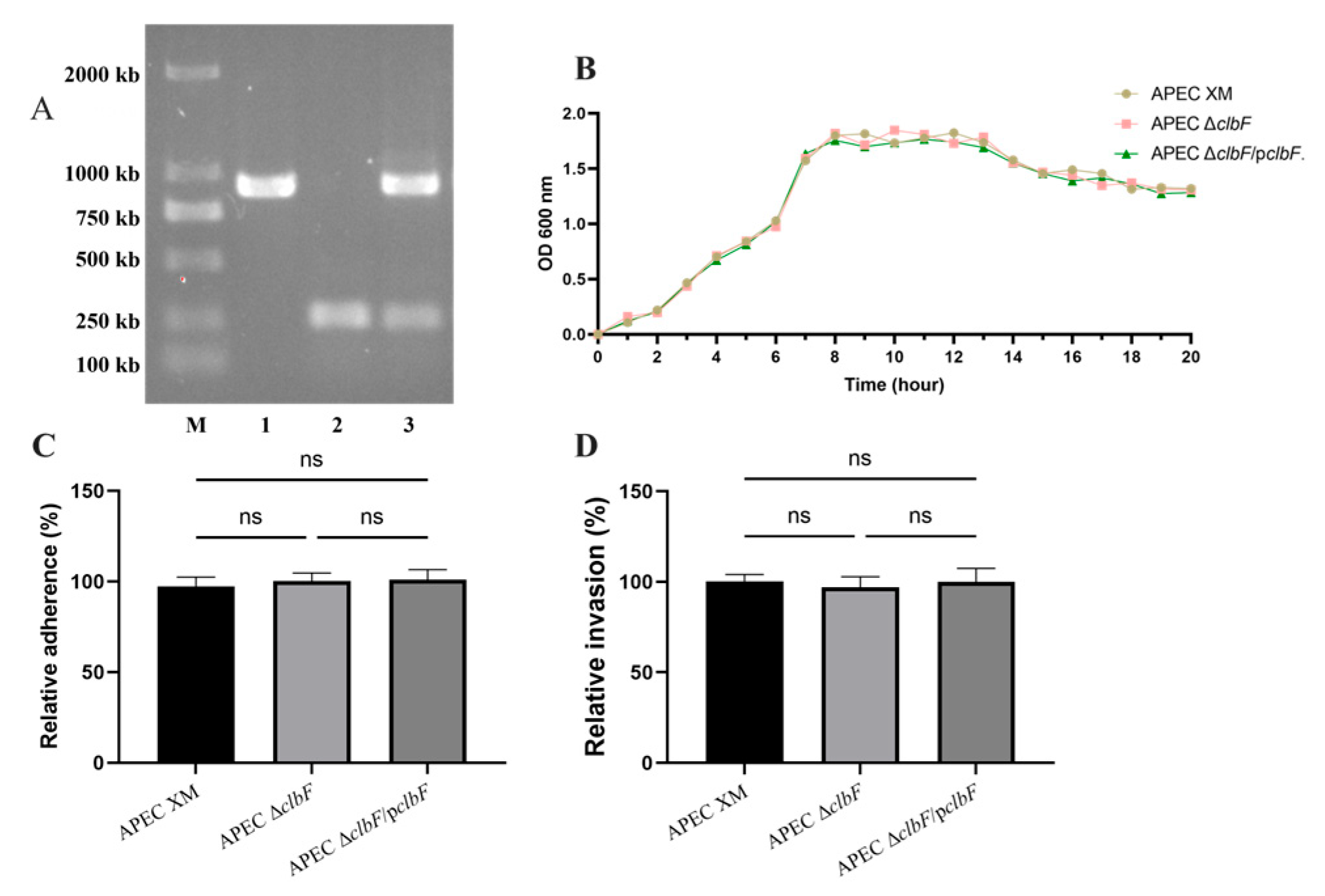
Figure 1. Verification of mutants and the ability of reproduction, adherence, and invasion. (A). Identification of mutants by PCR. The three strains, APEC, APEC ΔclbF, and APEC ΔclbF/pclbF, were amplified with primer P1/P2. (Lane M, DNA molecular size marker; Lane 1, APEC XM; Lane 2, APEC ΔclbF; and Lane 3, APEC ΔclbF/pclbF) (B). Growth curves analysis. The three strains, APEC, APEC ΔclbF, and APEC ΔclbF/pclbF, were grown in LB broth at 37 °C and the OD600 values were measured every 1 h over a period of 20 h. Each curve represents the averages of three independent experiments. Effects of clbF on adherence to (C) and invasion (D) of bEnd.3 cells (MOI = 100). All assays were carried out in triplicate (ns, p > 0.05).
Vetsci 12 00727 g001
Figure 2. ClbF is essential for colibactin biosynthesis. (A) Nuclear DNA and γH2AX were stained blue and green, respectively. (B) Percentage of γH2AX-positive cells at 72 hpi, normalized to the APEC XM group (set as 100%). (C) Megalocytosis in methylene blue-stained cells was quantified by absorbance (600 nm), with control group data normalized to 100% (**, p < 0.01; ns, p > 0.05).
Vetsci 12 00727 g002
Figure 3. ClbF affects the pathogenicity of APEC XM. ICR mice were inoculated intraperitoneally with a dose of 107 CFU bacteria or an equal volume of sterile saline. Bacterial load assay for the brain (A), blood (B), and spleen (C) by plate counting (**, p < 0.01; *, p < 0.05; ns, p > 0.05).
Vetsci 12 00727 g003
Figure 4. Related genes expression levels: (A) antiserum survival factor genes (ompA, ompW, and iss); (B) adherence and invasion genes (ibea, fimH, tsh, and fimA); (C) iron acquisition genes (ireA, fyuA, and ChuA); (D) secretion system genes (Sec ABGY). Transcript expression of the indicated genes was measured using RT-qPCR. gapA was used as the normalizing internal standard. Changes were calculated using APEC XM as the relative measure of comparison. (**, p < 0.01; *, p < 0.05).
Vetsci 12 00727 g004
Table 1. Calculations of LD50.
Dose of Challenge (CFU)No. of Dead Mice
APEC XMAPEC ΔclbFAPEC ΔclbF/pclbF
1 × 1075/53/54/5
1 × 1064/51/54/5
1 × 1051/51/51/5
1 × 1041/50/51/5
LD501.79 × 105 5.66 × 1063.16 × 105
Disclaimer/Publisher’s Note: The statements, opinions and data contained in all publications are solely those of the individual author(s) and contributor(s) and not of MDPI and/or the editor(s). MDPI and/or the editor(s) disclaim responsibility for any injury to people or property resulting from any ideas, methods, instructions or products referred to in the content.