Article

Evaluation of Cytokine Profile in Canine Malignant Oral Melanoma

by
Carmen G. Pérez-Santana
1,2,3,†,
Sara E. Cazorla-Rivero
2,4,†,
Ana A. Jiménez-Alonso
1,2,
Francisco Rodríguez-Esparragón
2,3,*,
Jesús María González Martín
2,5,6,
Ruth Henríquez-Cabrera
2,3,
Bernardino Clavo-Varas
2,3,6,7,8,9 and
Enrique Rodríguez Grau-Bassas
1,2
1
Instituto Universitario de Sanidad Animal y Seguridad Alimentaria (IUSA), Universidad de Las Palmas de Gran Canaria (ULPGC), 35400 Arucas, Spain
2
Unidad de Investigación Hospital Universitario de Gran Canaria Dr. Negrín, 35010 Las Palmas de Gran Canaria, Spain
3
Fundación Canaria Instituto de Investigación Sanitaria de Canarias (FIISC), Hospital Universitario de Gran Canaria Dr. Negrín, 35010 Las Palmas de Gran Canaria, Spain
4
Universidad de La Laguna, 38200 La Laguna, Spain
5
CIBER de Enfermedades Respiratorias, Instituto de Salud Carlos III, 28029 Madrid, Spain
6
Instituto Universitario de Enfermedades Tropicales y Salud Pública de Canarias, Universidad de La Laguna, 38296 La Laguna, Spain
7
CIBER de Enfermedades Infecciosas (CIBERINFEC), Instituto de Salud Carlos III, 28029 Madrid, Spain
8
Chronic Pain Unit, Hospital Universitario Dr. Negrín, 35010 Las Palmas de Gran Canaria, Spain
9
Radiation Oncology Department, Hospital Universitario de Gran Canaria, Dr. Negrín, 35010 Las Palmas de Gran Canaria, Spain
*
Author to whom correspondence should be addressed.
These authors contributed equally to this work.
Vet. Sci. 2025, 12(7), 627; https://doi.org/10.3390/vetsci12070627
Submission received: 16 April 2025 / Revised: 19 June 2025 / Accepted: 24 June 2025 / Published: 30 June 2025

Simple Summary

Canine models have been recognized as valuable tools for studying various human cancers, including melanoma. Research on carcinogenesis and the development of innovative cancer therapies is progressing rapidly, leading to improved treatment options in veterinary medicine. Despite this progress, the prognostic role of cytokines in canine oncology remains insufficiently investigated. This prospective study reports on 10 cases of oral malignant melanoma (OMM) in dogs that underwent surgical treatment. Among them, four were diagnosed with melanotic and six with amelanotic melanoma. Serum samples were collected at baseline (prior to surgery), on the day of surgery, and subsequently every 3–4 months, accompanied by clinical examinations and thoracic radiographs. Concentrations of GM-CSF, IFN-γ, IL-2, IL-6, IL-7, IL-8, IL-10, IL-15, IL-18, IP-10, KC-like, MCP-1, and TNFα were quantified. Follow-up samples indicated that after the removal of malignant melanoma, the serum levels of GM-CSF, IFN-γ, MCP-1, IL-18, and IL-2 increased significantly. In contrast, when comparing samples from dogs with oral malignant melanoma to those without the disease, concentrations of IL-7 and MCP-1 were significantly higher in the absence of disease samples than in the OMM samples. Furthermore, when comparing serum concentrations between samples from OMM patients with metastasis and those patients in remission, elevated levels of MCP-1 were associated with poorer overall survival due to the development of OMM metastasis. Lastly, a comparison of cytokines in samples from melanotic OMM and amelanotic OMM revealed that amelanotic OMM samples exhibited higher concentrations of IL-6, IL-10, and IL-15 compared to their melanotic counterparts. This study contributes to the evidence that canine models can offer valuable insights that may also translate into more effective and targeted treatments for human melanoma.

Abstract

Ten dogs with oral malignant melanoma were evaluated and treated with surgery, of which four dogs were diagnosed with melanotic melanoma and six were diagnosed with amelanotic melanoma. Serum samples from oral malignant melanoma (OMM) were collected at baseline, the day of the surgery, and every 3–4 months, during which time a clinical examination and chest X-rays were performed. Concentrations of GM-CSF, IFN-γ, IL-2, IL-6, IL-7, IL-8, IL-10, IL-15, IL-18, IP-10, KC-like, MCP-1, and TNFα were quantified. Follow-up samples indicated that after the removal of malignant melanoma, the serum levels of GM-CSF, IFN-γ, MCP-1, IL-18, and IL-2 increased significantly. In contrast, when comparing samples from dogs with OMM to those of patients in remission, the concentrations of IL-7 and MCP-1 were significantly higher in the remission samples than in the OMM samples. Furthermore, when comparing the serum concentrations between the OMM-metastasis samples and those patients in remission, elevated levels of MCP-1 were associated with poorer overall survival due to the development of OMM metastasis. Finally, a comparison of cytokines in the melanotic OMM and amelanotic OMM samples revealed that the amelanotic OMM samples exhibited higher concentrations of IL-6, IL-10, and IL-15 compared to the melanotic OMM samples.

1. Introduction

Dogs have been suggested as a useful model for several types of human cancer, including melanoma [1,2]. The in-depth characterization of these models will expedite the development and application of new therapeutic strategies. Comparative genomic analyses between human and canine tumors underscore the value of the dog model in advancing knowledge of tumor initiation and progression in humans [3]. These studies have accelerated the identification of clinically relevant treatments for both species. Immunotherapy, in particular, shows promise as a strategy due to its potential for systemic but cancer-specific therapeutic effects [4,5].
Immunotherapy is now recognized as one of the fundamental pillars of human cancer treatment, with clinical importance equivalent to surgery, radiotherapy, and traditional chemotherapy [4,6,7]. The study of carcinogenesis and the development of different cancer therapies is an extremely rapidly advancing area of research [8,9]. New treatment modalities are being developed to provide better veterinary care for canines [10,11].
Immune checkpoint inhibitors (ICIs) are widely used to treat human cancers, and growing evidence suggests that ICIs are promising treatments for canine malignancies [12]. To date, several serum factors have been identified that are predictive of ICI benefit among human cancer patients, including reactive protein (CRP), IL-6, soluble PD-L1, and various cytokines and chemokines [13,14,15]. However, the predictive value of cytokines is still under-investigated in canine cancer. In veterinary medicine, several clinical studies have evaluated the efficacy of immune checkpoint blockade for dogs with cancer [11,12].
Therefore, we aimed to prospectively identify specific cytokine levels or profiles that could serve as biomarkers and provide information either on disease status, tumor presence or absence after surgery, or that would allow distinguishing between metastases and tumor subtypes throughout the follow-up period.

2. Materials and Methods

Patient samples. Dogs affected by oral malignant melanoma that were presented at the Veterinary Oncology Service of GICOREC IUSA (Instituto Universitario de Sanidad Animal y Seguridad Alimentaria) of the Universidad de Las Palmas de Gran Canaria (ULPGC, Gran Canaria, Spain) since 2021, with a minimum follow-up of 1 year to 2024, were prospectively considered for this study. The dogs were presented for surgical treatment and were treated according to the Good Clinical Practice guidelines for animal clinical studies and approved by the bioethics committee of ULPGC (OEBA-ULPGC 33/2020R1).
Ten dogs were staged by obtaining a thorough clinical examination, complete blood cell count, serum biochemistry profile, X-rays of the thorax (three views), and total body computed tomography (CT) when indicated. Canine candidates for surgical excision of the primary tumor with or without regional lymphadenectomy and a histopathologically confirmed diagnosis of oral melanoma were included.
A total of 4 mL of blood was obtained from the jugular vein and immediately centrifuged to separate the serum. The samples were collected for the first time from oral malignant melanoma (OMM) dogs on the day of surgery. Subsequent samples were obtained every 4 months, during which time a clinical examination and chest X-rays were performed, until the completion of the 1-year follow-up. Concentrations of granulocyte-macrophage colony-stimulating factor (GM-CSF), interferon γ (IFN-γ), interleukins (IL)-2, IL-6, IL-7, IL-8, IL-10, IL-15, IL-18, interferon gamma-induced protein 10 (IP-10), keratinocyte chemotactic-like (KC-like), monocyte chemotactic protein-1 (MCP-1), and tumor necrosis factor α (TNFα) were quantified with MILLIPLEX Canine Cytokine Magnetic Bead Panel (Merck Millipore) using Luminex technology LABScan3D (XMAP).
Statistical analysis. Results are expressed as median, standard deviation, percentiles, and quartiles. The Shapiro–Wilk test was applied to assess the distribution of the quantitative variables. Pearson’s or Spearman’s coefficients were employed to evaluate the correlations between numerical variables.
Cytokine values were compared between amelanotic oral melanoma and melanotic oral melanoma expressed at the time of surgery using the Mann–Whitney U test. Friedman’s non-parametric test was performed to explore if there were significant differences over different time points. Mean comparisons between the groups (absence of the disease, presence of the tumor, and metastasis) were evaluated using the Mann–Whitney U test or the Kruskal–Wallis test when appropriate.
The statistical program R was used for statistical analysis (R version 4.3.2).

3. Results

Animals and serum samples. The main characteristics of the affected dogs are depicted in Table 1. The collected data includes breed, sex, age, reproductive state, diagnosis, TNM classification [16], and tumor size. Among the 10 oral melanomas measured on the day of surgical excision, 1 case had a maximum dimension of less than 2 cm, 8 cases ranged from 2 to 4 cm, and 1 case exceeded 4 cm, regardless of the stage. Histological examination of the excision margins revealed that all the dogs had non-infiltrated margins. Additionally, the mitotic index was found to be ≥4/10 high-power fields (HPF) in all dogs.
Out of a total of 10 patients, 5 remained alive until the completion of the 1-year follow-up, and 5 died due to metastasis (Table 2). In total, 31 serum samples (Table 3) were collected and analyzed, including 10 from patients with OMM on the day of surgery (T0) and 21 from follow-ups (T1, T2.1, T2.2). Of these, 16 were from patients in remission, 2 from those with OMM recurrence, and 3 from patients with metastasis, with 2 patients dying before their scheduled blood extraction. Samples were collected until patients completed 1 year of follow-up or died, with T0 indicating OMM presence, T1 representing the first 4 months post-tumor removal, and T2.1 and T2.2 referring to samples taken every 4 months until the conclusion of the one-year follow-up.
Cytokines evaluation and follow-up. The samples were categorized as OMM-diseased dog samples (Figure 1) and revision samples, which included samples from patients with metastasis and samples from OMM dogs in remission.
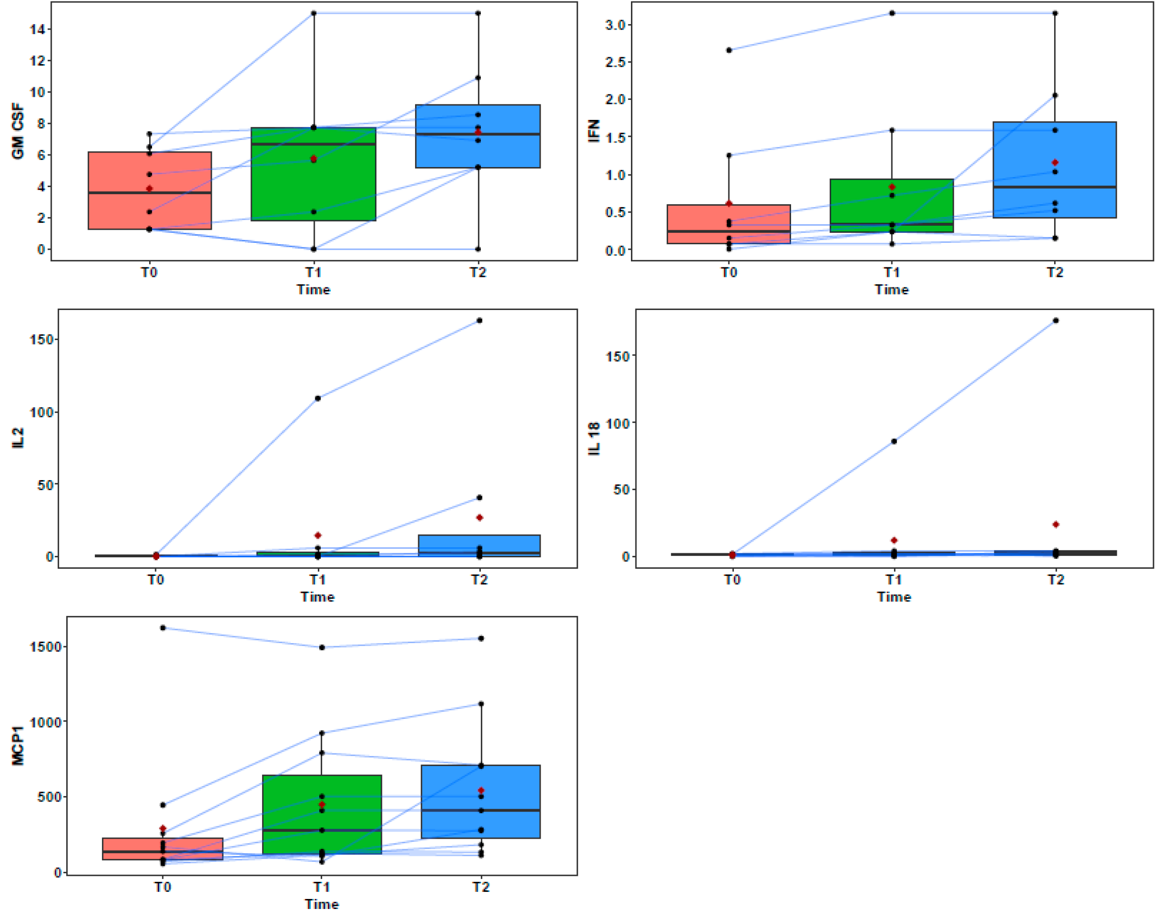
Pairwise comparisons indicate that there were significant higher serum cytokine levels between the T2 and initial T0 values for GM-CSF (7.31 pg/mL and 3.56 pg/mL; p = 0.043), IFN-γ (0.83 pg/mL and 0.24 pg/mL; p = 0.002), MCP-1 (408.55 pg/mL and 136.49 pg/mL; p = 0.024), IL-2 (2.64 pg/mL and 0 pg/mL; p = 0.014) and IL-18 (2.46 pg/mL and 1.23 pg/mL; p = 0.04). Figure 2 shows the GM-CSF, IFN-γ, MCP-1, IL-2, and IL-18 values over time.
When the patients were evaluated individually, it was observed that, although not statistically significant, the serum levels of GM-CSF, IFN-γ, IL-2, and IL-18 were negatively associated with the patients who presented with disease recurrence and/or metastasis. In contrast, MCP-1 increased in all the revision samples, with particularly elevated levels observed in the metastatic samples (Table 4, Table 5, Table 6, Table 7 and Table 8).
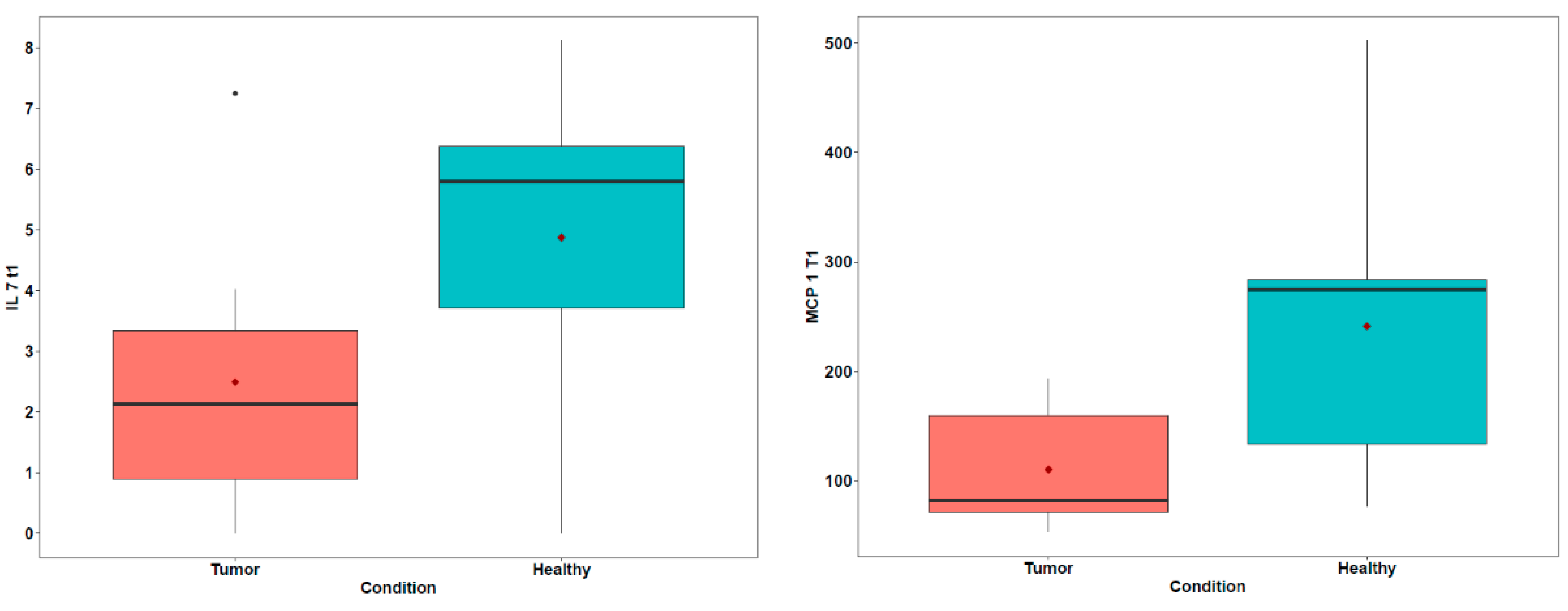
Evaluation of variability in different sample groups. To examine whether there were significant differences between the OMM-diseased dogs samples, the OMM-metastasis dogs samples, and the OMM dogs in remission samples, we first compared the cytokine concentrations between the OMM dogs in remission and the OMM-diseased dogs samples. IL-7 and MCP-1 increased in patients who were in remission after one year of follow-up (5.8 pg/mL and 2.12 pg/mL, p = 0.078; 274.68 pg/mL and 81.92 pg/mL, p = 0.051, respectively; Table 9, Figure 3). Although both differences did not result in the threshold value of significance, which is a p value < 0.05, the small sample size should be taken into consideration, with the statistical analysis showing p values between 0.05 to 0.1, supporting a trend towards significance [17,18].
To explore the serum biomarkers predictive of clinical outcome among dogs with OMM, the OMM-diseased dogs samples were compared with the OMM-metastasis samples. The results showed that the dogs with OMM metastasis displayed higher serum MCP-1 than the OMM-diseased dogs (702.88 pg/mL and 136.49 pg/mL, p = 0.036, respectively; Table 10). There were no significant associations between the other serum factors and clinical prognosis.
There were no significant differences between the OMM-metastasis samples and the OMM dogs in remission samples.
Study of variability among OMM subtypes. Consistent with previous studies demonstrating biological differences between amelanotic tumor and melanotic tumor, comparisons were performed between six amelanotic and four melanotic OMM. IL-6, IL-10, and IL-15 concentrations showed a trend towards higher levels in the amelanotic OMM samples (4.22 pg/mL and 0.74 pg/mL, p = 0.042; 13.21 pg/mL and 1.67 pg/mL, p = 0.038; 3.89 pg/mL and 0.71 pg/mL, p = 0.066, respectively; Table 11).
Blood leukocyte analysis. The neutrophil-to-lymphocyte ratio (NLR) showed a moderate inverse correlation with the survival time in OMM patients (p-value = 0.033). Furthermore, Cox regression analysis between the NEU/LYM ratio outcomes indicated that a higher ratio is a risk factor for mortality (Table 12).

4. Discussion

This study highlights the significant potential of using canine oral malignant melanoma (OMM) as a model for understanding the mechanisms of immune evasion and developing novel immunotherapeutic strategies that could be relevant to both veterinary and human medicine [9]. Given the histopathological and clinical similarities between canine and human malignant melanomas [19,20,21], the findings provide valuable insights into the immunological dynamics at play during tumor progression and post-treatment.
Our findings demonstrate a significant increase in cytokines such as IL-2, IL-18, GM-CSF, IFN-γ, and MCP-1 following tumor removal. This suggests an activation of the immune response that may contribute to the antitumor effects observed in the post-surgical phase. Notably, high serum IL-2 levels were associated with prolonged survival, underscoring its crucial role in the immune response against melanoma [22,23,24]. Additionally, patients with prolonged overall survival exhibited high serum concentrations of GM-CSF, while those with recurrence of OMM and/or metastasis showed significantly lower levels of this cytokine. Consistent with previous studies [12,25], both IL-2 and GM-CSF appear to play a role in significantly increasing survival time. IFN-γ is a key mediator in enabling the immune system to recognize and control tumor development. Consequently, the impaired immune activity commonly observed in melanoma patients may facilitate immune evasion by tumor cells, promoting their proliferation and contributing to disease progression [26]. This coincides with our study, in which an increase in this cytokine can be observed once the tumor has been removed. Contrary to previous research suggesting that the presence of tumor cells or tumor-related stimuli could stimulate IL-18 production [27], our findings suggest that high concentrations play an antitumor role, and their absence predisposes patients to develop disease. MCP-1 also showed elevated levels post-surgery, although its dual role was evident—being associated with both positive outcomes and metastasis in different contexts [28,29,30,31,32]. This highlights the complex nature of MCP-1’s involvement in tumor dynamics. A possible explanation may relate to the involvement of MCP-1 in chronic inflammation or immune regulation, which could persist or even intensify during remission or advanced disease stages. However, we acknowledge that further studies are needed to fully understand the biological significance of this pattern.
The potential of IL-7 as a predictor of survival and a therapeutic agent [33,34] emerges as a significant finding in this study. Its role in enhancing immune responses suggests that it could be an important target for future therapeutic interventions [35,36,37]. On the other hand, MCP-1, while showing promise in some contexts, requires more in-depth exploration to fully understand its contribution to OMM progression and metastasis. The complex behavior of MCP-1 suggests that it could both inhibit and promote tumor growth [29,30,31,32] depending on the tumor microenvironment and stage of disease.
This study also underscores the biological differences between melanotic and amelanotic OMM [38], with the latter exhibiting higher levels of IL-6, IL-10, and IL-15. These findings reinforce the hypothesis that melanotic and amelanotic melanomas may behave differently, although no significant differences in aggressiveness were observed during follow-up. Understanding these differences is crucial for developing more targeted and effective therapies that consider the unique biological characteristics of each tumor subtype.
The neutrophil-to-lymphocyte ratio (NLR) is an inexpensive marker derived from basic blood tests. Given the interest in new inexpensive and simple predictive tools, blood NLR has demonstrated diagnostic and prognostic potential in human patients with inflammatory and neoplastic diseases [39,40,41,42]. This study also evaluated the diagnostic potential of the NLR in canine OMM and found the NLR to be significantly higher in patients who experienced shorter survival times.
This prospective study acknowledges its small sample size, primarily due to the reliance on dog owners’ commitment to attend scheduled follow-up visits, with 60% of dogs dying due to OMM-disease before the 1-year follow-up. Despite this limitation, we believe the findings hold significant value for guiding future research in this area.
Despite the inherent limitations in statistical power, canine clinical trials provide important insights into drug efficacy, safety, and resistance, and allow for faster data acquisition compared to human studies.
Prospective, randomized, double-blind clinical trials, free from confounding factors and reported according to established guidelines, are needed to enable meaningful comparisons and treatment recommendations for canine oral malignant melanoma (OMM). Additionally, increasing the sample size would help improve the statistical power and reliability of the findings.

Future Directions

The identification and understanding of serum biomarkers in OMM could greatly enhance disease staging and prognosis, leading to more personalized treatment strategies. Continued research into these biomarkers is essential for improving survival rates and patient quality of life. This study contributes to the growing body of evidence that canine models can offer valuable insights that may also translate into more effective and targeted treatments for human melanoma.

Author Contributions

C.G.P.-S.: Resources, Project administration, Investigation, Supervision, Writing—original draft preparation, Writing—review and editing. S.E.C.-R.: Project administration, Investigation, Supervision, Writing—review and editing. A.A.J.-A.: Resources, Writing—review and editing. F.R.-E.: Resources, Methodology, Project administration, Investigation, Writing—review and editing. J.M.G.M.: Statistical analysis, Writing—review and editing. R.H.-C.: Resources, Writing—review and editing. B.C.-V.: Resources, Writing—review and editing. E.R.G.-B.: Conceptualization, Resources, Methodology, Project administration, Funding acquisition. Investigation. All authors have read and agreed to the published version of the manuscript.

Funding

This article has been funded by the G. PASCH project; CANCERSICO Siftung, a charitable foundation under Liechtenstein law, holder of the tax identification number 0002217375, and it is registered at Neugasse 15, 9490 Vaduz, Liechtenstein. C.P.S. is a recipient of an Investigo 23/SP3; [FIISC; Fondos Next Generation EU]. S.C.R. is a recipient of a Margarita Salas postdoctoral grant [Ministerio de Universidades (UNI/551/2021); Fondos Next Generation EU; Universidad de la Laguna].

Institutional Review Board Statement

This study was conducted according to the guidelines of the Declaration of Helsinki and approved by the Institutional Review Board of ULPGC (protocol code OEBA-ULPGC 33/2020R1; date: 23 October 2020).

Informed Consent Statement

Informed consent was obtained from all the subjects involved in this study.

Data Availability Statement

The original contributions presented in this study are included in the article. Further inquiries can be directed to the corresponding author.

Acknowledgments

The authors would like to thank CANCERSICO Siftung for supporting this article.

Conflicts of Interest

The authors declare no conflicts of interest.

References

  1. Nance, R.L.; Sajib, A.M.; Smith, B.F. Canine models of human cancer: Bridging the gap to improve precision medicine. Prog. Mol. Biol. Transl. Sci. 2022, 189, 67–99. [Google Scholar] [CrossRef]
  2. Khanna, C.; Lindblad-Toh, K.; Vail, D.; London, C.; Bergman, P.; Barber, L.; Breen, M.; Kitchell, B.; McNeil, E.; Modiano, J.F.; et al. The dog as a cancer model. Nat. Biotechnol. 2006, 24, 1065–1066. [Google Scholar] [CrossRef]
  3. Wu, K.; Rodrigues, L.; Post, G.; Harvey, G.; White, M.; Miller, A.; Lambert, L.; Lewis, B.; Lopes, C.; Zou, J. Analyses of canine cancer mutations and treatment outcomes using real-world clinico-genomics data of 2119 dogs. npj Precis. Oncol. 2023, 7, 8. [Google Scholar] [CrossRef] [PubMed]
  4. Brahmer, J.R.; Tykodi, S.S.; Chow, L.Q.; Hwu, W.-J.; Topalian, S.L.; Hwu, P.; Drake, C.G.; Camacho, L.H.; Kauh, J.; Odunsi, K.; et al. Safety and activity of anti-PD-L1 antibody in patients with advanced cancer. N. Engl. Med. Rev. J. 2012, 366, 2455–2465. [Google Scholar] [CrossRef] [PubMed]
  5. Topalian, S.L.; Hodi, F.S.; Brahmer, J.R.; Gettinger, S.N.; Smith, D.C.; McDermott, D.F.; Powderly, J.D.; Carvajal, R.D.; Sosman, J.A.; Atkins, M.B.; et al. Safety, activity, and immune correlates of anti-PD-1 antibody in cancer. N. Engl. Med. Rev. J. 2012, 366, 2443–2454. [Google Scholar] [CrossRef]
  6. Paoloni, M.; Davis, S.; Lana, S.; Withrow, S.; Sangiorgi, L.; Picci, P.; Hewitt, S.; Triche, T.; Meltzer, P.; Khanna, C. Canine tumor cross-species genomics uncovers targets linked to osteosarcoma progression. BMC Genom. 2009, 10, 625. [Google Scholar] [CrossRef] [PubMed]
  7. Scott, M.C.; Sarver, A.L.; Gavin, K.J.; Thayanithy, V.; Getzy, D.M.; Newman, R.A.; Cutter, G.R.; Lindblad-Toh, K.; Kisseberth, W.C.; Hunter, L.E.; et al. Molecular subtypes of osteosarcoma identified by reducing tumor heterogeneity through an interspecies comparative approach. Bone 2011, 49, 356–367. [Google Scholar] [CrossRef]
  8. Gardner, H.L.; Fenger, J.M.; London, C.A. Dogs as a Model for Cancer. Annu. Rev. Anim. Biosci. 2016, 4, 199–222. [Google Scholar] [CrossRef] [PubMed]
  9. Hernandez, B.; Adissu, H.A.; Wei, B.R.; Michael, H.T.; Merlino, G.; Simpson, R.M. Naturally Occurring Canine Melanoma as a Predictive Comparative Oncology Model for Human Mucosal and Other Triple Wild-Type Melanomas. Int. J. Mol. Sci. 2018, 19, 394. [Google Scholar] [CrossRef]
  10. Almela, R.M.; Ansón, A. A Review of Immunotherapeutic Strategies in Canine Malignant Melanoma. Vet. Sci. 2019, 6, 15. [Google Scholar] [CrossRef]
  11. Deguchi, T.; Maekawa, N.; Konnai, S.; Owaki, R.; Hosoya, K.; Morishita, K.; Nakamura, M.; Okagawa, T.; Takeuchi, H.; Kim, S.; et al. Enhanced Systemic Antitumour Immunity by Hypofractionated Radiotherapy and Anti-PD-L1 Therapy in Dogs with Pulmonary Metastatic Oral Malignant Melanoma. Cancers 2023, 15, 3013. [Google Scholar] [CrossRef]
  12. Maekawa, N.; Konnai, S.; Asano, Y.; Sajiki, Y.; Deguchi, T.; Okagawa, T.; Watari, K.; Takeuchi, H.; Takagi, S.; Hosoya, K.; et al. Exploration of serum biomarkers in dogs with malignant melanoma receiving anti-PD-L1 therapy and potential of COX-2 inhibition for combination therapy. Sci. Rep. 2022, 12, 9265. [Google Scholar] [CrossRef] [PubMed]
  13. Iivanainen, S.; Ahvonen, J.; Knuuttila, A.; Tiainen, S.; Koivunen, J.P. Elevated CRP levels indicate poor progression-free and overall survival on cancer patients treated with PD-1 inhibitors. ESMO Open 2019, 4, e000531. [Google Scholar] [CrossRef] [PubMed]
  14. Laino, A.S.; Woods, D.; Vassallo, M.; Qian, X.; Tang, H.; Wind-Rotolo, M. Jeffrey Weber Serum interleukin-6 and C-reactive protein are associated with survival in melanoma patients receiving immune checkpoint inhibition. J. Immunother. Cancer 2020, 8, e000842. [Google Scholar] [CrossRef] [PubMed]
  15. Zhou, J.; Mahoney, K.M.; Giobbie-Hurder, A.; Zhao, F.; Lee, S.; Liao, X.; Rodig, S.; Li, J.; Wu, X.; Butterfield, L.H.; et al. Soluble P.D-L1 as a Biomarker in Malignant Melanoma Treated with Checkpoint Blockade. Cancer Immunol. Res. 2017, 5, 480–492. [Google Scholar] [CrossRef]
  16. MacEwen, E.G.; Patnaik, A.K.; Harvey, H.J.; Hayes, A.A.; Matus, R. Canine oral melanoma: Comparison of surgery versus surgery plus Corynebacterium parvum. Cancer Investig. 1986, 4, 397–402. [Google Scholar] [CrossRef]
  17. Button, K.S.; Ioannidis, J.P.A.; Mokrysz, C.; Nosek, B.A.; Flint, J.; Robinson, E.S.J.; Munafò, M.R. Power failure: Why small sample size undermines the reliability of neuroscience. Nat. Rev. Neurosci. 2013, 14, 365–376. [Google Scholar] [CrossRef]
  18. These, M.S.; Ronna, B.; Ott, U. P value interpretations and considerations. J. Thorac. Dis. 2016, 8, E928–E931. [Google Scholar] [CrossRef]
  19. Carvajal, R.D.; Spencer, S.A.; Lydiatt, W. Mucosal melanoma: A clinically and biologically unique disease entity. J. Natl. Compr. Cancer Netw. 2012, 10, 345–356. [Google Scholar] [CrossRef]
  20. Gillard, M.; Cadieu, E.; De Brito, C.; Abadie, J.; Vergier, B.; Devauchelle, P.; Degorce, F.; Dréano, S.; Primot, A.; Dorso, L.; et al. Naturally occurring melanomas in dogs as models for non-UV pathways of human melanomas. Pigment. Cell Melanoma Res. 2014, 27, 90–102. [Google Scholar] [CrossRef]
  21. Simpson, R.M.; Bastian, B.C.; Michael, H.T.; Webster, J.D.; Prasad, M.L.; Conway, C.M.; Prieto, V.M.; Gary, J.M.; Goldschmidt, M.H.; Esplin, D.G.; et al. Sporadic naturally occurring melanoma in dogs as a preclinical model for human melanoma. Pigment. Cell Melanoma Res. 2014, 27, 37–47. [Google Scholar] [CrossRef] [PubMed]
  22. Briukhovetska, D.; Dörr, J.; Endres, S.; Libby, P.; Dinarello, C.A.; Kobold, S. Interleukins in cancer: From biology to therapy. Nat. Rev. Cancer 2021, 21, 481–499. [Google Scholar] [CrossRef]
  23. Stinson, J.A.; Barbosa, M.M.P.; Sheen, A.; Momin, N.; Fink, E.; Hampel, J.; Selting, K.A.; Kamerer, R.L.; Bailey, K.L.; Wittrup, K.D.; et al. Tumor-localized interleukin-2 and interleukin-12 combine with radiation therapy to safely potentiate regression of advanced malignant melanoma in pet dogs. Clin. Cancer Res. 2024, 30, 4029–4043. [Google Scholar] [CrossRef]
  24. Wigginton, J.M.; Wiltrout, R.H. IL-12/IL-2 combination cytokine therapy for solid tumours: Translation from bench to bedside. Expert Opin. Biol. Ther. 2002, 2, 513–524. [Google Scholar] [CrossRef] [PubMed]
  25. Finocchiaro, L.M.; Fiszman, G.L.; Karara, A.L.; Glikin, G.C. Suicide gene and cytokines combined nonviral gene therapy for spontaneous canine melanoma. Cancer Gene Ther. 2008, 15, 165–172. [Google Scholar] [CrossRef] [PubMed]
  26. Horiuchi, Y.; Tominaga, M.; Ichikawa, M.; Yamashita, M.; Okano, K.; Jikumaru, Y.; Nariai, Y.; Nakajima, Y.; Kuwabara, M.; Yukawa, M. Relationship between regulatory and type 1 T cells in dogs with oral malignant melanoma. Microbiol. Immunol. 2010, 54, 152–159. [Google Scholar] [CrossRef]
  27. Günel, N.; Coşkun, U.; Sancak, B.; Günel, U.; Hasdemir, O.; Bozkurt, S. Clinical importance of serum interleukin-18 and nitric oxide activities in breast carcinoma patients. Cancer 2002, 95, 663–667. [Google Scholar] [CrossRef]
  28. Graves, D.T.; Barnhill, R.; Galanopoulos, T.; Antoniades, H.N. Expression of monocyte chemotactic protein-1 in human melanoma in vivo. Am. J. Pathol. 1992, 140, 9–14. [Google Scholar]
  29. Manome, Y.; Wen, P.Y.; Hershowitz, A.; Tanaka, T.; Rollins, B.J.; Kufe, D.W.; Fine, H.A. Monocyte chemoattractant protein-1 (MCP-1) gene transduction: An effective tumor vaccine strategy for non-intracranial tumors. Cancer Immunol. Immunother. 1995, 41, 227–235. [Google Scholar] [CrossRef]
  30. Mantovani, A.; Allavena, P.; Sozzani, S.; Vecchi, A.; Locati, M.; Sica, A. Chemokines in the recruitment and shaping of the leukocyte infiltrate of tumors. Semin. Cancer Biol. 2004, 14, 155–160. [Google Scholar] [CrossRef]
  31. Nesbit, M.; Schaider, H.; Miller, T.H.; Herlyn, M. Low-level monocyte chemoattractant protein-1 stimulation of monocytes leads to tumor formation in nontumorigenic melanoma cells. J. Immunol. 2001, 166, 6483–6490. [Google Scholar] [CrossRef] [PubMed]
  32. Rollins, B.J.; Sunday, M.E. Suppression of tumor formation in vivo by expression of the JE gene in malignant cells. Mol. Cell. Biol. 1991, 11, 3125–3131. [Google Scholar] [CrossRef]
  33. Rosenberg, S.A.; Restifo, N.P.; Yang, J.C.; Morgan, R.A.; Dudley, M.E. Adoptive cell transfer: A clinical path to effective cancer immunotherapy. Nat. Rev. Cancer 2008, 8, 299–308. [Google Scholar] [CrossRef]
  34. Yee, C.; Thompson, J.A.; Byrd, D.; Riddell, S.R.; Roche, P.; Celis, E.; Greenberg, P.D. Adoptive T cell therapy using antigen-specific CD8+ T cell clones for the treatment of patients with metastatic melanoma: In vivo persistence, migration, and antitumor effect of transferred T cells. Proc. Natl. Acad. Sci. USA 2002, 99, 16168–16173. [Google Scholar] [CrossRef]
  35. Alpdogan, Ö.; Muriglan, S.J.; Eng, J.M.; Willis, L.M.; Greenberg, A.S.; Kappel, B.J.; Brink, M.R.v.D. IL-7 enhances peripheral T cell reconstitution after allogeneic hematopoietic stem cell transplantation. J. Clin. Investig. 2003, 112, 1095–1107. [Google Scholar] [CrossRef] [PubMed]
  36. Rosenberg, S.A.; Sportès, C.; Ahmadzadeh, M.; Fry, T.J.; Ngo, L.T.; Schwarz, S.L.; Stetler-Stevenson, M.; Morton, K.E.; Mavroukakis, S.A.; Morre, M.; et al. IL-7 administration to humans leads to expansion of CD8+ and CD4+ cells but a relative decrease of CD4+ T-regulatory cells. J. Immunother. 2006, 29, 313–319. [Google Scholar] [CrossRef] [PubMed]
  37. Sportès, C.; Hakim, F.T.; Memon, S.A.; Zhang, H.; Chua, K.S.; Brown, M.R.; Fleisher, T.A.; Krumlauf, M.C.; Babb, R.R.; Chow, C.K.; et al. Administration of rhIL-7 in humans increases in vivo TCR repertoire diversity by preferential expansion of naive T cell subsets. J. Exp. Med. 2008, 205, 1701–1714. [Google Scholar] [CrossRef] [PubMed]
  38. Pérez-Santana, C.G.; Jiménez-Alonso, A.A.; Rodríguez-Esparragón, F.; Cazorla-Rivero, S.; Rodríguez Grau-Bassas, E. Canine Oral Melanoma: Questioning the Existing Information through a Series of Clinical Cases. Vet. Sci. 2024, 11, 226. [Google Scholar] [CrossRef]
  39. Li, D.-X.; Wang, X.-M.; Tang, Y.; Yang, Y.-B.; Feng, D.-C.; Li, A.; Zhang, F.-C.; Bai, Y.-J.; Han, P. Prognostic value of preoperative neutrophil-to-lymphocyte ratio in histological variants of non-muscle-invasive bladder cancer. Investig. Clin. Urol. 2021, 62, 641–649. [Google Scholar] [CrossRef]
  40. Mjaess, G.; Chebel, R.; Karam, A.; Moussa, I.; Pretot, D.; Tayeh, G.A.; Sarkis, J.; Semaan, A.; Peltier, A.; Aoun, F.; et al. Prognostic role of neutrophil-to-lymphocyte ratio (NLR) in urological tumors: An umbrella review of evidence from systematic reviews and meta-analyses. Acta. Oncol. 2021, 60, 704–713. [Google Scholar] [CrossRef]
  41. Wang, Y.; Wang, K.; Ni, J.; Zhang, H.; Yin, L.; Zhang, Y.; Shi, H.; Zhang, T.; Zhou, N.; Mao, W.; et al. Combination of C-Reactive Protein and Neutrophil-to-Lymphocyte Ratio as a Novel Prognostic Index in Patients with Bladder Cancer After Radical Cystectomy. Front. Oncol. 2021, 11, 762470. [Google Scholar] [CrossRef] [PubMed]
  42. Kool, R.; Marcq, G.; Shinde-Jadhav, S.; Mansure, J.J.; Saleh, R.; Rajan, R.; Aprikian, A.; Tanguay, S.; Cury, F.L.; Brimo, F.; et al. Role of Serum Lymphocyte-derived Biomarkers in Nonmetastatic Muscle-invasive Bladder Cancer Patients Treated with Trimodal Therapy. Eur. Urol. Open Sci. 2021, 36, 26–33. [Google Scholar] [CrossRef] [PubMed]
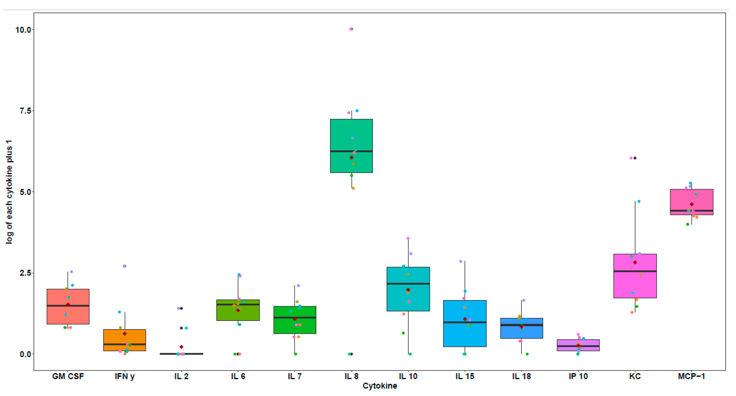
Figure 1. Cytokines at time 0 (T0), OMM samples.
Vetsci 12 00627 g001
Figure 2. Significant differences between cytokines over time. This figure displays cytokine concentrations using box plots. The central line in each box indicates the median, the boxes show the interquartile range (IQR), and the whiskers represent the range excluding outliers. The red dots represent the mean value for each time point, and the connecting lines trace the individual trajectories of each dog across time.
Vetsci 12 00627 g002
Figure 3. Comparison of cytokine concentrations between the OMM samples and the OMM dogs in remission samples. This figure presents the mean cytokine concentrations over time, with shaded areas representing the standard deviation.
Vetsci 12 00627 g003
Table 1. Data collection.
Pt BreedSexAge (y)Reproductive StateDiagnosisTNMTumor Size (cm)
1American staffordshireF10SpayedOral melanomaStage II2
2BeagleF13SpayedOral amelanotic melanomaStage III3.2
3Cocker spanielM13SpayedOral melanomaStage III2.5
4Labrador retrieverM14SpayedOral melanomaStage II2
5MixedF12SpayedOral melanomaStage I1.5
6Presa canarioM9IntactOral amelanotic melanomaStage III7.7
7Yorkshire terrierM14IntactOral amelanotic melanomaStage II2.1
8Yorkshire terrierM12SpayedOral amelanotic melanomaStage II2.8
9Yorkshire terrierF14SpayedOral amelanotic melanomaStage III2.5
10Yorkshire terrierM14SpayedOral amelanotic melanomaStage II2
Abbreviations. Pt: patient. F: female. M: male. y: years, TNM: tumor–node–metastasis classification.
Table 2. Survival time in days.
PtDiagnosisSurvival Time (days)
1OMM365
2OAM43
3OMM131
4OMM310
5OMM365
6OAM365
7OAM365
8OAM155
9OAM240
10OAM365
Abbreviations. OMM: Oral malignant melanoma. OAM: Oral amelanotic melanoma.
Table 3. Serum samples collected.
PtT0T1T2.1T2.2
1OMMRemissionRemissionRemission
2OMM
3OMMMetastasis
4OMMRemissionOMMMetastasis
5OMMRemissionRemissionOMM
6OMMRemissionRemissionRemission
7OMMRemissionRemissionRemission
8OMM
9OMMRemissionMetastasis
10OMMRemissionRemissionRemission
Abbreviations. T0: sample extraction on the day of surgery. T1: first sample extraction after tumor removal. T2.1 and T2.1: subsequent sample extractions. OMM: oral malignant melanoma diseased dogs.
Table 4. Evolution over time of the GM-CSF variable concentration in pg/mL.
Pt C0T0C1T1C2T2
1OMM1.264Remission2.367Remission5.190
2 *
3OMM6.477Metastasis4.969
4OMM1.264Remission0.000OMM0.000
5OMM4.747Remission5.625Remission10.868
6OMM2.367Remission7.717Remission8.523
7OMM7.308Remission7.717Remission7.717
8 *
9OMM1.264Remission0.000Metastasis0.000
10OMM6.054Remission7.717Remission6.895
Abbreviations. C0: condition at the time of sample collection on the day of surgery (T0). C1–C2: condition of the subsequent sample collection (T1, T2). *: missing values.
Table 5. Evolution over time of the IFN-γ variable concentration in pg/mL.
Pt C0T0C1T1C2T2
1OMM0.007Remission0.240Remission0.154
2 *
3OMM1.255Metastasis1.591
4OMM0.330Remission0.330OMM0.620
5OMM0.075Remission0.240Remission2.055
6OMM0.154Remission0.330Remission0.521
7OMM2.655Remission3.149Remission3.149
8 *
9OMM0.075Remission0.075Metastasis0.154
10OMM0.377Remission0.722Remission1.037
*: missing values.
Table 6. Evolution over time of the IL-2 variable concentration in pg/mL.
Pt C0T0C1T1C2T2
1OMM0.000Remission0.000Remission3.366
2 *
3OMM0.00Metastasis5.930
4OMM0.00Remission0.000OMM0.000
5OMM0.000Remission0.000Remission40.683
6OMM1.23Remission109.302Remission163.074
7OMM0.000Remission0.000Remission0.000
8 *
9OMM0.000Remission0.000Metastasis0.000
10OMM0.000Remission1.583Remission1.911
*: missing values.
Table 7. Evolution over time of the IL-18 variable concentration in pg/mL.
Pt C0T0C1T1C2T2
1OMM0.490Remission1.019Remission2.869
2 *
3OMM2.061Metastasis4.244
4OMM0.000Remission0.256OMM1.304
5OMM1.160Remission0.055Remission3.892
6OMM1.600Remission86.041Remission176.235
7OMM1.905Remission2.061Remission2.061
8 *
9OMM1.304Remission2.219Metastasis0.747
10OMM0.490Remission1.304Remission
*: missing values.
Table 8. Evolution over time of the MCP-1 variable concentration in pg/mL.
Pt C0T0C1T1C2T2
1OMM69.391Remission136.491Remission131.637
2 *
3OMM83.387Metastasis408.552
4OMM53.473Remission112.197OMM180.804
5OMM136.491Remission104.868Remission281.735
6OMM79.710Remission276.785Remission274.679
7OMM193.172Remission502.113Remission502.113
8 *
9OMM166.879Remission66.819Metastasis702.880
10OMM80.457Remission123.636Remission109.529
*: missing values.
Table 9. Comparison between OMM samples, regardless of metastatic status, and samples from OMM dogs in remission.
CytokinesOMM-P50
(pg/mL)
OMM P25-P75
(pg/mL)
DIR-P50
(pg/mL)
DIR P25-P75
(pg/mL)
p-Value
IL-72.120.89–3.335.83.71–6.380.078
MCP-181.9271.97–159.28274.68134.08–283.850.051
Abbreviations. OMM: oral malignant melanoma. DIR: OMM dogs in remission. P25: percentile 25. P50: median. P75: percentile 75.
Table 10. Comparison between the OMM samples and OMM-metastasis samples.
CytokinesOMMr-P50
(pg/mL)
OMMr P25-P75
(pg/mL)
MET-P50
(pg/mL)
MET P25-P75
(pg/mL)
p-Value
MCP-1136.4979.71–193.17813.18629.3–972.260.015
Abbreviations. OMM: oral malignant melanoma. MET: OMM-metastasis. P25: percentile 25. P50: median. P75: percentile 75.
Table 11. Comparison between the OMM subtype and cytokines.
CytokinesTumorP50
(pg/mL)
P25–P75
(pg/mL)
TumorP50
(pg/mL)
P25-P75
(pg/mL)
p-Value
IL-6A4.223.59–8.66M0.740–2.040.042
IL-10A13.217.31–19.3M1.670.68–4.490.038
IL-15A3.892.2–5.61M0.710–1.420.066
Abbreviations. A: amelanotic tumor. M: melanotic tumor. P25: percentile 25. P50: median. P75: percentile 75.
Table 12. Neutrophil-to-lymphocyte ratio (NLR).
Pt NEU (K/µL)LYM (K/µL)NLR
17.141.933.699
24.680.67.800
38.261.296.403
42.3080.892.593
5 * 1.2
68.733.212.719
713.531.767.687
87.071.45.050
95.81.793.240
108.272.343.534
Abbreviations. NEU: neutrophil. LYM: lymphocyte. *: missing values.
Disclaimer/Publisher’s Note: The statements, opinions and data contained in all publications are solely those of the individual author(s) and contributor(s) and not of MDPI and/or the editor(s). MDPI and/or the editor(s) disclaim responsibility for any injury to people or property resulting from any ideas, methods, instructions or products referred to in the content.