Quantitative Conspicuity of Pancreatic Canine Insulinoma: A Comparison of Dynamic 4D CT and Dual-Source, Dual-Energy Bolus-Triggered Multiphase CT Imaging
Simple Summary
Abstract
1. Introduction
2. Materials and Methods
2.1. Patient Selection
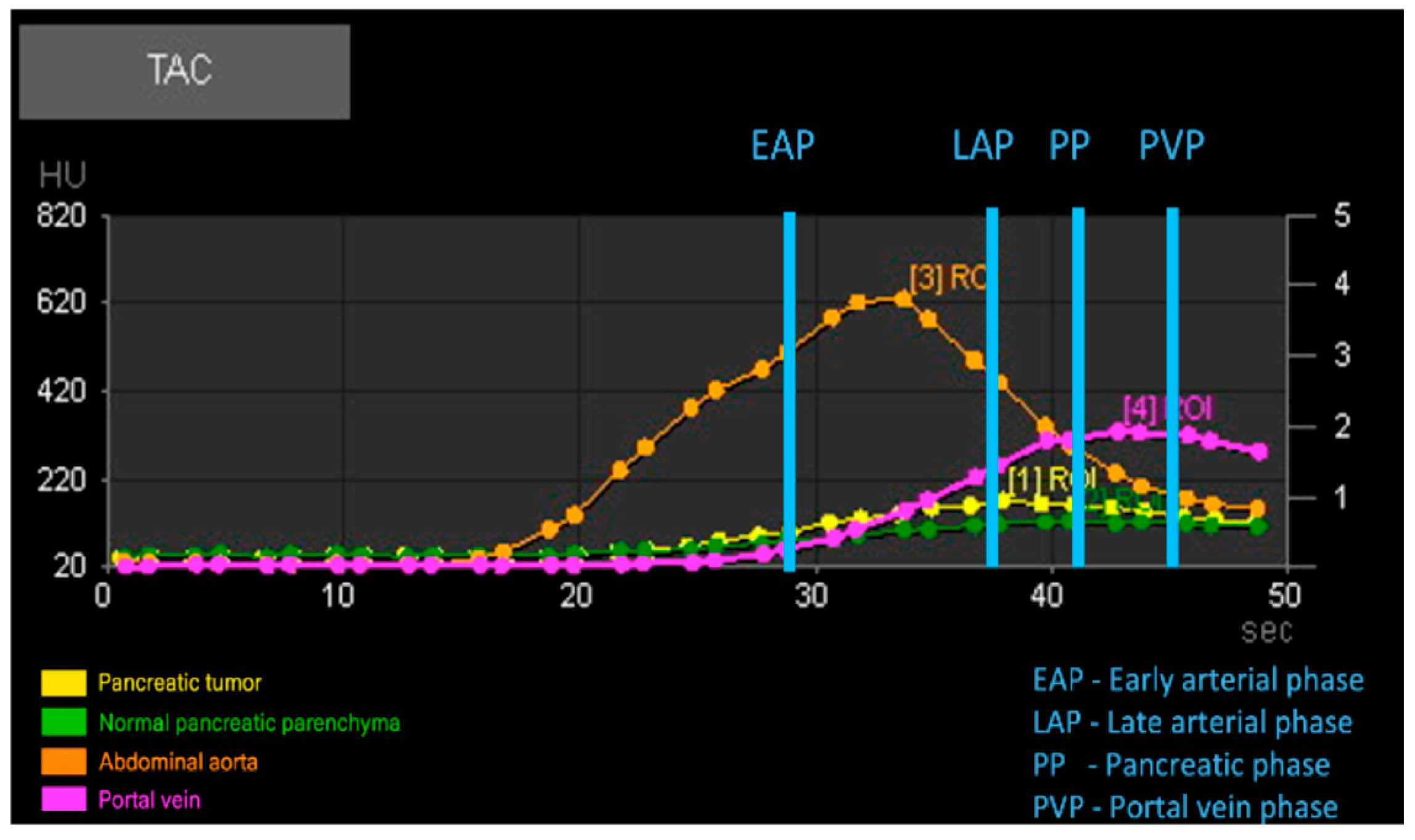
2.2. CT Technique
2.2.1. Perfusion Group (Group P)
2.2.2. Multiphase Group (Group M)
2.3. Diagnostic Imaging Analyses
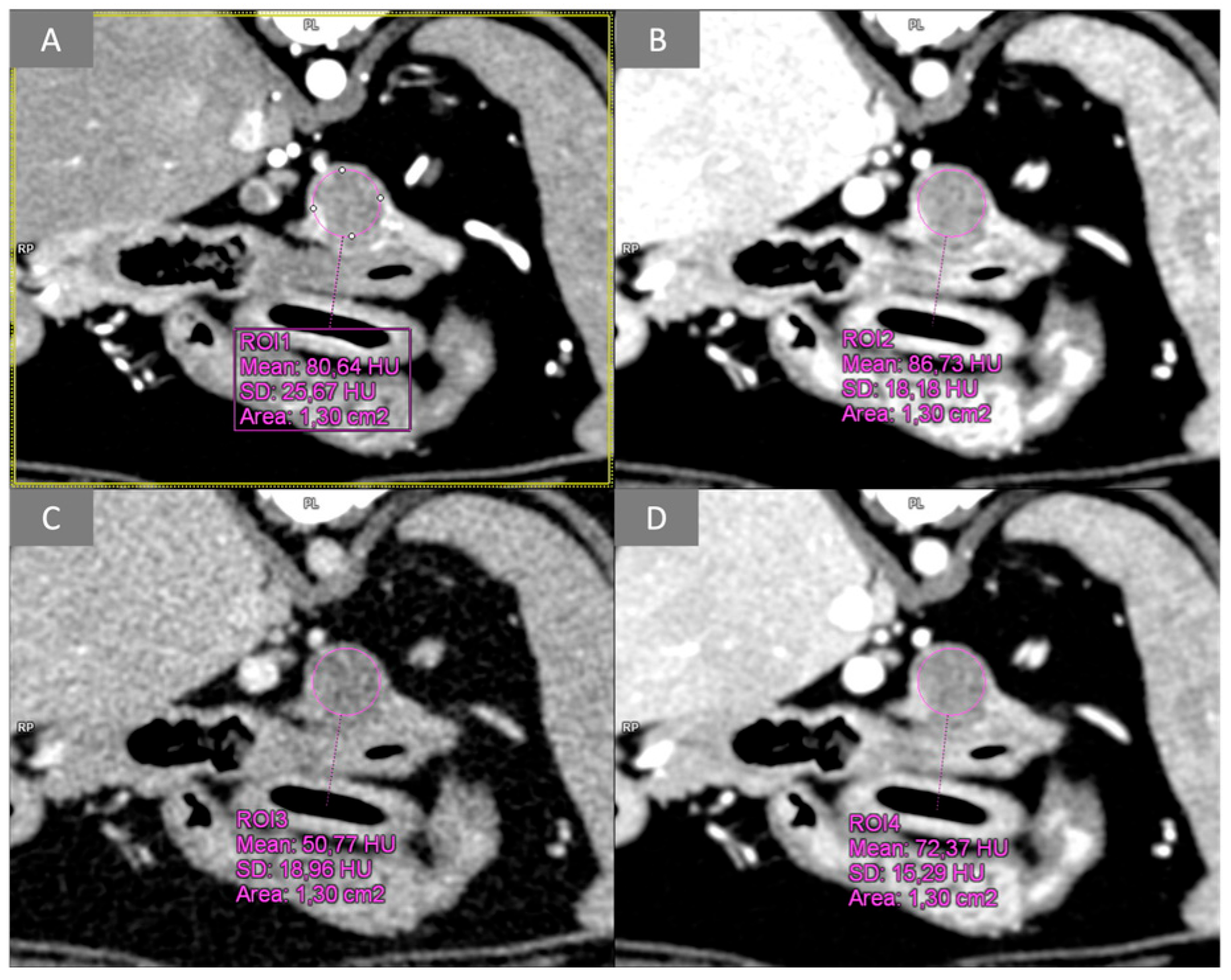
2.3.1. Quantitative Analysis
2.3.2. Qualitative Analysis
2.4. Statistical Analysis
3. Results
3.1. Population
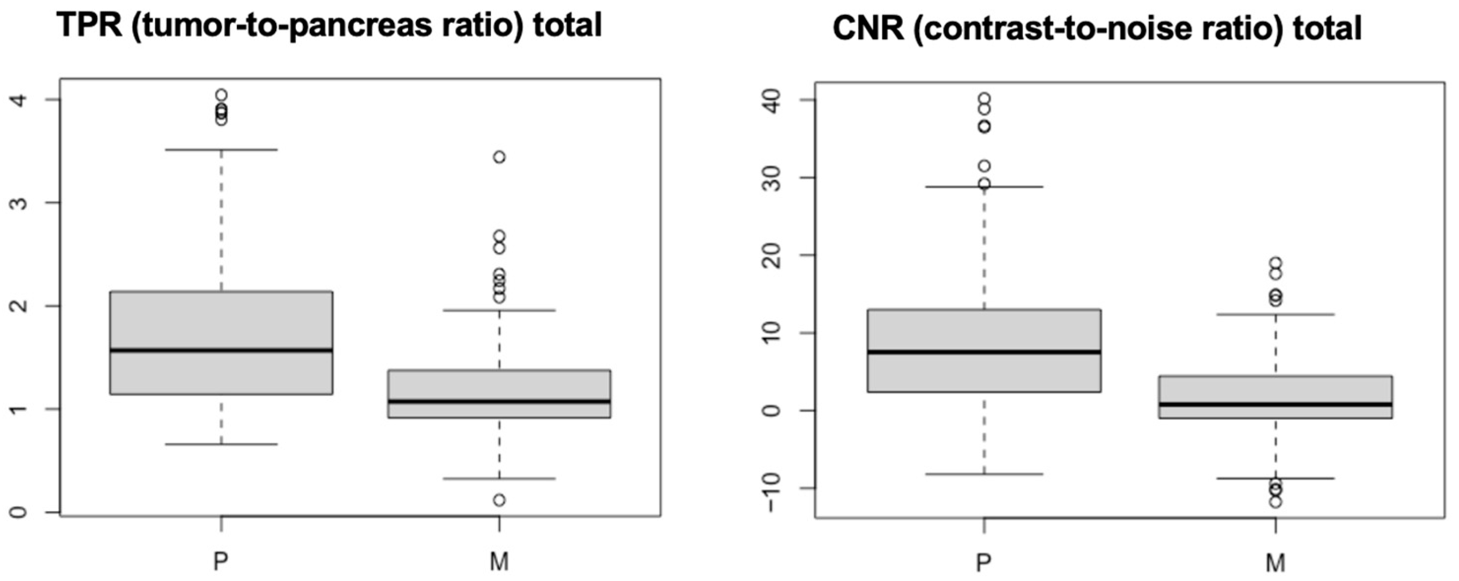
3.2. Quantitative Analysis
3.3. Qualitative Analysis
4. Discussion
5. Conclusions
Supplementary Materials
Author Contributions
Funding
Institutional Review Board Statement
Informed Consent Statement
Data Availability Statement
Acknowledgments
Conflicts of Interest
References
- Zhu, L.; Xue, H.D.; Sun, H.; Wang, X.; He, Y.L.; Jin, Z.Y.; Zhao, Y.-P. Isoattenuating insulinomas at biphasic contrast-enhanced CT: Frequency, clinicopathologic features and perfusion characteristics. Eur. Radiol. 2016, 26, 3697–3705. [Google Scholar] [CrossRef]
- Kraai, K.; O’Neill, D.G.; Davison, L.J.; Brodbelt, D.C.; Galac, S.; Buishand, F.O. Incidence and risk factors for insulinoma diagnosed in dogs under primary veterinary care in the UK. Sci. Rep. 2025, 15, 2463. [Google Scholar] [CrossRef]
- Buishand, F.O. Current trends in diagnosis, treatment and prognosis of canine insulinoma. Vet. Sci. 2022, 9, 540. [Google Scholar] [CrossRef] [PubMed]
- Buishand, F.O.; Vilaplana Grosso, F.R.; Kirpensteijn, J.; Van Nimwegen, S.A. Utility of contrast-enhanced computed tomography in the evaluation of canine insulinoma location. Vet. Q. 2018, 38, 53–62. [Google Scholar] [CrossRef]
- Hofland, J.; Refardt, J.C.; Feelders, R.A.; Christ, E.; de Herder, W.W. Approach to the Patient: Insulinoma. J. Clin. Endocrinol. Metab. 2024, 109, 1109–1118. [Google Scholar] [CrossRef]
- Zhu, L.; Wu, W.M.; Xue, H.D.; Liu, W.; Wang, X.; Sun, H.; Li, P.; Zhao, Y.; Jin, Z. Sporadic insulinomas on volume perfusion CT: Dynamic en-hancement patterns and timing of optimal tumor–parenchyma contrast. Eur Radiol. 2017, 27, 3491–3498. [Google Scholar] [CrossRef]
- Fu, J.; Zhang, J.; Wang, Y.; Yan, J.; Yuan, K.; Wang, M. Comparison of angio-CT versus multidetector CT in the detection and location for insulinomas. Clin. Radiol. 2020, 75, 796.E11–796.E16. [Google Scholar] [CrossRef] [PubMed]
- Decker, J.A.; Becker, J.; Härting, M.; Jehs, B.; Risch, F.; Canalini, L.; Wollny, C.; Scheurig-Muenkler, C.; Kroencke, T.; Schwarz, F. Optimal conspicuity of pancreatic ductal adenocarcinoma in virtual monochromatic imaging reconstructions on a photon-counting detector CT: Comparison to conventional MDCT. Abdom. Radiol. 2023, 49, 103–116. [Google Scholar] [CrossRef]
- Almeida, R.R.; Lo, G.C.; Patino, M.; Bizzo, B.; Canellas, R.; Sahani, D.V. Advances in pancreatic CT imaging. Am. J. Roentgenol. 2018, 211, 52–66. [Google Scholar] [CrossRef]
- Garcia, T.S.; Engelholm, J.L.; Vouche, M.; Hirakata, V.N.; Leitão, C.B. Intra- and interobserver reproducibility of pancreatic perfusion by computed tomography. Sci. Rep. 2019, 9, 6043. [Google Scholar] [CrossRef] [PubMed]
- Li, J.; Chen, X.Y.; Xu, K.; Zhu, L.; He, M.; Sun, T.; Zhang, W.-J.; Flohr, T.G.; Jin, Z.-Y.; Xue, H.-D. Detection of insulinoma: One-stop pancreatic perfusion CT with calculated mean temporal images can replace the combination of bi-phasic plus perfusion scan. Eur. Radiol. 2020, 30, 4164–4174. [Google Scholar] [CrossRef]
- Von Stade, L.; Rao, S.; Marolf, A.J. Computed tomographic evaluation of pancreatic perfusion in 10 dogs with acute pancreatitis. Vet. Radiol. Ultrasound 2023, 64, 823–833. [Google Scholar] [CrossRef]
- George, E.; Wortman, J.R.; Fulwadhva, U.P.; Uyeda, J.W.; Sodickson, A.D. Dual energy CT applications in pancreatic pathologies. BJR 2017, 90, 20170411. [Google Scholar] [CrossRef]
- Brook, O.R.; Gourtsoyianni, S.; Brook, A.; Siewert, B.; Kent, T.; Raptopoulos, V. Split-bolus spectral multidetector CT of the pancreas: Assessment of radiation dose and tumor conspicuity. Radiology 2013, 269, 139–148. [Google Scholar] [CrossRef]
- Iseri, T.; Yamada, K.; Chijiwa, K.; Nishimura, R.; Matsunaga, S.; Fujiwara, R.; Sasaki, N. Dynamic computed tomography of the pancreas in normal dogs and in a dog with pancreatic insulinoma. Vet. Radiol. Ultrasound 2007, 48, 328–331. [Google Scholar] [CrossRef] [PubMed]
- Mai, W.; Cáceres, A.V. Dual-phase computed tomographic angiography in three dogs with pancreatic insulinoma. Vet. Radiol. Ultrasound 2008, 49, 141–148. [Google Scholar] [CrossRef] [PubMed]
- Fukushima, K.; Fujiwara, R.; Yamamoto, K.; Kanemoto, H.; Ohno, K.; Tsuboi, M.; Uchida, K.; Matsuki, N.; Nishimura, R.; Tsujimoto, H. Characterization of triple-phase computed tomography in dogs with pancreatic insulinoma. J. Vet. Med. Sci. 2015, 77, 1549–1553. [Google Scholar] [CrossRef] [PubMed]
- Coss, P.; Gilman, O.; Warren-Smith, C.; Major, A.C. The appearance of canine insulinoma on dual phase computed tomographic angiography. J. Small Anim. Pract. 2021, 62, 540–546. [Google Scholar] [CrossRef]
- Skarbek, A.; Fouriez-Lablée, V.; Dirrig, H.; Llabres-Diaz, F. Confirmed and presumed canine insulinomas and their presumed metastases are most conspicuous in the late arterial phase in a triple arterial phase CT protocol. Vet. Radiol. Ultrasound 2023, 64, 834–843. [Google Scholar] [CrossRef]
- Rodriguez, D.; Rylander, H.; Vigen, K.K.; Adams, W.M. Influence of field strength on intracranial vessel conspicuity in canine magnetic resonance angiography. Vet. Radiol. Ultrasound 2009, 50, 477–482. [Google Scholar] [CrossRef]
- Pollard, R.E.; Johnson, E.G.; Pesavento, P.A.; Baker, T.W.; Cannon, A.B.; Kass, P.H.; Marks, S.L. Effects of corn oil administered orally on conspicuity of ultrasonographic small intestinal lesions in dogs with lymphangiectasia. Vet. Radiol. Ultrasound 2013, 54, 390–397. [Google Scholar] [CrossRef]
- Fitzgerald, E.; Lam, R.; Drees, R. Improving conspicuity of the canine gastrointestinal wall using dual phase contrast-enhanced computed tomography: A retrospective cross-sectional study. Vet. Radiol. Ultrasound 2017, 58, 151–162. [Google Scholar] [CrossRef]
- Thierry, F.; Chau, J.; Makara, M.; Specchi, S.; Auriemma, E.; Longo, M.; Handel, I.; Schwarz, T. Vascular conspicuity differs among injection protocols and scanner types for canine multiphasic abdominal computed tomographic angiography. Vet. Radiol. Ultrasound 2018, 59, 677–686. [Google Scholar] [CrossRef] [PubMed]
- Lee, S.; Jang, S.; Kim, S.; Lee, J.; Hyeong, S.; Choi, J. Feasibility of low dose CT protocols for evaluating the sinonasal cavity and reducing radiation exposure in dogs. Vet. Radiol. Ultrasound 2022, 63, 414–421. [Google Scholar] [CrossRef] [PubMed]
- Uosyte, R.; Shaw, D.J.; Gunn-Moore, D.A.; Fraga-Manteiga, E.; Schwarz, T. Effects of fluid and computed tomographic technical factors on conspicuity of canine and feline nasal turbinates. Vet. Radiol. Ultrasound 2015, 56, 494–502. [Google Scholar] [CrossRef] [PubMed]
- Peterson, M. Insulin producing islet cell neoplasms: Surgical considerations and general management in 35 dogs. J. Am. Anim. Hosp. Assoc. 1984, 20, 151–163. [Google Scholar]
- Lin, X.Z.; Wu, Z.Y.; Tao, R.; Guo, Y.; Li, J.Y.; Zhang, J.; Chen, K.M. Dual energy spectral CT imaging of insulinoma—Value in preoperative diagnosis compared with conventional multi-detector CT. Eur. J. Radiol. 2012, 81, 2487–2494. [Google Scholar] [CrossRef]
- Yamada, Y.; Jinzaki, M.; Hosokawa, T.; Tanami, Y.; Abe, T.; Kuribayashi, S. Abdominal CT: An intra-individual comparison between virtual monochromatic spectral and polychromatic 120-kVp images obtained during the same examination. Eur. J. Radiol. 2014, 83, 1715–1722. [Google Scholar] [CrossRef]
- Del Busto, I.; German, A.J.; Treggiari, E.; Romanelli, G.; O’Connell, E.M.; Batchelor, D.J.; Silvestrini, P.; Murtagh, K. Incidence of postoperative complications and outcome of 48 dogs undergoing surgical management of insulinoma. Vet. Intern. Med. 2020, 34, 1135–1143. [Google Scholar] [CrossRef]


| Scan Mode | Arterial Phase | Dual-Energy Portal Venous Phase | Dynamic 4D CT |
|---|---|---|---|
| (Group M) | (Group M) | (Group P) | |
| Scanner | SOMATOM Force | SOMATOM Force | SOMATOM Force |
| Tube current (mAs) | 600 | 400–800 | 220 |
| Tube voltage (kV) | 120 | 100–150 | 120 |
| Slices | 0.66 mm (Acq. 192 × 0.66 mm) | 0.66 mm (Acq. 192 × 0.66 mm) | 0.6 mm (Acq. 192 × 0.66 mm) |
| Direction | Cranio-caudal | Cranio-caudal | Caudo-cranial |
| Rotation time | 0.5 s | 1 s | 0.33 s |
| Pitch | 0.6 | 0.5 | 0.5 |
| Reconstruction kernel | Br40 | Br40 | Bv36 |
| CT Dose Index (mGy) | 15–48 | 13–43 | 474–788 |
| Cycle time | — | — | 1.5 s |
| Position increment | — | — | 0.3 |
| Field of view (FOV) | — | — | 200 × 200 mm |
Disclaimer/Publisher’s Note: The statements, opinions and data contained in all publications are solely those of the individual author(s) and contributor(s) and not of MDPI and/or the editor(s). MDPI and/or the editor(s) disclaim responsibility for any injury to people or property resulting from any ideas, methods, instructions or products referred to in the content. |
© 2025 by the authors. Licensee MDPI, Basel, Switzerland. This article is an open access article distributed under the terms and conditions of the Creative Commons Attribution (CC BY) license (https://creativecommons.org/licenses/by/4.0/).
Share and Cite
Camosci, V.; Canton, C.; Ventura, L.; Bertolini, G. Quantitative Conspicuity of Pancreatic Canine Insulinoma: A Comparison of Dynamic 4D CT and Dual-Source, Dual-Energy Bolus-Triggered Multiphase CT Imaging. Vet. Sci. 2025, 12, 1102. https://doi.org/10.3390/vetsci12111102
Camosci V, Canton C, Ventura L, Bertolini G. Quantitative Conspicuity of Pancreatic Canine Insulinoma: A Comparison of Dynamic 4D CT and Dual-Source, Dual-Energy Bolus-Triggered Multiphase CT Imaging. Veterinary Sciences. 2025; 12(11):1102. https://doi.org/10.3390/vetsci12111102
Chicago/Turabian StyleCamosci, Veronica, Claudia Canton, Laura Ventura, and Giovanna Bertolini. 2025. "Quantitative Conspicuity of Pancreatic Canine Insulinoma: A Comparison of Dynamic 4D CT and Dual-Source, Dual-Energy Bolus-Triggered Multiphase CT Imaging" Veterinary Sciences 12, no. 11: 1102. https://doi.org/10.3390/vetsci12111102
APA StyleCamosci, V., Canton, C., Ventura, L., & Bertolini, G. (2025). Quantitative Conspicuity of Pancreatic Canine Insulinoma: A Comparison of Dynamic 4D CT and Dual-Source, Dual-Energy Bolus-Triggered Multiphase CT Imaging. Veterinary Sciences, 12(11), 1102. https://doi.org/10.3390/vetsci12111102


