Review

Targeting the Gut–Mammary Axis for Understanding Mastitis Pathogenesis and Therapeutic Strategies

1
College of Agriculture and Biology, Liaocheng University, Liaocheng 252000, China
2
Yili Kazak Autonomous Prefecture Livestock General Station, Yili 835000, China
3
Department of Clinical Laboratory Sciences, College of Applied Medical Sciences, King Khalid University, Abha 61421, Saudi Arabia
4
Department of Clinical Laboratories Sciences, College of Applied Medical Sciences, Taif University, Taif 21944, Saudi Arabia
*
Authors to whom correspondence should be addressed.
Vet. Sci. 2025, 12(11), 1049; https://doi.org/10.3390/vetsci12111049 (registering DOI)
Submission received: 13 October 2025 / Revised: 28 October 2025 / Accepted: 28 October 2025 / Published: 1 November 2025
(This article belongs to the Special Issue Mammary Development and Health: Challenges and Advances)

Simple Summary

This review explores the gut–mammary axis in bovine mastitis pathogenesis and management. Mastitis development involves systematic microbiome disruption characterized by altered Firmicutes-to-Proteobacteria ratios, depletion of beneficial bacteria (Lactobacillus, Bifidobacterium, Akkermansia), and pathogenic expansion of Staphylococcus and Streptococcus species. Intestinal dysbiosis promotes mastitis through compromised barrier integrity, reduced protective metabolite production (short-chain fatty acids, secondary bile acids), and inflammatory pathway activation. Fecal microbiota transplantation experiments definitively established causality by transferring dysbiotic gut communities from mastitis-affected animals to healthy recipients, successfully inducing mammary inflammation. These findings provide direct evidence of a causal relationship between gut microbiome disturbances and the development of mastitis. Therapeutic interventions including probiotics, prebiotics, polysaccharides, and plant-derived compounds restore microbiome homeostasis and reduce mastitis severity, offering sustainable alternatives to antibiotics for dairy production.

Abstract

Mastitis represents one of the most economically devastating diseases in dairy production, causing billions of dollars in annual losses through reduced milk quality and quantity. Recent advances in microbiome research have unveiled a critical gut–mammary axis that fundamentally influences mastitis susceptibility and pathogenesis in dairy cattle. Through comprehensive analysis of microbial communities across multiple anatomical sites, we demonstrate that mastitis development involves systematic disruption of both mammary and gastrointestinal microbiomes, characterized by reduced beneficial bacterial populations and increased pathogenic species. Healthy animals maintain balanced microbial ecosystems dominated by protective taxa including Firmicutes, Bacteroidetes, and beneficial Lactobacillus species, while mastitis-affected animals exhibit dysbiotic shifts toward Proteobacteria dominance, elevated Streptococcus and Staphylococcus populations, and compromised microbial diversity. Mechanistic investigations reveal that gut microbiota disruption compromises systemic immune competence, alters metabolite production including short-chain fatty acids and bile acids, and influences inflammatory mediators that circulate to mammary tissue. Therapeutic interventions targeting this axis, including probiotics, prebiotics, and plant-derived compounds, demonstrate significant efficacy in restoring microbiome homeostasis and reducing mastitis severity. These findings establish the gut–mammary axis as a fundamental regulatory mechanism in mastitis pathogenesis, opening new avenues for microbiome-based prevention and treatment strategies that could significantly enhance dairy health management while addressing antimicrobial resistance concerns.

1. Introduction

Mastitis, characterized by inflammation of the mammary gland, represents one of the most economically significant and clinically challenging diseases affecting dairy cattle worldwide [1,2]. This multifactorial condition is primarily caused by bacterial pathogens that invade the udder tissue through the teat canal, leading to a cascade of inflammatory responses that compromise both milk quality and quantity [3]. Clinical manifestations of mastitis range from subclinical cases with no visible signs but elevated somatic cell counts and reduced milk production, to acute clinical presentations characterized by swelling, heat, pain, and redness of the affected mammary quarters, often accompanied by abnormal milk appearance including clots, flakes, or discoloration [4,5,6,7].
The economic burden imposed by this disease on the global dairy industry is staggering, with annual losses estimated between US$19.7 billion and US$32 billion, stemming from decreased milk yield, discarded milk, increased veterinary costs, premature culling, and reduced reproductive performance [8,9,10,11,12,13,14,15]. The United States alone accounts for approximately US$2 billion in annual losses [9,15], while Canada’s dairy industry faces Can$400 million (US$318 million) in losses each year, and China experiences fiscal impacts ranging from 15 to 45 billion Chinese Yuan (US$2.1–6.3 billion) [14]. These substantial economic consequences underscore the critical importance of effective prevention and treatment strategies for sustainable dairy production systems. While traditionally viewed as a localized bacterial infection, emerging evidence reveals that mastitis pathogenesis involves complex interactions between disrupted microbial communities and host immune responses [16,17,18].
This inflammatory condition can result not only from external microbial contamination but also from internal translocation of gut microbiota via the entero-mammary pathway (Figure 1) [16]. In this process, dendritic cells in the gut lamina propria capture microorganisms and transport them to mesenteric lymph nodes, where antigenic information is subsequently transmitted to mammary-associated lymph nodes through migrating lymphocytes, dendritic cell trafficking via lymphatic vessels, and soluble bacterial antigens in lymphatic fluid. Additionally, bacteria may translocate hematogenously through compromised intestinal barriers. These immune cell-mediated mechanisms ultimately facilitate microbial colonization of the mammary gland, contributing to mammary dysbiosis and milk microbiota composition. Recent microbiome research has fundamentally shifted our understanding of mastitis etiology, demonstrating that both mammary and intestinal microbial dysbiosis contribute to disease susceptibility [17,18]. Under normal physiological conditions, the mammary microbiota provides colonization resistance against pathogens through competitive exclusion and immune modulation, while the gut microbiome regulates systemic immune function and metabolic homeostasis [18,19,20,21]. However, environmental stressors, antibiotic exposure, and management practices can destabilize these microbial ecosystems, compromise natural defense mechanisms and create conditions favorable for pathogen establishment.
The gut–mammary axis, an emerging concept in veterinary and human medicine, suggests that the composition and metabolic activity of intestinal microbial communities can significantly influence mammary gland health and susceptibility to infectious diseases [22,23,24]. Gut dysbiosis can impair systemic immune competence and alter circulating metabolites that influence mammary gland susceptibility to infection. Conversely, mammary microbiota disruption directly undermines local immune responses, potentially leading to either excessive inflammation or inadequate pathogen clearance. Despite these advances, significant knowledge gaps persist regarding the mechanistic basis of gut–mammary communication, optimal microbiome restoration strategies, and the clinical translation of microbiome-targeted interventions [25,26,27]. Critical research priorities include identifying microbial biomarkers for early disease detection, characterizing temporal dynamics of microbiota changes during infection, and developing evidence-based therapeutic approaches that can be integrated into existing management protocols.
This review synthesizes current understanding of the gut–mammary axis and its role in mastitis pathogenesis, examining how bidirectional communication between intestinal and mammary microbiomes influences disease susceptibility and progression. We critically evaluate microbiome-mediated mechanisms underlying mastitis development, including disrupted colonization resistance, compromised barrier integrity, altered metabolite production, and dysregulated immune responses. Furthermore, we assess emerging microbiota-based therapeutic strategies—including probiotics, prebiotics, nutritional interventions, and plant-derived bioactive compounds—that target these mechanistic pathways to enhance udder health, reduce antimicrobial dependence, and promote sustainable dairy production systems. By integrating descriptive microbiome profiles with mechanistic insights into gut–mammary communication and immune modulation, this review provides a comprehensive framework for understanding mastitis pathogenesis and advancing precision microbiome-based interventions.

2. Comparative Microbiota of Milk and Gut in Healthy and Mastitis Animals

The contemporary understanding of mastitis pathogenesis has undergone a paradigmatic shift from the traditional conceptualization of a localized mammary gland infection to recognition of a complex multisystem disorder characterized by intricate microbiota–host interactions. This fundamental transformation in perspective has been driven by comprehensive investigations utilizing advanced sequencing technologies and metabolomic analyses, which have consistently demonstrated that mastitis development and progression are intimately linked to dynamic changes in microbial communities across multiple anatomical sites, thereby establishing the concept of a gut–mammary axis in dairy animal health.

2.1. Milk Microbiota Association with Mastitis

Across diverse dairy species and breeds, the most consistent hallmark of mastitis-associated dysbiosis is the dramatic alteration in Firmicutes-to-Proteobacteria ratios. However, the directional nature of this shift varies with pathogen type and disease severity. Multiple studies have documented this fundamental ecological disruption. For instance, Zhang L et al. [28] showed in yaks’ progression from balanced 39.7% Firmicutes and 60.17% Proteobacteria in healthy animals to 95.36% Proteobacteria dominance in clinical mastitis. In contrast, Salman et al. [29,30] found healthy cattle dominated by Proteobacteria (56.48%) but shifted to Firmicutes dominance (64%) during clinical mastitis. Furthermore, Secchi et al. [31] demonstrated pathogen-specific patterns. Specifically, Streptococcus agalactiae infections increased Firmicutes to 55.6% compared to 39.2% in controls. These findings, corroborated by Steinberg et al. [32] showing increased Firmicutes in subclinical mastitis across multiple breeds and Khasapane et al. [33] identifying Proteobacteria dominance in clinical cases, establish universal inflammatory-driven ecological shifts that transcend species boundaries while maintaining pathogen-specific characteristics.
The depletion of beneficial taxa represents another universal feature of mastitis dysbiosis, with multiple investigations identifying consistent losses of protective microorganisms across species. Healthy animals consistently harbor beneficial bacteria including Leuconostoc mesenteroides, Lactococcus piscium, Carnobacterium maltaromaticum, and Lactococcus raffinolactis in yaks [28], Akkermansia muciniphila and Lactobacillus fermentum in cattle [29,30], and protective taxa such as Bifidobacterium pseudolongum, Jeotgalibaca porci, and Romboutsia species [34]. Concurrently, studies by Tong et al. [35] and Burakova et al. [36] demonstrated that mastitis progression involves specific losses of Proteobacteria, Actinobacteria, Acidobacteria, as well as protective genera including Ralstonia, Lachnospiraceae NK3A20 group, Acetitomaculum, Massilia, and Atopostipes. The beneficial hub species identified by Derakhshani et al. [37], including Bacteroidaceae and Phascolarctobacterium, further emphasize the critical role of protective microbial networks in maintaining mammary health through anti-inflammatory metabolite production and immune homeostasis regulation.
Pathogenic microbial expansion follows predictable patterns across species, with certain taxa consistently emerging as mastitis-associated pathogens regardless of host species. Staphylococcus aureus appears as a dominant pathogen across studies, identified in yak mastitis [28], cattle investigations [38,39], and goat studies [40] where it maintained dominance across all disease states. Streptococcus species, including S. agalactiae [31,35] and S. dysgalactiae [29,30], consistently correlate with mastitis development, while opportunistic pathogens such as Escherichia coli, Staphylococcus uberis [39], Moraxella osloensis, Psychrobacter cibarius [28], and Pseudomonas fluorescens emerge with increasing disease severity. The pathogen-specific ecological signatures identified by Secchi et al. [31], where Prototheca spp. infections uniquely elevated Cyanobacteria to 17.3% versus 0.7% in controls, demonstrate that different etiological agents create distinct environmental niches favoring particular microbial communities.
Reduced microbial diversity emerges as a universal consequence of mastitis development, consistently documented across all species and study designs. Alpha diversity analysis by Salman et al. [29,30] showed reduced microbial diversity in mastitis cases across both Sahiwal cattle and Nili Ravi buffalo, while Zhang, L. et al. [28] documented progressive diversity loss correlating with yak mastitis severity. Maslennikova et al. [41] found mastitis-affected cows consistently harbored fewer bacterial taxa compared to healthy controls, with Polveiro et al. [40] demonstrating in goats that diversity disruption intensified with disease severity, reaching maximum alteration in gangrenous mastitis. This pattern is further supported by temporal studies showing that diversity reduction persists throughout lactation phases, suggesting that microbiome instability represents both a consequence and a perpetuating factor in mastitis pathogenesis.
The polymicrobial nature of mastitis has been consistently demonstrated across investigations, fundamentally challenging traditional single-pathogen therapeutic paradigms. Rudenko et al. [39] revealed that mastitis involves microbial consortia comprising two to seven distinct isolates with significant biofilm-forming capacity in 63.6% of cases, particularly among Staphylococcus aureus, Escherichia coli, and Staphylococcus uberis. This polymicrobial complexity is supported by Hoque et al. [42], who identified that mastitis-associated microbes carry genes for bacterial invasion, immune system disruption, antibiotic resistance, and oxidative stress induction, with 68% representing previously unreported opportunistic strains. The functional gene enrichment creates self-reinforcing inflammatory environments resistant to conventional therapies, while the reduced biofilm-forming capabilities of protective Lactobacillus species (22.5%) suggest competitive exclusion mechanisms that are disrupted during polymicrobial infections.
Specific diagnostic biomarkers have emerged from multiple studies, providing taxonomic signatures that could revolutionize mastitis detection and monitoring strategies. Complementary investigations identified overlapping biomarker panels including Hymenobacter and Lachnospiraceae NK4A136 group as mastitis indicators, increased Bifidobacterium and Lachnospiraceae_AC2044_group correlating with somatic cell counts of 100,000 cells/mL, and Romboutsia, Turicibacter, and Paeniclostridium as predictive markers for inflammatory development when somatic cell counts exceed 2 × 10⁵ [24,36,43]. These quantitative thresholds, combined with phylum-level shifts from beneficial Actinobacteriota to inflammation-associated Firmicutes and Proteobacteria dominance, provide integrated diagnostic approaches combining traditional inflammatory markers with microbial profiling.
Host genetic factors significantly influence microbiome composition and mastitis susceptibility, with multiple studies documenting breed-specific and genetic-dependent microbial patterns. However, mastitis has low heritability at approximately 0.16, indicating that while genetic factors play a role, environmental and management factors are more substantial contributors to mastitis development [44]. Tarrah et al. [45] demonstrated that Holstein cows with different estimated breeding values for feed efficiency and mastitis resilience harbored distinct communities, with Aerococcus, Corynebacterium, Facklamia, and Psychrobacter more abundant in high feed efficiency animals, while Mycoplana and Rhodococcus predominated in low mastitis resilience groups. These genomic–microbiome interactions, combined with species-specific baseline differences documented across cattle, buffalo, yak, and goat studies, suggest that effective mastitis prevention strategies must integrate universal microbiome principles with host-adapted therapeutic approaches.
The mechanistic validation linking specific taxa to inflammatory tissue destruction provides direct evidence for microbiome-mastitis causality. Chen et al. [46] established quantitative correlations between Corynebacterium kroppenstedtii abundance and breast abscess formation rates, while Sokolov et al. [38] identified pathogenic consortia including Staphylococcus aureus, Aerococcus spp., and Streptococcus spp. directly associated with elevated somatic cell counts. These findings, combined with the temporal dynamics documented by Maslennikova et al. [41] showing consistent taxonomic reduction throughout lactation phases, establish that specific microbial signatures both predict and mechanistically contribute to inflammatory mastitis development. The relationship between pathogenic microbial expansion and mastitis development is primarily causative rather than merely correlational. Bacterial challenge studies demonstrate that introducing specific pathogens (S. aureus, E. coli, Streptococcus species) into healthy mammary glands successfully induces mastitis and associated dysbiotic shifts, confirming that pathogenic bacteria drive disease development. Additionally, successful antimicrobial treatment restores balanced microbial communities [36], with pathogen elimination preceding microbiome normalization. While bidirectional effects between systemic inflammation and microbial composition are theoretically possible, experimental evidence—including challenge studies, antibiotic trials, and restoration interventions—supports a predominantly unidirectional causative pathway where pathogenic proliferation drives inflammatory dysbiosis and disease progression.
Collectively, these comprehensive investigations establish mastitis as a predictable ecosystem-level disruption characterized by consistent dysbiotic patterns across species: Firmicutes-Proteobacteria ratio alterations, beneficial taxa depletion (particularly Lactobacillus, Leuconostoc, Lactococcus, Akkermansia, and Bifidobacterium species), pathogenic expansion (dominated by Staphylococcus aureus, Streptococcus species, and opportunistic organisms), reduced microbial diversity, and polymicrobial biofilm formation. The identification of universal protective and pathogenic taxa, combined with species-specific variations and host genetic influences, provides a comprehensive foundation for developing precision microbiome-targeted interventions that restore protective networks while eliminating pathogenic consortia through integrated therapeutic approaches.

2.2. Gut Microbiota Association with Mastitis

The concept of a gut–mammary axis has gained substantial empirical support through multiple independent studies demonstrating that gastrointestinal microbial dysbiosis plays a crucial role in mastitis development. Yu J et al. [47] provided compelling evidence for this axis by demonstrating that cows with subclinical mastitis harbored distinctly altered gut microbial communities characterized by increased abundance of potentially harmful bacteria including Cyanobacteria, Proteobacteria, Succinivibrio, and Lactobacillus iners, while beneficial symbiotic bacteria such as Paraprevotella, Coprococcus, Succiniclasticum, Desulfovibrio, and Bifidobacterium pseudolongum were significantly reduced. This microbial imbalance extends beyond compositional changes to functional metabolic disruptions, as evidenced by elevated pro-inflammatory metabolites in both feces and plasma, including 9(S)-HPODE, 25-hydroxycholesterol, and histamine, while anti-inflammatory compounds such as mandelic acid, gamma-tocotrienol, and deoxycholic acid were notably decreased.
The rumen microbiota has emerged as a particularly critical component of this gut–mammary axis, with Zhang, H. et al. [48] demonstrating that cows with high somatic cell counts exhibited significantly compromised milk quality alongside altered rumen microbial communities characterized by increased abundances of Bacteroidetes, Firmicutes, Lachnospiraceae, Prevotella, and Rumiclostridium. These animals also showed elevated serum inflammatory markers and enrichment in metabolic pathways related to glutathione metabolism and cellular signaling, with specific bacterial families serving as key mediators linking rumen fermentation status, inflammatory responses, and milk quality parameters. This finding aligns with the work of Chuang et al. [49], who identified specific ruminal bacterial taxa as potential biomarkers for mastitis, with pathogenic genera such as Sharpea correlating positively with somatic cell counts and pro-inflammatory cytokines, while beneficial bacteria including Ruminococcaceae UCG-014, Ruminococcus flavefaciens, and Treponema saccharophilum demonstrated negative associations with inflammatory markers. Critical metabolic mediators have been identified that link gut dysbiosis to mastitis pathogenesis. Zhao, C. et al. [50] identified sialic acid as a crucial metabolic fuel that exacerbates gut dysbiosis and drives mastitis development, with cows experiencing subacute ruminal acidosis-associated mastitis exhibiting elevated sialic acid levels and distinct microbial communities characterized by increased pathogenic Moraxellaceae and reduced beneficial Prevotellaceae. Experimental validation demonstrated that sialic acid treatment specifically induced mastitis only when gut dysbiosis was present, associated with enhanced growth of harmful bacteria including Escherichia coli, Enterobacteriaceae, and Akkermansiaceae, while interventions targeting pathogenic Enterobacteriaceae or supplementing with beneficial Lactobacillus reuteri effectively alleviated mastitis symptoms. Furthermore, sialic acid potentiated serum LPS levels caused by antibiotic treatment, leading to increased activation of the TLR4-NF-κB/NLRP3 pathways in the mammary gland and colon.
The translocation of pathogenic microorganisms from the gastrointestinal tract to the mammary gland represents a key mechanism underlying the gut–mammary axis. Hu et al. [51] demonstrated that subacute rumen acidosis increased permeability across multiple barriers including blood-milk, gut, and rumen barriers, allowing harmful substances to translocate between body compartments. Lipopolysaccharides from the disrupted gut microbiota entered the bloodstream and accumulated in mammary glands, triggering inflammatory responses that led to mastitis symptoms, with Stenotrophomonas bacteria identified as particularly important in this process. Oral administration of this organism to lactating mice successfully induced mastitis, confirming its pathogenic role in endogenous infection routes.
Neuronal–microbial interactions add another layer of complexity to gut–mammary axis regulation. He, Y. et al. [52] demonstrated that vagotomy induces blood–milk barrier disruption and mammary gland inflammation through gut microbiota-dependent mechanisms, with vagotomy significantly altering gut microbial composition by reducing beneficial bacteria (Firmicutes) while increasing pathogenic bacteria (Proteobacteria, Campylobacterota, and Rikenellaceae_RC9_gut_group). The study revealed that vagotomy disrupts tryptophan metabolism, reducing 5-hydroxyindole acetic acid levels, and that supplementation with this metabolite effectively alleviates mastitis through activation of the aryl hydrocarbon receptor (AhR) and subsequent inhibition of the NF-κB inflammatory pathway.
The severity of mastitis correlates directly with the degree of gut microbiota dysbiosis, as demonstrated by Wang, Y. et al. [53], who found that cows with clinical mastitis showed increased inflammation-associated bacteria including Lachnospiraceae, Moraxella, and Neisseriaceae, accompanied by elevated inflammatory metabolites. Subclinical mastitis exhibited increased opportunistic pathogens (Ruminiclostridium_9 and Enterorhabdus) along with antibacterial compound-producing metabolites, while beneficial microbes were depleted across both mastitis groups, specifically short-chain fatty acid-producing bacteria (Prevoterotoella_1) and probiotics (Bifidobacterium). This experimental proof-of-concept is mechanistically supported by Hu et al. [54], who showed that gut microbiota dysbiosis significantly worsened S. aureus-induced mastitis severity through compromised blood–milk barrier function, with restoration achieved through short-chain fatty acid supplementation that modulated local inflammatory responses.
The cellular and molecular mechanisms underlying microbiota-mediated immune protection involve complex interactions between commensal bacteria and immune surveillance systems. Pan et al. [55] demonstrated that commensal microbiota-dependent γδ T cells (Tregs) mediate IL-17A-driven neutrophil recruitment essential for defense against S. aureus mastitis. Antibiotic treatment revealed that microbiota disruption caused significant downregulation of IL-17A signaling pathway-related genes, with the relative abundance of Firmicutes decreasing significantly while Proteobacteria, Bacteroidota, and Campylobacterota increased, establishing that beneficial microbes maintain protective inflammatory surveillance through the γδ T cells/IL-17A axis.
Systemic inflammatory cascades represent the downstream consequences of gut–mammary axis disruption. Guo et al. [56] demonstrated that mastitic cows exhibited significantly elevated concentrations of inflammatory markers including TNF-α, IL-1, and LPS compared to healthy animals, with these systemic inflammatory changes correlating directly with structural differences in rumen microbial communities including increased Aeriscardovia, Lactococcus, and Bacillus in healthy cow feces compared to mastitic animals. The identification of Moryella as a characteristic bacterial signature in mastitic animals suggests that specific microbial taxa may trigger or sustain inflammatory cascades that extend beyond the mammary gland. Chen et al. [57] further revealed that mastitis-associated gut microbiota dysbiosis, particularly increased Proteobacteria abundance, triggered cascading metabolic disruptions affecting bile acid metabolism, amino acid metabolism, and arachidonic acid pathways in mammary tissue, ultimately activating inflammatory proteins including IFIH1, Tnfaip8l2, IRGM, and IRF5.
The diversity–inflammation relationship represents a critical determinant of mastitis susceptibility, with reduced microbial diversity correlating with enhanced inflammatory activity. Zhu et al. [58] revealed that clinical mastitis cows showed significantly reduced fecal microbial diversity and disturbed milk microbiota compared to healthy controls, with metabolomic analysis identifying extensive disruptions in amino acid and energy metabolism pathways that fuel inflammatory processes. Clinical mastitis cows showed increased fecal abundance of UCG-010, Bacteroides, Prevotella, Ruminococcus, Ralstonia, and seven other genera, but decreased dgA-11 gut group, while their milk had elevated pathogenic Proteobacteria and reduced Bilophila and Clostridium innocuum group compared to healthy cows with higher beneficial Firmicutes, Actinobacteriota, Bifidobacterium, and Streptococcus.
High-throughput sequencing studies demonstrate that mastitis development involves paradoxical changes in microbial communities, characterized by decreased overall diversity coupled with increased species abundance, suggesting selective proliferation of specific bacterial populations rather than generalized microbial overgrowth [59]. The identification of significantly altered genera in both milk (Sphingomonas, Stenotrophomonas) and fecal samples (Alistipes, Flavonifractor, Agathobacter, Pygmaiobacter) provides strong evidence for an endogenous microbial pathway linking intestinal and mammary gland health. In buffalo, mastitis-associated microbiota changes manifest primarily through specific taxa alterations, with increased abundance of uncultured_bacterium_f_Muribaculaceae and Eubacterium_nodatum_group, alongside decreased beneficial genera including Ruminococcus_2 and Turicibacter [60].
A critical mechanistic pathway linking gut health to mastitis susceptibility involves bile acid metabolism, as demonstrated in subacute ruminal acidosis (SARA) models. Zhao, C. et al. [61] showed that SARA cows exhibited significantly reduced levels of both primary bile acid cholic acid (CA) and secondary bile acid deoxycholic acid (DCA) in their milk compared to healthy cows, directly associated with increased susceptibility to Staphylococcus aureus-induced mastitis. The mechanistic pathway involves healthy gut microbiota converting CA to DCA, leading to TGR5 activation, cAMP-PKA signaling, inhibition of NF-κB/NLRP3 pathways, and ultimately reduced inflammation and mastitis protection. Vancomycin-induced gut dysbiosis recapitulated this phenotype by causing marked alterations in gut microbiota structure, functionally resulting in reduced DCA levels, impaired TGR5 activation in mammary tissue, and more severe mastitis symptoms.

2.3. Fecal Microbiota Transplantation Causes Mastitis

The transmissible nature of mastitis-associated microbial communities has been conclusively demonstrated through fecal microbiota transplantation experiments, providing definitive proof that dysbiotic microbial communities possess inherent pathogenic properties that can directly cause disease across mammalian hosts. Hoque et al. [62] successfully induced mastitis in mice by transferring microbiota from clinical mastitis cows, with whole metagenome sequencing revealing significant differences in microbiome diversity and composition between clinical mastitis and healthy samples. The mastitis-associated microbiomes were characterized by specific bacterial species such as Pseudomonas aeruginosa and Lactobacillus crispatus in cows, while mastitis-induced mice showed enrichment of Muribaculum spp., Escherichia coli, and Staphylococcus aureus among others, establishing that microbiome dysbiosis and genomic functional perturbations serve as central mechanisms in mastitis pathogenesis. Despite the successful transmission of mastitis syndrome across species, the microbial specificity reveals important host-dependent variations in pathogenic community structure. Cows and mice shared only 1.14% of mastitis-associated microbial taxa, indicating species-specific microbiome patterns while maintaining conserved mastitis syndromes across mammalian hosts [62]. This finding suggests that while the functional consequences of dysbiosis are conserved, the specific bacterial actors may vary according to host physiology and environmental factors, pointing to universal mechanisms of microbiota-mediated disease pathogenesis rather than specific pathogen-driven processes.
The mechanistic pathways underlying microbiota transplantation-induced mastitis have been elucidated through detailed molecular analysis of recipient animals. Zhao C et al. [63] demonstrated that cows with clinical mastitis exhibited marked systemic inflammation associated with significant ruminal dysbiosis, particularly characterized by enriched Proteobacteria in the rumen. Critically, ruminal microbiota transplantation from mastitis cows to mice resulted in recipient mice developing mastitis symptoms along with increased mammary pro-inflammatory signature activation through TLR4-cGAS-STING-NF-κB/NLRP3 pathways, establishing that the inflammatory cascade activation represents a conserved response to dysbiotic microbial signals regardless of the original microbiota source.
The protective role of beneficial commensal bacteria emerges as a crucial counterbalance to pathogenic microbiota effects. Zhao C et al. [23] demonstrated that cows with mastitis had marked gut dysbiosis characterized by enrichment of opportunistic pathogenic Escherichia_Shigella and depletion of commensal Roseburia. When fecal microbiota transplantation from donor cows with mastitis was performed in recipient mice, it significantly induced mastitis and facilitated the translocation of pathobionts from the gut to the mammary gland. However, supplementation with commensal Roseburia intestinalis improved mastitis and microbial dysbiosis by producing butyrate, which was associated with inflammatory signaling inhibition and barrier repair, effectively limiting bacterial translocation. This finding demonstrates that the balance between pathogenic and protective microbial populations determines the severity of transplantation-induced mastitis. The causal relationship between microbiota dysbiosis and inflammatory mastitis susceptibility extends beyond bovine models to human lactation mastitis. Kong et al. [64] provided definitive evidence that dysbiotic gut microbiota actively contribute to lactation mastitis by demonstrating that fecal microbiota transplantation from mastitis patients induced systemic inflammation in recipient mice, characterized by elevated pro-inflammatory cytokines IL-4, IL-17, MPO, IL-6, IL-1β, and TNF-α. The microbiota alterations in mastitis patients included an increased Firmicutes/Bacteroidetes ratio, indicating dysbiosis, with elevated Actinobacteria and reduced Verrucomicrobia at the phylum level. At the genus level, beneficial bacteria Ruminococcus and Faecalibacterium were significantly decreased, whereas Parabacteroides were increased in both mastitis patients and mice that received their fecal transplants, establishing conserved dysbiotic patterns across human and experimental animal models.
The relationship between metabolic dysfunction and mastitis susceptibility has been further clarified through microbiota transplantation studies linking subclinical ketosis to mammary gland inflammation. Tang R et al. [65] demonstrated that subclinical ketosis in dairy cows exhibits profound associations with elevated somatic cell counts in milk, accompanied by distinct alterations in rumen microbiota diversity and volatile fatty acid profiles. Innovative microbiota transplantation experiments provided compelling evidence that transferring rumen microbiota from subclinical ketosis and mastitis-affected cows into mice successfully induced mammary inflammation and liver function impairment, whereas transplants from healthy animals produced no such deleterious effects. Linear discriminant analysis identified key bacterial genera including Chrysobacterium, Christensenellaceae_R-7_group, ceae_RC9_gut_group, and Prevotella as significant biomarkers distinguishing the rumen microbiota of ketotic cows from healthy animals, indicating that metabolic disorders create specific dysbiotic signatures that predispose to mastitis development.
While the predominant evidence supports gut dysbiosis as a primary driver of mastitis susceptibility, potential bidirectional interactions cannot be entirely excluded. Systemic inflammation from mastitis could theoretically influence gut microbiota composition through altered immune signaling and metabolic changes. However, experimental evidence from fecal microbiota transplantation studies definitively demonstrates that gut dysbiosis alone is sufficient to induce mastitis in healthy recipients [62,63,64,65], establishing causality in the gut-to-mammary direction. Moreover, temporal sequence studies show that induced gut dysbiosis precedes mastitis development [50,61], and antibiotic-mediated microbiota depletion exacerbates mastitis severity [54], collectively indicating that gut microbiome disturbances represent a primary causative factor rather than merely a consequence of mammary inflammation.
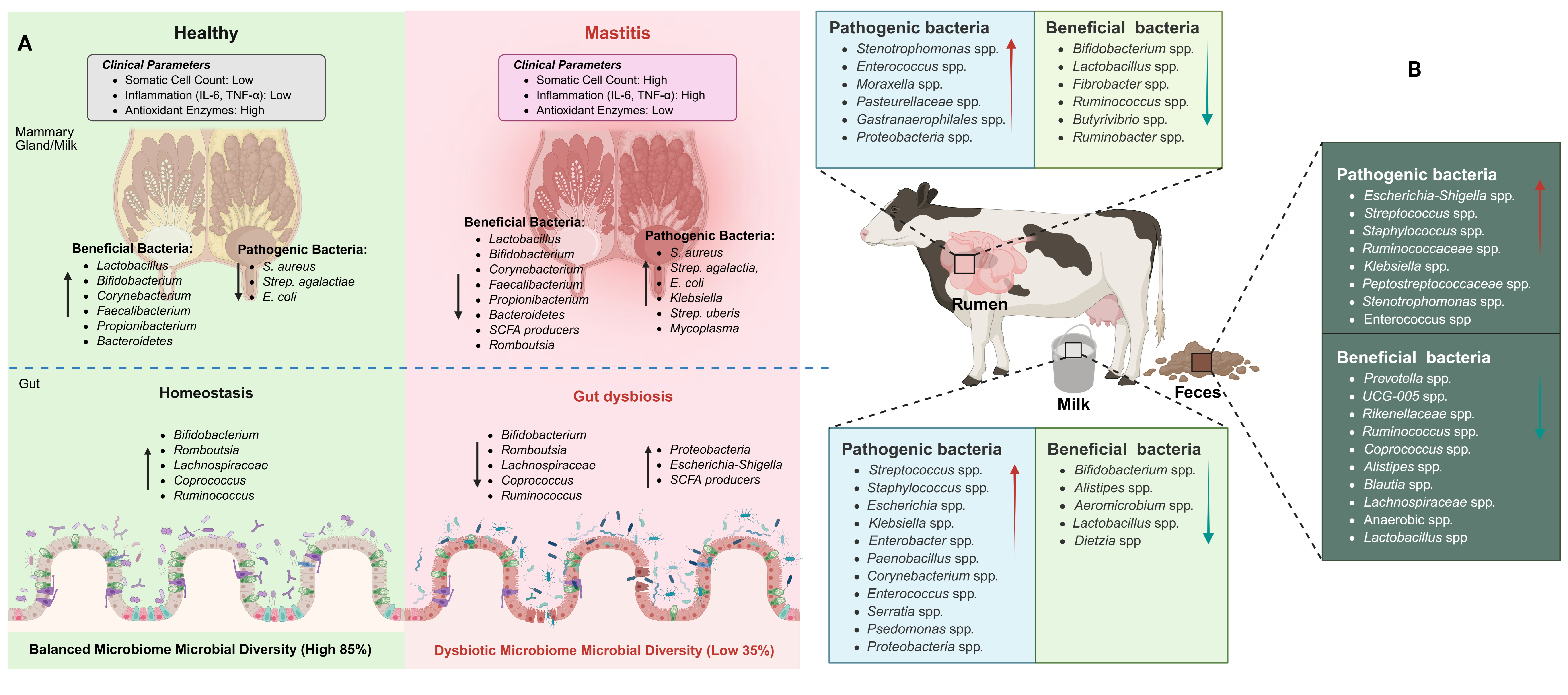
While establishing this causal relationship, it is essential to contextualize the role of gut microbiota within the broader landscape of bovine health. It is important to note that gut microbiota dysbiosis has been implicated in numerous other disease conditions beyond mastitis, including metabolic disorders, lameness, respiratory infections, and reproductive diseases in dairy cattle. The gut microbiome serves as a central regulator of systemic health through modulation of immune function, metabolic homeostasis, and barrier integrity across multiple organ systems. Nevertheless, the specific mechanisms linking gut dysbiosis to mammary gland pathology—including bile acid metabolism disruption, bacterial translocation via dendritic cells, and alterations in circulating immunomodulatory metabolites—represent unique pathways that distinguish the gut–mammary axis from other microbiome–disease relationships. To provide a comprehensive overview of these relationships, a summary of microbiota associations between milk, gut, and fecal samples in relation to mastitis is provided in Table 1. In addition, Figure 2 presents the taxonomic classification of microbiota present in milk, fecal, and gut samples from both healthy and mastitis-affected animals.

2.4. Functional Correlation Between Gut and Milk Microbiota

The emerging concept of the gut–mammary axis posits that intestinal and mammary microbial communities are not isolated ecosystems but rather functionally interconnected through complex metabolic and immunological communication networks. This bidirectional crosstalk fundamentally influences mammary gland health and mastitis susceptibility, thereby establishing the gut microbiome as a critical determinant of udder immunity and disease resistance [17]. Central to understanding this axis is the recognition that the gut microbiome produces a diverse array of bioactive metabolites that enter systemic circulation and directly influence mammary gland physiology and immune function. Among these metabolites, short-chain fatty acids (SCFAs), particularly butyrate, propionate, and acetate, represent primary mediators of gut–mammary metabolic communication. These microbial fermentation products cross the intestinal epithelium, enter the bloodstream, and reach mammary tissue, where they exert multiple protective effects. Notably, butyrate has been shown to activate histone deacetylase 3 (HDAC3)-mediated antimicrobial programs in mammary macrophages, thereby enhancing pathogen clearance capacity [66]. Significantly, during mastitis, gut dysbiosis leads to reduced SCFA production, particularly butyrate depletion, which consequently compromises mammary antimicrobial defense mechanisms and creates permissive conditions for pathogen establishment.
In addition to SCFAs, secondary bile acids constitute another critical metabolic link between gut and mammary microbiomes. These compounds, produced through microbial transformation of primary bile acids in the intestinal lumen, modulate systemic inflammation through interactions with farnesoid X receptor (FXR) and TGR5, thereby influencing immune cell trafficking and cytokine production patterns relevant to mammary gland immunity [61]. Consistent with this mechanism, research has demonstrated that mastitis-associated gut dysbiosis alters bile acid metabolism profiles, reducing protective secondary bile acid production and contributing to systemic inflammatory status that predisposes animals to mammary infections [61].
Furthermore, tryptophan metabolism represents an additional metabolic axis connecting intestinal and mammary health. Gut microbiota converts dietary tryptophan into indole derivatives, including indole-3-propionic acid (IPA) and indole-3-acetic acid (IAA), which possess potent anti-inflammatory and barrier-protective properties [19,50]. These indole metabolites activate the AhR in immune cells, thereby promoting regulatory T cell differentiation and suppressing excessive inflammatory responses. Importantly, during mastitis development, alterations in gut microbial composition disrupt tryptophan metabolism, reducing protective indole metabolite production and compromising systemic immune regulation, which subsequently increases mammary inflammation susceptibility [50]. Building upon these findings, recent metabolomic investigations have identified succinate as a pro-inflammatory metabolite elevated during mastitis-associated gut dysbiosis. Specifically, Qiu et al. [67] demonstrated that gut microbiota-derived extracellular vesicles containing elevated succinate levels exacerbate mastitis severity in mice, thereby establishing a direct mechanistic link between intestinal metabolic dysregulation and mammary pathology. This finding exemplifies how altered gut microbial metabolism can actively promote mastitis development through systemic metabolite signaling.
Beyond these metabolic pathways, the gut–mammary axis operates through sophisticated immunological communication mechanisms that coordinate local and systemic immune responses. The intestinal immune system, housing approximately 70% of the body’s immune cells, serves as a primary training ground for immune competence that extends to peripheral tissues including the mammary gland [61]. Accordingly, gut microbiota composition critically influences the development, maturation, and functional capacity of immune cell populations that subsequently migrate to mammary tissue [21,50]. Within this immunological framework, dendritic cells (DCs) represent key cellular mediators of gut–mammary immunological communication [51,68]. These professional antigen-presenting cells sample intestinal microbial antigens, undergo maturation and activation, and migrate through lymphatic circulation to peripheral tissues including the mammary gland [69]. During this transit, DCs can carry viable commensal bacteria from the intestinal lumen to distant sites through a process termed bacterial translocation [70,71]. This enteromammary pathway allows beneficial gut microorganisms, particularly Lactobacillus and Bifidobacterium species, to colonize mammary tissue and contribute to the milk microbiome composition, providing natural protection against pathogens through competitive exclusion and immune modulation. However, it is important to note that gut dysbiosis can pervert this beneficial translocation mechanism, allowing pathogenic or opportunistic bacteria to access mammary tissue and contribute to mastitis pathogenesis [72]. Furthermore, they demonstrated that mastitis-associated changes in gut microbiota composition correlate with altered bacterial translocation patterns, potentially introducing dysbiotic microbial signatures into the mammary environment and compromising local immune homeostasis [72].
Complementing the role of dendritic cells, T lymphocyte populations represent another critical immunological link between gut and mammary compartments. Gut microbiota composition profoundly influences the balance between pro-inflammatory T helper 17 (Th17) cells and anti-inflammatory regulatory T cells. Specifically, a healthy, diverse gut microbiome promotes Treg development through mechanisms involving SCFA-mediated histone modifications and transforming growth factor-β (TGF-β) signaling, establishing systemic immune tolerance that prevents excessive inflammatory responses in peripheral tissues [73,74]. Conversely, dysbiotic gut microbiota skews differentiation toward Th17 cells, thereby promoting systemic inflammation and enhancing mammary tissue susceptibility to infection-induced damage. Moreover, immunoglobulin A (IgA), the predominant antibody isotype in mucosal secretions including milk, provides additional evidence of gut–mammary immunological connectivity [75]. The mammary gland receives IgA-producing plasma cells that originate from gut-associated lymphoid tissue (GALT) through a process termed the common mucosal immune system. These plasma cells, initially primed by intestinal microbial antigens, traffic to the mammary gland where they secrete IgA antibodies that recognize gut-derived microorganisms, thereby providing passive immunity to offspring while simultaneously contributing to mammary colonization resistance. Disruption of gut microbiome composition impairs optimal IgA responses, consequently compromising this critical protective mechanism [76].
Expanding our understanding of these gut–mammary communication mechanisms, recent research has identified microbiome-derived extracellular vesicles (mEVs) as novel mediators of this axis. These nanoscale lipid-bound particles, secreted by both commensal and pathogenic bacteria, contain diverse cargoes including proteins, lipopolysaccharides, nucleic acids, and metabolites that can exert biological effects on distant tissues [77]. In a seminal study, Qiu et al. [67] demonstrated that gut microbiota-derived EVs accumulate in mammary tissue during mastitis and contribute to disease pathology through delivery of pro-inflammatory molecules including succinate. Conversely, EVs derived from beneficial bacteria such as Akkermansia muciniphila have shown therapeutic potential in mastitis management, suggesting that mEVs represent both pathogenic mediators and potential therapeutic vehicles within the gut–mammary axis.
Critically, longitudinal studies employing fecal microbiota transplantation (FMT) have provided definitive evidence of causality in gut–mammary axis function, thereby substantiating the mechanistic relationships described above. Experiments demonstrating that transfer of dysbiotic gut microbiota from mastitis-affected animals to healthy recipients successfully induces mammary inflammation establish that gut microbiome composition directly influences mastitis susceptibility rather than merely reflecting disease-associated changes [76,78]. These causal relationships are further supported by complementary evidence examining the mammary microbiome itself. Interestingly, another study reported that changes in milk microbiota were significantly associated with mastitis [59], employing 16S rDNA high-throughput sequencing to perform correlation analysis of milk and gut microbial communities in mastitis-affected Holstein cows. This investigation revealed that alterations in the gut microbiome were paralleled by corresponding shifts in the mammary gland microbiota composition, with specific bacterial taxa showing concurrent enrichment or depletion across both anatomical sites [59].
These findings fundamentally validate the gut–mammary axis concept and provide strong rationale for therapeutic interventions targeting intestinal microbiome restoration as a strategy for mastitis prevention and management. In conclusion, the functional correlation between gut and milk microbiota establishes the intestinal microbiome as a critical determinant of mammary health, operating through integrated metabolic and immunological communication networks. Understanding these mechanistic connections provides an essential foundation for developing precision microbiota-based interventions that target the gut–mammary axis for mastitis prevention and treatment.

3. Enhancement of Microbiota and Their Association with Mastitis Management

The emerging understanding of the gut–mammary axis has fundamentally transformed our comprehension of mastitis pathogenesis, revealing intricate interconnections between gastrointestinal microbiota and mammary gland health that extend far beyond traditional infectious disease paradigms. Contemporary research demonstrates that mastitis development involves not only direct pathogenic microbial infections of mammary tissue but also encompasses sophisticated endogenous pathways mediated by rumen and gut microbiota dysbiosis, thereby establishing novel therapeutic targets for preventing and controlling this economically significant disease in dairy cattle operations [65]. This paradigm shift has opened new therapeutic avenues focused on restoring beneficial microbial communities and their protective metabolites rather than simply eliminating pathogenic organisms.
Restoration of beneficial microbiota represents a fundamental therapeutic strategy, with spore-forming bacteria transplantation demonstrating remarkable efficacy in reversing dysbiosis-induced mastitis susceptibility. Zhao C et al. [61] showed that spore-forming bacteria transplantation in dysbiotic mice restored gut microbial diversity, normalized DCA production and TGR5 activation, while specific supplementation with Clostridium scindens increased mammary TGR5 expression and DCA levels, reduced S. aureus-induced mammary damage, and restored tight junction proteins. This approach directly addresses the mechanistic deficits created by dysbiosis, particularly the disruption of secondary bile acid metabolism that normally provides anti-inflammatory protection to mammary tissue.
Understanding of harmful metabolic pathways has revealed specific targets for therapeutic intervention, particularly the role of succinate in exacerbating mastitis through novel microbiota-extracellular vesicle mechanisms. Qiu et al. [67] demonstrated that succinate, a metabolite elevated in SARA-associated mastitis, induces significant gut microbiota dysbiosis and exacerbates mammary inflammation through a microbiota-extracellular vesicle (mEV) mechanism. Succinate administration resulted in characteristic dysbiotic shifts featuring increased Bacteroidetes and decreased Firmicutes populations, while genus-level analysis revealed decreased beneficial commensals (Lactobacillus and Phascolarctobacterium) and increased potentially pathogenic taxa (norank_f_Muribaculaceae and Prevotellaceae_UCG-001). The dysbiotic microbiota produced increased quantities of mEVs containing lipopolysaccharides, which translocated across compromised intestinal barriers to mammary tissue and activated the TLR4/NF-κB inflammatory pathway.

3.1. Nutritional Interventions

Nutritional intervention strategies, including bioactive compounds, phytogenic substances, and probiotics, have demonstrated promising efficacy in mastitis management through modulation of immune function, metabolic processes, and microbiota composition [16,79,80]. Yeast fermentation products have emerged as effective interventions for correcting SARA-associated microbiota disruptions. Wu K. et al. [81] found that SARA decreased the abundance of Firmicutes and increased Actinobacteriota and Bacteroidota at the phylum level, with yeast fermentation product (YFF) supplementation effectively mitigating these detrimental changes by restoring microbial diversity and richness, increasing Firmicutes and Actinobacteria abundance, promoting beneficial genera like Ruminococcus and Olsenella, and suppressing SARA-associated genera. Specific bacterial genera correlated with inflammation, with genera like unclassified_o__Oscillospirales and Denitrobacterium showing negative correlation with inflammatory markers, while genera like Lachnospiraceae_ND3007_group and Acetitomaculum showed positive correlation, implicating them in mastitis promotion.

3.2. Probiotic and Prebiotic Interventions

Probiotics and prebiotics have emerged as promising therapeutic and preventive strategies for mastitis by modulating the mammary gland microbiome, enhancing immune function, and reducing pathogenic bacterial colonization [22,82,83,84]. Among these interventions, polysaccharide-based treatments represent a particularly promising therapeutic strategy, with multiple compounds demonstrating efficacy through gut microbiota modulation and short-chain fatty acid production. Li K et al. [85] showed that Astragalus polysaccharide (APS) effectively mitigated S. aureus infection in murine mammary glands through gut microbiota modulation, uniquely enriching Ruminococcus bromii in the gut and facilitating metabolism of SCFAs, particularly acetate and butyrate, which proved pivotal to APS’s protective effects. Similarly, Ran et al. [86] demonstrated that Angelica sinensis polysaccharide (ASP) effectively mitigated S. aureus-induced mastitis through targeted modulation of intestinal microbiota, with protective effects being entirely dependent on gut microbiota integrity, as evidenced by complete negation of benefits following antibiotic-induced microbiota disruption. ASP enhanced gut microbial diversity while promoting beneficial compositional shifts, specifically increasing Lachnospiraceae_NK4A136 abundance and reducing Erysipelatoclostridium populations, with metabolomic analysis revealing elevated levels of bioactive metabolites including tabersonine and riboflavin.
Probiotic interventions have shown compelling therapeutic results through direct introduction of beneficial bacterial species and their bioactive products. Zhang B et al. [87] demonstrated that Akkermansia muciniphila and its derived outer membrane vesicles (AOMVs) showed significant potential, with Spearman’s correlation analysis revealing significant negative correlation between the relative abundances of Verrucomicrobia and Akkermansia and inflammatory cytokine levels in mastitis milk. Live A. muciniphila exhibited stronger anti-inflammatory effects compared to pasteurized preparations, notably inhibiting TLR4 and NF-κB signaling pathways, while administration significantly reduced somatic cell counts in mastitis-affected dairy cows, with four of five treated animals showing decreased counts below clinical thresholds and negative California Mastitis Test results. Marine-derived Bacillus amyloliquefaciens-9 (GB-9) demonstrated similar therapeutic potential in Saanen dairy goats, significantly reducing somatic cell counts and inflammatory markers including malondialdehyde, immunoglobulins (IgA, IgM), and pro-inflammatory cytokines (IL-2, IL-4, IL-6), while amplicon sequencing revealed alterations in fecal bacterial communities with Bacteroides and Phascolarctobacterium as primary responding genera [75].
Prebiotic supplementation has emerged as highly effective, with inulin demonstrating dose-dependent benefits through comprehensive modulation of both rumen and hindgut microbiota. Wang Y et al. [88] determined optimal therapeutic effects at 300 g/day, resulting in increased milk yield, protein, and lactose content alongside reduced somatic cell counts and decreased pro-inflammatory cytokines while enhancing anti-inflammatory IL-4 and antioxidant activity. Mechanistically, inulin enhanced rumen fermentation and promoted beneficial bacteria including Prevotella, Butyrivibrio, Muribaculaceae, and Bifidobacterium while suppressing pro-inflammatory taxa such as Clostridia UCG-014, Streptococcus, and Escherichia-Shigella. Wang Y et al. [89] further demonstrated that dietary inulin supplementation resulted in substantial alterations to milk microbiota composition, significantly reducing mastitis-causing and pro-inflammatory microbes including Escherichia-Shigella, Pseudomonas, Rhodococcus, and Burkholderia-Caballeronia-Paraburkholderia, while simultaneously increasing beneficial probiotics such as Lactobacillus and Bifidobacterium. Wang, Y. et al. [90] complemented these findings by investigating hindgut microbiome responses, showing increased abundance of beneficial Bacteroides and Bifidobacteria while decreasing potentially harmful taxa including Paeniclostridium, Ruminococcaceae, Coprococcus, and Clostridia, with elevated concentrations of beneficial short-chain fatty acids and reduced pro-inflammatory lipid oxidation products.
The mechanistic basis for prebiotic efficacy has been definitively established through microbiota transplantation studies. Zhao, C. et al. [66] showed that fiber-enriched high-inulin diet significantly alleviated S. aureus-induced mastitis in mice through gut microbiota-dependent mechanisms, as protective effects were completely abolished when gut microbiota was depleted with antibiotics, and fecal microbiota transplantation from high-inulin-treated mice successfully transferred protection to recipient animals. The protective mechanism involved increased production of short-chain fatty acids, particularly butyrate, which enhanced antimicrobial programs in macrophages by inhibiting histone deacetylase 3, while specific beneficial bacterial genera including Prevotellaceae, Bacteroides, and Lachnospiraceae were enriched and negatively correlated with mammary bacterial load and inflammation.

3.3. Plant-Derived Bioactive Compounds

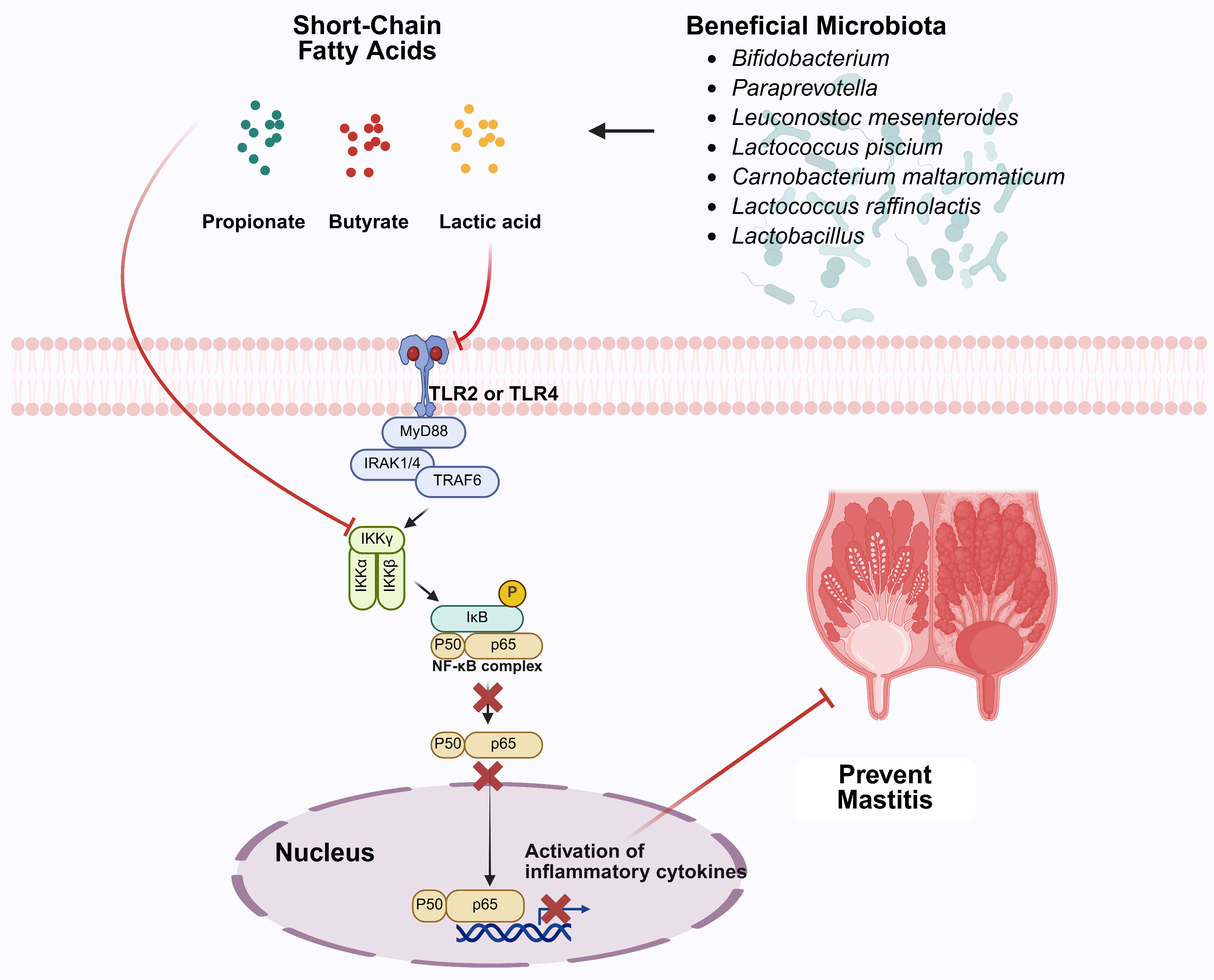
Plant-derived bioactive compounds have demonstrated significant therapeutic potential through multiple interconnected mechanisms involving microbiota modulation, anti-inflammatory signaling, and barrier function restoration. Xie et al. [91] showed that hordenine, a phenylethylamine alkaloid naturally extracted from malt, effectively attenuated LPS-induced mammary tissue damage and restored blood–milk barrier integrity by inhibiting TLR4-MAPK/NF-κB signaling while simultaneously activating beneficial AMPK/Nrf2/HO-1 signaling pathways (Figure 3). Critically, hordenine modulated intestinal microbiota composition to increase beneficial Lactobacillus populations while reducing harmful inflammatory bacteria from Desulfovibrionaceae and Enterobacteriaceae families, leading to restoration of short-chain fatty acid levels, with butyrate demonstrating direct enhancement of tight junction protein expression. Similarly, Gao et al. [92] demonstrated that Forsythiaside A (FTA), a phenylethanol glycoside extracted from Forsythia, effectively mitigated LPS-induced mastitis through multiple interconnected protective mechanisms, significantly reducing inflammation and apoptosis levels, modulating PI3K/AKT/mTOR signaling pathways, and enhancing antioxidant capacity. FTA restored gut microbiota diversity by increasing beneficial bacteria such as Lactobacillus and Bifidobacterium while reducing pro-inflammatory taxa including Helicobacteraceae and Desulfovibrionaceae, normalizing the Firmicutes/Bacteroidetes ratio and elevating butyrate-producing genera like Turicibacter.
Additional plant compounds have shown similar efficacy through targeted microbiota modulation. Li K et al. [93] demonstrated that maslinic acid, an olive-derived compound, significantly altered gut microbiota structure, specifically increasing the abundance of potentially beneficial Enterobacteriaceae while decreasing harmful Streptococcaceae. This modulation towards a more favorable microbial profile coincided with reduced mammary inflammation and protection of the blood–milk barrier, alongside direct inhibition of mammary inflammatory pathways including NLRP3, AKT/NF-κB, and MAPK. Ran et al. [94] showed that hesperetin treatment restored healthy gut microbiota by reversing α-diversity loss, restructuring the microbial community, specifically suppressing harmful Enterobacteriaceae while enriching beneficial taxa like Lactobacillaceae (including Lactobacillus johnsonii) and Lachnospiraceae (SCFA-producers), with microbiota rebalancing correlating with inhibition of mammary TLR4/NF-κB inflammatory signaling pathway and preservation of tight-junction proteins crucial for blood–milk barrier integrity (Figure 3). Yu et al. [95] demonstrated that dihydromyricetin (DMY), a flavonoid monomeric compound, effectively ameliorated subclinical mastitis in dairy cows through complementary antioxidant and microbiota-modulating mechanisms. DMY supplementation significantly reduced milk somatic cell counts while enhancing serum antioxidant capacity by increasing total antioxidant capacity (T-AOC) and catalase (CAT) activities and reducing malondialdehyde (MDA) levels. The compound modulated gut microbiota composition by promoting beneficial genera including Coprococcus and Roseburia while reducing potentially harmful Cyanobacteria, Proteobacteria, and Dehalobacterium abundance. Metabolomic analysis revealed that DMY intervention decreased pro-inflammatory metabolites including arachidonic acid analogs and ω-6 polyunsaturated fatty acids while increasing anti-inflammatory compounds such as secondary bile acids and antioxidant vitamins, demonstrating its therapeutic potential as a dietary supplement for managing mammary inflammation through enhanced antioxidant activity and improved gut microbial community structure [95].
Traditional herbal compounds have provided additional therapeutic mechanisms. Sun et al. [96] demonstrated that Abrus cantoniensis total flavonoids (ATF) effectively reduced mammary tissue damage and inhibited secretion of inflammatory cytokines including TNF-α, IL-1β, and IL-6 in LPS-induced mastitis models, suppressing the CD14/TLR4/NF-κB/MAPK signaling pathway while enhancing tight junction protein expression in the blood–milk barrier, with intestinal microbiota analysis revealing regulatory effects on microbial composition. Xiang Y et al. [97] showed that Pulsatilla chinensis extract (PCE) demonstrated alternative treatment potential for S. aureus-induced mastitis through gut microbiota modulation, with principal coordinate analysis revealing that PCE treatment maintained intestinal microbiota structures closely resembling healthy controls, while mastitis model groups exhibited significant structural deviations. Mastitis development was characterized by significant increases in potentially pathogenic populations including Proteobacteria, Deferribacterota, and notably Planctomycetota, which appeared exclusively in diseased states, with comprehensive correlation analysis demonstrating that Actinobacteriota and Bacteroidota showed significant negative correlations with inflammatory markers, while Firmicutes, Fusobacteriota, and Proteobacteria demonstrated significant positive correlations with disease parameters.
Building on these findings, research into traditional Chinese herbal formulations has revealed multi-target therapeutic mechanisms that offer comprehensive treatment strategies [98]. Zhao G et al. [99] demonstrated that Gongying San (GYS) significantly increased milk yield, lactose, and milk protein while decreasing somatic cell counts, decreasing serum levels of lipopolysaccharide, interleukins (IL-2, IL-4, IL-8, IL-10), tumor necrosis factor-α, and malondialdehyde while increasing superoxide dismutase concentration. Microbiota analysis showed increases in UCG-010 and Blautia and decreases in Bacteroides, Lachnospiraceae, and Agathobacter, with fecal untargeted metabolomics revealing that GYS supplementation primarily downregulated inflammation-related metabolism, including arachidonic acid and choline metabolism.
Direct intramammary interventions have also shown promise through local microbiome modulation. Zhang H et al. [100] reported that matrine-chitosan hydrogels demonstrated efficacy in modulating local microbiomes in dairy cows with subclinical mastitis, achieving significant reductions in somatic cell counts and microbial diversity, with treatment decreasing pathogenic microorganisms (Aerococcus, Corynebacterium_1, Staphylococcus, Firmicutes) while increasing beneficial Proteobacteria. Metabolomic analysis identified 74 differentially expressed metabolites, including upregulated sphingolipids, glycerophospholipids, flavonoids, and fatty acyls associated with glycerophospholipid and sphingolipid metabolic pathways, consistent with matrine’s antimicrobial and anti-inflammatory properties. The effects of various therapeutic interventions—namely nutritional interventions, probiotic and prebiotic interventions, plant-derived bioactive compounds, and direct intramammary interventions—on microbiota composition and subsequent mastitis development are presented in Table 2.

4. Conclusions

Mastitis extends beyond localized mammary infection, involving systemic microbiome disruption across mammary and intestinal sites, characterized by altered Firmicutes-to-Proteobacteria ratios and depletion of beneficial taxa (Lactobacillus, Bifidobacterium). Fecal microbiota transplantation experiments confirm that dysbiotic gut communities directly induce mammary inflammation, demonstrating the gut–mammary axis’s critical role.
This axis operates through metabolite signaling (SCFAs, bile acids), immune cell trafficking, bacterial translocation, and neuroendocrine coordination. Therapeutic interventions show significant efficacy: dietary fiber, probiotics (Lactobacillus, Bifidobacterium, Bacillus species), prebiotics, and plant-derived bioactives restore microbiome homeostasis while providing antimicrobial and immunomodulatory effects.
This paradigm shift reframes mastitis as a systems-level dysbiosis disorder rather than a simple infection. Future management strategies emphasize microbiome optimization and colonization resistance over antimicrobial pathogen elimination, offering sustainable alternatives that address antimicrobial resistance while promoting animal health and productivity.

Author Contributions

Y.L., M.Z.K., Q.M. and C.W.: Conceptualization, writing—original draft; M.Z.K., M.W., Y.L., M.G., W.L., M.A., C.W., F.M.A., K.J.A. and Q.M.: writing—review and editing, literature search, proofreading, data curation, software, M.Z.K., Q.M. and C.W.: supervision, validation, resources and funding. All authors have read and agreed to the published version of the manuscript.

Funding

This research was funded by the National Key R&D Program of China (grant numbers 2023YFD1302004; 2022YFD1600103), Livestock and Poultry Breeding Industry Project of the Ministry of Agriculture and Rural Affairs (grant number 19211162), the Shandong Province Agricultural Major Technology Collaborative Promotion Plan (SDNYXTTG-2024-13), the Deanship of Scientific Research at King Khalid University, the large research group program under grant number (R.G.P.02/709/46) and the Liaocheng Municipal Bureau of Science and Technology, High-talented Foreign Expert Introduction Program (GDWZ202401).

Institutional Review Board Statement

Not applicable.

Informed Consent Statement

Not applicable.

Data Availability Statement

No new data were created or analyzed in this study. Data sharing is not applicable to this article.

Conflicts of Interest

The authors declare no conflicts of interest.

References

  1. Khan, M.Z.; Li, L.; Zhan, Y.; Binjiang, H.; Liu, X.; Kou, X.; Khan, A.; Qadeer, A.; Ullah, Q.; Alzahrani, K.J.; et al. Targeting Nrf2/KEAP1 signaling pathway using bioactive compounds to combat mastitis. Front. Immunol. 2025, 16, 1425901. [Google Scholar] [CrossRef]
  2. Khan, M.Z.; Wang, J.; Ma, Y.; Chen, T.; Ma, M.; Ullah, Q.; Khan, I.M.; Khan, A.; Cao, Z.; Liu, S. Genetic polymorphisms in immune-and inflammation-associated genes and their association with bovine mastitis resistance/susceptibility. Front. Immunol. 2023, 14, 1082144. [Google Scholar] [CrossRef]
  3. Tong, X.; Barkema, H.W.; Nobrega, D.B.; Xu, C.; Han, B.; Zhang, C.; Yang, J.; Li, X.; Gao, J. Virulence of Bacteria Causing Mastitis in Dairy Cows: A Literature Review. Microorganisms 2025, 13, 167. [Google Scholar] [CrossRef] [PubMed]
  4. McDougall, S.; Williamson, J.; Lacy-Hulbert, J.; Steele, N.; Eastwood, C. Clinical and subclinical mastitis incidence in pasture-based dairy cows. N. Z. Vet. J. 2025, 73, 316–327. [Google Scholar] [CrossRef]
  5. Stanek, P.; Żółkiewski, P.; Januś, E. A review on mastitis in dairy cows research: Current status and future perspectives. Agriculture 2024, 14, 1292. [Google Scholar] [CrossRef]
  6. Cobirka, M.; Tancin, V.; Slama, P. Epidemiology and classification of mastitis. Animals 2020, 10, 2212. [Google Scholar] [CrossRef]
  7. Martins, L.; Barcelos, M.M.; Cue, R.I.; Anderson, K.L.; Dos Santos, M.V.; Gonçalves, J.L. Chronic subclinical mastitis reduces milk and components yield at the cow level. J. Dairy Res. 2020, 87, 298–305. [Google Scholar] [CrossRef] [PubMed]
  8. Adriaens, I.; Van Den Brulle, I.; Geerinckx, K.; D’Anvers, L.; De Vliegher, S.; Aernouts, B. Milk losses linked to mastitis treatments at dairy farms with automatic milking systems. Prev. Vet. Med. 2021, 194, 105420. [Google Scholar] [CrossRef]
  9. Puerto, M.A.; Shepley, E.; Cue, R.I.; Warner, D.; Dubuc, J.; Vasseur, E. The hidden cost of disease: I. Impact of the first incidence of mastitis on production and economic indicators of primiparous dairy cows. J. Dairy Sci. 2021, 104, 7932–7943. [Google Scholar] [CrossRef] [PubMed]
  10. Azooz, M.F.; El-Wakeel, S.A.; Yousef, H.M. Financial and economic analyses of the impact of cattle mastitis on the profitability of Egyptian dairy farms. Vet. World 2020, 13, 1750. [Google Scholar] [CrossRef]
  11. Dalanezi, F.M.; Joaquim, S.F.; Guimarães, F.F.; Guerra, S.T.; Lopes, B.C.; Schmidt, E.M.; Cerri, R.L.; Langoni, H. Influence of pathogens causing clinical mastitis on reproductive variables of dairy cows. J. Dairy Sci. 2020, 103, 3648–3655. [Google Scholar] [CrossRef]
  12. Dahl, M.O.; De Vries, A.; Galvão, K.N.; Maunsell, F.P.; Risco, C.A.; Hernandez, J.A. Combined effect of mastitis and parity on pregnancy loss in lactating Holstein cows. Theriogenology 2020, 143, 57–63. [Google Scholar] [CrossRef] [PubMed]
  13. Raboisson, D.; Ferchiou, A.; Pinior, B.; Gautier, T.; Sans, P.; Lhermie, G. The use of meta-analysis for the measurement of animal disease burden: Losses due to clinical mastitis as an example. Front. Vet. Sci. 2020, 7, 149. [Google Scholar] [CrossRef]
  14. Wang, L.; Yang, F.; Wei, X.J.; Luo, Y.J.; Guo, W.Z.; Zhou, X.Z.; Guo, Z.T. Prevalence and risk factors of subclinical mastitis in lactating cows in Northwest China. Isr. J. Vet. Med. 2019, 74, 17–22. [Google Scholar]
  15. Rollin, E.; Dhuyvetter, K.C.; Overton, M.W. The cost of clinical mastitis in the first 30 days of lactation: An economic modeling tool. Prev. Vet. Med. 2015, 122, 257–264. [Google Scholar] [CrossRef] [PubMed]
  16. Wang, Y.; Zhao, Y.; Tang, X.; Nan, X.; Jiang, L.; Wang, H.; Liu, J.; Yang, L.; Yao, J.; Xiong, B. Nutrition, gastrointestinal microorganisms and metabolites in mastitis occurrence and control. Anim. Nutr. 2024, 17, 220–231. [Google Scholar] [CrossRef]
  17. Luo, S.; Wang, Y.; Kang, X.; Liu, P.; Wang, G. Research progress on the association between mastitis and gastrointestinal microbes in dairy cows and the effect of probiotics. Microb. Pathog. 2022, 173, 105809. [Google Scholar] [CrossRef]
  18. Gryaznova, M.V.; Syromyatnikov, M.Y.; Dvoretskaya, Y.D.; Solodskikh, S.A.; Klimov, N.T.; Mikhalev, V.I.; Zimnikov, V.I.; Mikhaylov, E.V.; Popov, V.N. Microbiota of cow’s milk with udder pathologies. Microorganisms 2021, 9, 1974. [Google Scholar] [CrossRef]
  19. Zhao, C.; Hu, X.; Bao, L.; Wu, K.; Feng, L.; Qiu, M.; He, Y.; He, Z.; Zhang, N.; Fu, Y. Aryl hydrocarbon receptor activation by Lactobacillus reuteri tryptophan metabolism alleviates Escherichia coli-induced mastitis in mice. PLoS Pathog. 2021, 17, e1009774. [Google Scholar] [CrossRef] [PubMed]
  20. Fu, Y.; He, Y.; Xiang, K.; Zhao, C.; He, Z.; Qiu, M.; Hu, X.; Zhang, N. The role of rumen microbiota and its metabolites in subacute ruminal acidosis (SARA)-induced inflammatory diseases of ruminants. Microorganisms 2022, 10, 1495. [Google Scholar] [CrossRef]
  21. Bronzo, V.; Lopreiato, V.; Riva, F.; Amadori, M.; Curone, G.; Addis, M.F.; Cremonesi, P.; Moroni, P.; Trevisi, E.; Castiglioni, B. The role of innate immune response and microbiome in resilience of dairy cattle to disease: The mastitis model. Animals 2020, 10, 1397. [Google Scholar] [CrossRef]
  22. Matera, M.; Palazzi, C.M.; Bertuccioli, A.; Di Pierro, F.; Zerbinati, N.; Cazzaniga, M.; Gregoretti, A.; Cavecchia, I. The Role of Targeted Microbiota Therapy in the Prevention and Management of Puerperal Mastitis. Diseases 2025, 13, 176. [Google Scholar] [CrossRef]
  23. Zhao, C.; Bao, L.; Qiu, M.; Wu, K.; Zhao, Y.; Feng, L.; Xiang, K.; Zhang, N.; Hu, X.; Fu, Y. Commensal cow Roseburia reduces gut-dysbiosis-induced mastitis through inhibiting bacterial translocation by producing butyrate in mice. Cell Rep. 2022, 41, 108345. [Google Scholar] [CrossRef]
  24. Zhang, J.; Li, W.; Tang, Y.; Liu, X.; Zhang, H.; Zhou, Y.; Wang, Y.; Xiao, W.; Yu, Y. Testing two somatic cell count cutoff values for bovine subclinical mastitis detection based on milk microbiota and peripheral blood leukocyte transcriptome profile. Animals 2022, 12, 1694. [Google Scholar] [CrossRef]
  25. Gao, J.; Liu, Y.C.; Wang, Y.; Li, H.; Wang, X.M.; Wu, Y.; Zhang, D.R.; Gao, S.; Qi, Z.L. Impact of yeast and lactic acid bacteria on mastitis and milk microbiota composition of dairy cows. AMB Express 2020, 10, 22. [Google Scholar] [CrossRef]
  26. Catozzi, C.; Cusco’, A.; Lecchi, C.; De Carlo, E.; Vecchio, D.; Martucciello, A.; Francino, O.; Grilli, G.; Ceciliani, F. Impact of intramammary inoculation of inactivated Lactobacillus rhamnosus and antibiotics on the milk microbiota of water buffalo with subclinical mastitis. PLoS ONE 2019, 14, e0210204. [Google Scholar] [CrossRef] [PubMed]
  27. Hu, X.; Li, S.; Fu, Y.; Zhang, N. Targeting gut microbiota as a possible therapy for mastitis. Eur. J. Clin. Microbiol. Infect. Dis. 2019, 38, 1409–1423. [Google Scholar] [CrossRef] [PubMed]
  28. Zhang, L.; Ma, H.; Tang, W.; Zeng, J.; Kulyar, M.F.; Hu, J. Changes in the Microbiome in Yak Mastitis: Insights Based on Full-Length 16S rRNA Sequencing. Vet. Sci. 2024, 11, 335. [Google Scholar] [CrossRef]
  29. Salman, M.M.; Nawaz, M.; Yaqub, T.; Mushtaq, M.H. Investigation of milk microbiota of healthy and mastitic Sahiwal cattle. BMC Microbiol. 2023, 23, 304. [Google Scholar] [CrossRef]
  30. Salman, M.M.; Nawaz, M.; Yaqub, T.; Mushtaq, M.H. Exploring the milk microbiota of healthy and Mastitic Nili Ravi Buffalo using 16S rRNA gene base metagenomic analysis. Animals 2023, 13, 2298. [Google Scholar] [CrossRef] [PubMed]
  31. Secchi, G.; Bisutti, V.; Toscano, A.; Pegolo, S.; Giannuzzi, D.; Cecchinato, A.; Bittante, G.; Franciosi, E. Changes in the milk and fecal microbiota in Holstein cows with subclinical intramammary infection. J. Dairy Sci. 2025, 108, 10220–10236. [Google Scholar] [CrossRef]
  32. Steinberg, R.S.; Silva e Silva, L.C.; De Souza, M.R.; Reis, R.B.; Da Silva, P.C.; Lacorte, G.A.; Nicoli, J.R.; Neumann, E.; Nunes, Á.C. Changes in bovine milk bacterial microbiome from healthy and subclinical mastitis affected animals of the Girolando, Gyr, Guzera, and Holstein breeds. Int. Microbiol. 2022, 25, 803–815. [Google Scholar] [CrossRef] [PubMed]
  33. Khasapane, N.G.; Khumalo, Z.T.; Kwenda, S.; Nkhebenyane, S.J.; Thekisoe, O. Characterisation of milk microbiota from subclinical mastitis and apparently healthy dairy cattle in Free State Province, South Africa. Vet. Sci. 2023, 10, 616. [Google Scholar] [CrossRef]
  34. Alessandri, G.; Sangalli, E.; Facchi, M.; Fontana, F.; Mancabelli, L.; Donofrio, G.; Ventura, M. Metataxonomic analysis of milk microbiota in the bovine subclinical mastitis. FEMS Microbiol. Ecol. 2023, 99, fiad136. [Google Scholar] [CrossRef] [PubMed]
  35. Tong, J.; Zhang, H.; Zhang, Y.; Xiong, B.; Jiang, L. Microbiome and metabolome analyses of milk from dairy cows with subclinical Streptococcus agalactiae mastitis—Potential biomarkers. Front. Microbiol. 2019, 10, 2547. [Google Scholar] [CrossRef]
  36. Burakova, I.; Gryaznova, M.; Smirnova, Y.; Morozova, P.; Mikhalev, V.; Zimnikov, V.; Latsigina, I.; Shabunin, S.; Mikhailov, E.; Syromyatnikov, M. Association of milk microbiome with bovine mastitis before and after antibiotic therapy. Vet. World 2023, 16, 2389. [Google Scholar] [CrossRef]
  37. Derakhshani, H.; Plaizier, J.C.; De Buck, J.; Barkema, H.W.; Khafipour, E. Composition and co-occurrence patterns of the microbiota of different niches of the bovine mammary gland: Potential associations with mastitis susceptibility, udder inflammation, and teat-end hyperkeratosis. Anim. Microbiome 2020, 2, 11. [Google Scholar] [CrossRef]
  38. Sokolov, S.; Fursova, K.; Shulcheva, I.; Nikanova, D.; Artyemieva, O.; Kolodina, E.; Sorokin, A.; Dzhelyadin, T.; Shchannikova, M.; Shepelyakovskaya, A.; et al. Comparative analysis of milk microbiomes and their association with bovine mastitis in two farms in Central Russia. Animals 2021, 11, 1401. [Google Scholar] [CrossRef] [PubMed]
  39. Rudenko, P.; Sachivkina, N.; Vatnikov, Y.; Shabunin, S.; Engashev, S.; Kontsevaya, S.; Karamyan, A.; Bokov, D.; Kuznetsova, O.; Vasilieva, E. Role of microorganisms isolated from cows with mastitis in Moscow region in biofilm formation. Vet. World 2021, 14, 40. [Google Scholar] [CrossRef]
  40. Polveiro, R.C.; Vidigal, P.M.; de Oliveira Mendes, T.A.; Yamatogi, R.S.; da Silva, L.S.; Fujikura, J.M.; Da Costa, M.M.; Moreira, M.A. Distinguishing the milk microbiota of healthy goats and goats diagnosed with subclinical mastitis, clinical mastitis, and gangrenous mastitis. Front. Microbiol. 2022, 13, 918706. [Google Scholar] [CrossRef]
  41. Maslennikova, I.L.; Nechaeva, Y.I.; Ilina, L.A.; Laptev, G.Y.; Ponomareva, E.S.; Zhdanova, I.N.; Kuznetsova, M.V. A decline in taxonomic diversity of milk microbiome is linked to clinical mastitis and physiological states of cow. Gene Rep. 2025, 39, 102169. [Google Scholar] [CrossRef]
  42. Hoque, M.N.; Istiaq, A.; Clement, R.A.; Sultana, M.; Crandall, K.A.; Siddiki, A.Z.; Hossain, M.A. Metagenomic deep sequencing reveals association of microbiome signature with functional biases in bovine mastitis. Sci. Rep. 2019, 9, 13536. [Google Scholar] [CrossRef]
  43. Liu, J.; Liu, H.; Cao, G.; Cui, Y.; Wang, H.; Chen, X.; Xu, F.; Li, X. Microbiota characterization of the cow mammary gland microenvironment and its association with somatic cell count. Vet. Sci. 2023, 10, 699. [Google Scholar] [CrossRef] [PubMed]
  44. Urioste, J.I.; Franzén, J.; Windig, J.J.; Strandberg, E. Genetic relationships among mastitis and alternative somatic cell count traits in the first 3 lactations of Swedish Holsteins. J. Dairy Sci. 2012, 95, 3428–3434. [Google Scholar] [CrossRef] [PubMed]
  45. Tarrah, A.; Callegaro, S.; Pakroo, S.; Contiero, B.; Magrin, L.; Cassandro, M.; Corich, V. New insights into the raw milk microbiota diversity from animals with a different genetic predisposition for feed efficiency and resilience to mastitis. Sci. Rep. 2022, 12, 13498. [Google Scholar] [CrossRef]
  46. Chen, W.; Zhang, D.; Zeng, Y.; Cui, J.; Yu, J.; Wang, J.; Li, S.; Huang, Q.; Mansoor, K.M. Clinical characteristics and microbiota analysis of 44 patients with granulomatous mastitis. Front. Microbiol. 2023, 14, 1175206. [Google Scholar] [CrossRef] [PubMed]
  47. Yu, J.; Liu, C.; Wang, D.; Wan, P.; Cheng, L.; Yan, X. Integrated microbiome and metabolome analysis reveals altered gut microbial communities and metabolite profiles in dairy cows with subclinical mastitis. BMC Microbiol. 2025, 25, 115. [Google Scholar] [CrossRef]
  48. Zhang, H.; Lu, T.; Guo, S.; He, T.; Shin, M.K.; Luo, C.; Tong, J.; Zhang, Y. Rumen microbes affect the somatic cell counts of dairy cows by modulating glutathione metabolism. mSystems 2025, 10, e01093-24. [Google Scholar] [CrossRef]
  49. Chuang, S.T.; Li, K.Y.; Tu, P.W.; Ho, S.T.; Hsu, C.C.; Hsieh, J.C.; Chen, M.J. Investigating the reciprocal interrelationships among the ruminal microbiota, metabolome, and mastitis in early lactating Holstein dairy cows. Animals 2021, 11, 3108. [Google Scholar] [CrossRef]
  50. Zhao, C.; Hu, X.; Qiu, M.; Bao, L.; Wu, K.; Meng, X.; Zhao, Y.; Feng, L.; Duan, S.; He, Y.; et al. Sialic acid exacerbates gut dysbiosis-associated mastitis through the microbiota-gut–mammary axis by fueling gut microbiota disruption. Microbiome 2023, 11, 78. [Google Scholar] [CrossRef]
  51. Hu, X.; Li, S.; Mu, R.; Guo, J.; Zhao, C.; Cao, Y.; Zhang, N.; Fu, Y. The rumen microbiota contributes to the development of mastitis in dairy cows. Microbiol. Spectr. 2022, 10, e02512-21. [Google Scholar] [CrossRef]
  52. He, Y.; Zhao, C.; Su, N.; Yang, W.; Yang, H.; Yuan, C.; Zhang, N.; Hu, X.; Fu, Y. Disturbances of the gut microbiota-derived tryptophan metabolites as key actors in vagotomy-induced mastitis in mice. Cell Rep. 2024, 43, 114559. [Google Scholar] [CrossRef]
  53. Wang, Y.; Nan, X.; Zhao, Y.; Jiang, L.; Wang, M.; Wang, H.; Zhang, F.; Xue, F.; Hua, D.; Liu, J.; et al. Rumen microbiome structure and metabolites activity in dairy cows with clinical and subclinical mastitis. J. Anim. Sci. Biotechnol. 2021, 12, 36. [Google Scholar] [CrossRef]
  54. Hu, X.; Guo, J.; Zhao, C.; Jiang, P.; Maimai, T.; Yanyi, L.; Cao, Y.; Fu, Y.; Zhang, N. The gut microbiota contributes to the development of Staphylococcus aureus-induced mastitis in mice. ISME J. 2020, 14, 1897–1910. [Google Scholar] [CrossRef]
  55. Pan, N.; Xiu, L.; Xu, Y.; Bao, X.; Liang, Y.; Zhang, H.; Liu, B.; Feng, Y.; Guo, H.; Wu, J.; et al. Mammary γδ T cells promote IL-17A-mediated immunity against Staphylococcus aureus-induced mastitis in a microbiota-dependent manner. iScience 2023, 26, 108489. [Google Scholar] [CrossRef]
  56. Guo, C.; Liu, J.; Wei, Y.; Du, W.; Li, S. Comparison of the gastrointestinal bacterial microbiota between dairy cows with and without mastitis. Front. Microbiol. 2024, 15, 1332497. [Google Scholar] [CrossRef]
  57. Chen, K.; Hu, B.; Ren, J.; Deng, X.; Li, Q.; Zhang, R.; Zhang, Y.; Shen, G.; Liu, S.; Zhang, J.; et al. Enhanced protein-metabolite correlation analysis: To investigate the association between Staphylococcus aureus mastitis and metabolic immune pathways. FASEB J. 2024, 38, e23587. [Google Scholar] [CrossRef] [PubMed]
  58. Zhu, C.; Zhao, Y.; Yang, F.; Zhang, Q.; Zhao, X.; Yang, Z.; Dao, X.; Laghi, L. Microbiome and metabolome analyses of milk and feces from dairy cows with healthy, subclinical, and clinical mastitis. Front. Microbiol. 2024, 15, 1374911. [Google Scholar] [CrossRef] [PubMed]
  59. Jiang, C.; Hou, X.; Gao, X.; Liu, P.; Guo, X.; Hu, G.; Li, Q.; Huang, C.; Li, G.; Fang, W.; et al. The 16S rDNA high-throughput sequencing correlation analysis of milk and gut microbial communities in mastitis Holstein cows. BMC Microbiol. 2023, 23, 180. [Google Scholar] [CrossRef] [PubMed]
  60. Chen, X.; An, M.; Zhang, W.; Li, K.; Kulyar, M.F.; Duan, K.; Zhou, H.; Wu, Y.; Wan, X.; Li, J.; et al. Integrated bacteria-fungi diversity analysis reveals the gut microbial changes in buffalo with mastitis. Front. Vet. Sci. 2022, 9, 918541. [Google Scholar] [CrossRef]
  61. Zhao, C.; Wu, K.; Hao, H.; Zhao, Y.; Bao, L.; Qiu, M.; He, Y.; He, Z.; Zhang, N.; Hu, X.; et al. Gut microbiota-mediated secondary bile acid alleviates Staphylococcus aureus-induced mastitis through the TGR5-cAMP-PKA-NF-κB/NLRP3 pathways in mice. npj Biofilms Microbiomes 2023, 9, 8. [Google Scholar] [CrossRef] [PubMed]
  62. Hoque, M.N.; Rahman, M.S.; Islam, T.; Sultana, M.; Crandall, K.A.; Hossain, M.A. Induction of mastitis by cow-to-mouse fecal and milk microbiota transplantation causes microbiome dysbiosis and genomic functional perturbation in mice. Anim. Microbiome 2022, 4, 43. [Google Scholar] [CrossRef]
  63. Zhao, C.; Hu, X.; Bao, L.; Wu, K.; Zhao, Y.; Xiang, K.; Li, S.; Wang, Y.; Qiu, M.; Feng, L.; et al. Gut dysbiosis induces the development of mastitis through a reduction in host anti-inflammatory enzyme activity by endotoxemia. Microbiome 2022, 10, 205. [Google Scholar] [CrossRef]
  64. Kong, C.Y.; Yang, Y.Q.; Han, B.; Chen, H.L.; Mao, Y.Q.; Huang, J.T.; Wang, L.S.; Li, Z.M. Fecal microbiome transplant from patients with lactation mastitis promotes mastitis in conventional lactating mice. Front. Microbiol. 2023, 14, 1123444. [Google Scholar] [CrossRef]
  65. Tang, R.; Yang, W.; Song, J.; Xiang, K.; Li, S.; Zhao, C.; Zhang, N.; Fu, Y.; Hu, X. The rumen microbiota contributed to the development of mastitis induced by subclinical ketosis. Microb. Pathog. 2024, 187, 106509. [Google Scholar] [CrossRef] [PubMed]
  66. Zhao, C.; Bao, L.; Zhao, Y.; Wu, K.; Qiu, M.; Feng, L.; Zhang, N.; Hu, X.; Fu, Y. A fiber-enriched diet alleviates Staphylococcus aureus-induced mastitis by activating the HDAC3-mediated antimicrobial program in macrophages via butyrate production in mice. PLoS Pathog. 2023, 19, e1011108. [Google Scholar] [CrossRef] [PubMed]
  67. Qiu, M.; Ye, C.; Zhao, X.; Zou, C.; Tang, R.; Xie, J.; Liu, Y.; Hu, Y.; Hu, X.; Zhang, N.; et al. Succinate exacerbates mastitis in mice via extracellular vesicles derived from the gut microbiota: A potential new mechanism for mastitis. J. Nanobiotechnol. 2024, 22, 712. [Google Scholar] [CrossRef]
  68. Walker, W.A.; Iyengar, R.S. Breast milk, microbiota, and intestinal immune homeostasis. Pediatr. Res. 2015, 77, 220–228. [Google Scholar] [CrossRef]
  69. Castro, I.; García-Carral, C.; Furst, A.; Khwajazada, S.; García, J.; Arroyo, R.; Ruiz, L.; Rodríguez, J.M.; Bode, L.; Fernández, L. Interactions between human milk oligosaccharides, microbiota and immune factors in milk of women with and without mastitis. Sci. Rep. 2022, 12, 1367. [Google Scholar] [CrossRef]
  70. Rescigno, M.; Urbano, M.; Valzasina, B.; Francolini, M.; Rotta, G.; Bonasio, R.; Granucci, F.; Kraehenbuhl, J.P.; Ricciardi-Castagnoli, P. Dendritic cells express tight junction proteins and penetrate gut epithelial monolayers to sample bacteria. Nat. Immunol. 2001, 2, 361–367. [Google Scholar] [CrossRef]
  71. Macpherson, A.J.; Uhr, T. Induction of protective IgA by intestinal dendritic cells carrying commensal bacteria. Science 2004, 303, 1662–1665. [Google Scholar] [CrossRef]
  72. Wang, Z.; Ma, Z.; Tian, Z.; Jia, H.; Zhang, L.; Mao, Y.; Yang, Z.; Liu, X.; Li, M. Microbial dysbiosis in the gut–mammary axis as a mechanism for mastitis in dairy cows. Int. J. Dairy Technol. 2025, 78, e13150. [Google Scholar] [CrossRef]
  73. Duan, H.; Wang, L.; Huangfu, M.; Li, H. The impact of microbiota-derived short-chain fatty acids on macrophage activities in disease: Mechanisms and therapeutic potentials. Biomed. Pharmacother. 2023, 165, 115276. [Google Scholar] [CrossRef] [PubMed]
  74. Rooks, M.G.; Garrett, W.S. Gut microbiota, metabolites and host immunity. Nat. Rev. Immunol. 2016, 16, 341–352. [Google Scholar] [CrossRef] [PubMed]
  75. Li, Y.; Jiang, N.; Zhang, W.; Lv, Z.; Liu, J.; Shi, H. Bacillus amyloliquefaciens-9 reduces somatic cell count and modifies fecal microbiota in lactating goats. Mar. Drugs 2021, 19, 404. [Google Scholar] [CrossRef] [PubMed]
  76. Hu, X.; He, Z.; Zhao, C.; He, Y.; Qiu, M.; Xiang, K.; Zhang, N.; Fu, Y. Gut/rumen-mammary gland axis in mastitis: Gut/rumen microbiota–mediated “gastroenterogenic mastitis”. J. Adv. Res. 2024, 55, 159–171. [Google Scholar] [CrossRef]
  77. Li, Y.; Khan, M.Z.; Wang, C.; Ma, Q. The emerging role of milk-derived extracellular vesicles in gut pathology and cancer management. Crit. Rev. Food Sci. Nutr. 2025, 1–8. [Google Scholar] [CrossRef]
  78. Ma, C.; Sun, Z.; Zeng, B.; Huang, S.; Zhao, J.; Zhang, Y.; Su, X.; Xu, J.; Wei, H.; Zhang, H. Cow-to-mouse fecal transplantations suggest intestinal microbiome as one cause of mastitis. Microbiome 2018, 6, 200. [Google Scholar] [CrossRef]
  79. Khan, M.Z.; Huang, B.; Kou, X.; Chen, Y.; Liang, H.; Ullah, Q.; Khan, I.M.; Khan, A.; Chai, W.; Wang, C. Enhancing bovine immune, antioxidant and anti-inflammatory responses with vitamins, rumen-protected amino acids, and trace minerals to prevent periparturient mastitis. Front. Immunol. 2024, 14, 1290044. [Google Scholar] [CrossRef]
  80. Khan, M.Z.; Li, L.; Wang, T.; Liu, X.; Chen, W.; Ma, Q.; Zahoor, M.; Wang, C. Bioactive compounds and probiotics mitigate mastitis by targeting NF-κB signaling pathway. Biomolecules. 2024, 14, 1011. [Google Scholar] [CrossRef]
  81. Wu, K.; Sun, X.; Xu, J.; Guan, Z.; Yuan, W.; Bao, L.; Zhao, Y.; Shan, R.; Chen, H.; Zhao, C.; et al. Yeast-fermented feed improves high-concentrate diet-induced mastitis in dairy goats by regulating rumen microbiota. Front. Microbiol. 2025, 16, 1582314. [Google Scholar] [CrossRef]
  82. He, Z.; Li, W.; Yuan, W.; He, Y.; Xu, J.; Yuan, C.; Zhao, C.; Zhang, N.; Fu, Y.; Hu, X. Lactobacillus reuteri inhibits Staphylococcus aureus-induced mastitis by regulating oxytocin releasing and gut microbiota in mice. FASEB J. 2024, 38, e23383. [Google Scholar] [CrossRef]
  83. Li, Y.; Zhu, Y.; Chu, B.; Liu, N.; Chen, S.; Wang, J. Lactobacillus rhamnosus GR-1 prevents Escherichia coli-induced apoptosis through PINK1/Parkin-mediated mitophagy in bovine mastitis. Front. Immunol. 2021, 12, 715098. [Google Scholar] [CrossRef]
  84. Barker, M.; Adelson, P.; Peters, M.D.; Steen, M. Probiotics and human lactational mastitis: A scoping review. Women Birth 2020, 33, e483–e491. [Google Scholar] [CrossRef] [PubMed]
  85. Li, K.; Ran, X.; Han, J.; Ding, H.; Wang, X.; Li, Y.; Guo, W.; Li, X.; Guo, W.; Fu, S.; et al. Astragalus polysaccharide alleviates mastitis disrupted by Staphylococcus aureus infection by regulating gut microbiota and SCFAs metabolism. Int. J. Biol. Macromol. 2025, 286, 138422. [Google Scholar] [CrossRef]
  86. Ran, X.; Li, Y.; Guo, W.; Li, K.; Guo, W.; Wang, X.; Liu, J.; Bi, J.; Fu, S. Angelica sinensis polysaccharide alleviates Staphylococcus aureus-induced mastitis by regulating the intestinal flora and gut metabolites. J. Agric. Food Chem. 2024, 72, 24504–24517. [Google Scholar] [CrossRef]
  87. Zhang, B.; Wang, N.; He, G.; Chen, T.; Zong, J.; Shen, C.; Wang, Y.; Li, C.; Yin, X.; Meng, Y.; et al. Akkermansia muciniphila-Derived Outer Membrane Vesicles as a Novel Therapeutic Approach for Mastitis: Insights From In Vitro and Vivo Studies. FASEB J. 2025, 39, e70770. [Google Scholar] [CrossRef]
  88. Wang, Y.; Nan, X.; Zhao, Y.; Jiang, L.; Wang, H.; Zhang, F.; Hua, D.; Liu, J.; Yao, J.; Yang, L.; et al. Dietary supplementation of inulin ameliorates subclinical mastitis via regulation of rumen microbial community and metabolites in dairy cows. Microbiol. Spectr. 2021, 9, e00105-21. [Google Scholar] [CrossRef] [PubMed]
  89. Wang, Y.; Nan, X.; Zhao, Y.; Jiang, L.; Wang, H.; Zhang, F.; Hua, D.; Liu, J.; Yao, J.; Yang, L.; et al. Consumption of supplementary inulin modulates milk microbiota and metabolites in dairy cows with subclinical mastitis. Appl. Environ. Microbiol. 2022, 88, e02059-21. [Google Scholar] [CrossRef]
  90. Wang, Y.; Nan, X.; Zhao, Y.; Jiang, L.; Wang, H.; Zhang, F.; Hua, D.; Liu, J.; Yang, L.; Yao, J.; et al. Changes in the profile of fecal microbiota and metabolites as well as serum metabolites and proteome after dietary inulin supplementation in dairy cows with subclinical mastitis. Front. Microbiol. 2022, 13, 809139. [Google Scholar] [CrossRef] [PubMed]
  91. Xie, Y.; Li, X.; Xu, D.; He, D.; Wang, J.; Bi, J.; Liu, J.; Fu, S. Hordenine Alleviates Lipopolysaccharide-Induced Mastitis by Suppressing Inflammation and Oxidative Stress, Modulating Intestinal Microbiota, and Preserving the Blood–Milk Barrier. J. Agric. Food Chem. 2024, 72, 21503–21519. [Google Scholar] [CrossRef]
  92. Gao, Y.; Hao, Z.; Zhang, H.; Liu, J.; Zhou, G.; Wen, H.; Su, Q.; Tong, C.; Huang, S.; Wang, X. Forsythiaside A attenuates lipopolysaccharide-induced mouse mastitis by activating autophagy and regulating gut microbiota and metabolism. Chem. Biol. Interact. 2024, 396, 111044. [Google Scholar] [CrossRef]
  93. Li, K.; Ran, X.; Zeng, Y.; Li, S.; Hu, G.; Wang, X.; Li, Y.; Yang, Z.; Liu, J.; Fu, S. Maslinic acid alleviates LPS-induced mice mastitis by inhibiting inflammatory response, maintaining the integrity of the blood-milk barrier and regulating intestinal flora. Int. Immunopharmacol. 2023, 122, 110551. [Google Scholar] [CrossRef] [PubMed]
  94. Ran, X.; Hu, G.; Guo, W.; Li, K.; Wang, X.; Liu, J.; Fu, S. Hesperetin regulates the intestinal flora and inhibits the TLR4/NF-κB signaling axis to protect the blood-milk barrier and prevent mastitis. Life Sci. 2024, 342, 122533. [Google Scholar] [CrossRef] [PubMed]
  95. Yu, J.; Ao, Y.; Chen, H.; Deng, T.; Liu, C.; Wang, D.; Wan, P.; Xiang, M.; Cheng, L. Investigating the Alleviating Effects of Dihydromyricetin on Subclinical Mastitis in Dairy Cows: Insights from Gut Microbiota and Metabolomic Analysis. Microorganisms 2025, 13, 1890. [Google Scholar] [CrossRef]
  96. Sun, W.J.; Wu, E.Y.; Zhang, G.Y.; Xu, B.C.; Chen, X.G.; Hao, K.Y.; Wang, Y.; He, L.Z.; Lv, Q.Z. Total flavonoids of Abrus cantoniensis inhibit CD14/TLR4/NF-κB/MAPK pathway expression and improve gut microbiota disorders to reduce lipopolysaccharide-induced mastitis in mice. Front. Microbiol. 2022, 13, 985529. [Google Scholar] [CrossRef]
  97. Xiang, Y.; Li, Z.; Liu, C.; Wei, Z.; Mo, X.; Zhong, Y.; He, R.; Liang, Z.; He, Y.; He, J. Pulsatilla chinensis extract alleviate Staphylococcus aureus induced mastitis in mice by regulating the inflammatory response and gut microbiota. Front. Vet. Sci. 2025, 12, 1603107. [Google Scholar] [CrossRef]
  98. Fan, X.; Qadeer, A.; Asiri, M.; Alzahrani, F.M.; Alzahrani, K.; Alsharif, K.F.; Khan, M.Z.; Jiang, X. Traditional Chinese Medicine and Plant-Derived Bioactive Compounds as Sustainable Alternatives to Antibiotics in Bovine Mastitis: A Review. Front. Vet. Sci. 2025, 12, 1642647. [Google Scholar] [CrossRef]
  99. Zhao, G.; Li, H.; Huang, L.; Cheng, Y.; Liu, J.; Song, R.; Wang, X. Integrated multi-omics analysis reveals that Gongying San ameliorates subclinical mastitis by modulating intestinal microbiota and metabolites in dairy cows. Front. Vet. Sci. 2025, 12, 1589900. [Google Scholar] [CrossRef] [PubMed]
  100. Zhang, H.; Wang, Z.; Yao, H.; Jiang, L.; Tong, J. Intramammary infusion of matrine-chitosan hydrogels for treating subclinical bovine mastitis—Effects on milk microbiome and metabolites. Front. Microbiol. 2022, 13, 950231. [Google Scholar] [CrossRef]
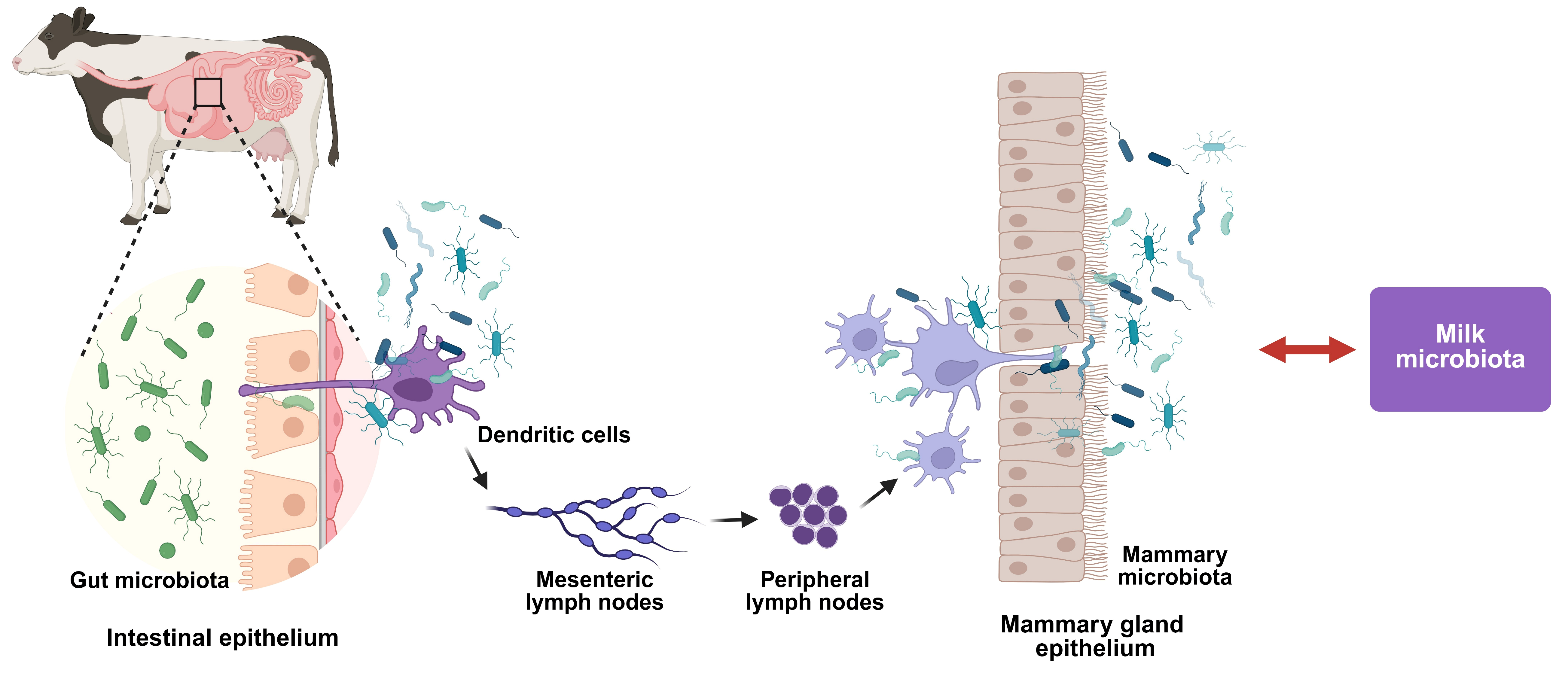
Figure 1. The enteromammary pathway: microbial translocation from gut to mammary gland. The figure presents the schematic representation of the enteromammary pathway showing how gut microorganisms can translocate across the intestinal epithelium, be captured by dendritic cells, migrate through lymph nodes, and ultimately colonize the mammary gland, contributing to milk microbiota composition.
Vetsci 12 01049 g001
Figure 2. Bovine microbiome dysbiosis in mastitis: mammary gland pathology and systemic microbial distribution. (A): Comparison of mammary gland health status and associated microbiome profiles. Left panel shows healthy mammary tissue with low somatic cell count, reduced inflammation (IL-6, TNF-α), and high antioxidant enzyme activity, characterized by predominant beneficial bacteria (Lactobacillus, Bifidobacterium, Faecalibacterium, Propionibacterium, Brevibacterium) and maintained gut homeostasis with balanced microbiome diversity (85%). Right panel depicts mastitis condition with elevated somatic cell count, increased inflammation markers, decreased antioxidant enzymes, dominated by pathogenic bacteria (S. aureus, Strep. agalactiae, E. coli, Klebsiella, Strep. uberis, Strep. dysgalactiae), and associated gut dysbiosis with reduced microbial diversity (35%). (B): Anatomical distribution of beneficial and pathogenic bacteria across different bovine body sites. The diagram illustrates site-specific microbial communities in the rumen, mammary gland (milk), and intestinal tract (feces), showing the distinct bacterial populations that colonize each anatomical niche. Beneficial bacteria include Bifidobacterium, Lactobacillus, Faecalibacter, Ruminococcus, and Butyrrivibrio species, while pathogenic bacteria comprise Staphylococcus, Enterococcus, Escherichia-Shigella, Streptococcus, and other potentially harmful species specific to each body compartment.
Vetsci 12 01049 g002
Figure 3. Short-chain fatty acids from beneficial microbiota modulate inflammatory pathways to prevent mastitis. This diagram illustrates how beneficial microbiota species (Bifidobacterium, Parasutterella, Lactobacillus acidophilus, Lactococcus piscium, Garcionitricum multicarbonum, Lactococcus raffinolactis, and Lactobacillus spp.) produce short-chain fatty acids (propionate-blue dots, butyrate-red dots, lactic acid-yellow dots) that modulate immune signaling pathways. These SCFAs interact with toll-like receptors (TLR2/TLR4) at the cell membrane (pink dotted line), triggering cytoplasmic signaling cascades involving MyD88, TRIF, and other intermediates that regulate nuclear transcription factors (NF-κB, IRF) to suppress inflammatory cytokine activation, ultimately preventing mastitis in mammary tissue.
Vetsci 12 01049 g003
Table 1. Microbiota differences/associations with mastitis.
Animal SpeciesSample TypeHealth Status ComparisonKey Microbiota Differences/AssociationsReference
Cows/MiceGutMastitis vs. HealthyEscherichia_Shigella, ↓ Roseburia; R. intestinalis protective via butyrate production[23]
Dairy cowsMilkSCC levels (<100 K vs. 100–200 K vs. >200 K)SCC < 100 K: ↑ Bifidobacterium, Lachnospiraceae_AC2044; Higher SCC: increased inflammatory gene expression[24]
YaksMilkHealthy vs. Subclinical vs. Clinical mastitisHealthy: Firmicutes (39.7%), Proteobacteria (60.17%); Mastitis: Proteobacteria ↑ 89.32% (subclinical), 95.36% (clinical); Firmicutes ↓ 10.49%, 2.92%[28]
Sahiwal cattleMilkHealthy vs. Subclinical vs. Clinical mastitisHealthy: Proteobacteria (56.48%), Firmicutes (15.87%); Clinical: Proteobacteria (2.68%), Firmicutes (64%); Subclinical: intermediate patterns[29]
Nili Ravi buffaloMilkHealthy vs. Subclinical vs. Clinical mastitisHealthy: Proteobacteria dominant, Streptococcus (11.60%); Clinical: ↑ Streptococcus (33.96%), Staphylococcus, Corynebacterium[30]
Holstein cowsMilk and FecesHealthy vs. mastitisMilk: ↑ Firmicutes, ↑ Cyanobacteria, ↑ Streptococcus, ↓ Macrococcus caseolyticus.
Feces: No significant differences
[31]
Multiple breedsMilkHealthy vs. Subclinical mastitisFirmicutes in subclinical; ↑ Proteobacteria in healthy; 45 taxonomic biomarkers identified[32]
Dairy cowsMilkNon-subclinical vs. Subclinical mastitisNon-subclinical: Anthropi spp., P. azotoformans, P. fragi dominant; Subclinical: P. azotoformans, Mycobacterium bovis, P. koreensis[33]
Dairy cowsMilkHealthy vs. Subclinical mastitisCorynebacterium bovis, C. xerosis (10-fold), Streptococcus dysgalactiae, S. uberis; Core mastitis microbiota: Lactobacillus acidipiscis, Staphylococcus hominis[34]
Dairy cowsMilkHealthy vs. GBS subclinical mastitisProteobacteria, Actinobacteria, Acidobacteria;Firmicutes, Streptococcus, trends toward ↑ Turicibacter, Enterococcus[35]
Dairy cowsMilkHealthy vs. Subclinical vs. Clinical mastitisHymenobacter, Lachnospiraceae NK4A136 in mastitis; ↓ Ralstonia, Lachnospiraceae NK3A20, Acetitomaculum, Massilia, Atopostipes[36]
Dairy cowsMilk and Teat canalHealthy vs. Clinical mastitisInverse relationship: ↓ milk diversity = ↑ inflammation; Sphingobacterium negatively associated with diversity[37]
Dairy cowsMilkHealthy vs. MastitisMastitis: S. aureus, Aerococcus spp., Streptococcus spp.; Surprising S. thermophilus in high SCC[38]
Dairy cowsMilkClinical mastitis polymicrobial486 cultures, 11 genera; 63.6% biofilm-forming; S. aureus most prolific biofilm former (18.8%)[39]
GoatsMilkHealthy vs. Subclinical vs. Clinical vs. GangrenousStaphylococcus dominant all groups; ↑ Mycoplasma in clinical; Novel Escherichia/Shigella-Enterococcus association in gangrenous[40]
Dairy cowsMilkHealthy vs. Mastitis across lactationMastitis: fewer bacterial taxa, ↓ diversity throughout lactation, Proteobacteria (9.1–95.4% vs. 24.0–92.9% healthy), Firmicutes (1.4–50.7% vs. 3.1–35.9% healthy)[41]
Dairy cowsMilkClinical mastitis vs. HealthyMastitis: 363 vs. 146 strains in healthy; 68% unreported/opportunistic strains; 14 archaeal, 14 viral genera unique[42]
Dairy cowsMilkSCC levels (<2 × 10⁵ vs. >2 × 10⁵ vs. >5 × 10⁵)SCC <2 × 10⁵: Actinobacteriota dominant; SCC 2 − 5 × 10⁵: Firmicutes dominant; SCC > 5 × 10⁵: Firmicutes = Proteobacteria[43]
Holstein cowsMilkHigh vs. Low resilience to mastitisLow resilience: ↑ Mycoplana, Rhodococcus; High efficiency: ↑ Aerococcus, Corynebacterium, Facklamia, Psychrobacter[45]
HumansBreast tissueGranulomatous vs. Acute mastitis vs. ControlsCorynebacterium >1% in 34.1% GM patients, C. kroppenstedtii predominant species[46]
Dairy cowsGutHealthy vs. Subclinical mastitisCyanobacteria, Proteobacteria, Succinivibrio, Lactobacillus_iners;
Paraprevotella, Coprococcus, Succiniclasticum, Desulfovibrio, and Bifidobacterium_pseudolongum
[47]
Dairy cowsRumenLow vs. High SCCBacteroidetes, Firmicutes, Lachnospiraceae, Prevotella, Rumiclostridium in H-SCC group[48]
Dairy cowsRumenSubclinical vs. Clinical mastitisClinical: ↑ Lachnospiraceae, Moraxella, Neisseriaceae; ↓ beneficial SCFA producers[49]
Cows/MiceRumen/GutSARA-mastitis vs. HealthyMoraxellaceae, ↓ Prevotellaceae; Sialic acid promotes ↑ Enterobacteriaceae, Akkermansiaceae[50]
Holstein cowsRumen/Feces/MilkSARA vs. ControlsStenotrophomonas in rumen; Barrier disruption allowing bacterial translocation[51]
MiceGutVagotomy-induced mastitisFirmicutes, Proteobacteria;Campylobacterota, Rikenellaceae_RC9_gut_group[52]
Dairy cowsRumenHealthy vs. Subclinical vs. Clinical mastitisClinical: ↑ Lachnospiraceae, Moraxella, Neisseriaceae; ↓ Prevoterotoella_1, Bifidobacterium[53]
MiceGutS. aureus mastitis vs. ControlsEnterobacter, ↓ short-chain fatty acids (SCFA)-producing bacteria (Firmicutes, Bacteroidetes)[54]
MiceGut and Mammary glandAntibiotic-treated vs. ControlsFirmicutes, ↓ Lactobacillaceae;Proteobacteria, Bacteroidota, Campylobacterota[55]
Dairy cowsRumen and FecesHealthy vs. MastitisRumen: Moryella characteristic in mastitis; Feces: Aeriscardovia, Lactococcus, Bacillus in healthy[56]
RatsGutHealthy vs. MastitisProteobacteria phylum triggering metabolic disruptions[57]
Dairy cowsMilk and FecesHealthy vs. Subclinical vs. Clinical mastitisMilk: ↑ Proteobacteria, ↓ Firmicutes, Actinobacteriota, Bifidobacterium in mastitis; Feces: ↑ UCG-010, Bacteroides, Prevotella in clinical mastitis[58]
Dairy cowsMilk and GutHealthy vs. MastitisMilk: ↑ Sphingomonas, Stenotrophomonas; Feces: ↑ Alistipes, Flavonifractor, Agathobacter, Pygmaiobacter[59]
BuffaloGutHealthy vs. MastitisMuribaculaceae, Eubacterium_nodatum, Lachnoclostridium_10, Pichia; ↓ Ruminococcus_2, Candidatus_Stoquefichus, Turicibacter[60]
Cows/MiceMultipleClinical mastitis vs. HealthyCows: P. aeruginosa, L. crispatus, K. oxytoca; Mice: Muribaculum, Duncaniella, B. animalis, E. coli, S. aureus[62]
Cows/MiceRumenClinical mastitis vs. HealthyProteobacteria in rumen; Dysbiosis-derived LPS promotes mastitis via TLR4-cGAS-STING-NF-κB/NLRP3[63]
Humans/MiceGutMastitis vs. ControlsFirmicutes/Bacteroidetes ratio, ↑ Actinobacteria, ↓ Verrucomicrobia, ↓ Ruminococcus, Faecalibacterium;Parabacteroides[64]
Dairy cows and miceFecesMastitis infected cow’s feces transplantation to miceIdentified key bacterial genera (Chrysobacterium, Christensenellaceae_R-7_group, Prevotella) as biomarkers.
Revealed endogenous pathway mediated by rumen microbiota dysbiosis in mastitis development.
Transplantation from mastitis cows induced mammary inflammation in mice.
[65]
“↓” shows decrease, “↑” indicate increase.
Table 2. Effects of various treatments on microbiota and mastitis management.
TreatmentAnimal ModelKey Findings Associated with Mastitis TreatmentReference
Fiber-enriched dietMice
Prevent Staphylococcus aureus-induced mastitis
Enriched Prevotellaceae, Bacteroides, and Lachnospiraceae.
Increased butyrate production, enhanced antimicrobial programs in macrophages.
Effects abolished with antibiotic-induced microbiota depletion.
[66]
Secondary Bile Acids/Clostridium scindensMice
Restored secondary bile acid-producing bacteria.
Increased mammary TGR5 expression and DCA levels. Activated TGR5-cAMP-PKA signaling, inhibited NF-κB/NLRP3. Restored tight junction proteins.
[67]
Bacillus amyloliquefaciens-9 (GB-9)Saanen dairy goats
Enhanced Bacteroides and Phascolarctobacterium populations.
Reduced SCC, inflammatory markers (IL-2, IL-4, IL-6). Decreased immunoglobulins (IgA, IgM) and malondialdehyde.
Effects mediated through Bacteroides-dependent immune modulation.
[75]
Yeast fermentation product (YFF)Dairy goats
Restored microbial diversity, increased Firmicutes and Actinobacteria.
Promoted beneficial genera (Ruminococcus, Olsenella), suppressed SARA-associated genera. Reduced LPS levels, inflammatory cytokines, and oxidative stress.
[81]
Astragalus Polysaccharide (APS)Mice
Enriched Ruminococcus bromii, facilitated SCFA metabolism (acetate and butyrate). Reduced S. aureus infection, suppressed inflammation, restored blood–milk barrier integrity.
Protective effects dependent on gut microbiota.
[85]
Angelica sinensis Polysaccharide (ASP)Mice
Increased Lachnospiraceae_NK4A136, reduced Erysipelatoclostridium.
Enhanced gut microbial diversity.
Elevated bioactive metabolites (tabersonine, riboflavin).
Effects entirely dependent on gut microbiota integrity.
[86]
Akkermansia muciniphilaDairy cows and mice
Enhanced Verrucomicrobia abundance, negatively correlated with inflammatory cytokines.
Inhibited TLR4 and NF-κB pathways.
Reduced SCC
Effects mediated through outer membrane vesicles (AOMVs).
[87]
Inulin (Rumen Microbiome)Dairy cows
Promoted propionate/butyrate-producing species (Prevotella, Butyrivibrio).
Enhanced commensal bacteria (Muribaculaceae, Bifidobacterium).
Suppressed pro-inflammatory taxa (Clostridia UCG-014, Streptococcus).
Increased milk yield and quality.
[88]
Inulin SupplementationDairy cows
Reduced mastitis-causing microbes (Escherichia-Shigella, Pseudomonas).
Increased beneficial probiotics (Lactobacillus, Bifidobacterium).
Enhanced SCFA production, reduced inflammatory mediators.
[89,90]
HordenineMice
Increased beneficial Lactobacillus, reduced Desulfovibrionaceae and Enterobacteriaceae.
Restored SCFA levels (butyrate, acetate, propionate).
Inhibited TLR4-MAPK/NF-κB, activated AMPK/Nrf2/HO-1 pathways.
Enhanced tight junction proteins.
[91]
Forsythiaside A (FTA)Mice
Increased Lactobacillus and Bifidobacterium, reduced Helicobacteraceae and Desulfovibrionaceae.
Normalized Firmicutes/Bacteroidetes ratio. Enhanced SCFA levels, reduced circulating LPS.
Modulated PI3K/AKT/mTOR pathways.
[92]
Maslinic acidMice
Increased beneficial Enterobacteriaceae, decreased harmful bacteria like Streptococcaceae.
Altered gut microbiota structure (β-diversity changes).
Reduced mammary inflammation (IL-6, IL-1β, TNF-α). Inhibited NLRP3, AKT/NF-κB, MAPK pathways.
[93]
HesperetinMice
Restored α-diversity, enriched Lactobacillaceae and Lachnospiraceae.
Suppressed harmful Enterobacteriaceae.
Inhibited TLR4/NF-κB pathway. Preserved tight-junction proteins (occludin, claudin-3, ZO-1).
[94]
Abrus cantoniensis total flavonoids (ATF)Mice
Increased beneficial Lactobacillus and norank_f_Muribaculaceae.
Reduced harmful taxa (Lachnospiraceae, Bacteroides, Helicobacter).
Suppressed CD14/TLR4/NF-κB/MAPK pathway. Enhanced tight junction proteins.
[95]
Pulsatilla chinensis extract (PCE)Mice
Maintained healthy microbiota structure, prevented pathological shifts.
Reduced Proteobacteria, Deferribacterota, and Planctomycetota.
Enhanced anti-inflammatory microbiota communities.
Dual mechanism: pathogen inhibition + microbiota preservation.
[97]
Gongying San (GYS)Holstein cows
Increased UCG-010 and Blautia, decreased Bacteroides and Lachnospiraceae.
Reduced SCC, LPS, inflammatory cytokines (IL-2, IL-4, IL-8, TNF-α).
Enhanced milk yield, lactose, and protein. Downregulated arachidonic acid metabolism.
[99]
Matrine-chitosan HydrogelsDairy cows
Decreased pathogenic microorganisms (Aerococcus, Corynebacterium_1, Staphylococcus).
Increased beneficial Proteobacteria.
Reduced SCC and microbial diversity.
Enhanced sphingolipids and glycerophospholipids.
[100]
Disclaimer/Publisher’s Note: The statements, opinions and data contained in all publications are solely those of the individual author(s) and contributor(s) and not of MDPI and/or the editor(s). MDPI and/or the editor(s) disclaim responsibility for any injury to people or property resulting from any ideas, methods, instructions or products referred to in the content.