
Simple Summary
Yujin powder is a polyherbal formulation that has been used to treat the large intestine dampness-heat syndrome for a long time. Escherichia coli is one of the most common pathogenic bacteria to cause this syndrome in diarrheal chickens, leading to serious economic losses in the poultry industry. The limited effect of antibiotics on antibiotic-resistant E. coli makes this bacterium a potential threat to human health. The present study is designed to explore the treatment effects and mechanisms of the Yujin powder and its potential bioactive components on lethal diarrhea caused by clinical isolated multidrug-resistant avian pathogenic E. coli and demonstrate the potential bioactive component of YJP. A multidrug-resistant E. coli was isolated and identified from a clinical diarrheal chick. Yujin powder and its components could directly inhibit the growth of this strain at high concentrations in vitro, and presents obvious anti-bacterial effects by reducing the bacterial loads, the release of endotoxin, and inflammation in vivo, which was much more effective than the resistant antibiotic ciprofloxacin. Based on our results, Yujin powder and its components, Scutellariae Radix and Baicalin, could be used as novel treatment against the diarrhea caused by this isolated multidrug-resistant E. coli.
Abstract
Escherichia coli is one of the most common pathogenic bacteria in diarrheal chickens, leading to serious economic losses in the poultry industry. The limited effect of antibiotics on antibiotic-resistant E. coli makes this bacterium a potential threat to human health. Yujin powder (YJP) has been reported as an agent that releases the symptoms caused by E. coli for a long time. The objective of this study is to investigate the effect of Yujin powder (YJP) and its components, Scutellariae Radix (SR) and Baicalin (Bac), anti-against multi-drug-resistant E. coli in vitro and in vivo. A multi-drug-resistant bacteria was isolated and identified from a clinical diarrheal chick. Then, the anti-bacterial effects of drugs were assessed in vitro and in vivo by analyzing the bacteria loads of organs, the levels of endotoxin, TNF-α, IL-1β, and IL-6 of the serum. Results found that the pathogenic E. coli was resistant to 19 tested antibiotics. YJP, SR, and Bac could directly inhibit the growth of this strain at high concentrations in vitro, and presents obvious anti-bacterial effects by reducing the bacterial loads, the release of endotoxin, and inflammation in vivo, which was much more effective than the resistant antibiotic ciprofloxacin. This study demonstrates that those natural medicines have the potential to be used as novel treatments to treat the disease caused by this isolated MDREC strain.
1. Introduction
Avian pathogenic Escherichia coli (APEC) causes bacterial infections through the digestive and respiratory tract in poultry, leading to colibacillosis [1]. Nowadays, colibacillosis caused by APEC is one of the major causes of high mortality and morbidity, which causes great economic losses in the poultry industry [2,3]. Antibiotics were used to promote the growth of animals or treat diarrhea caused by pathogenic bacteria in the chicken industry [4,5]. However, the abuse and long-term use of antibiotics lead to the production of drug-resistant strains, which makes antibiotics ineffective, brings food safety to human beings, and also formats a serious threat to human health [6,7]. Nowadays, more and more reports found that multiple strains of APEC isolated from farms with several antibiotic-resistant genes are resistant to multiple antibiotics, even APEC strains isolated from one farm are all resistant to some commonly used antibiotics in clinical practice [8,9,10]. Therefore, it is urgent to develop safe, efficacious agents with limited harmful effects for the treatment of multi-drug-resistant E. coli (MDREC) infected diarrhea.
Clinical practices in traditional Chinese medicine (TCM) and traditional Chinese veterinary medicine (TCVM) showed that the use of TCMs exerted great effects to treat diarrheal diseases under the guidance of TCM theory, especially the use of formulas containing several herbs [11,12]. For instance, Yujin Powder (YJP), one classical prescription composed of various herbs in TCM and TCVM, showed effective therapeutic effects on “Chang Huang”, characterized by fever, diarrhea, and abdominal pain, caused by excessive labor and diet, indigestible food, or contamination of food and drinking water in hot climates [13,14,15]. YJP was first recorded in Yuan Heng’s classical Collection on the Treatment of Equine Diseases with eight herbs, Curcumae Radix, Chebulae Fructus, Scutellariae Radix (SR), Rhei Radix Et Rhizoma (Rhubarb, Rb), Coptids Rhizoma, Phellodendri Chinensis Cortex, Gardenia Fructus, and Paeoniae Radix Alba, based on a Huanglianjiedu decoction [16,17]. According to TCVM theory, chicken colibacillosis belongs to the syndrome of internal heat caused by the accumulation of damp heat. Excessive water and dampness damage the spleen with the decrease of its transport function, leading to symptoms such as diarrhea [13,14]. Coptids Rhizoma, Scutellariae Radix, Phellodendri Chinensis Cortex, and Gardenia Fructus could clear the fire of “Sanjiao” and eliminate damp heat, act as minister herbs in this formula; Curcumae Radix could promote Qi circulation and relieve depression, cool and mix blood, normalize gallbladder to cure jaundice, and act as monarch herb; Paeoniae Radix Alba and Chebulae Fructus could astringe intestines to stop diarrhea, Rhubarb could remove accumulation with purgation, cool the blood, and remove the toxic materials; these three herbs act as adjuvant herbs [18,19,20,21]. Although these herbs have some similar effects, Scutellariae Radix and Rhei et Radix Rhizoma were chosen for further research according to the consumption, the differences among their “flavor and meridian tropism”, and our former research [22,23,24,25]. Nowadays, more and more reports demonstrate that these TCMs are shown to be great treatments and inhibit tissue injury through different pathways according to modern pharmacological research [26,27]. Scutellariae Radix (SR) and its component, baicalin, could decrease the levels of pro-inflammatory cytokines in serum, it also could inhibit the growth of Staphylococcus saprophyticus and Pseudomonas aeruginosa [24,28,29]. Curcumae Radix could not only directly inhibit bacteria, but also improve the effectiveness of antibiotics on inhibiting the growth of bacteria; so, this herb was not used as the research object of this study [30,31]. Rhubarb demonstrated broad-spectrum antibacterial activity against the Gram-negative bacteria, E. coli, and can be used to protect the gut barrier, maintain the intestinal micro-ecological environment, and prevent bacterial translocation as well [32,33]. In these aspects, the concepts of clearing away heat and detoxicating in TCM and TVCM are consistent with that of antibacterial, anti-inflammatory, and diarrhea in modern pharmacological research.
However, the treatment mechanisms of YJP and its potential bioactive components on diarrhea infected by a single MDREC are uncleared. In the present study, we isolate the pathogenic bacteria causing diarrhea in a chicken farm and tested its antibiotic-resistant spectrum. Then the anti-bacterial activities of YJP and its components to this strain were tested and the mechanisms of the effects were demonstrated in vitro and in vivo.
2. Materials and Methods
2.1. Bacterial Strains
The isolated multi-drug-resistant E. coli (MDREC) was obtained from the heart sample collected from a sick chick in a suspected colibacillosis layer farm in Dangtu County, Anhui Province, China. The standard strain of E. coli ATCC25922 was presented by a professor of the Department of Basic Veterinary Medicine, Nanjing Agricultural University.
2.2. Isolation and Identification of the Pathogen
The heart sample from the sick chickens was inoculated on nutrient broth agar medium (Qingdao Haibo, Qingdao, China) and incubated at 37 °C for 24 h. Then, the bacteria were purified and inoculated on McConkey’s agar medium and eosin methylene blue agar medium (Qingdao Haibo, Qingdao, China). After incubation at 37 °C for 24 h, the isolated bacterium was tentatively identified based on the colony and cell morphology, Gram staining, and biochemical activity detection. Molecular identification of the isolated E. coli was performed based on the nucleotide sequence analysis of 16S rRNA (27F: AGAGTTTGATCCTGGCTCAG; 1492R: TACGACTTAACCCCAATCGC). The PCR products were purified and sequenced after being subjected to agarose gel electrophoresis. The sequences were compared with the data using the nucleotide basic local alignment search tool (http://blast.ncbi.nlm.nih.gov/Blast.cgi, accessed on 26 October 2022). Phylogenetic analysis of the 16S rRNA gene was carried out using the MEGA version 7.0 software. The dendrogram was generated by the neighbor-joining method.
2.3. Antimicrobial Susceptibility Testing
Nineteen antibiotics were tested by the Kirby–Bauer method on Mueller–Hinton agar according to the guidelines of the Clinical and Laboratory Standards Institute [29]. Interpretive criteria for the inhibition zone diameters were provided by CLSI as well. It was considered as MDREC if the isolates were resistant to more than three classes of antibiotics. E. coli ATCC25922 was used as a standard strain to detect the susceptibility of antibiotics disks.
2.4. Drug Extracting Methods
The herbs for YJP were purchased from Nanjing Tianyuan Pharmacy (Nanjing, Jiangsu). All these herbs were genuine medicinal materials to ensure the best curative effects and authenticated by prof. Jiaguo Liu in the College of Veterinary Medicine of Nanjing Agriculture University. The composition of YJP was listed in Table 1 [17,34]. The plant names have been checked with “World Flora Online” (www.worldfloraonline.org, accessed on accessed on 17 January 2023). YJP and SR were extracted by the decocting method.
Table 1.
Herbs of Yujin Powder (YJP).
When extracting YJP, all herbs (160 g) except Radix et Rhizoma Rhei (60 g) and Curcumae Radix (30 g), were mixed and immersed in distilled water (2500 mL) for 30 min and then boiled for 30 min; after filtered with 8 layers of gauze, distilled water (1500 mL) was added to the herbs and boiled again; the third extraction is the same as the second extraction; at last, the filtrates from the three times were mixed, filtered by a Buchner funnel, concentrated with a rotary evaporator at 60 °C, and then freeze-dried with a vacuum freeze dryer. Radix et Rhizoma Rhei (60 g) and Curcumae Radix (30 g) were crushed, put into bags, and added in at the last five minutes. SR (30 g) was directly decocted with distilled water (300 mL) as the main method of YJP. When extracting Rb, it (60 g) was soaked in distilled water (600 mL) for 45 min, then decocted in boiling water for 5 min, and repeated three times. The filtrates were treated as YJP. These freeze-dried powders were stored at −20 °C for standby.
2.5. The MICs of YJP and Its Components
The minimal inhibit concentrations (MICs) of ciprofloxacin (CIP), YJP, SR, Rb, and Baicalin (Bac) against the MDREC and ATCC25922 were determined by broth microdilution method according to the guideline by the CLSI [35]. The test medium was Mueller–Hinton broth (MHB) and the isolated bacteria were added to the 96-well microtiter plate at an initial concentration of 2 × 10⁵ CFU/mL. Each well contains 50 μL of bacteria and 50 μL of different concentrations of drugs. After incubation at 37 °C for 24 h, the lowest concentration of the drugs without visual growth was recorded as the MIC values of these drugs.
2.6. Compliance with Ethical Standards
All animal research and facilities were carried out according to the experimental practices and standards. All experiments comply with the manual of the care and use of laboratory animals published by the US National Institutes of Health. All experimental protocols were approved by the Laboratory Animal Center of Nanjing Agricultural University.
2.7. YJP and Its Components against MDREC In Vivo
Institute of Cancer Research (ICR) mice (18~20 g) were purchased from Qing Long Shan animal breeding farm (SCXK (Su) 2017-0001). After 7 d of adaptive feedings, they were divided into six groups, including the blank control (BC) group, the E. coli control (EC) group, the CIP group, the YJP group, the SR group, and the Bac group. Each mouse was inoculated with 0.2 mL 4.49 × 10⁸ CFU/mL bacteria by intraperitoneal injection according to the toxicity test results of isolated bacteria (Table S2), except the BC group with normal saline. After one hour, the treatment groups were treated with YJP (3.75 g/kg) and SR (1 g/kg) by intragastric administration, Bac (0.0375 g/kg) by hypodermic injection near the back, and CIP (0.02 g/kg) by intramuscular injection in the thighs, respectively. The drugs were given once a day for 3 d continuously. The BC group and EC group were treated with the same amount of normal saline at the same time. The clinical symptoms and mortality of mice were observed and recorded for 7 d continuously.
2.8. Histopathological Evaluation
These mice were anesthetized with CO2 and the spleen tissues were obtained in each group at 18 and 72 h after injecting the isolated bacteria liquid. Sections (5-mm thickness) of fully fixed spleens were obtained and stained with hematoxylin and eosin, and then examined under a light microscope (Olympus Corporation, Tokyo, Japan).
2.9. Bacterial Loads in Organs
The heart, liver, and spleen were ground with 1 mL of normal saline by the homogenizer. The upper suspensions were diluted 10 times and inoculated to MacConkey agar plates with 20 μg/mL ampicillin. After incubation at 37 °C for 18 h, the numbers of viable bacteria in 1 g tissue were measured and calculated.
2.10. Endotoxin and Indexes of Inflammatory
The blood samples at 18 and 72 h after injecting the isolated bacteria were centrifuged to collect serum after coagulation at 37 °C and stood for 30 min. The levels of endotoxin, TNF-α, IL-1β, and IL-6 in the serum were determined according to the instructions of ELISA kits (Nanjing Aoqing, China).
2.11. Statistical Analysis
All data were analyzed by IBM SPSS statistics 19.0 software (IBM Corporation, Armonk, NY, USA). The statistical results were expressed as mean ± standard deviation. The difference among the mean values of experimental groups was analyzed by one-way ANOVA and Duncan’s multiple range test. The survival rate was tested by the chi-square test. p-values < 0.05 were considered statistically significant.
3. Results
3.1. The Colony Morphology of the Isolate
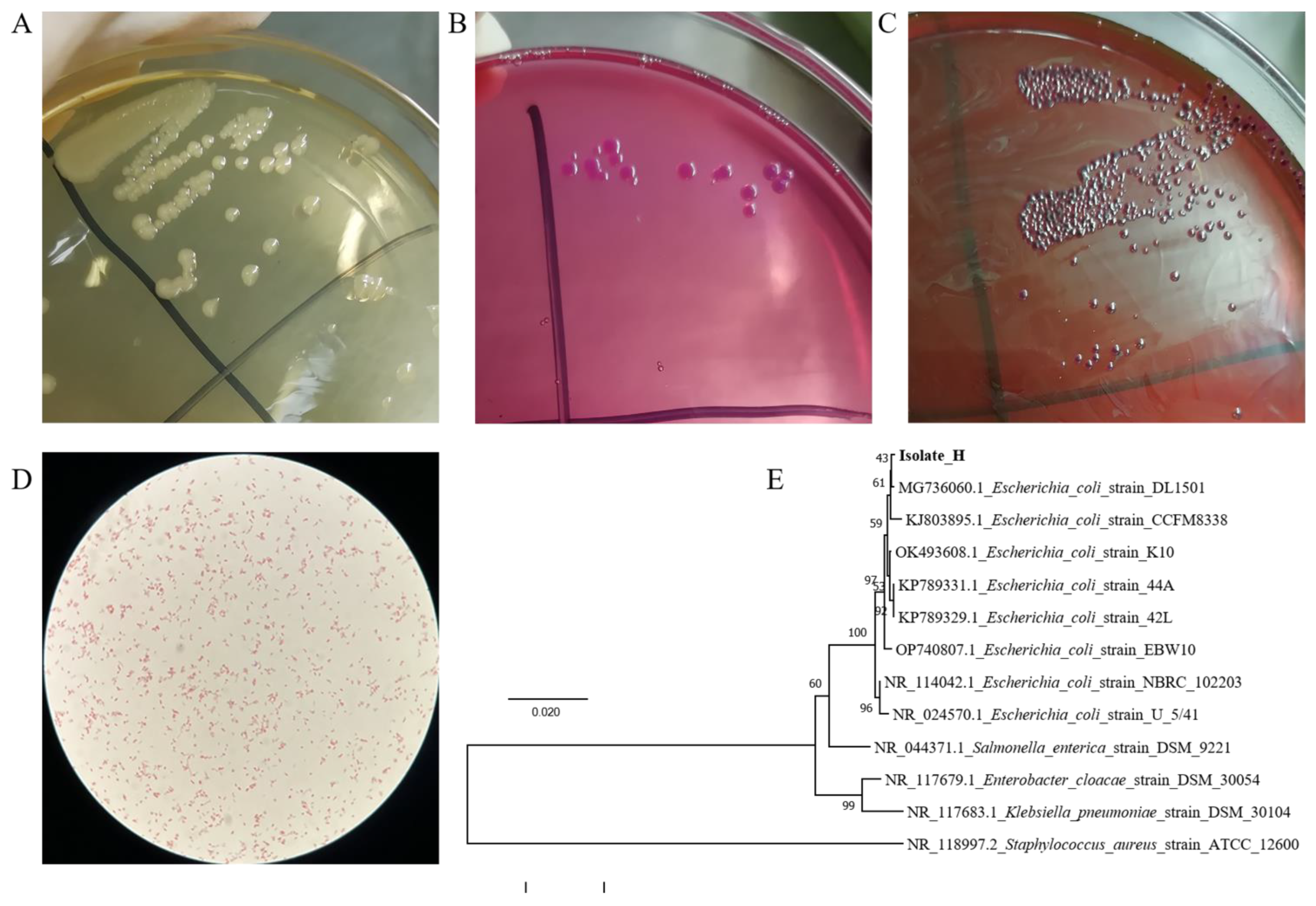
A large number of colonies with consistent morphology indicated that the diarrhea was caused by a single pathogen infection. On nutrient agar medium, the colonies are different in size but consistent in colony morphology, about 1–3 mm in diameter, moist and smooth on the surface, and neat in edges (Figure 1A). The diameters of colonies on McConkey’s medium are about 2 mm, the colonies are smooth, flat and moist, neat round, with neat edges, and a single colony is light red (Figure 1B). On eosin-methylene blue agar medium, the colonies’ diameters are about 1 mm, the purple-black colonies are round and convex, with smooth surfaces and typical metallic luster (Figure 1C). The typical dominant single colonies on the identification medium were selected for purification and culture for subsequent experiments.
Figure 1.
The characteristics of the isolate. (A–C). The colony morphology of the isolate from the heart on various media. (A). Nutrient Agar medium; (B). MacConkey agar medium (C). Eosin-methylene blue agar medium; (D). Morphology of isolates light microscope (10 × 100). The Gram-negative, short rod-shaped bacteria with blunt ends were seen under a light microscope. (E). Phylogenetic tree analysis of isolate and other bacteria. Phylogenetic tree analysis of this strain was based on 16S rRNA sequences (1433 bp fragments).
3.2. Biochemical Identification of the Isolate
The results of the biochemical identification of the isolate are shown in Table 2. On trisaccharide iron medium (TSI), the isolate can produce acid and gas on both the slope and the bottom layer, but cannot produce H2S. Indole can be produced with the forming of red ring. Glucose, lactose, maltose, mannitol, and sucrose can be fermented with the appearance of acid and gas. It can ferment honey disaccharide, cannot ferment side marigold alcohol and inositol, cannot utilize citrate, and cannot decompose urea. The VP test is negative, and the MR is positive. The lysine decarboxylase test is positive, and the phenylalanine deaminase is negative. It is athletic and can grow along the puncture line in semi-solid agar.
Table 2.
Biochemical identification results of pathogenic bacteria.
3.3. Microscopic Identification and PCR Identification
The Gram-negative, short rod-shaped bacteria with blunt ends were seen under the light microscope (Figure 1D). Then the molecular method was applied to identify the isolate. The 16S rRNA gene was amplified and sequenced. The isolate was closely similar to E. coli with the highest scoring 99% from the BLAST program and the phylogenetic tree analysis (Figure 1F). Combined with the results of other identification tests mentioned above, it can be proved that the isolated pathogen is E. coli.
3.4. Antimicrobial Susceptibility Testing
The diameters of the inhibition zone and the antimicrobial susceptibility results were shown in Table 3. The isolate is resistant to all the tested 19 antibiotics, indicating that the pathogen is MDREC.
Table 3.
The antimicrobial susceptibility test and resistance parameters on the isolated strain and quality control strain.
3.5. Antimicrobial Activity of CIP, YJP, and Its Components
We detected the MICs of YJP, Rb, SR, and Bac to assess the antibacterial effect of YJP and its components on the isolated MDREC. YJP and its components demonstrated antibacterial activity against MDREC and ATCC25922 in vitro, with MIC of YJP values 250.0 mg/mL, Rb 250.0 mg/mL, SR 62.5 mg/mL, and Bac 2.0 mg/mL. The MIC of CIP for MDREC was 8.0 × 10⁻² mg/mL, while for ATCC25922 was 1.0 × 10⁻⁵ mg/mL, indicating the severe resistance of the isolate to CIP (Table 4). After co-culture with 1/2 MIC concentration of Rb and SR for 24 h, the number of viable bacteria was significantly less than that in the blank group (Table S1).
Table 4.
The results of MIC of TCM and CIP (unit: mg/mL).
3.6. The Survival Rate of Each Group of Infected Mice
The survival rate of mice in each group is shown in Table 5. Survival rate (%) = number of survivors/total number of effective samples × 100%. There is no death case in the BC group, and the survival rate in the EC group is significantly lower than that in the BC group. The survival rate of the YJP group (66.7%), the SR group (66.7%), and the Bac (73.3%) group were significantly higher than that of the EC group (26.7%) (p < 0.05). In addition, the survival rate of the Bac group was significantly higher than that of the CIP group.
Table 5.
The survival situation of each group in the experiment.
3.7. Histopathological Changes of the Spleen of Mice
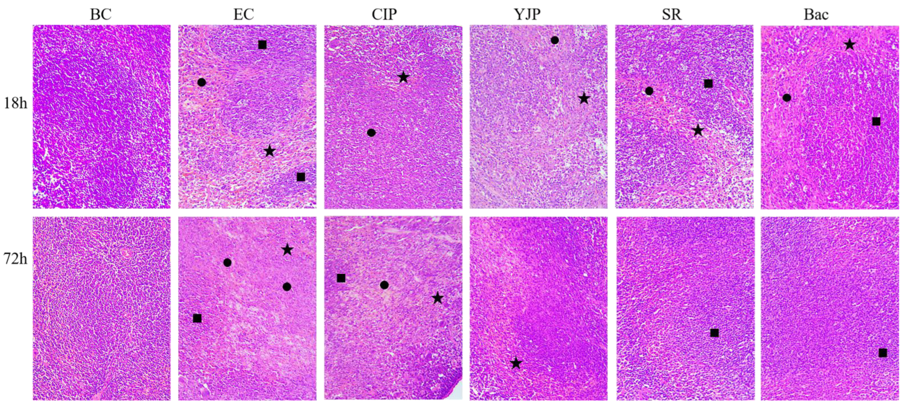
The sections of spleens in each group were evaluated microscopically using H + E staining, and the results were shown in Figure 2. At 18 h, the boundary between red pulp and white pulp in the BC group was clear, and the structure of the splenic corpuscle was complete. In the CIP group and EC Group, the boundary between red pulp and white pulp of spleen tissue was blurred, the tissue structure was lost. Increased red blood cells and pink amyloid in the red pulp were observed as well. Many inflammatory cells infiltrated in the EC group. The boundary between red pulp and white pulp in the YJP group was blurred, and that in the SR group and Bac group were clear. There were pink amyloid substances in the red pulp of the three groups, accompanied by a large number of red blood cells and inflammatory cells.
Figure 2.
Effects of CIP, YJP and its components on splenic histopathological changes of mice with E. coli infection (H&E staining, ×200, n = 6). BC, Blank Control; EC, E. coli control; YJP, Yujin Powder; SR, Scutellariae Radix; Bac, Baicalin; CIP, ciprofloxacin. ★, blood cells; ●, pink amyloid; ■, inflammatory cell infiltration.
At 72 h, the unique structure of normal spleen tissue disappeared, no boundary between red pulp and white pulp was found, a large number of red blood cells and inflammatory cells were found, and pink amyloid diffused in the CIP group and EC Group. In the YJP group, SR group, and Bac group, the boundary between red pulp and white pulp of spleen tissue was clear, and there were still many red cells and inflammatory cells.
3.8. Bacteria Loads in Various Organs of Mice
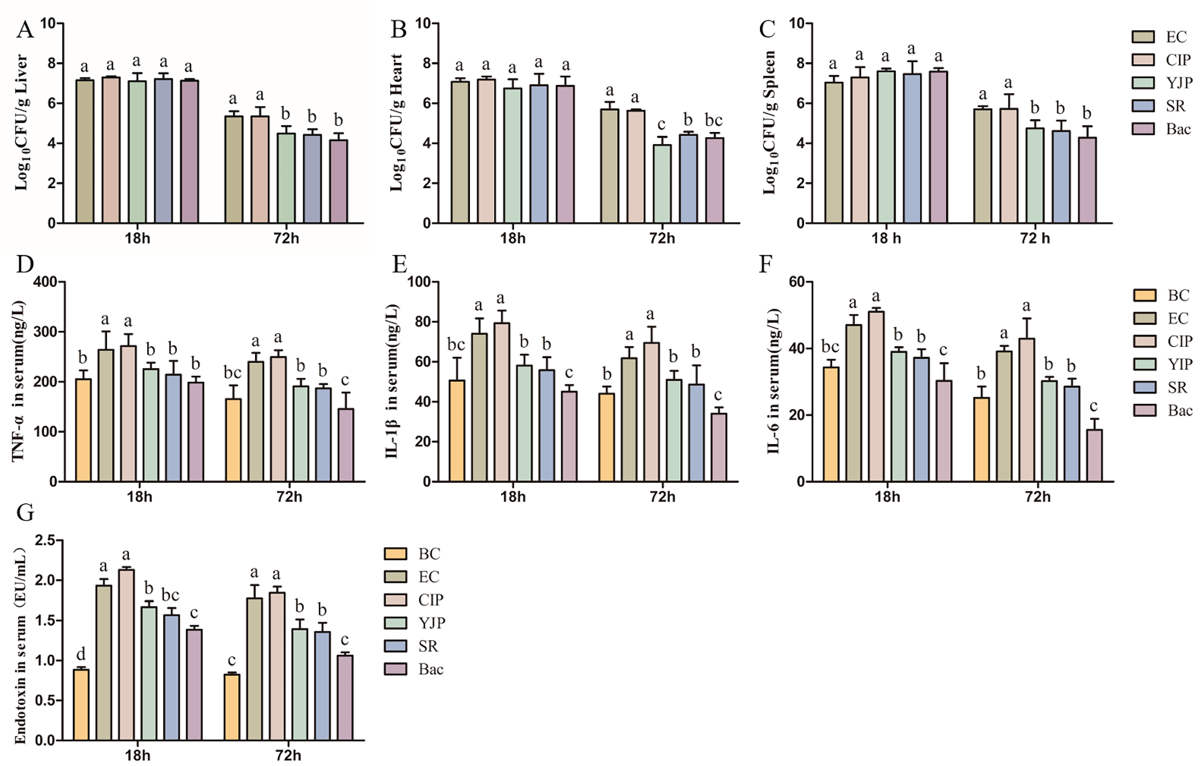
At 18 h, the initial stage of disease, the numbers of colonies in the YJP group, SR group, and Bac group in the liver (Figure 3A), heart (Figure 3B), and spleen (Figure 3C) were not significantly different from that in the EC group (p > 0.05). At 72 h, the number of bacteria in the three organs in the YJP group, SR group, and Bac group was significantly lower than that in the EC Group and CIP group (p < 0.05), and there was no significant difference among the three TCMs-treated groups (p > 0.05). In the YJP group, SR group, and Bac group, which are significantly different from the EC Group, Bac has a more obvious effect in reducing the total number of colonies.
Figure 3.
Treatment effects of CIP, YJP and its components on the mice with Escherichia coli infection (n = 6). (A–C). The logarithm of the number of bacteria in different organs ((A) Liver; (B) Heart; (C) Spleen). (D–F). The contents of inflammatory factors in the serum of different treated groups. ((D) TNF-α; (E) IL-1β; (F) IL-6). (G). The contents of endotoxin in serum of different treated groups. BC, Blank Control; EC, E. coli control; YJP, Yujin Powder; SR, Scutellariae Radix; Bac, Baicalin; CIP, ciprofloxacin. The data was represented by the mean ± standard deviation. The letter “a” indicates the maximum value, “a”, “b”, “c” and “d” decrease in turn. The same lowercase letter indicates no significant differences between groups, while completely different lowercase letters show significant differences with a p < 0.05.
3.9. Assessment of Inflammatory Cytokines Levels
The finding of the serum levels of TNF-α (Figure 3D), IL-1β (Figure 3E), and IL-6 (Figure 3F) demonstrated that the YJP, SR, and Bac significantly decreased the inflammation index levels compared with the EC group (p < 0.05). Bac could even reduce these three inflammatory factors to the normal level. While, CIP failed to reduce the levels of these three inflammatory factors at 18 h and 72 h, and even slightly increased compared with the EC group with no significance (p > 0.05).
3.10. The Levels of Bacterial Endotoxin in Serum
As shown in Figure 3G, plasma was significantly affected by the treatment of YJP, SR, and Bac (p < 0.05). In YJP, SR, and Bac groups, compared with the BC group, the YJP, SR, and Bac decreased the plasma endotoxin content of mice. In addition, the Bac significantly decreased the plasma endotoxin of mice at 18 and 72 h after inoculating the isolated E. coli, compared with the YJP group. While, CIP slightly increased the endotoxin in serum compared with the EC group (p > 0.05).
4. Discussion
Chicken colibacillosis caused by APEC is one of the important infectious diseases endangering the poultry industry in the world, accounting for the first place of bacterial diseases [2,36]. We received sick chickens from Anhui Dangtu laying hen farm. A large number of chickens on this farm suffered from diarrhea repeatedly, with ineffective use of several antibiotics and a lot of death. In this experiment, the pathogenic bacteria were successfully isolated, identified, and analyzed for drug resistance, and the pathogen was determined to be MDREC.
Efficient alternative treatment must be developed for serious infections caused by multi-drug-resistant bacteria. The current therapeutic strategy by enhancing the dosage of useless antibiotics is problematic due to the development of antibiotic resistance. Antibiotics were always used as growth stimulants, disease prevention, and treatment in animals, which may leave antibiotic residues in foodstuffs such as eggs and meat in the poultry industry, and antibiotic-resistant bacteria appeared at the same time [6,37]. Not only do the residual antibiotics in food produce a series of toxic side effects when they enter the human body, but also the development of drug-resistant bacteria poses challenges to the treatment of human diseases [38,39]. In this study, the isolated pathogenic E. coli was resistant to all tested bacteria, and the MIC of CIP to this strain was 8000 times the amount of that to ATCC 25922, indicating the severe antibiotic resistance caused the ineffectiveness of several antibiotics in the treatment.
Recent studies have reported that several Chinese herbal compounds exert potent activities against pathogenic bacteria [40,41]. Previously, it was confirmed that YJP could effectively treat diarrhea caused by E. coli [13,14,42]. However, whether YJP exerted its effects and which was the bio-effective component remained unknown. To further explore the potential mechanisms, the MICs of YJP, SR, Rb, and Bac were investigated. MIC was typically used to reveal the anti-bacterial effect of drugs [29,38]. Results of MICs showed that YJP, SR Rb, and Bac could directly suppress the bacteria growth at high concentrations in vitro.
As an important indicator of the drug protection effect, the survival rate of experimental animals can directly indicate the therapeutic significance of drugs [43,44,45]. The treatment of YJP, SR, and Bac significantly increased the number of surviving mice. The results showed that YJP, SR, and Bac could effectively reduce the mortality of the diseased mice, showing a good anti-infection effect, among which Bac had the best effect. The pathogenic mechanism of pathogenic E. coli is a multi-step mechanism, including the colonization of mucosal sites, escape from host defense, bacterial proliferation, and damage to host tissue and cells [46]. At the beginning of the disease (18 h), bacterial proliferation and colonization were dominant, there was no difference in the bacteria loads between all groups in all tissues. At the late stage (72 h), the total number of colonies in the YJP group, SR group, and Bac group was significantly lower than that in the EC group and CIP group. It can be seen that the total number of colonies in various organs could be effectively reduced by YJP, SR, and Bac. In addition, the groups with high survival rates also have fewer bacterial loads in the organs.
Endotoxin exists in the cell wall of Gram-negative bacteria and can be released when the cell wall disintegrates after the death of the bacteria [47]. When E. coli invade the body, it will release endotoxin [48]. At 18 and 72 h after inoculation, the serum endotoxin of the YJP, SR, and Bac groups was reduced to the normal level. Bac had a better effect on endotoxin reduction than YJP and SR at 72 h. After CIP treatment, the content of endotoxin slightly increased compared with that of the EC group (p > 0.05), corresponding to the conclusion that antibiotic-treated animals may show higher endotoxin levels than untreated animals [49,50]. All TCMs can effectively eliminate endotoxin in the body and improve the survival rate. When coliform infection is clinically treated with antibiotics, it may increase the release of endotoxin, leading to further deterioration of the disease [51].
The release of endotoxin can activate the monocyte-macrophage system and induce secretion of various mediators, including TNF-α, Cytokines such as IL-1β and IL-6, which are most closely related to the pathological response of the body [52,53]. The data from this study showed that the MDREC infection can cause severe inflammatory reactions in the body as evidenced by increased plasma TNF-α, IL-1β, and IL-6 levels and spleen pathological changes of inflammatory reaction. At 18 and 72 h after inoculation, the inflammatory factors can be significantly reduced with the treatment of YJP, SR, and Bac, corresponding to the results of spleen section staining. Among them, Bac-reduced IL-1β and IL-6 was more effective than YJP and SR. However, these effects were not observed in the CIP group, indicating the failure of CIP treatment because of antibiotic resistance [54]. The results were in accordance with the conclusion that reducing the levels of inflammatory factors is one of the mechanisms by which drugs play a therapeutic role in infectious diseases [55].
Our results indicated that Bac may be the main bioactive component of SR, which may be an important active drug of YJP. In our study, the effect of Bac is best, followed by SR, and YJP. However, in clinical practice, there are few cases of single bacterial infection, and it is generally multiple bacterial mixed infections. Therefore, the use of YJP always has a better effect than a single drug in clinical practice, which is always different from the results of the laboratory [56,57]. If diarrhea caused by a single bacterial infection is confirmed in clinical practice, the use of baicalin may have a better therapeutic effect and less cost than the use of formula.
5. Conclusions
In conclusion, the pathogenic MDR E. coli causing chicken diarrhea was successfully isolated and identified, and YJP and its components could inhibit this strain in vitro and in vivo. Moreover, the antibacterial effects of YJP, SR, and Bac were achieved by reducing the bacterial loads, the release of endotoxin, and inflammation. YJP, SR, and Bac could be potent antibiotic substitutes to treat colibacillosis caused by this strain.
Supplementary Materials
The following supporting information can be downloaded at: https://www.mdpi.com/article/10.3390/vetsci10050328/s1. Supplementary Method S1: Numbers of the isolate after the treatment of drugs. Supplementary Methods S2: Determination of median lethal dose of isolated MDREC. Supplementary Table S1: The numbers of colonies of MDREC after 24 h. Supplementary Table S2: The death situation of mice caused by different amounts of infected bacteria.
Author Contributions
J.L., J.M. and J.W. conceived and designed the experiments. J.M., J.W., J.Z. and S.L. performed the experiments. J.D. and W.W. (Wenjia Wang) analyzed the data. J.M. contributed to the reagents/materials/analysis tools. J.M., T.Q. and W.W. (Weiran Wang) wrote the manuscript. All authors have read and agreed to the published version of the manuscript.
Funding
This work was supported by the Fundamental research funds for the central universities (KYYJ201803), the National Natural Science Foundation of China (Grant Nos. 31772784 and 31572557), Key Special Projects for the 14th Five Year Plan (2022YFD1801102) and the Project Funded by the Priority Academic Program Development of Jiangsu Higher Education Institutions (PAPD).
Institutional Review Board Statement
The animal study protocols was approved by the Laboratory Animal Center of Nanjing Agricultural University (IACUC2019056, 20 December 2019).
Informed Consent Statement
Written informed consent has been obtained from the master of the farm to publish this paper.
Data Availability Statement
Not applicable.
Acknowledgments
We thank Dawei Guo for providing us with ATCC25922 from the Department of Basic Veterinary Medicine, Nanjing Agricultural University.
Conflicts of Interest
The authors declare that there are no conflict of interest.
Abbreviation
| YJP | Yujin powder |
| SR | Scutellariae Radix |
| Rb | Rhubarb |
| Bac | Baicalin |
| CIP | Ciprofloxacin |
| APEC | Avian pathogenic Escherichia coli |
| MDREC | Multi-drug-resistant E. coli |
| MIC | Minimum inhibitory concentration |
| TCM | Traditional Chinese medicine |
| TCVM | Traditional Chinese veterinary medicine |
| CLSI | Clinical and Laboratory Standards Institute |
References
- Kathayat, D.; Lokesh, D.; Ranjit, S.; Rajashekara, G. Avian Pathogenic Escherichia coli (APEC): An Overview of Virulence and Pathogenesis Factors, Zoonotic Potential, and Control Strategies. Pathogens 2021, 12, 467. [Google Scholar] [CrossRef] [Scilit] [PubMed]
- Newman, D.M.; Barbieri, N.L.; de Oliveira, A.L.; Willis, D.; Nolan, L.K.; Logue, C.M. Characterizing avian pathogenic Escherichia coli (APEC) from colibacillosis cases, 2018. PeerJ 2021, 9, e11025. [Google Scholar] [CrossRef] [Scilit]
- Zhuang, Q.Y.; Wang, S.C.; Li, J.P.; Liu, D.; Liu, S.; Jiang, W.M.; Chen, J.M. A clinical survey of common avian infectious diseases in China. Avian Dis. 2014, 58, 297–302. [Google Scholar] [CrossRef] [Scilit] [PubMed]
- Bacanli, M.; Basaran, N. Importance of antibiotic residues in animal food. Food Chem. Toxicol. 2019, 125, 462–466. [Google Scholar] [CrossRef] [Scilit] [PubMed]
- Costa, M.C.; Bessegatto, J.A.; Alfieri, A.A.; Weese, J.S.; Filho, J.A.; Oba, A. Different antibiotic growth promoters induce specific changes in the cecal microbiota membership of broiler chicken. PLoS ONE 2017, 12, e171642. [Google Scholar] [CrossRef] [Scilit]
- Lobanovska, M.; Pilla, G. Penicillin’s Discovery and Antibiotic Resistance: Lessons for the Future? Yale J. Biol. Med. 2017, 90, 135–145. [Google Scholar]
- Muurinen, J.; Richert, J.; Wickware, C.L.; Richert, B.; Johnson, T.A. Swine growth promotion with antibiotics or alternatives can increase antibiotic resistance gene mobility potential. Sci. Rep. 2021, 11, 5485. [Google Scholar] [CrossRef] [Scilit]
- Xu, X.; Sun, Q.; Zhao, L. Virulence Factors and Antibiotic Resistance of Avian Pathogenic Escherichia coli in Eastern China. J. Vet. Res. 2019, 63, 317–320. [Google Scholar] [CrossRef] [Scilit]
- Gao, J.; Duan, X.; Li, X.; Cao, H.; Wang, Y.; Zheng, S.J. Emerging of a highly pathogenic and multi-drug resistant strain of Escherichia coli causing an outbreak of colibacillosis in chickens. Infect. Genet. Evol. J. Mol. Epidemiol. Evol. Genet. Infect. Dis. 2018, 65, 392–398. [Google Scholar] [CrossRef] [Scilit]
- Yoon, M.Y.; Kim, Y.B.; Ha, J.S.; Seo, K.W.; Noh, E.B.; Son, S.H.; Lee, Y.J. Molecular characteristics of fluoroquinolone-resistant avian pathogenic Escherichia coli isolated from broiler chickens. Poult. Sci. 2020, 99, 3628–3636. [Google Scholar] [CrossRef] [Scilit]
- Guo, Y.; Li, J.X.; Wang, Y.L.; Mao, T.Y.; Chen, C.; Xie, T.H.; Han, Y.F.; Tan, X.; Han, H.X. Yinchen Linggui Zhugan Decoction Ameliorates Nonalcoholic Fatty Liver Disease in Rats by Regulating the Nrf2/ARE Signaling Pathway. Evid. Based Complement. Altern. Med. 2017, 2017, 6178358. [Google Scholar] [CrossRef] [Scilit] [PubMed]
- Tao, Y.; Li, C.; Gao, T.; Huo, J. Molecular mechanism of Sishen pills in the treatment of diarrheal diabetic enteropathy based on network pharmacology. Medicine 2022, 101, e30096. [Google Scholar] [CrossRef] [Scilit] [PubMed]
- Yao, W.; Yang, C.; Wen, Y.; Zhang, W.; Zhang, X.; Ma, Q.; Ji, P.; Hua, Y.; Wei, Y. Treatment effects and mechanisms of Yujin Powder on rat model of large intestine dampness-heat syndrome. J. Ethnopharmacol. 2017, 202, 265–280. [Google Scholar] [CrossRef] [Scilit]
- Yao, W.; Zhang, Y.; Zhang, W.; Wen, Y.; Yang, R.; Dong, J.; Zhang, X.; Hua, Y.; Ji, P.; Wei, Y. Pathological mechanism of intestinal mucosal barrier injury of large intestine dampness-heat syndrome rats and the protective effect of Yujin powder. Res. Vet. Sci. 2022, 152, 485–496. [Google Scholar] [CrossRef] [Scilit] [PubMed]
- Song, C.; Ji, M.; Xu, J.; Huo, B.; Chi, Y.; Shen, X. Study on the Therapeutic Effect of Modified Yujin Powder on Transimmissible Gastroenteritis in Landrace Pigs. China Anim. Husb. Vet. Med. 2019, 46, 600–607. [Google Scholar]
- Yu, B.; Yu, Y. Yuan Heng’s Classical Collection on the Treatment of Equine Diseases; Agriculture Press: Beijing, China, 1957; p. 138. (In Chinese) [Google Scholar]
- Chinese Veterinary Pharmacopoeia Commission. Veterinary Pharmacopoeia of People’s Republic of China; Chinese Agriculture Press: Beijing, China, 2020; Volume II. (In Chinese) [Google Scholar]
- Wang., D. Practical Chinese Veterinary Medicine, 1st ed.; Chinese Agriculture University Press: Beijing, China, 2006; p. 68. (In Chinese) [Google Scholar]
- Zhang, Y.; Yao, W.; Wen, Y.; Wen, Y.; Hua, Y.; Peng, J.; Wei, Y. Regulation of Yujin Powder on Serum Immunoglobulin and Antioxidant Related Factors of Large Intestine Dampness-heat Syndrome Rat. Chin. J. Anim. Vet. Sci. 2018, 49, 2044–2053. [Google Scholar]
- Yang, Y.; Kang, N.; Xia, H.; Li, J.; Chen, L.; Qiu, F. Metabolites of protoberberine alkaloids in human urine following oral administration of Coptidis Rhizoma decoction. Planta Med. 2010, 76, 1859–1863. [Google Scholar] [CrossRef] [Scilit]
- Liao, F. Diagnostics of Chinese Medicine, 2nd ed.; People’s Medical Publishing House: Beijing, China, 2013; p. 108. (In Chinese) [Google Scholar]
- Qiao, Y.; Zhang, Y.; Peng, S.; Huo, M.; Li, J.; Cao, Y.; Chen, Z. Property theory of Chinese materia medica: Clinical pharmacodynamics of traditional Chinese medicine. J. Tradit. Chin. Med. Sci. 2022, 9, 7–12. [Google Scholar] [CrossRef] [Scilit]
- Bao, H. The Science of Chinese Materia Medicine, 1st ed.; Chinese Agriculture Press: Beijing, China, 2008. (In Chinese) [Google Scholar]
- Wang, J.; Qiao, M.; Zhou, Y.; Du, H.; Bai, J.; Yuan, W.; Liu, J.; Wang, D.; Hu, Y.; Wu, Y. In vitro synergistic effect of baicalin with azithromycin against Staphylococcus saprophyticus isolated from francolins with ophthalmia. Poult. Sci. 2019, 98, 373–380. [Google Scholar] [CrossRef] [Scilit]
- Xiang, J.Y.; Chi, Y.Y.; Han, J.X.; Xiang, H.; Xie, Q. The Toxicity and Attenuation Methods of Toxic Chinese Materia Medica for its Reasonable Application: A Review. Am. J. Chin. Med. 2020, 49, 41–67. [Google Scholar] [CrossRef] [Scilit]
- Cheng, X.; Cao, Z.; Luo, J.; Hu, R.; Cao, H.; Guo, X.; Xing, C.; Yang, F.; Zhuang, Y.; Hu, G. Baicalin ameliorates APEC-induced intestinal injury in chicks by inhibiting the PI3K/AKT-mediated NF-kappaB signaling pathway. Poult. Sci. 2022, 101, 101572. [Google Scholar] [CrossRef] [Scilit] [PubMed]
- Peng, L.Y.; Shi, H.T.; Tan, Y.R.; Shen, S.Y.; Yi, P.F.; Shen, H.Q.; Fu, B.D. Baicalin inhibits APEC-induced lung injury by regulating gut microbiota and SCFA production. Food Funct. 2021, 12, 12621–12633. [Google Scholar] [CrossRef] [Scilit] [PubMed]
- Wan, F.; Wang, M.; Zhong, R.; Chen, L.; Han, H.; Liu, L.; Zhao, Y.; Lv, H.; Hou, F.; Yi, B.; et al. Supplementation with Chinese Medicinal Plant Extracts from Lonicera hypoglauca and Scutellaria baicalensis Mitigates Colonic Inflammation by Regulating Oxidative Stress and Gut Microbiota in a Colitis Mouse Model. Front. Cell. Infect. Microbiol. 2022, 11, 798052. [Google Scholar] [CrossRef] [Scilit] [PubMed]
- Luo, J.; Dong, B.; Wang, K.; Cai, S.; Liu, T.; Cheng, X.; Lei, D.; Chen, Y.; Li, Y.; Kong, J.; et al. Baicalin inhibits biofilm formation, attenuates the quorum sensing-controlled virulence and enhances Pseudomonas aeruginosa clearance in a mouse peritoneal implant infection model. PLoS ONE 2017, 12, e0176883. [Google Scholar] [CrossRef] [Scilit]
- Luo, J.Y.; Yan, D.; Yang, M.H. Study of the anti-MRSA activity of Rhizoma coptidis by chemical fingerprinting and broth microdilution methods. Chin. J. Nat. Med. 2014, 12, 393–400. [Google Scholar] [CrossRef] [Scilit]
- Mun, S.H.; Joung, D.K.; Kim, Y.S.; Kang, O.H.; Kim, S.B.; Seo, Y.S.; Kim, Y.C.; Lee, D.S.; Shin, D.W.; Kweon, K.T.; et al. Synergistic antibacterial effect of curcumin against methicillin-resistant Staphylococcus aureus. Phytomedicine 2013, 20, 714–718. [Google Scholar] [CrossRef] [Scilit]
- Bhattacharjee, M.K.; Bommareddy, P.K.; DePass, A.L. A Water-Soluble Antibiotic in Rhubarb Stalk Shows an Unusual Pattern of Multiple Zones of Inhibition and Preferentially Kills Slow-Growing Bacteria. Antibiotics 2021, 10, 951. [Google Scholar] [CrossRef] [Scilit]
- Chen, D.C.; Wang, L. Mechanisms of therapeutic effects of rhubarb on gut origin sepsis. Chin. J. Traumatol. 2009, 12, 365–369. [Google Scholar]
- Hu, Y. Traditional Chinese Veterinary Medicine, 1st ed.; Science Press: Beijing, China, 2013. (In Chinese) [Google Scholar]
- M100-S11; Performance Standards for Antimicrobial Susceptibility Testing. Clinical and Laboratory Standards Institute: Wayne, PA, USA, 2018; pp. 34–46.
- Monson, M.S.; Lamont, S.J. Genetic resistance to avian pathogenic Escherichia coli (APEC): Current status and opportunities. Avian Pathol. 2021, 50, 392–401. [Google Scholar] [CrossRef] [Scilit]
- Ghimpeteanu, O.M.; Pogurschi, E.N.; Popa, D.C.; Dragomir, N.; Dragotoiu, T.; Mihai, O.D.; Petcu, C.D. Antibiotic Use in Livestock and Residues in Food-A Public Health Threat: A Review. Foods 2022, 11, 1430. [Google Scholar] [CrossRef] [Scilit]
- Chen, J.; Ying, G.G.; Deng, W.J. Antibiotic Residues in Food: Extraction, Analysis, and Human Health Concerns. J. Agric. Food Chem. 2019, 67, 7569–7586. [Google Scholar] [CrossRef] [Scilit] [PubMed]
- Donoghue, D.J. Antibiotic residues in poultry tissues and eggs: Human health concerns? Poult. Sci. 2003, 82, 618–621. [Google Scholar] [CrossRef] [Scilit] [PubMed]
- Khan, R.U.; Khan, A.; Naz, S.; Ullah, Q.; Laudadio, V.; Tufarelli, V.; Ragni, M. Potential Applications of Moringa oleifera in Poultry Health and Production as Alternative to Antibiotics: A Review. Antibiotics 2021, 10, 1540. [Google Scholar] [CrossRef] [Scilit] [PubMed]
- Khan, R.U.; Fatima, A.; Naz, S.; Ragni, M.; Tarricone, S.; Tufarelli, V. Perspective, Opportunities and Challenges in Using Fennel (Foeniculum vulgare) in Poultry Health and Production as an Eco-Friendly Alternative to Antibiotics: A Review. Antibiotics 2022, 20, 278. [Google Scholar] [CrossRef] [Scilit]
- Zhao, T.; Tang, H.; Xie, L.; Zheng, Y.; Ma, Z.; Sun, Q.; Li, X. Scutellaria baicalensis Georgi. (Lamiaceae): A review of its traditional uses, botany, phytochemistry, pharmacology and toxicology. J. Pharm. Pharmacol. 2019, 71, 1353–1369. [Google Scholar] [CrossRef] [Scilit]
- Zhang, M.; Li, H.; Agyekumwaa, A.K.; Yu, Y.; Xiao, X. Effects of citronellal on growth and enterotoxins production in Staphylococcus aureus ATCC 29213. Toxicon Off. J. Int. Soc. Toxinology 2022, 213, 92–98. [Google Scholar] [CrossRef] [Scilit]
- Shen, W.C.; Wang, X.; Qin, W.T.; Qiu, X.F.; Sun, B.W. Exogenous carbon monoxide suppresses Escherichia coli vitality and improves survival in an Escherichia coli-induced murine sepsis model. Acta Pharm. Sin. 2014, 35, 1566–1576. [Google Scholar] [CrossRef] [Scilit]
- Choi, Y.Y.; Kim, M.H.; Han, J.M.; Hong, J.; Lee, T.H.; Kim, S.H.; Yang, W.M. The anti-inflammatory potential of Cortex Phellodendron in vivo and in vitro: Down-regulation of NO and iNOS through suppression of NF-κB and MAPK activation. Int. Immunopharmacol. 2014, 19, 214–220. [Google Scholar] [CrossRef] [Scilit]
- Saldana, Z.; Erdem, A.L.; Schuller, S.; Okeke, I.N.; Lucas, M.; Sivananthan, A.; Phillips, A.D.; Kaper, J.B.; Puente, J.L.; Giron, J.A. The Escherichia coli common pilus and the bundle-forming pilus act in concert during the formation of localized adherence by enteropathogenic E. coli. J. Bacteriol. 2009, 191, 3451–3461. [Google Scholar] [CrossRef] [Scilit]
- Schletter, J.; Heine, H.; Ulmer, A.J.; Rietschel, E.T. Molecular mechanisms of endotoxin activity. Arch. Microbiol. 1995, 164, 383–389. [Google Scholar] [CrossRef]
- Luo, G.; Zhang, J.; Sun, Y.; Wang, Y.; Wang, H.; Cheng, B.; Shu, Q.; Fang, X. Nanoplatforms for Sepsis Management: Rapid Detection/Warning, Pathogen Elimination and Restoring Immune Homeostasis. Nano-Micro Lett. 2021, 13, 88. [Google Scholar] [CrossRef] [Scilit] [PubMed]
- Holzheimer, R.G. Antibiotic induced endotoxin release and clinical sepsis: A review. J. Chemother. 2001, 13, 159–172. [Google Scholar] [CrossRef] [Scilit]
- Shenep, J.L. Antibiotic-induced bacterial cell lysis: A therapeutic dilemma. Eur. J. Clin. Microbiol. 1986, 5, 11–12. [Google Scholar] [CrossRef] [Scilit] [PubMed]
- Morrison, D.C.; Bucklin, S.E. Evidence for antibiotic-mediated endotoxin release as a contributing factor to lethality in experimental gram-negative sepsis. Scand. J. Infect. Dis. Suppl. 1996, 101, 3–8. [Google Scholar] [PubMed]
- Galanos, C.; Freudenberg, M.A. Mechanisms of endotoxin shock and endotoxin hypersensitivity. Immunobiology 1993, 187, 346–356. [Google Scholar] [CrossRef] [Scilit]
- Schedlowski, M.; Engler, H.; Grigoleit, J.S. Endotoxin-induced experimental systemic inflammation in humans: A model to disentangle immune-to-brain communication. Brain Behav. Immun. 2014, 35, 1–8. [Google Scholar] [CrossRef] [Scilit]
- Seidavi, A.; Tavakoli, M.; Asroosh, F.; Scanes, C.G.; Abd, E.M.; Naiel, M.; Taha, A.E.; Aleya, L.; El-Tarabily, K.A.; Swelum, A.A. Antioxidant and antimicrobial activities of phytonutrients as antibiotic substitutes in poultry feed. Environ. Sci. Pollut. Res. Int. 2022, 29, 5006–5031. [Google Scholar] [CrossRef] [Scilit]
- Qiu, X.; Shen, W.; Wang, X.; Qin, W.; Sun, B. A novel strategy for antimicrobial agent: Role of exogenous carbon monoxide on suppressing Escherichia coli vitality and toxicity. Pak. J. Pharm. Sci. 2015, 28, 281–292. [Google Scholar]
- Wang, G.; Wu, M.; Si, Z. Clinical Observation on Curative Effect of Compound Yujin Powder on Chicken Colibacillosis. Shandong Anim. Husb. Vet. Med. 2003, 3, 9–10. [Google Scholar]
- Xu, Y.; Guo, S.; Chen, G.; Zhang, M.; Zhang, X.; Dou, D. Evaluation of anti-sepsis activity by compounds with high affinity to lipid a from HuanglianJiedu decoction. Immunopharmacol. Immunotoxicol. 2017, 39, 364–370. [Google Scholar] [CrossRef] [Scilit]
Disclaimer/Publisher’s Note: The statements, opinions and data contained in all publications are solely those of the individual author(s) and contributor(s) and not of MDPI and/or the editor(s). MDPI and/or the editor(s) disclaim responsibility for any injury to people or property resulting from any ideas, methods, instructions or products referred to in the content. |
© 2023 by the authors. Licensee MDPI, Basel, Switzerland. This article is an open access article distributed under the terms and conditions of the Creative Commons Attribution (CC BY) license (https://creativecommons.org/licenses/by/4.0/).