
Impact of Chlorpyrifos on Cytopathological Indices in Mangrove Crab, Episesarma tetragonum (Fabricius)

 , ,
, , 
Abstract
Simple Summary
Abstract
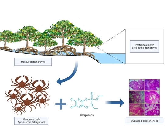
1. Introduction
2. Materials and Methods
2.1. Ethical Approval Declarations
2.2. Test Animal Collection and Maintenance
2.3. Tested Chemical
2.4. Test Procedure
2.5. Cytological Analysis
3. Results
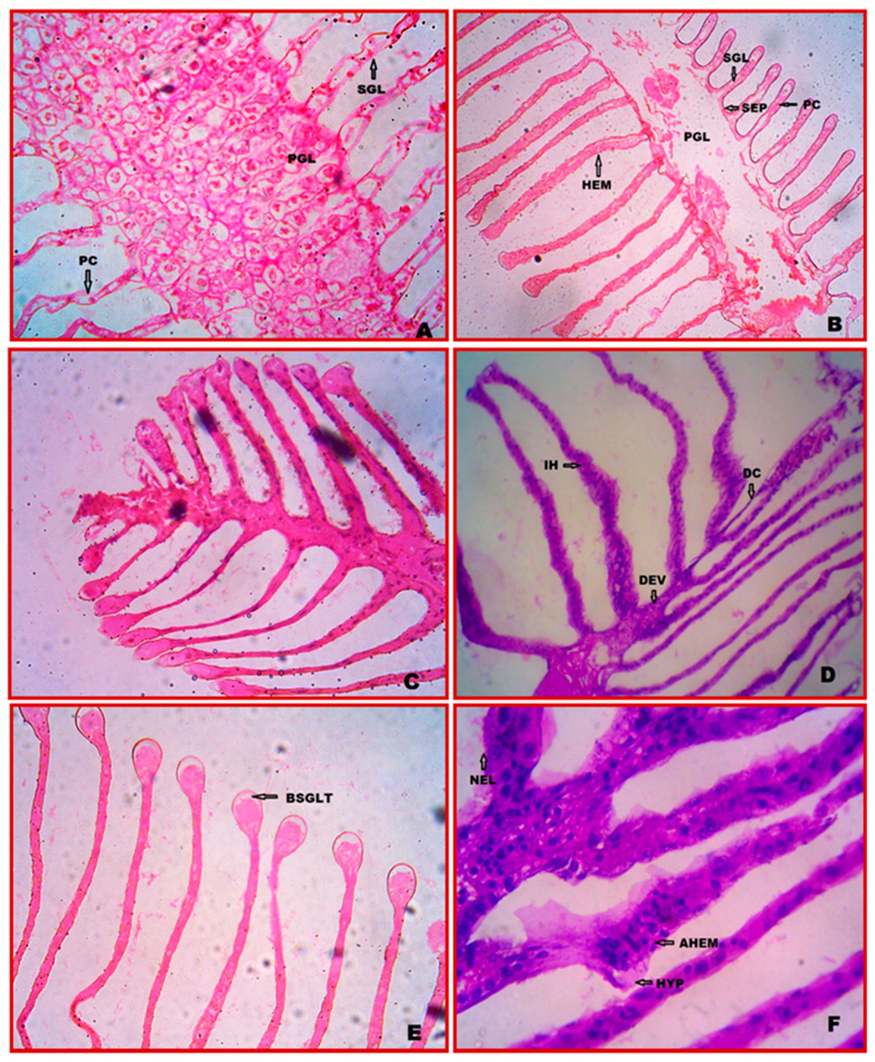
3.1. Cytology of Gill
3.2. Cytopathology of Gill
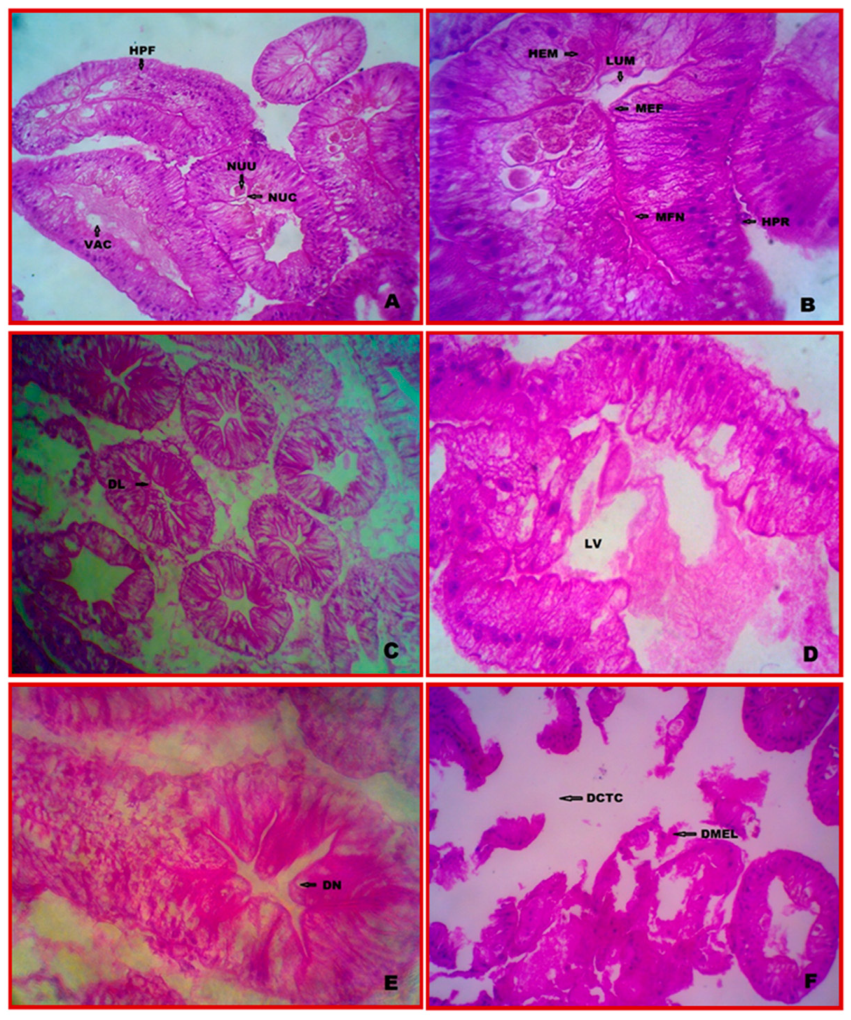
3.3. Cytology of Hepatopancreas
3.4. Cytopathology of Hepatopancreas
3.5. Cytology of Muscle
3.6. Cytopathology of Muscle
4. Discussion
5. Conclusions
Author Contributions
Funding
Institutional Review Board Statement
Informed Consent Statement
Data Availability Statement
Acknowledgments
Conflicts of Interest
References
- Abhilash, P.C.; Singh, N. Pesticide Use and Application: An Indian Scenario. J. Hazard Mater. 2009, 165, 1–12. [Google Scholar] [CrossRef] [PubMed]
- United Nations Environment Programme (UNEP). Stockholm Convention on Persistent Organic Pollutants (POPs). Available online: https://www.unep.org/explore-topics/chemicals-waste/what-we-do/persistent-organic-pollutants-pops (accessed on 10 November 2022).
- United Nations Children’s Fund (UNICEF). Understanding the Impacts of Pesticides on Children: A Discussion Paper; UNICEF: New York, NY, USA, 2018. [Google Scholar]
- Tudi, M.; Daniel Ruan, H.; Wang, L.; Lyu, J.; Sadler, R.; Connell, D.; Chu, C.; Phung, D.T. Agriculture Development, Pesticide Application and Its Impact on the Environment. Int. J. Environ. Res. Public Health 2021, 18, 1112. [Google Scholar] [CrossRef] [PubMed]
- NPIC. Chlorpyrifos-Technical Fact Sheet; NPIC: Corvallis, OR, USA, 2017. [Google Scholar]
- Ali, M.; Majid, M.; Hussain, I.; Kali, S.; Naz, T.; Niazi, M.B.K.; Khan, M.R.A.; Zafar, M.I. Chlorpyrifos Mediated Oxidative Damage and Histopathological Alterations in Freshwater Fish Oncorhynchus mykiss in Northern Pakistan. Aquac. Res. 2020, 51, 4583–4594. [Google Scholar] [CrossRef]
- Hossain, M.A.; Sutradhar, L.; Sarker, T.R.; Saha, S.; Iqbal, M.M. Toxic Effects of Chlorpyrifos on the Growth, Hematology, and Different Organs Histopathology of Nile Tilapia, Oreochromis niloticus. Saudi J. Biol. Sci. 2022, 29, 103316. [Google Scholar] [CrossRef] [PubMed]
- Mahmood, I.; Imadi, S.R.; Shazadi, K.; Gul, A.; Hakeem, K.R. Effects of pesticides on environment. In Plant, Soil and Microbes; Springer: Cham, Switzerland, 2016; pp. 253–269. [Google Scholar]
- Al-Saleh, I.A. Pesticides: A review article. J. Environ. Pathol. Toxicol. Oncol. Off. Organ Int. Soc. Environ. Toxicol. Cancer 1994, 13, 151–161. [Google Scholar]
- Malaj, E.; von der Ohe, P.C.; Grote, M.; Kühne, R.; Mondy, C.P.; Usseglio-Polatera, P.; Brack, W.; Schäfer, R.B. Organic Chemicals Jeopardize the Health of Freshwater Ecosystems on the Continental Scale. Proc. Natl. Acad. Sci. USA 2014, 111, 9549–9554. [Google Scholar] [CrossRef]
- Hussain, A.; Asi, M.R. Pesticides as water pollutants. In Groundwater for Sustainable Development; CRC Press: Boca Raton, FL, USA, 2008; pp. 119–126. [Google Scholar]
- Queyrel, W.; Habets, F.; Blanchoud, H.; Ripoche, D.; Launay, M. Pesticide Fate Modeling in Soils with the Crop Model STICS: Feasibility for Assessment of Agricultural Practices. Sci. Total Environ. 2016, 542, 787–802. [Google Scholar] [CrossRef]
- Stara, A.; Pagano, M.; Capillo, G.; Fabrello, J.; Sandova, M.; Albano, M.; Zuskova, E.; Velisek, J.; Matozzo, V.; Faggio, C. Acute Effects of Neonicotinoid Insecticides on Mytilus galloprovincialis: A Case Study with the Active Compound Thiacloprid and the Commercial Formulation Calypso 480 SC. Ecotoxicol. Environ. Saf. 2020, 203, 110980. [Google Scholar] [CrossRef]
- Banaee, M.; Sureda, A.; Faggio, C. Protective Effect of Protexin Concentrate in Reducing the Toxicity of Chlorpyrifos in Common Carp (Cyprinus carpio). Environ. Toxicol. Pharmacol. 2022, 94, 103918. [Google Scholar] [CrossRef]
- Kasmin, S. Enforcing Ship-Based Marine Pollution for Cleaner Sea in the Strait of Malacca. Environ. Asia 2010, 3, 61–65. [Google Scholar]
- Ettore, C.; Dimitrios, K. (Eds.) Pesticide Risk Assessment in Rice Paddies: Theory and Practice, 1st ed.; Elsevier: Amsterdam, The Netherlands, 2007. [Google Scholar]
- Norton, G.W.; Heong, K.L.; Johnson, D.; Savary, S. Rice Pest Management: Issues and Opportunities. In Rice in the 21st Century Global Economy: Strategic Research and Policy Issues for Food Security; IRRI: Los Banos, Philippines, 2010; pp. 297–332. [Google Scholar]
- Sehonova, P.; Tokanova, N.; Hodkovicova, N.; Kocour Kroupova, H.; Tumova, J.; Blahova, J.; Marsalek, P.; Plhalova, L.; Doubkova, V.; Dobsikova, R.; et al. Oxidative Stress Induced by Fluoroquinolone Enrofloxacin in Zebrafish (Danio rerio) Can Be Ameliorated after a Prolonged Exposure. Environ. Toxicol. Pharmacol. 2019, 67, 87–93. [Google Scholar] [CrossRef] [PubMed]
- Tresnakova, N.; Stara, A.; Velisek, J. Effects of Glyphosate and Its Metabolite AMPA on Aquatic Organisms. Appl. Sci. 2021, 11, 9004. [Google Scholar] [CrossRef]
- Vali, S.; Majidiyan, N.; Azadikhah, D.; Varcheh, M.; Tresnakova, N.; Faggio, C. Effects of Diazinon on the Survival, Blood Parameters, Gills, and Liver of Grass Carp (Ctenopharyngodon idella Valenciennes, 1844; Teleostei: Cyprinidae). Water 2022, 14, 1357. [Google Scholar] [CrossRef]
- Impellitteri, F.; Curpăn, A.S.; Plăvan, G.; Ciobica, A.; Faggio, C. Hemocytes: A Useful Tool for Assessing the Toxicity of Microplastics, Heavy Metals, and Pesticides on Aquatic Invertebrates. Int. J. Environ. Res. Public Health 2022, 19, 16830. [Google Scholar] [CrossRef]
- Pagano, M.; Savoca, S.; Impellitteri, F.; Albano, M.; Capillo, G.; Faggio, C. Toxicological Evaluation of Acetylsalicylic Acid in Non-Target Organisms: Chronic Exposure on Mytilus galloprovincialis (Lamarck, 1819). Front. Physiol. 2022, 13, 920952. [Google Scholar] [CrossRef] [PubMed]
- Banaee, M.; Impellitteri, F.; Evaz-Zadeh Samani, H.; Piccione, G.; Faggio, C. Dietary Arthrospira platensis in Rainbow Trout (Oncorhynchus mykiss): A Means to Reduce Threats Caused by CdCl2 Exposure? Toxics 2022, 10, 731. [Google Scholar] [CrossRef] [PubMed]
- Oulmi, Y.; Negele, R.D.; Braunbeck, T. Cytopathology of liver and kidney in rainbow trout Oncorhynchus mykiss after long-term exposure to sublethal concentrations of linuron. Dis. Aquat. Org. 1995, 21, 35–52. [Google Scholar] [CrossRef]
- Maharajan, A.; Ganapirya, V.; Shanmugavel, K.; Amsath, A.; Kashuri, S. Histopathological Changes in Gill and Hepatopancreas of Mangrove Crab, Perisesarma bidens Exposed to Profenofos. Int. J. Zool. Appl. Biosci. 2016, 1, 283–290. [Google Scholar]
- Uğurlu, P.; Satar, E.I.; Çiçek, T. The histopathological, cytopathological and ultrastructural effects of carbaryl on gills of Oreochromis niloticus (Linnaeus, 1758). Environ. Toxicol. Pharmacol. 2019, 71, 103217. [Google Scholar] [CrossRef]
- Duarte-Restrepo, E.; Jaramillo-Colorado, B.E.; Duarte-Jaramillo, L. Effects of Chlorpyrifos on the Crustacean Litopenaeus vannamei. PLoS ONE 2020, 15, e0231310. [Google Scholar] [CrossRef] [PubMed]
- Stara, A.; Bellinvia, R.; Velisek, J.; Strouhova, A.; Kouba, A.; Faggio, C. Acute Exposure of Common Yabby (Cherax destructor) to the Neonicotinoid Pesticide. Sci. Total Environ. 2019, 665, 718–723. [Google Scholar] [CrossRef] [PubMed]
- Velisek, J.; Stara, A.; Zuskova, E.; Kubec, J.; Buric, M.; Kouba, A. Effects of s-metolachlor on early life stages of marbled crayfish. Pestic. Biochem. Physiol. 2019, 153, 87–94. [Google Scholar] [CrossRef] [PubMed]
- Johnston, D.J.; Alexander, C.G.; Yellowlees, D. Epithelial Cytology and Function in the Digestive Gland of Thenus orientalis (Decapoda, Scyllaridae). J. Crust. Biol. 1998, 18, 271–278. [Google Scholar]
- Sousa, L.G.; Petriella, A.M. Changes in the Hepatopancreas Histology of Palaemonetes argentinus (Crustacea, Caridea) during Moult. Biocell 2001, 25, 275–281. [Google Scholar] [PubMed]
- APHA. Standard Methods for the Estimation of Water and Wastewater; American Public Health Association: New York, NY, USA, 1995; pp. 4–127. [Google Scholar]
- Cengiz, E.I.; Ünlü, E. Histopathology of Gills in Mosquitofish, Gambusia affinis After Long-Term Exposure to Sublethal Concentrations of Malathion. J. Environ. Sci. Health Part B 2003, 38, 581–589. [Google Scholar] [CrossRef] [PubMed]
- Kamble, N.A.; Potdar, V.V. Hepatopancreatic Damages in Snail Bellamya bengalensis Tested against Lead Acetate Toxicity. Indian J. Comp. Anim. Physiol. 2010, 28, 7–10. [Google Scholar]
- Sucitha, C.; Muthukumaravel, K.; Kumarasamy, P.; Vasanthi, N. Studies on the Impact of Heavy Metal Copper on The Ultrastructure of Gill in the Mangrove Crab Sesarma brocki. IJPBS 2019, 9, 109–111. [Google Scholar]
- Joshi, P.P. Physiological Responses of Freshwater Fish and Crab to Pyrethroid Pesticides: A Comparative Study. Ph.D. Thesis, Marathwada University, Aurangabad, India, 2006. [Google Scholar]
- Jadhav, T.J.; Kulkarni, G.K.; Joshi, P.P.; Jawale, C.J. Effect of Endosulfan and Thimet on Histological Profile of Gills and Ovary of a Freshwater Crab, Bary-Telphusa Cunicularis. In Aquaculture; APH Publishing Corporation: New Delhi, India, 2007; pp. 85–92. [Google Scholar]
- Li, N.; Zhao, Y.; Yang, J. Impact of Waterborne Copper on the Structure of Gills and Hepatopancreas and Its Impact on the Content of Metallothionein in Juvenile Giant Freshwater Prawn Macrobrachium rosenbergii (Crustacea: Decapoda). Arch. Environ. Contam. Toxicol. 2007, 52, 73–79. [Google Scholar] [CrossRef] [PubMed]
- Richmonds, C.; Dutta, H.M. Histopathological Changes Induced by Malathion in the Gills of Bluegill Lepomis macrochirus. Bull. Environ. Contam. Toxicol. 1989, 43, 123–130. [Google Scholar] [CrossRef] [PubMed]
- Katuli, K.K.; Amiri, B.M.; Massarsky, A.; Yelghi, S. Impact of a short term diazinon exposure on the osmoregulation potentiality of Caspian roach (Rutilus rutilus) fingerlings. Chemosphere 2014, 108, 396–404. [Google Scholar] [CrossRef]
- Ghasemzadeh, J.; Sinaeei, M.; Bolouki, M. Biochemical and Histological Changes in Fish, Spotted Scat (Scatophagus Argus) Exposed to Diazinon. Bull. Environ. Contam. Toxicol. 2015, 94, 164–170. [Google Scholar] [CrossRef]
- Bhide, M.; Gupta, P.; Khan, M.A.; Dubey, U.; Thakur, P.; Nema, P.; Jain, S. Morphological and Biochemical Studies on the Different Developmental Stages of a Freshwater Snail, Lymnaea stagnalis (Lymnaedae) after Treatment with Some Pesticides. J. Environ. Biol. 2006, 27, 359–366. [Google Scholar]
- Wu, J.-P.; Chen, H.-C.; Huang, D.-J. Histopathological and Biochemical Evidence of Hepatopancreatic Toxicity Caused by Cadmium and Zinc in the White Shrimp, Litopenaeus vannamei. Chemosphere 2008, 73, 1019–1026. [Google Scholar] [CrossRef]
- Magalhães, C.; Campos, M.R.; Collins, P.A.; Mantelatto, F.L. Diversity, Distribution and Conservation of Freshwater Crabs and Shrimps in South America. In A Global Overview of the Conservation of Freshwater Decapod Crustaceans; Springer International Publishing: Cham, Switzerland, 2016; pp. 303–322. [Google Scholar]
- Maharajan, A.; Rajalakshmi, S.; Vijayakumaran, M.; Kumarasamy, P. Sublethal Effect of Copper Toxicity against Histopathological Changes in the Spiny Lobster, Panulirus homarus (Linnaeus, 1758). Biol. Trace Elem. Res. 2012, 145, 201–210. [Google Scholar] [CrossRef]
- Negro, L.; Senkman, E.; Montagna, M.; Collins, P. Freshwater Decapods and Pesticides: An Unavoidable Relation in the Modern World. In Pesticides in the Modern World Risks and Benefits; Intech Open: London, UK, 2011; pp. 208–210. [Google Scholar]
- Saravana Bhavan, P. Histopathology of the Hepatopancreas and Gills of the Prawn Macrobrachium malcolmsonii Exposed to Endosulfan. Aquat. Toxicol. 2000, 50, 331–339. [Google Scholar] [CrossRef]
- Caceci, T.; Neck, K.F.; Lewis, D.D.H.; Sis, R.F. Ultrastructure of the Hepatopancreas of the Pacific White Shrimp, Penaeus vannamei (Crustacea: Decapoda). J. Mar. Biol. Assoc. U. K. 1988, 68, 323–337. [Google Scholar] [CrossRef]
- Ceccaldi, H.J. A Synopsis Of The Morphology And Physiology of the Digestive System of Some Crustacean Species Studied in France. Rev. Fish. Sci. 1998, 6, 13–39. [Google Scholar] [CrossRef]
- Tehrani, A.A.G.; Sadeghi, N.H.; Badamchi, Z.; Sanjou, S.; Mansoub, N.; Azhari, A. Effect of Carbamates pesticides on Instar III larvae and Adult Artemia urumiana. Ann. Biol. Res. 2011, 2, 515–525. [Google Scholar]
- Das, B.K.S.C.; Mukherjee, A. A Histopathological Study of Carp Labeo rohita Exposed to Hexachlorocyclohexane. Vet. Arch. 2000, 70, 169–180. [Google Scholar]
- Fatma, A.S.M. Histopathological studies on Tilapia zillii and Solea vulgaris fromLake Qarun, Egypt. World J. Fish Mar. Sci. 2009, 1, 29–39. [Google Scholar]
- Rakhi, S.F.; Reza, A.H.M.M.; Hossen, M.S.; Hossain, Z. Alterations in Histopathological Features and Brain Acetylcholinesterase Activity in Stinging Catfish Heteropneustes fossilis Exposed to Polluted River Water. Int. Aquat. Res. 2013, 5, 7. [Google Scholar] [CrossRef]
- Parikh, H.; Rangrez, P.; Bagchi, A.; Desai, B.N. Effect of Dimethote on some histoarchitecture of Freshwater Fish Oreochromis mossambicus (Peters, 1852). Bioscan 2010, 5, 55–58. [Google Scholar]


| Cytopathology | Experimental Groups | |||||
|---|---|---|---|---|---|---|
| Tissue | Indices | Control | 0.0294 ppm | 0.0588 ppm | ||
| 7-d | 28-d | 7-d | 28-d | |||
| Gills | Detached cuticle (DC) | – | – | ++ | – | – |
| Necrosis (NEC) | – | – | – | – | +++ | |
| Infiltration of haemocytes (IH) | – | – | ++ | – | – | |
| Damaged efferent vessel (DEV) | – | + | ++ | ++ | – | |
| Bulging of secondary gill lamellae tip (BSGLT) | – | – | – | – | – | |
| Aggregation of haemocytes (AHEM) | – | – | – | – | ++ | |
| Hypertrophy (HYP) | – | – | – | – | ++ | |
| Hepatopancreas | Distended lumen (DL) | – | + | – | ++ | – |
| Large vacuole (LV) | – | + | ++ | – | – | |
| Damaged connective tissue layer (DCTL) | – | – | – | – | ++ | |
| The disappearance of the nucleus (DN) | – | – | ++ | ++ | +++ | |
| Damaged myoepithelial layer (DMTL) | – | – | – | + | +++ | |
| Muscle | Large vacuole (LV) | – | ++ | – | – | – |
| Gap formation (GF) | – | ++ | ++ | – | – | |
| Rupture of muscle bundle (RMB) | – | – | ++ | – | – | |
| Wavy muscle bundle (WMB) | – | – | – | – | – | |
| Vacuole formation (VF) | – | – | – | ++ | – | |
| Fusion of muscle bundle (FMB) | – | – | – | +++ | +++ | |
| Shortening of muscle bundle (SMB) | – | – | – | – | +++ | |
| Loosen of muscle bundle (LMB) | – | – | – | – | – | |
Disclaimer/Publisher’s Note: The statements, opinions and data contained in all publications are solely those of the individual author(s) and contributor(s) and not of MDPI and/or the editor(s). MDPI and/or the editor(s) disclaim responsibility for any injury to people or property resulting from any ideas, methods, instructions or products referred to in the content. |
© 2023 by the authors. Licensee MDPI, Basel, Switzerland. This article is an open access article distributed under the terms and conditions of the Creative Commons Attribution (CC BY) license (https://creativecommons.org/licenses/by/4.0/).
Share and Cite
Ravi, R.; Athisuyambulingam, M.; Kanagaraj, S.; Tresnakova, N.; Impellitteri, F.; Viswambaran, G.; Faggio, C. Impact of Chlorpyrifos on Cytopathological Indices in Mangrove Crab, Episesarma tetragonum (Fabricius). Vet. Sci. 2023, 10, 53. https://doi.org/10.3390/vetsci10010053
Ravi R, Athisuyambulingam M, Kanagaraj S, Tresnakova N, Impellitteri F, Viswambaran G, Faggio C. Impact of Chlorpyrifos on Cytopathological Indices in Mangrove Crab, Episesarma tetragonum (Fabricius). Veterinary Sciences. 2023; 10(1):53. https://doi.org/10.3390/vetsci10010053
Chicago/Turabian StyleRavi, Rajesh, Maharajan Athisuyambulingam, Shanmugavel Kanagaraj, Nikola Tresnakova, Federica Impellitteri, Ganapiriya Viswambaran, and Caterina Faggio. 2023. "Impact of Chlorpyrifos on Cytopathological Indices in Mangrove Crab, Episesarma tetragonum (Fabricius)" Veterinary Sciences 10, no. 1: 53. https://doi.org/10.3390/vetsci10010053
APA StyleRavi, R., Athisuyambulingam, M., Kanagaraj, S., Tresnakova, N., Impellitteri, F., Viswambaran, G., & Faggio, C. (2023). Impact of Chlorpyrifos on Cytopathological Indices in Mangrove Crab, Episesarma tetragonum (Fabricius). Veterinary Sciences, 10(1), 53. https://doi.org/10.3390/vetsci10010053