Review

Comparative Aspects of Human, Canine, and Feline Obesity and Factors Predicting Progression to Diabetes

by
Margarethe Hoenig
Department of Veterinary Clinical Medicine, College of Veterinary Medicine, University of Illinois at Urbana-Champaign, 1008 W. Hazelwood Drive, Urbana, IL 61802, USA
Vet. Sci. 2014, 1(2), 121-135; https://doi.org/10.3390/vetsci1020121
Submission received: 25 April 2014 / Revised: 12 August 2014 / Accepted: 14 August 2014 / Published: 21 August 2014
(This article belongs to the Special Issue Animal Models of Disease)

Abstract

:
Obesity and diabetes mellitus are common diseases in humans, dogs and cats and their prevalence is increasing. Obesity has been clearly identified as a risk factor for type 2 diabetes in humans and cats but recent data are missing in dogs, although there is evidence that the unprecedented rise in canine obesity in the last decade has led to a rise in canine diabetes of similar magnitude. The insulin resistance of obesity has often been portrayed as major culprit in the loss of glucose control; however, insulin resistance alone is not a good indicator of progression to diabetes in people or pets. A loss of beta cell function is necessary to provide the link to impaired fasting and post-prandial plasma glucose. Increased endogenous glucose output by the liver is also a prerequisite for the increase in fasting blood glucose when non-diabetic obese humans and pets develop diabetes. This may be due to decreased hepatic insulin sensitivity, decreased insulin concentrations, or a combination of both. While inflammation is a major link between obesity and diabetes in humans, there is little evidence that a similar phenomenon exists in cats. In dogs, more studies are needed to examine this important issue.

1. Introduction

Obesity is the most common nutritional disorder in dogs and cats and has been shown to be a risk factor for the development of type 2 diabetes in cats and humans. More recent epidemiological studies examining if obesity is a risk factor for diabetes in dogs as well are missing, although obesity has also been reported to have increased dramatically in dogs in the last decade and the increase was paralleled by an increase in diabetes of similar magnitude suggesting a cause/effect relationship [1]. Obesity leads to insulin resistance, which, if not accompanied by an appropriate increase in insulin concentrations, sets the path for glucose intolerance and eventually overt diabetes. In the veterinary literature, especially when primary data from pets are missing, it is not uncommon to find attempts to project from the human data connecting obesity to diabetes. Rather than do this, this review will examine the evidence available for similarities and differences among these three species. Specifically, we will examine insulin secretion, regulation of endogenous glucose production by the liver, and the role of inflammation.

2. Obesity-Associated Changes in Insulin Secretion: A Risk Factor for Diabetes Mellitus in Dogs and Cats?
It was shown in a hallmark study by Kealy and coworkers over a decade ago that higher food intake and body fat is detrimental for a dog’s health and longevity and leads to changes in lipid and glucose metabolism [2]; however, there are no recent epidemiological studies examining if obesity in the dog also leads to overt diabetes. The fact that data from a large Swedish retrospective study of the prevalence of diabetes, which was carried out from 2000 to 2003, showed that 55% of dogs were overweight or obese at first diagnosis of diabetes compared to 20% of normal-weight dogs of the same age group suggests that obesity may be a risk factor in dogs [3]. A connection between obesity and diabetes was also implied by a recent summary by Banfield Hospitals, in which it was reported that the large increase in canine obesity, which they observed from 2007 to 2012, was accompanied by an equally large increase in diabetes [1], strongly suggesting that obesity might be causing the increase in diabetes. That a form of diabetes similar to type 2 diabetes in humans is present in some dogs has already been shown four decades ago by Kaneko and coworkers who were able to distinguish a group of obese diabetic dogs from lean diabetic dogs based on their insulin and glucose profiles [4]. The obese diabetic dogs were characterized by high glucose and high fasting insulin concentrations but were unable to mount an appropriate insulin response when challenged with high glucose concentrations.
It was suggested in a recent study of a small number of obese non-diabetic dogs which had normal fasting plasma glucose (FPG) and normal glucose tolerance that obese dogs do not develop a type of diabetes similar to type 2 seen in humans because they increase insulin output, including first phase, in response to obesity, whereas people do not [5]. However, such statement about a difference between dog and human insulin secretion is not supported by evidence from human clinical studies. It has been known for decades that in people obesity by itself leads to a robust response in insulin secretion of both phases [6,7,8]. Results from a large Finnish study showed that obese subjects with an approximately 40% reduction in insulin sensitivity, but FPG and 2 h plasma glucose (2-h PG) concentrations within the reference categories had increases of over 50% in early and late phase insulin secretion [9]. This has been called the Stage 1 or compensation phase of obesity [10] and results from an increase in β-cell mass [10,11,12]. Insulin sensitivity decreased further when subjects developed impaired fasting glucose and impaired glucose tolerance. However, early-phase insulin release only reached a substantial decrease in those subjects in whom FPG and 2-h PG increased into the diabetic range. This indicates that a decrease in early (first) phase insulin secretion would not be expected in a dog or person with normal FPG and normal glucose tolerance. Instead, non-diabetic obese dogs or people with normal FPG have higher early and late phase insulin secretion compared to lean subjects in this compensation phase, and one can conclude that glucose tolerant people and dogs compensate well for the increase in insulin resistance.
Over 80% of obese insulin-resistant humans remain euglycemic and do not progress to type 2 diabetes [13]. It is not known how many obese dogs progress to diabetes, although, as stated above, new evidence suggests that it is an important risk factor. It was shown in a recent study that several prerequisites have to be present for an insulin-resistant dog to progress to diabetes [14]. In that study, diet-induced obesity led to a decrease in peripheral insulin sensitivity, which was perfectly met by an increase in insulin concentrations. Hepatic insulin sensitivity was maintained and glucose tolerance remained normal, as would be expected in a non-diabetic obese dog. It was not until beta cell mass was greatly reduced by the administration of various doses of the beta cell toxin streptozotocin, that insulin secretion decreased in a dose dependent manner, and impaired fasting glucose and diabetes ensued. It can be seen from that study that insulin resistance alone elicited the appropriate regulatory responses: Insulin concentrations increased to counteract the peripheral resistance, allowing normal glucose control. The loss in beta cells had to be severe before a deterioration of FPG and glucose tolerance was seen. In fact, when fasting blood glucose increased and glucose tolerance became abnormal, insulin concentrations had decreased by 90%.
The compensatory hyperinsulinemia, which is seen in obese non-diabetic dogs, has been thought to be the result of upregulation of beta cell sensitivity to glucose and subsequent increase in insulin secretion. However, based on recent data from a large number of dogs with varying fat mass, it was proposed that changes in insulin clearance resulting in higher insulin levels feed back onto insulin action of peripheral and hepatic tissues and are the primary cause for the hyperinsulinemia of obesity [15].
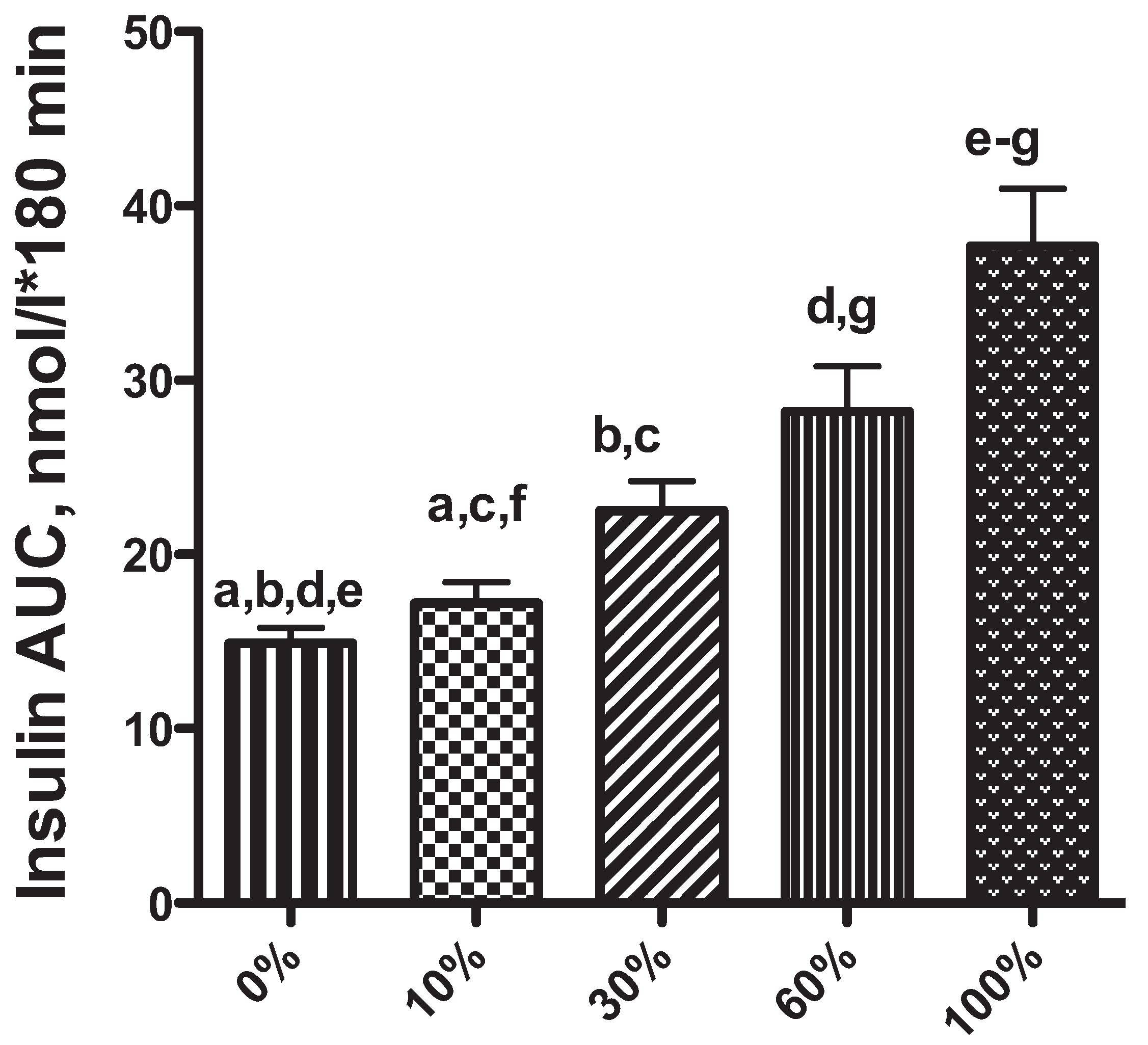
Figure 1. Area under the curve for insulin during a low dose intravenous glucose tolerance test (0.5 g/kg body weight; Mean ± SEM; 1B) in 20 adult neutered cats (equal gender distribution) at 0%–60% weight increases and in 12 cats at 100%. Identical superscripts indicate significant differences (reprinted with permission from Obesity [21]).
Vetsci 01 00121 g001
Using the gold-standard method to evaluate insulin sensitivity, the euglycemic hyperinsulinemic clamp, we found that in cats obesity leads to a decrease in glucose effectiveness and an increase in insulin resistance, similar to what has been shown in people and dogs [16,17]. Development of obesity in cats is not associated with a change in FPG, even when obesity is severe [18,19,20]. This was recently shown in a longitudinal study, where 20 cats were fed ad libitum and became obese [21]. Even with an increase in fat mass of 100%, fasting glucose concentrations remained normal. Early phase and total insulin concentrations increased until 100% increase in fat mass was reached (Figure 1), when a blunting of early insulin secretion and altered glucose clearance was seen, indicating that under extreme conditions (high degree of resistance and high glucose dose), the response of the beta cell to glucose becomes abnormal. These results are similar to those described in people by Matsumoto and coworkers, who compared glucose tolerance, insulin secretion, and insulin sensitivity in a large number of non-obese and obese Japanese subjects [22]. They found a blunted early phase of insulin secretion only in those obese people who had impaired glucose tolerance, and suggested that impaired early phase insulin release may be the initial abnormality in the development of glucose intolerance. The worsening from impaired glucose tolerance to diabetes in their study was associated with a greater decrease in early-phase insulin secretion, a significant decrease in total insulin secretion, and a significant increase in insulin resistance. There is no long-term longitudinal study in obese cats, which has examined if results from intravenous glucose tolerance testing can be used as biomarkers for progression to diabetes. It is questionable if intravenous glucose tolerance testing is appropriate to answer this question. This skepticism is supported by the fact that a difference in glucose tolerance, characterized by a lower glucose clearance, could not be seen between lean and obese cats until a supramaximal dose of glucose (1.3 g/kg) was administered [18]. This suggests that testing beta cells with high glucose concentrations is indicated to evaluate their maximal secretory capacity but the same test might not be a good indicator of an animal’s or person’s beta cell function during their normal daily routine. One needs to appreciate that glucose values seen during IV glucose tolerance testing with high glucose doses are never seen after a meal in a cat or dog and represent an extreme challenge for the endocrine pancreas, as well as for peripheral tissues and the liver, and results are likely not good indicators of progression. Intravenous glucose tolerance tests are rarely used in human medicine to examine factors that lead to a deterioration of glucose control and the dose is much lower (0.3 g/kg) than that used in cats. In people, glucose tolerance is usually tested with a dose of orally administered glucose (75 g), which leads to blood glucose concentrations not too dissimilar to what a person experiences after normal food intake. This may explain why results from tests in humans are more predictive of progression because the abnormality is already present at lower levels of glucose, i.e., levels that are actually present in a person under normal physiological conditions. It is glucose concentrations seen under those conditions, which influence beta cell function long-term. We were unable to show a difference between lean and obese cats when we followed their daily glucose concentrations with continuous glucose monitoring for 7 days. Only one of the obese cats in that study showed a glucose profile, which fluctuated between the upper and mildly high glucose range after having been severely obese for many years [19]. It would be speculation to make a prediction whether this cat might be a “progressor”. It is well known from human subjects that generally about 35% to 50% of those with impaired fasting blood glucose convert to diabetes after 10–20 years of follow-up [23]. While reference values for glucose tolerance tests have been published for client-owned cats, the results vary greatly, even within one and the same laboratory [24,25,26]. This indicates that these tests are not useful when applied to the general cat population and should only be used in strictly controlled research populations. Clearly, long-term longitudinal studies are needed to examine which biochemical changes might be biomarkers for progression in cats, i.e., healthy cats should be followed until they develop diabetes.
In a recent study of normal-weight, overweight, obese, and diabetic cats, we showed that insulin but not glucose concentrations were significantly higher in overweight and obese non-diabetic cats compared to normal weight non-diabetic cats and were significantly lower in naïve diabetic cats [20], supporting the notion that marked insulin deficiency is necessary for the progression to diabetes. This notion is also supported by the finding that a combination of both insulin resistance and insulin deficiency due to a reduction in beta cell mass were needed for the development of experimental diabetes in cats [27]. In that study, overt diabetes was not seen until insulin secretion was about 80% lower than that seen in healthy cats.
In summary: Insulin resistance alone is not a good indicator of progression to diabetes in people or pets. A loss of beta cell function is necessary to provide the link to impaired fasting and post-prandial plasma glucose.

3. The Role of the Liver in Glucose Control
The liver plays a crucial role in the disposal of exogenous glucose and the control of endogenous glucose production (EGP). As with the evaluation of insulin secretion profiles, one needs to be careful to distinguish obese non-diabetic subjects from obese diabetic subjects when evaluating liver function. Usually, in insulin-resistant obese non-diabetic humans with normal FPG and normal glucose tolerance, the inhibitory effect of insulin on endogenous glucose output is preserved [28,29]. In obese non-diabetic people, the maintenance of euglycemia was found to be due to a reduction in glycogenolysis, whereas gluconeogenesis was increased in both, non-diabetic and diabetic obese people, although more in diabetic subjects. These changes resulted in increased endogenous glucose production in diabetic but not in obese non-diabetic patients [29]. No change in overall glucose production was also seen by Basu and coworkers in obese non-diabetic compared to lean non-diabetic patients [30], although they found that only a change in gluconeogenesis but not glycogenolysis led to the maintenance of euglycemia in obese non-diabetic patients [30]. Progression to impaired glucose tolerance and diabetes was characterized by increased endogenous glucose production and, concomitantly, endogenous glucose production became less suppressible by insulin. No change in endogenous glucose production or suppressibility by insulin occurred in those people, who did not progress to the diabetic state [31].
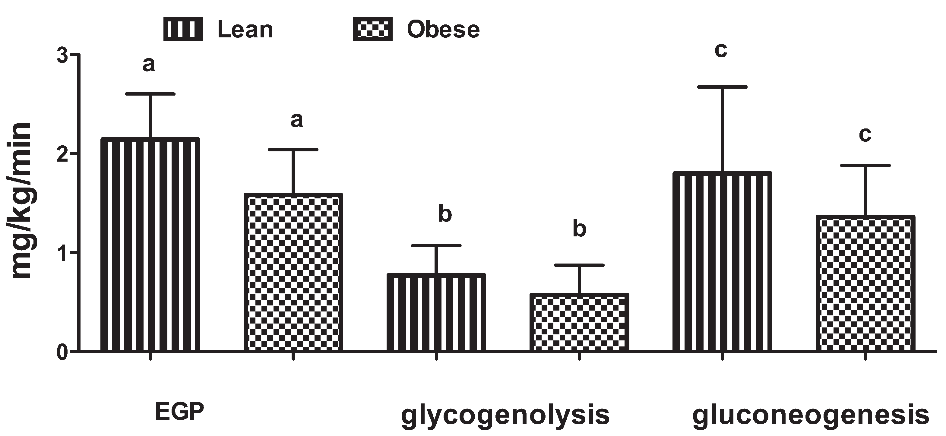
In our own studies in cats, we found that endogenous glucose production was decreased in obese cats in the fasted and post-prandial state, counteracting the effect of insulin resistance on peripheral glucose uptake and resulting in the maintenance of euglycemia in long-term obese animals [32,33]. In one study of fasted cats, the lower EGP in the obese cats was due to a lower glycogenolysis and gluconeogenesis (Figure 2) [32]. There was a strong negative correlation between plasma insulin concentrations and endogenous glucose production and between endogenous glucose production and girth, as well as body mass index, suggesting that the decreased endogenous glucose production in our long-term obese cats was due to the higher insulin concentrations of obesity. To our knowledge, glucose turnover and metabolic fluxes have not been evaluated in diabetic cats. However, one might expect to also see a progressive loss of insulin sensitivity of the liver and an increase in endogenous glucose production when cats become diabetic, similar to what has been described in humans [31]. However, the switch from normal glucose regulation to diabetes appears to occur quickly in obese cats because there was no difference in fructosamine concentrations, an indicator of glucose control in the previous 2 weeks, among lean, overweight and obese cats [20]. Fructosamine concentrations, which are tightly correlated with blood glucose concentrations (Figure 3), were clearly separated from naïve diabetics and treated diabetics. If progression from the obese to the diabetic state would occur slowly in obese cats, a sliding scale of glucose and fructosamine concentrations would have been expected in this study of client-owned cats.
Figure 2. Mean ± SEM of endogenous glucose production (EGP), glycogenolysis and gluconeogenesis in lean and obese cats. Identical superscripts indicate significant differences (modified from Kley et al. Am J Physiol Regul Integr Comp Physiol 296: R936-R943, 2009 [32]).
Vetsci 01 00121 g002
Figure 3. Correlation between serum fructosamine and glucose concentrations in 117 cats (25 normal body condition, 27 overweight, 24 obese, 21 naïve diabetic, and 20 treated diabetic cats) (reprinted with permission from JAVMA [20]).
Vetsci 01 00121 g003
The authors are not aware of a comparison of hepatic metabolism among healthy, obese, and spontaneously diabetic dogs. In 18 hour-fasted lean healthy dogs, glycogenolysis was found to be the major contributor to EGP, and gluconeogenesis accounted for approximately one third of the glucose production [34]. This is different from what has been described in cats and in people for a similar postprandial time period. In both cats and people, gluconeogenesis contributes about two thirds and glycogenolysis contributes about one third to endogenous glucose production [32,33,35]. Glycogenolysis appears to be more sensitive than gluconeogenesis to suppression by insulin in dogs [30,36], whereas both are already suppressed almost completely in lean people by a 50% lower dose of insulin [30]. It is not known what physiological effect this difference might have. Perhaps it should be reflected in a difference in liver glycogen content. It is, however, known, that liver glycogen is similar among the three species. The glycogen content in healthy dogs and people is approximately 4% of liver wet weight [37,38]. We have found that the liver of lean and obese cats contains about 5% of glycogen [33].
It has been suggested that insulin action in the liver is in part determined by the intrahepatic triglyceride content. In people, obesity is associated with an increase in intrahepatic triglycerides, an abnormality known as nonalcoholic fatty liver disease [39]. Its prevalence increases linearly with increasing body mass index but also occurs in about 15% of normal-weight subjects [40]. The amount of triglycerides in the liver was shown in one study not only to correlate with insulin resistance in the liver, but also in muscle and adipose tissue [39]. The mechanism by which hepatic lipids induce hepatic insulin resistance is not well understood and it is unclear if hepatic triglycerides are the cause or effect of insulin resistance. A 2 hits theory has been proposed: The first hit would be lower fatty acid oxidation, higher fatty acid synthesis, and lower VLDL efflux from the liver due to the oversupply of fatty acids because of lower lipoprotein lipase activity and the hyperinsulinemia of obesity. The result would be increased esterification of fatty acids and deposition of triglycerides in the liver. The second hit would be oxidative stress resulting in lipid peroxidation leading to hepatocellular degeneration and necrosis [41].
Traditionally, excessive hepatic triglyceride content, or steatosis, has been defined as exceeding 5% of liver volume or liver weight, or histologically when 5% of hepatocytes contain visible intracellular triglycerides [42]. In a recent study the threshold for a normal amount of intrahepatic triglycerides was reported to be 5.6% of liver volume, whereas others have not found any evidence of an obvious threshold that can be used to define normality [40].
We recently measured liver fat in lean and obese cats using nuclear magnetic resonance spectroscopy. Median liver fat percentages in lean and obese cats were 1.3% and 6.8%, respectively [43]. In a different group of lean and obese cats, we employed a chemical assay with similar results (1.7% for lean cats and 6% for obese cats [43]. A previous publication involving chemical liver triglyceride measurement in wedge biopsies from cats reported a mean liver triglyceride content of 1% for 5 cats of ideal body weight [44]. A 2–3 fold increase in liver fat was also reported in cats after weight gain [45]. This shows that obese cats have higher triglyceride accumulation in the liver compared to lean cats; however, as in the human studies, a cause/effect relationship to insulin sensitivity cannot be established. To our knowledge, liver fat content in obese or diabetic dogs has not been quantified.
Liver steatosis is responsive to thiazolidinediones (TZD) in people. TZDs are agonists of peroxisome proliferator-activated receptor gamma (PPARγ), a nuclear transcription factor that is a key regulator of adipogenesis [46]. The natural ligands for PPARγ are unknown, but the receptor possesses a low affinity for various endogenous fatty acids and eicosanoids [47]. PPARγ is expressed abundantly in adipose tissue, and at low levels in other tissues, including pancreatic β-cells [46,47]. In humans, dogs and cats, TZD administration leads to an increase in whole-body insulin sensitivity [48,49,50]. It is known that in people this occurs parallel to a reduction of triglyceride content in non-adipose tissues such as liver and skeletal muscle, and lowering of serum triglyceride and/or non-esterified fatty acid concentrations [50]. There are no similar studies in dogs and cats to show if hepatic triglyceride content changes with thiazolidinediones, although one might also expect a reduction in liver steatosis because of the similarities of its positive effect on metabolism and insulin sensitivity in those species.
In summary: Increased endogenous glucose output by the liver is a prerequisite for the increase in fasting blood glucose when non-diabetic obese humans and pets develop diabetes. This may be due to decreased hepatic insulin sensitivity, decreased insulin concentrations, or a combination of both.

4. The Role of Inflammation in Obesity and Diabetes

There is abundant scientific evidence of a link between obesity/diabetes and inflammation in people [51,52,53]. Inflammatory cells and other markers of inflammation increase with the expansion of fat mass in white adipose tissue and also in the liver in human subjects [39,54]. White adipose tissue secretes a variety of factors, which influence metabolism and are involved in the regulation of inflammation. Some of these, like leptin and adiponectin, are primarily produced by fat tissue. Other cytokines, like TNF-a, MCP-1, and IL-6 are produced by macrophages and other cells involved in the immune response, as well as adipocytes. Lipid accumulation in the adipocyte increases reactive oxygen species (ROS) and cellular stress pathways are activated leading to the secretion of pro-inflammatory cytokines [55]. The increased ROS leads to a decrease in activities of anti-oxidant enzymes, such as glutathione peroxidase, Cu,Zn-superoxide dismutase (SOD), and catalase. When cats were followed longitudinally during the development of obesity in one study, it was shown that there was no change in the anti-oxidant enzymes catalase, glutathione peroxidase, and red blood cell as well as heparin-releasable superoxide dismutase. There was also no change in the inflammatory cytokines, IL-1, IL-6 and TNF-α suggesting that development of obesity in cats does not lead to the same changes in circulating inflammatory markers and activity of antioxidant enzymes that is seen in other species. Urinary isoprostane concentrations were also not different in long-term obese cats and there was no infiltration of white fat with inflammatory cells [21]. Jeusette and coworkers found higher urinary isoprostane concentrations in obese cats compared to lean after both were given ad libitum food for 8 weeks, but no difference in other oxidative stress markers was seen [56]. No difference was also seen before and after successful weight loss of acute phase proteins, and enzymes associated with inflammation (buturylcholinesterase and paraoxonase) [57]. The notion that inflammation does not play a role in the development of insulin resistance with increasing obesity in cats is also supported by results from a study of immune responses in lean and obese cats [58]. In that study, complete blood counts and lymphocyte distribution were examined and immune function was assessed by measuring the proliferative activity of different cellular fractions in response to several polyclonal mitogens. Phagocytosis and natural killer cell cytotoxicity was examined to test innate immune functions. It was shown that a similar immune innate and adaptive immune response was elicited regardless of body condition and concluded that obesity does not alter immune responses in cats. It is unclear why insulin resistance of obesity in cats is not associated with an inflammatory response and a change in immune function. But it may explain the fact that obese cats do not develop cardiovascular disease and clinical hypertension.
Few studies have examined inflammatory cytokines in obese and lean dogs. Anti-oxidant enzyme activity has not been compared between lean and obese dogs, to the author’s knowledge. In laboratory dogs, weight loss did not lead to a change in acute phase proteins [59]. The cytokine IL-6 was below the detection limit of the assay in that study. In another study, TNF-α as well as leptin were below the detectable limit in the majority of dogs. Haptoglobin and serum amyloid A were lower after weight loss than before suggesting that obese dogs have higher concentrations but no comparison to normal-weight dogs was included [60]. In both studies, a comparison was not made between lean and obese dogs before weight loss. It appears that a comparison of anti-oxidant enzymes, inflammatory cytokines, and immune function using assays that have been validated for canine serum or plasma are important next steps to elucidate the role of inflammation in obese dogs.
Concentrations of the adipokines, adiponectin and leptin, have been reported in many studies in human subjects and several studies in dogs and cats. Adiponectin, an insulin sensitizing adipokine, has an important role in glucose and lipid metabolism and its expression is regulated by PPAR-γ [57,61,62]. It has anti-inflammatory activity and anti-atherogenic properties, and suppresses NK cytotoxicity. It also inhibits phagocytic activity and TNF-α production, and inhibits lipid accumulation in a variety of cells [63]. In addition, adiponectin has anti-apoptotic effects on beta cells in vitro, which appear to be mediated by reduction of ceramide concentrations [64,65]. In people [66] and cats [17,32], fat cells secrete higher amounts of adiponectin in the lean state but concentrations decrease with increasing fat mass. In obese dogs, plasma adiponectin concentrations have been found to be lower [62,67] or unchanged [60,68] compared to lean dogs. In one study, adiponectin was only lower in those obese dogs with metabolic syndrome, i.e., dogs with high glucose and lipid values, systolic blood pressure and body condition scoring but not in obese dogs without signs of metabolic syndrome [69]. In another study, adiponectin levels remained unchanged when dogs lost weight [60]. However, in that study the average adiponectin levels were reported to be very low (10 pg/mL), whereas other studies show adiponectin concentrations in dogs to be several magnitudes higher (ug/mL) and similar to those of other species. This makes the low results questionable.
Leptin is an adipokine, which acts in the hypothalamus and suppresses food intake and increases energy expenditure. Contrary to adiponectin, it is positively correlated with fat mass in humans, dogs, and cats [17,70,71,72]. Increased circulating leptin in obesity is a marker of leptin resistance. Leptin acts as a positive regulator of immune functions and cytotoxicity of splenic NK cells at physiological concentrations [73]. In human subjects, it stimulates proliferation and cytokine synthesis in peripheral lymphocytes [74]. Plasma levels of leptin and inflammatory markers are positively correlated in people and predict cardiovascular risk [75,76]. In fact, leptin has a structural and functional relation to proinflammatory cytokines [77]. It is not known if high leptin in dogs is associated with an increased cardiovascular risk as it is in people. Obesity has been found to only be a secondary cause of hypertension in dogs in association with other primary diseases in one study [78]. In another study, approximately 10/35 obese dogs had mild hypertension, which only normalized in 2/35 with weight loss [59]. A comparison to normal-weight dogs was not made and the clinical significance is not known. And in yet another study, obese dogs had on average higher systemic blood pressure than lean dogs but their blood pressure fell within the normal canine range and they were only at mild risk for target organ damage. They also had higher left ventricular free wall thickness than lean dogs based on echocardiographic Doppler measurements but not on postmortem examination. The authors concluded that longitudinal studies were needed to examine if obese dogs developed cardiomyopathy over time [79].
In summary: There is abundant scientific evidence of a link between obesity/diabetes and inflammation in people. There is little evidence to support that inflammation plays a role in the pathogenesis of obesity in cats. More controlled studies using validated assays are needed to address this issue in dogs.

5. Conclusions

The pathophysiologies of obesity and its progression to diabetes in people, dogs and cats share similarities, but also show differences. In many instances, information, which is available in humans, is still missing in pets. Researchers should not be content to apply data from humans, but rather seek to fill these gaps. Not only will this lead to crucial comparative information, but it will also aid the design of preventive and therapeutic strategies in domestic animals. Ideally, we would employ large multi-center studies to follow obese dogs and cats longitudinally for many years to find true biomarkers and make accurate assessments about factors involved in the progression from obesity to diabetes.

Conflicts of Interest

The author declares no conflict of interest.

References

  1. Howard, B. Banfield sees bump in fat pets. DVM Newsmag. Vet. Med. 2012, 43, 1. [Google Scholar]
  2. Kealy, R.D.; Lawler, D.F.; Ballam, J.M.; Mantz, S.L.; Biery, D.N.; Greeley, E.H.; Lust, G.; Segre, M.; Smith, G.K.; Stowe, H.D. Effects of diet restriction on life span and age-related changes in dogs. J. Am. Vet. Med. Assoc. 2002, 220, 1315–1320. [Google Scholar]
  3. Fall, T.; Hamlin, H.H.; Hedhammar, A.; Kampe, O.; Egenvall, A. Diabetes mellitus in a population of 180,000 insured dogs: Incidence, survival, and breed distribution. J. Vet. Intern. Med. 2007, 21, 1209–1216. [Google Scholar] [CrossRef]
  4. Kaneko, J.J.; Mattheeuws, D.; Rottiers, R.P.; Vermeulen, A. Glucose tolerance and insulin response in diabetes mellitus of dogs. J. Small Anim. Pract. 1978, 19, 85–94. [Google Scholar] [CrossRef]
  5. Verkest, K.R.; Fleeman, L.M.; Rand, J.S.; Morton, J.M. Evaluation of beta-cell sensitivity to glucose and first-phase insulin secretion in obese dogs. Am. J. Vet. Res. 2011, 72, 357–366. [Google Scholar] [CrossRef]
  6. Karam, J.H.; Grodsky, G.M.; Forsham, P.H. Excessive insulin response to glucose in obese subjects as measured by immunochemical assay. Diabetes 1963, 12, 197–204. [Google Scholar]
  7. Polonsky, K.S.; Given, B.D.; Hirsch, L.; Shapiro, E.T.; Tillil, H.; Beebe, C.; Galloway, J.A.; Frank, B.H.; Karrison, T.; van Cauter, E. Quantitative study of insulin secretion and clearance in normal and obese subjects. J. Clin. Invest. 1988, 81, 435–441. [Google Scholar] [CrossRef]
  8. Seino, S.; Shibasaki, T.; Minami, K. Dynamics of insulin secretion and the clinical implications for obesity and diabetes. J. Clin. Investig. 2011, 121, 2118–2125. [Google Scholar] [CrossRef]
  9. Stancakova, A.; Javorsky, M.; Kuulasmaa, T.; Haffner, S.M.; Kuusisto, J.; Laakso, M. Changes in insulin sensitivity and insulin release in relation to glycemia and glucose tolerance in 6414 Finnish men. Diabetes 2009, 58, 1212–1221. [Google Scholar]
  10. Weir, G.C.; Bonner-Weir, S. Five stages of evolving beta-cell dysfunction during progression to diabetes. Diabetes 2004, 53, S16–S21. [Google Scholar] [CrossRef]
  11. Maclean, N.; Ogilvie, R.F. Quantitative estimation of the pancreatic islet tissue in diabetic subjects. Diabetes 1955, 4, 367–376. [Google Scholar]
  12. Butler, A.E.; Janson, J.; Bonner-Weir, S.; Ritzel, R.; Rizza, R.A.; Butler, P.C. Beta-cell deficit and increased beta-cell apoptosis in humans with type 2 diabetes. Diabetes 2003, 52, 102–110. [Google Scholar] [CrossRef]
  13. Chan, J.M.; Rimm, E.B.; Colditz, G.A.; Stampfer, M.J.; Willett, W.C. Obesity, fat distribution, and weight gain as risk factors for clinical diabetes in men. Diabetes Care 1994, 17, 961–969. [Google Scholar]
  14. Ionut, V.; Liu, H.; Mooradian, V.; Castro, A.V.; Kabir, M.; Stefanovski, D.; Zheng, D.; Kirkman, E.L.; Bergman, R.N. Novel canine models of obese prediabetes and mild type 2 diabetes. Am. J. Physiol. Endocrinol. Metab. 2010, 298, E38–E48. [Google Scholar] [CrossRef]
  15. Ader, M.; Stefanovski, D.; Kim, S.P.; Richey, J.M.; Ionut, V.; Catalano, K.J.; Hucking, K.; Ellmerer, M.; van Citters, G.; Hsu, I.R.; et al. Hepatic insulin clearance is the primary determinant of insulin sensitivity in the normal dog. Obesity (Silver Spring) 2014, 22, 1238–1245. [Google Scholar] [CrossRef]
  16. Hoenig, M.; Thomaseth, K.; Brandao, J.; Waldron, M.; Ferguson, D.C. Assessment and mathematical modeling of glucose turnover and insulin sensitivity in lean and obese cats. Domest. Anim. Endocrinol. 2006, 31, 373–389. [Google Scholar] [CrossRef]
  17. Hoenig, M.; Thomaseth, K.; Waldron, M.; Ferguson, D.C. Insulin sensitivity, fat distribution, and adipocytokine response to different diets in lean and obese cats before and after weight loss. Am. J. Physiol. Regul. Integr. Comp. Physiol. 2007, 292, R227–R234. [Google Scholar]
  18. Hoenig, M.; Alexander, S.; Holson, J.; Ferguson, D.C. Influence of glucose dosage on interpretation of intravenous glucose tolerance tests in lean and obese cats. J. Vet. Intern. Med. 2002, 16, 529–532. [Google Scholar] [CrossRef]
  19. Hoenig, M.; Pach, N.; Thomaseth, K.; Devries, F.; Ferguson, D.C. Evaluation of long-term glucose homeostasis in lean and obese cats by use of continuous glucose monitoring. Am. J. Vet. Res. 2012, 73, 1100–1106. [Google Scholar] [CrossRef]
  20. Hoenig, M.; Traas, A.M.; Schaeffer, D.J. Evaluation of routine hematology profile results and fructosamine, thyroxine, insulin, and proinsulin concentrations in lean, overweight, obese, and diabetic cats. J. Am. Vet. Med. Assoc. 2013, 243, 1302–1309. [Google Scholar]
  21. Hoenig, M.; Pach, N.; Thomaseth, K.; Le, A.; Schaeffer, D.; Ferguson, D.C. Cats differ from other species in their cytokine and antioxidant enzyme response when developing obesity. Obesity (Silver Spring) 2013, 21, E407–E414. [Google Scholar]
  22. Matsumoto, K.; Miyake, S.; Yano, M.; Ueki, Y.; Yamaguchi, Y.; Akazawa, S.; Tominaga, Y. Glucose tolerance, insulin secretion, and insulin sensitivity in nonobese and obese Japanese subjects. Diabetes Care 1997, 20, 1562–1568. [Google Scholar]
  23. Dankner, R.; Abdul-Ghani, M.A.; Gerber, Y.; Chetrit, A.; Wainstein, J.; Raz, I. Predicting the 20-year diabetes incidence rate. Diabetes Metab. Res. Rev. 2007, 23, 551–558. [Google Scholar]
  24. Appleton, D.J.; Rand, J.S.; Priest, J.; Sunvold, G.D. Determination of reference values for glucose tolerance, insulin tolerance, and insulin sensitivity tests in clinically normal cats. Am. J. Vet. Res. 2001, 62, 630–636. [Google Scholar] [CrossRef]
  25. Link, K.R.; Rand, J.S.; Hendrikz, J.K. Evaluation of a simplified intravenous glucose tolerance test and a reflectance glucose meter for use in cats. Vet. Rec. 1997, 140, 253–256. [Google Scholar] [CrossRef]
  26. Link, K.R.; Rand, J.S. Reference values for glucose tolerance and glucose tolerance status in cats. J. Am. Vet. Med. Assoc. 1998, 213, 492–496. [Google Scholar]
  27. Hoenig, M.; Hall, G.; Ferguson, D.; Jordan, K.; Henson, M.; Johnson, K.; O’Brien, T. A feline model of experimentally induced islet amyloidosis. Am. J. Pathol. 2000, 157, 2143–2150. [Google Scholar] [CrossRef]
  28. Muller, C.; Assimacopoulos-Jeannet, F.; Mosimann, F.; Schneiter, P.; Riou, J.P.; Pachiaudi, C.; Felber, J.P.; Jequier, E.; Jeanrenaud, B.; Tappy, L. Endogenous glucose production, gluconeogenesisand liver glycogen concentration in obese non-diabetic patients. Diabetologia 1997, 40, 463–468. [Google Scholar] [CrossRef]
  29. Gastaldelli, A.; Baldi, S.; Pettiti, M.; Toschi, E.; Camastra, S.; Natali, A.; Landau, B.R.; Ferrannini, E. Influence of obesity and type 2 diabetes on gluconeogenesis and glucose output in humans: A quantitative study. Diabetes 2000, 49, 1367–1373. [Google Scholar] [CrossRef]
  30. Basu, R.; Chandramouli, V.; Dicke, B.; Landau, B.; Rizza, R. Obesity and type 2 diabetes impair insulin-induced suppression of glycogenolysis as well as gluconeogenesis. Diabetes 2005, 54, 1942–1948. [Google Scholar] [CrossRef]
  31. Weyer, C.; Bogardus, C.; Mott, D.M.; Pratley, R.E. The natural history of insulin secretory dysfunction and insulin resistance in the pathogenesis of type 2 diabetes mellitus. J. Clin. Investig. 1999, 104, 787–794. [Google Scholar] [CrossRef]
  32. Kley, S.; Hoenig, M.; Glushka, J.; Jin, E.S.; Burgess, S.C.; Waldron, M.; Jordan, E.T.; Prestegard, J.H.; Ferguson, D.C.; Wu, S.; et al. The impact of obesity, sex, and diet on hepatic glucose production in cats. Am. J. Physiol. Regul. Integr. Comp. Physiol. 2009, 296, R936–R943. [Google Scholar]
  33. Hoenig, M.; Jordan, E.T.; Glushka, J.; Kley, S.; Patil, A.; Waldron, M.; Prestegard, J.H.; Ferguson, D.C.; Wu, S.; Olson, D.E. Effect of macronutrients, age, and obesity on 6- and 24-h postprandial glucose metabolism in cats. Am. J. Physiol. Regul. Integr. Comp. Physiol. 2011, 301, R1798–R1807. [Google Scholar]
  34. Edgerton, D.S.; Cardin, S.; Emshwiller, M.; Neal, D.; Chandramouli, V.; Schumann, W.C.; Landau, B.R.; Rossetti, L.; Cherrington, A.D. Small increases in insulin inhibit hepatic glucose production solely caused by an effect on glycogen metabolism. Diabetes 2001, 50, 1872–1882. [Google Scholar] [CrossRef]
  35. Rothman, D.L.; Magnusson, I.; Katz, L.D.; Shulman, R.G.; Shulman, G.I. Quantitation of hepatic glycogenolysis and gluconeogenesis in fasting humans with 13C NMR. Science 1991, 254, 573–576. [Google Scholar]
  36. Chiasson, J.L.; Liljenquist, J.E.; Finger, F.E.; Lacy, W.W. Differential sensitivity of glycogenolysis and gluconeogenesis to insulin infusions in dogs. Diabetes 1976, 25, 283–291. [Google Scholar] [CrossRef]
  37. Sokal, J.E.; Gerszi, K.E. Human liver glycogen levels. J. Lab. Clin. Med. 1959, 53, 876–881. [Google Scholar]
  38. Fittschen, C.; Bellamy, J.E. Prednisone-induced morphologic and chemical changes in the liver of dogs. Vet. Pathol. 1984, 21, 399–406. [Google Scholar]
  39. Korenblat, K.M.; Fabbrini, E.; Mohammed, B.S.; Klein, S. Liver, muscle, and adipose tissue insulin action is directly related to intrahepatic triglyceride content in obese subjects. Gastroenterology 2008, 134, 1369–1375. [Google Scholar]
  40. Fabbrini, E.; Sullivan, S.; Klein, S. Obesity and nonalcoholic fatty liver disease: Biochemical, metabolic, and clinical implications. Hepatology 2010, 51, 679–689. [Google Scholar] [CrossRef]
  41. Hijona, E.; Hijona, L.; Arenas, J.I.; Bujanda, L. Inflammatory mediators of hepatic steatosis. Mediat. Inflamm. 2010. [Google Scholar] [CrossRef]
  42. Kleiner, D.E.; Brunt, E.M.; van Natta, M.; Behling, C.; Contos, M.J.; Cummings, O.W.; Ferrell, L.D.; Liu, Y.C.; Torbenson, M.S.; Unalp-Arida, A.; et al. Design and validation of a histological scoring system for nonalcoholic fatty liver disease. Hepatology 2005, 41, 1313–1321. [Google Scholar]
  43. Clark, M.H.; Larsen, R.; Lu, W.; Hoenig, M. Investigation of 1H MRS for quantification of hepatic triglyceride in lean and obese cats. Res. Vet. Sci. 2013, 95, 678–680. [Google Scholar] [CrossRef]
  44. Hall, J.A.; Barstad, L.A.; Connor, W.E. Lipid composition of hepatic and adipose tissues from normal cats and from cats with idiopathic hepatic lipidosis. J. Vet. Intern. Med. 1997, 11, 238–242. [Google Scholar]
  45. Ibrahim, W.H.; Bailey, N.; Sunvold, G.D.; Bruckner, G.G. Effects of carnitine and taurine on fatty acid metabolism and lipid accumulation in the liver of cats during weight gain and weight loss. Am. J. Vet. Res. 2003, 64, 1265–1277. [Google Scholar]
  46. Lehrke, M.; Lazar, M.A. The many faces of PPARgamma. Cell 2005, 123, 993–999. [Google Scholar]
  47. Walczak, R.; Tontonoz, P. PPARadigms and PPARadoxes: Expanding roles for PPARgamma in the control of lipid metabolism. J. Lipid Res. 2002, 43, 177–186. [Google Scholar]
  48. Hoenig, M.; Ferguson, D.C. Effect of darglitazone on glucose clearance and lipid metabolism in obese cats. Am. J. Vet. Res. 2003, 64, 1409–1413. [Google Scholar] [CrossRef]
  49. Ikeda, H.; Taketomi, S.; Sugiyama, Y.; Shimura, Y.; Sohda, T.; Meguro, K.; Fujita, T. Effects of pioglitazone on glucose and lipid metabolism in normal and insulin resistant animals. Arzneimittelforschung 1990, 40, 156–162. [Google Scholar]
  50. Yki-Jarvinen, H. Thiazolidinediones. N. Engl. J. Med. 2004, 351, 1106–1118. [Google Scholar] [CrossRef]
  51. Bastard, J.P.; Maachi, M.; Lagathu, C.; Kim, M.J.; Caron, M.; Vidal, H.; Capeau, J.; Feve, B. Recent advances in the relationship between obesity, inflammation, and insulin resistance. Eur. Cytokine Netw. 2006, 17, 4–12. [Google Scholar]
  52. Dandona, P.; Aljada, A.; Bandyopadhyay, A. Inflammation: The link between insulin resistance, obesity and diabetes. Trends Immunol. 2004, 25, 4–7. [Google Scholar] [CrossRef]
  53. Lumeng, C.N.; Saltiel, A.R. Inflammatory links between obesity and metabolic disease. J. Clin. Investig. 2011, 121, 2111–2117. [Google Scholar] [CrossRef]
  54. Shoelson, S.E.; Herrero, L.; Naaz, A. Obesity, inflammation, and insulin resistance. Gastroenterology 2007, 132, 2169–2180. [Google Scholar] [CrossRef]
  55. Houstis, N.; Rosen, E.D.; Lander, E.S. Reactive oxygen species have a causal role in multiple forms of insulin resistance. Nature 2006, 440, 944–948. [Google Scholar] [CrossRef]
  56. Jeusette, I.; Torre, C.; Salas, A.; Iraculis, N.; Compagnucci, M.; Romano, V.; Kirschvink, N. Effects of consuming diets containing various fats or citrus flavanones on plasma lipid and urinary F2-isoprostane concentrations in overweight cats. Am. J. Vet. Res. 2010, 71, 1039–1044. [Google Scholar]
  57. Tvarijonaviciute, A.; Ceron, J.J.; Holden, S.L.; Morris, P.J.; Biourge, V.; German, A.J. Effects of weight loss in obese cats on biochemical analytes related to inflammation and glucose homeostasis. Domest. Anim. Endocrinol. 2012, 42, 129–141. [Google Scholar] [CrossRef]
  58. Jaso-Friedmann, L.; Leary, J.H., 3rd; Praveen, K.; Waldron, M.; Hoenig, M. The effects of obesity and fatty acids on the feline immune system. Vet. Immunol. Immunopathol. 2008, 122, 146–152. [Google Scholar] [CrossRef]
  59. Tvarijonaviciute, A.; Tecles, F.; Martinez-Subiela, S.; Ceron, J.J. Effect of weight loss on inflammatory biomarkers in obese dogs. Vet. J. 2012, 193, 570–572. [Google Scholar] [CrossRef]
  60. German, A.J.; Hervera, M.; Hunter, L.; Holden, S.L.; Morris, P.J.; Biourge, V.; Trayhurn, P. Improvement in insulin resistance and reduction in plasma inflammatory adipokines after weight loss in obese dogs. Domest. Anim. Endocrinol. 2009, 37, 214–226. [Google Scholar] [CrossRef]
  61. Clark, M.; Thomaseth, K.; Dirikolu, L.; Ferguson, D.C.; Hoenig, M. Effects of pioglitazone on insulin sensitivity and serum lipids in obese cats. J. Vet. Intern. Med. 2014, 28, 166–174. [Google Scholar] [CrossRef]
  62. Gayet, C.; Leray, V.; Saito, M.; Siliart, B.; Nguyen, P. The effects of obesity-associated insulin resistance on mRNA expression of peroxisome proliferator-activated receptor-gamma target genes, in dogs. Br. J. Nutr. 2007, 98, 497–503. [Google Scholar] [CrossRef]
  63. Kim, K.Y.; Kim, J.K.; Han, S.H.; Lim, J.S.; Kim, K.I.; Cho, D.H.; Lee, M.S.; Lee, J.H.; Yoon, D.Y.; Yoon, S.R.; et al. Adiponectin is a negative regulator of NK cell cytotoxicity. J. Immunol. 2006, 176, 5958–5964. [Google Scholar] [CrossRef]
  64. Holland, W.L.; Miller, R.A.; Wang, Z.V.; Sun, K.; Barth, B.M.; Bui, H.H.; Davis, K.E.; Bikman, B.T.; Halberg, N.; Rutkowski, J.M.; et al. Receptor-mediated activation of ceramidase activity initiates the pleiotropic actions of adiponectin. Nat. Med. 2011, 17, 55–63. [Google Scholar] [CrossRef]
  65. Wijesekara, N.; Krishnamurthy, M.; Bhattacharjee, A.; Suhail, A.; Sweeney, G.; Wheeler, M.B. Adiponectin-induced ERK and Akt phosphorylation protects against pancreatic beta cell apoptosis and increases insulin gene expression and secretion. J. Biol. Chem. 2010, 285, 33623–33631. [Google Scholar]
  66. Weyer, C.; Funahashi, T.; Tanaka, S.; Hotta, K.; Matsuzawa, Y.; Pratley, R.E.; Tataranni, P.A. Hypoadiponectinemia in obesity and type 2 diabetes: Close association with insulin resistance and hyperinsulinemia. J. Clin. Endocrinol. Metab. 2001, 86, 1930–1935. [Google Scholar]
  67. Ishioka, K.; Omachi, A.; Sagawa, M.; Shibata, H.; Honjoh, T.; Kimura, K.; Saito, M. Canine adiponectin: cDNA structure, mRNA expression in adipose tissues and reduced plasma levels in obesity. Res. Vet. Sci. 2006, 80, 127–132. [Google Scholar] [CrossRef]
  68. Verkest, K.R.; Rand, J.S.; Fleeman, L.M.; Morton, J.M.; Richards, A.A.; Rose, F.J.; Whitehead, J.P. Distinct adiponectin profiles might contribute to differences in susceptibility to type 2 diabetes in dogs and humans. Domest. Anim. Endocrinol. 2011, 41, 67–73. [Google Scholar] [CrossRef]
  69. Tvarijonaviciute, A.; Ceron, J.J.; Holden, S.L.; Cuthbertson, D.J.; Biourge, V.; Morris, P.J.; German, A.J. Obesity-related metabolic dysfunction in dogs: A comparison with human metabolic syndrome. BMC Vet. Res. 2012, 8. [Google Scholar] [CrossRef]
  70. Considine, R.V.; Sinha, M.K.; Heiman, M.L.; Kriauciunas, A.; Stephens, T.W.; Nyce, M.R.; Ohannesian, J.P.; Marco, C.C.; McKee, L.J.; Bauer, T.L.; et al. Serum immunoreactive-leptin concentrations in normal-weight and obese humans. N. Engl. J. Med. 1996, 334, 292–295. [Google Scholar] [CrossRef]
  71. Backus, R.C.; Havel, P.J.; Gingerich, R.L.; Rogers, Q.R. Relationship between serum leptin immunoreactivity and body fat mass as estimated by use of a novel gas-phase Fourier transform infrared spectroscopy deuterium dilution method in cats. Am. J. Vet. Res. 2000, 61, 796–801. [Google Scholar] [CrossRef]
  72. Jeusette, I.C.; Lhoest, E.T.; Istasse, L.P.; Diez, M.O. Influence of obesity on plasma lipid and lipoprotein concentrations in dogs. Am. J. Vet. Res. 2005, 66, 81–86. [Google Scholar] [CrossRef]
  73. Tian, Z.; Sun, R.; Wei, H.; Gao, B. Impaired natural killer (NK) cell activity in leptin receptor deficient mice: Leptin as a critical regulator in NK cell development and activation. Biochem. Biophys. Res. Commun. 2002, 298, 297–302. [Google Scholar] [CrossRef]
  74. Sanchez-Margalet, V.; Martin-Romero, C.; Santos-Alvarez, J.; Goberna, R.; Najib, S.; Gonzalez-Yanes, C. Role of leptin as an immunomodulator of blood mononuclear cells: Mechanisms of action. Clin. Exp. Immunol. 2003, 133, 11–19. [Google Scholar]
  75. Soderberg, S.; Ahren, B.; Jansson, J.H.; Johnson, O.; Hallmans, G.; Asplund, K.; Olsson, T. Leptin is associated with increased risk of myocardial infarction. J. Intern. Med. 1999, 246, 409–418. [Google Scholar] [CrossRef]
  76. Wolk, R.; Berger, P.; Lennon, R.J.; Brilakis, E.S.; Johnson, B.D.; Somers, V.K. Plasma leptin and prognosis in patients with established coronary atherosclerosis. J. Am. Coll. Cardiol. 2004, 44, 1819–1824. [Google Scholar] [CrossRef]
  77. Zhang, F.; Basinski, M.B.; Beals, J.M.; Briggs, S.L.; Churgay, L.M.; Clawson, D.K.; DiMarchi, R.D.; Furman, T.C.; Hale, J.E.; Hsiung, H.M.; et al. Crystal structure of the obese protein leptin-E100. Nature 1997, 387, 206–209. [Google Scholar] [CrossRef]
  78. Bodey, A.R.; Michell, A.R. Epidemiological study of blood pressure in domestic dogs. J. Small Anim. Pract. 1996, 37, 116–125. [Google Scholar]
  79. Mehlman, E.; Bright, J.M.; Jeckel, K.; Porsche, C.; Veeramachaneni, D.N.; Frye, M. Echocardiographic evidence of left ventricular hypertrophy in obese dogs. J. Vet. Intern. Med. 2013, 27, 62–68. [Google Scholar] [CrossRef]