Article

Determination of Sage Tea Polyphenols and Their Antioxidant Effects Using an Electrochemical DNA-Based Biosensor

Faculty of Food Technology, J. J. Strossmayer University of Osijek, F. Kuhača 18, 31000 Osijek, Croatia
*
Author to whom correspondence should be addressed.
Beverages 2023, 9(3), 76; https://doi.org/10.3390/beverages9030076
Submission received: 25 July 2023 / Revised: 28 August 2023 / Accepted: 31 August 2023 / Published: 6 September 2023
(This article belongs to the Section Tea, Coffee, Water, and Other Non-Alcoholic Beverages)

Abstract

:
This study describes two polyphenols, caffeic acid (CA) and (+)-catechin, as well as their antioxidant effects, detected by cyclic voltammetry (CV) using an electrochemical deoxyribonucleic acid (DNA)-based biosensor in sage tea. Square-wave voltammetry (SWV) was applied to investigate CA, as one of the derivatives of phenolic acids, and (+)-catechin, a representative of flavonoids, in sage tea. Square-wave voltammograms (SWVs) of CA showed one peak (0.197 V) and the presence of anodic and cathodic peaks, which suggests an unfolded reversible process on the surface of the glassy carbon electrode (GCE). Furthermore, SWVs of (+)-catechin showed two peaks, which proposes a reversible process at the first peak (0.232 V) and an irreversible process at the second peak (0.6 V) on the surface of the GCE. The determination of the antioxidant effects of sage tea polyphenols was carried out by a DNA-based biosensor. The obtained results indicated that the addition of sage tea to the cleavage solution significantly reduces the degree of DNA degradation. The adopted methods have proved to be simple and applicable tools for the electrochemical characterization of sage tea polyphenols and their antioxidant effects. The study also discusses total phenolic content.

1. Introduction

Sage (Salvia officinalis L.) is a plant from the Lamiaceae family and is native to the Mediterranean region and beyond. The most common use of the Salvia species is as a spice, as well as a traditional herbal medicine [1,2,3]. In traditional herbal medicine, sage can be used as an herbal tea. Sage is a rich source of polyphenol compounds. Polyphenols are chemicals that naturally occur in the plant kingdom. They are secondary metabolites of plants derived from the shikimate, phenylpropanoid and pentose phosphate pathways. Polyphenols belong to a wide group of chemical substances having one or more aromatic rings with two or more hydroxyl groups. They can be divided into phenolic acids, flavonoids, stilbenes, lignans and others. One of the roles of plant polyphenols is protection against aggressors (sun, oxidation stress, pathogens and insects), but they are also responsible for the colors of plants. The main motives for the interest of researchers and consumers in polyphenols are their great abundance in the human diet, the recognition of their antioxidant properties, and their probable role in preventing various diseases. Epidemiological studies have shown different benefits of consuming sage in the form of a spice, in pharmaceutical drugs, or as tea for human health, primarily for the possible prevention of inflammatory processes, cardiovascular diseases, memory disorders, and diabetes, as well as in the improvement of the lipid profile, immune system, antioxidant defences, etc. [1,2,3,4].
The antioxidant effect of sage has been studied extensively and it was found to be related to the presence of polyphenols. This effect is possible because of the specific chemical structure of polyphenols, especially the presence of double bonds and aromatic rings. As sage is a natural source of polyphenols like carnosic acid, rosmarinic acid and caffeic acid (CA), it could possess strong antioxidant, radical-scavenging, antiviral and antibacterial influences. Most of the phenolic acids in sage are derivatives of caffeic acid. Caffeic acid is the building block of various plant metabolites. It plays a central role in the biochemistry of the Lamiaceae plants. Caffeic acid in sage occurs mainly in dimer form as rosmarinic acid. Carnosic acid and rosmarinic acid are the most abundant polyphenols in sage extracts and have shown possible strong antioxidant effects [1,2,3,4].
Therefore, several instrumental techniques have been developed for the identification and quantification of polyphenols in real samples, like sage tea. The most common techniques are spectrophotometric, liquid chromatographic (LC) and electrochemical [5,6,7,8]. Spectrophotometric techniques and liquid chromatography are more sensitive and selective but expensive techniques for the detection of polyphenols in real samples. Polyphenols, as electroactive compounds, can exist in different ionic forms and easily enter electron-transfer reactions [6]. This makes polyphenols suitable for electrochemical analyses.
Electrochemical techniques are increasingly being used for the analysis of polyphenols in real samples. They are simple, sensitive, easy, and low-cost methods. The electrochemical behavior of different electroactive compounds could be researched by cyclic, differential pulse and square-wave voltammetry [6,7,8,9,10,11,12,13,14,15,16]. Voltammetric techniques are advanced techniques that provide selective and sensitive analysis. Square-wave voltammetry is a significant electrochemical technique favorable for analytical applications, studies of the mechanism of electrode processes, and electrokinetic studies. In addition to voltammetric techniques, flow injection analysis and HPLC with an electrochemical detector (ECD) are also used [4,12].
At present, electrochemical sensors are applied for the characterization (identification and quantification) of polyphenol content in herbal tea and the detection of their antioxidant effects. The main reason for electrochemical sensor usage is their sensitivity and selectivity. Among the developed and applied electrochemical sensors are biosensors created with a DNA layer. Changes in the surface of electrochemical biosensors with a DNA layer are detected by some voltammetric techniques, such as cyclic [17,18] and differential pulse voltammetry [6,19], in real samples. Most studies on tea polyphenol composition are directly connected to their antioxidant effects. The antioxidant effects of polyphenols and their electrochemical behavior are connected, which means that the lower the anodic peak potential, the stronger the antioxidant effect [7,20]. Accordingly, electrochemical techniques can be suitable tools for the investigation of electrochemical properties and the determination of the antioxidant effects of herbal tea polyphenols [7,16,21,22,23,24].
Hence, the aim of this study was to investigate the electrochemical behavior of caffeic acid and (+)-catechin in sage tea by applying SWV. This was applied to studies of the influence of chemical structures, the effect of concentrations of CA and (+)-catechin and the enrichment process in sage tea. The contribution of polyphenols to the antioxidant effects of sage tea was investigated by cyclic voltammetry using an electrochemical, DNA-based biosensor. The total phenolic content was determined for comparison with the antioxidant effects of sage tea. To the best of our knowledge, and considering the results of an extensive literature search, this is the first study about the antioxidant effect of sage tea polyphenols applying this type of biosensor.

2. Materials and Methods

2.1. Chemicals

K3[Fe(CN)6] and K4[Fe(CN)6] were purchased from Merck (Darmstadt, Germany). For the preparation of the supporting electrolyte, 0.1 mol L−1 phosphate-buffer solutions (PBS), analytical-grade chemicals (sodium dihydrogen phosphate, and disodium hydrogen phosphate) were obtained from Kemika (Zagreb, Croatia). Polyphenols (+)-catechin and caffeic acid, and salmon sperm double-stranded DNA (dsDNA), were purchased from Sigma-Aldrich (St. Louis, MO, USA). Stock solutions of (+)-catechin and caffeic acid were prepared in methanol (HPLC grade; Merck, Germany) and stored at 4 °C. Working solutions of (+)-catechin and caffeic acid of different concentrations for electrochemical measurements were prepared by the dilution of an appropriate amount of stock solution in 0.1 mol L−1 PBS. Ultrapure water was used for the preparation of solutions.
Alumina powder of 0.05 μm for glassy carbon electrode (GCE) polishing was purchased from Buehler (Lake Bluff, IL, USA).

2.2. Preparation of Solutions for Electrochemical DNA Biosensor

A stock solution of dsDNA (1 mg mL−1) was prepared in a 0.1 mol L−1 phosphate-buffer solution (PB) of pH 7.0 containing 0.01 mol L−1 NaCl and stored at refrigerator temperature. PB was prepared from Na2HPO4, NaH2PO4, and NaCl in ultrapure water. The redox indicator solution [Fe(CN)6]3−/[Fe (CN)6]4− (c = 1 × 10⁻³ mol L−1) was prepared in PB of pH 7.0. The mixture of 2.5 × 10⁻⁴ mol L−1 H2O2 and 1 × 10⁻⁶ mol L−1 of CuSO4 was used for the generation of OH-radicals in situ.

2.3. Preparation of Sage Tea

Sage tea (Salvia officinalis L.) was purchased from a local family farm through an online platform. The sage tea was prepared as follows: two teaspoons of sage were infused with 200 mL of boiling ultrapure water (100 °C). After mixing with a magnetic stirrer for 3 min, the tea was cooled to ambient temperature and filtered through the filter paper before analysis. The sage tea was freshly prepared before measurement.
For the SWV measurements, sage tea was diluted at 1:70 (v/v), and for the electrochemical measurements performed by DNA biosensor at 1:10 (v/v) with the supporting electrolyte.

2.4. Instrumentation

The CV and SWV measurements were performed using an μAutolab potentiostat/galvanostat running with GPES software (Eco Chemie, Utrecht, The Netherlands). The SWV measurements were carried out in a standard three-electrode electrochemical cell (Metrohm, Switzerland). The working electrode was a glassy carbon electrode (GCE) of 3 mm diameter (model MF-2012, Bioanalytical Systems, West Lafayette, IN, USA), the counter-electrode was a Pt-wire electrode, and the reference electrode was Ag/AgCl (3 mol L−1 KCl). Both electrodes were made by Metrohm, Switzerland. All the potentials in the study concerning SWV were determined with respect to this reference electrode.
The CV measurements for the detection of DNA damage were performed on the commercially screen-printed carbon electrode modified with the carboxyl-functionalized single-walled carbon nanotubes (SWCNT-COOH/SPCE) purchased from Metrohm DropSens, Spain. SWCNT-COOH/SPCE is a three-electrode system. The working electrode is the SWCNT-COOH layer on the carbon substrate, the counter-electrode is carbon, the silver layer is the reference electrode and electric contacts are made of silver. In this study, the potentials are reported with respect to the silver layer reference electrode for the antioxidant effect of sage tea polyphenols.
All measurements in this study were performed at room temperature (298 K).

2.5. Methodology

In order to ensure reproducible SWV results, the surface of the working GCE was polished to a mirror-like surface with 0.05 μm alumina powder and rinsed with ultrapure water before each measurement. After mechanical polishing, the GCE was cleaned electrochemically by performing cyclic voltammetry (in the potential range of from −0.2 to +1.0 V, with a scan rate of 50 mV s−1) in the supporting electrolyte (0.1 mol L−1 PBS) until steady-state cyclic voltammograms were achieved.
The experimental conditions for SWV measurements were as follows: a pulse amplitude of 50 mV, a frequency of 50 Hz, and a potential increment of 2 mV, as well as an effective scan rate of 100 mV s−1.
All measurements were repeated at least three times.

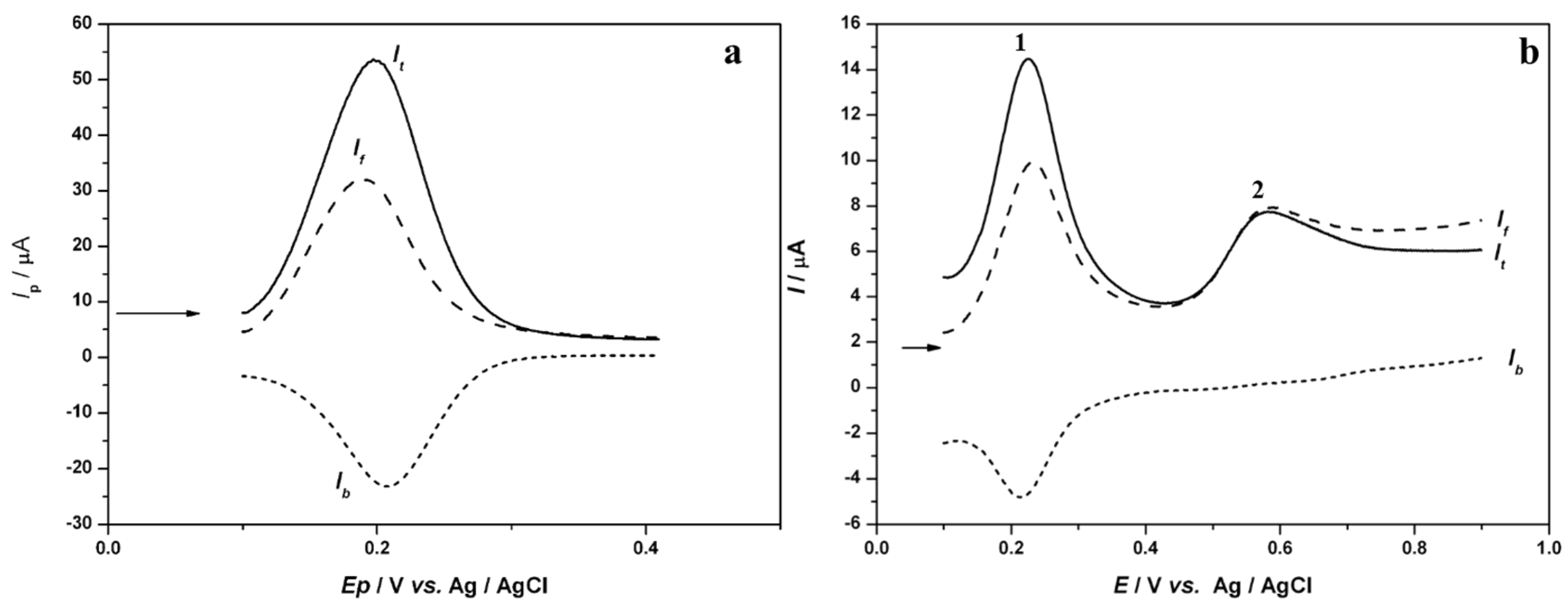
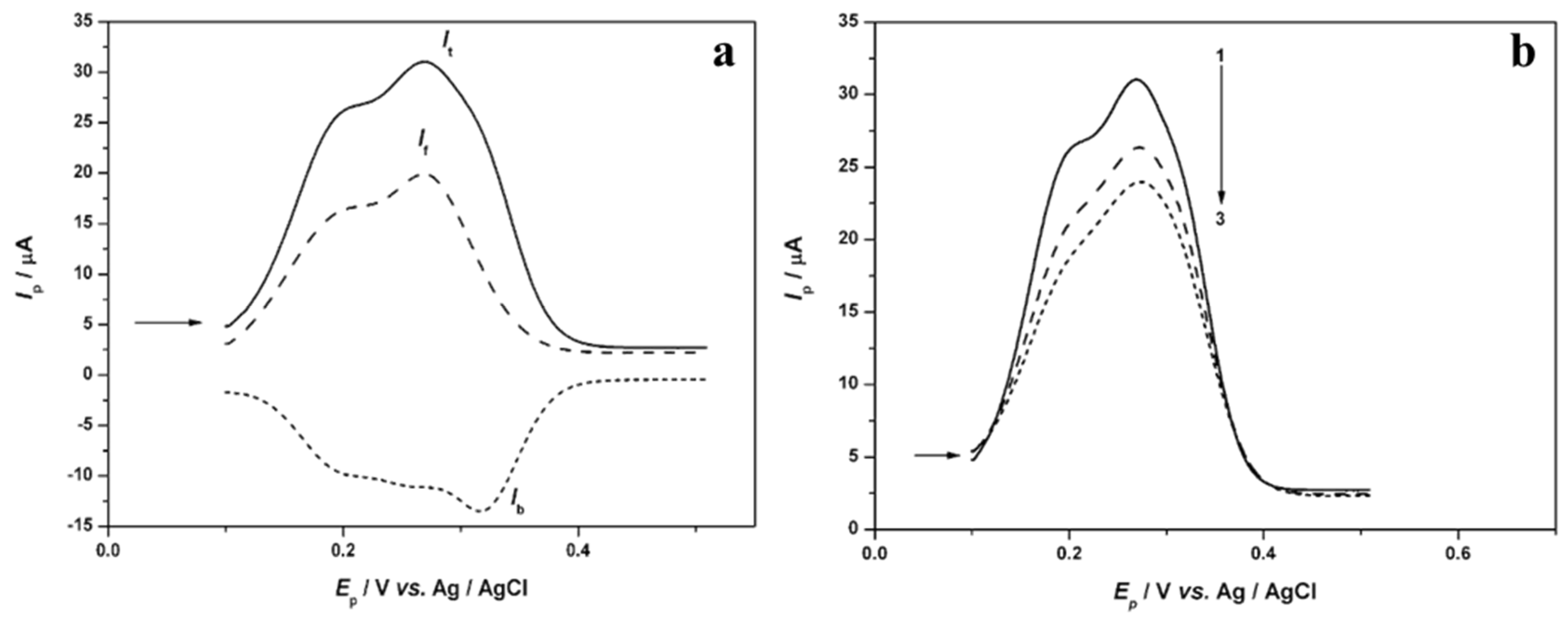
2.6. Determination of the Antioxidant Effect by Electrochemical DNA-Based Biosensor

The CV measurements were performed by applying the electrochemical DNA-based biosensor as it was described in our previous study [18]. In brief, a DNA-based biosensor was prepared by immersing SWCNT-COOH/SPC into the redox indicator solution [Fe(CN)6]3−/4− (c = 1 × 10⁻³ mol L−1). The CV measurements were achieved at the potential range from −0.4 V to +0.7 V and back to −0.4 V. The scan rate was 50 mV s−1. The CV response of the electrode was obtained without the DNA layer. After washing the electrode with ultrapure water, 5 μL of DNA solution was applied to the surface of the working electrode SWCNT-COOH/SPCE and left to dry for 30 min at room temperature to complete the immobilization of DNA onto the surface of the electrode (DNA/SWCNT-COOH/SPCE). The CV scan with DNA/SWCNT-COOH/SPCE was performed in the redox indicator solution. Next, the electrochemical biosensor was rinsed with ultrapure water and incubated in the cleavage solution for up to 15 min to detect DNA damage. The cleavage solution contained the 2.5 × 10⁻⁴ mol L−1 H2O2 and 1 × 10⁻⁶ mol L−1 of CuSO4 to produce hydroxyl ions that could damage the DNA layer. Finally, the CV scan was performed in a redox indicator [Fe(CN)6]3−/4− using the same experimental conditions as mentioned above. Before each experiment, a fresh cleavage mixture was used. The DNA damage experiments and the CV scans were repeated three times to achieve the mean response.
The same procedure was used for the detection of the antioxidant effects of sage tea polyphenols. The DNA-based biosensor, however, was incubated in the cleavage solution with the addition of (+)-catechin (50 mg L−1), caffeic acid (50 mg L−1), or sage tea (DF = 2). Results are presented as a relative portion of survived DNA (surv DNA, %), which was expressed as the normalized biosensor responses (ΔIa, rel; ΔIc, rel; Δ(ΔEp) calculated according to equations in the studies of Hlavatá et al., 2014 [25]; Ziyatdinova and Labuda, 2011 [22] and Tomac et al., 2020 [18].

2.7. Total Phenolic Content

The spectrophotometric measurements of total phenols from sage tea using Folin-Ciocalteu reagent (Merck, Germany) were performed with a UV-Vis spectrophotometer UV-1280 (Shimadzu, Japan) at 745 nm based on the study performed by Singleton et al. (1999) [26], with minor modifications. The measurements were compared to standard calibration curves of gallic and caffeic acids, and (+)-catechin solutions. The obtained results were expressed as milligrams of gallic acid (GAE), caffeic acid (CAE), and (+)-catechin (CE) equivalents per liter of sage tea. Stock solutions of polyphenols were prepared in methanol (HPLC grade).

2.8. Data Analysis

All measurements were performed at least three times. All CV and SWV voltammograms were analysed using GPES software (Eco Chemie, Utrecht, The Netherlands). All figures and statistical analysis of numerical results presented in this study were created with the OriginPro 8.0 (OriginLab Corporation, Northampton, MA, USA) software programme. The antioxidant effects were demonstrated as the mean values from at least three measurements. The statistical analysis of the antioxidant effect using a significance level of p < 0.05 was considered statistically significant.

3. Results and Discussion

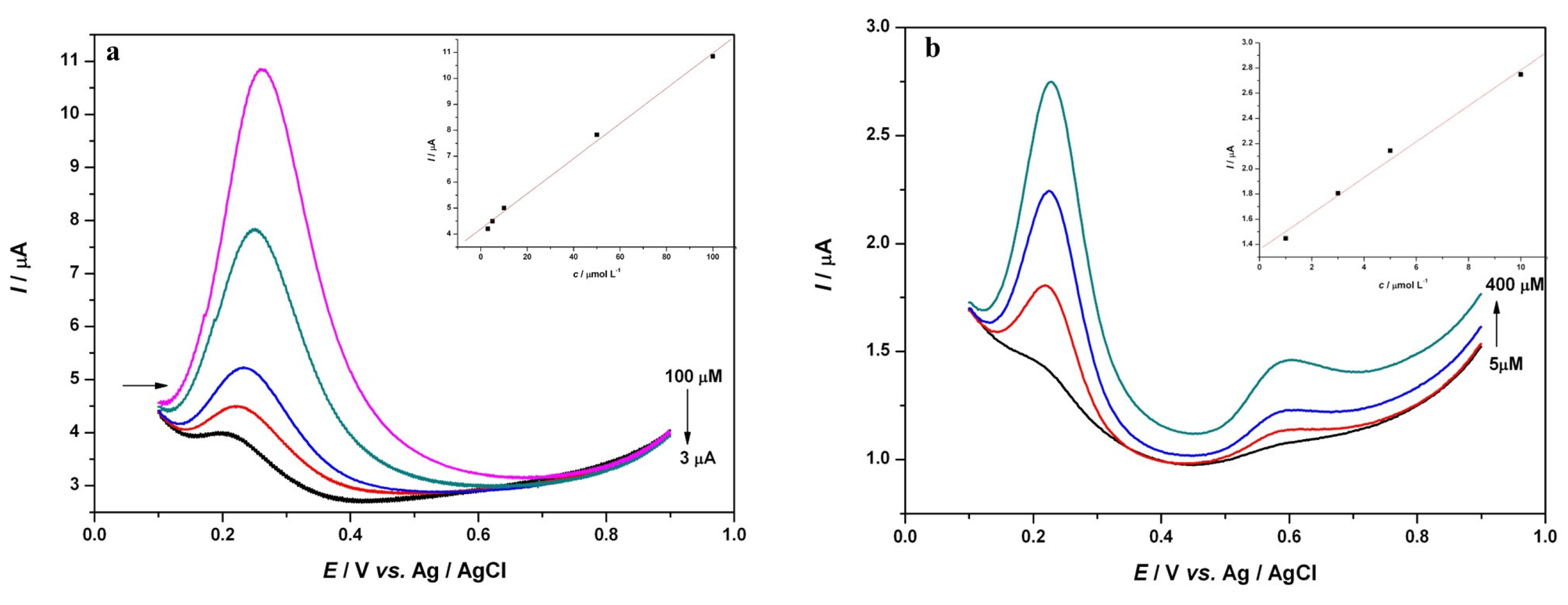
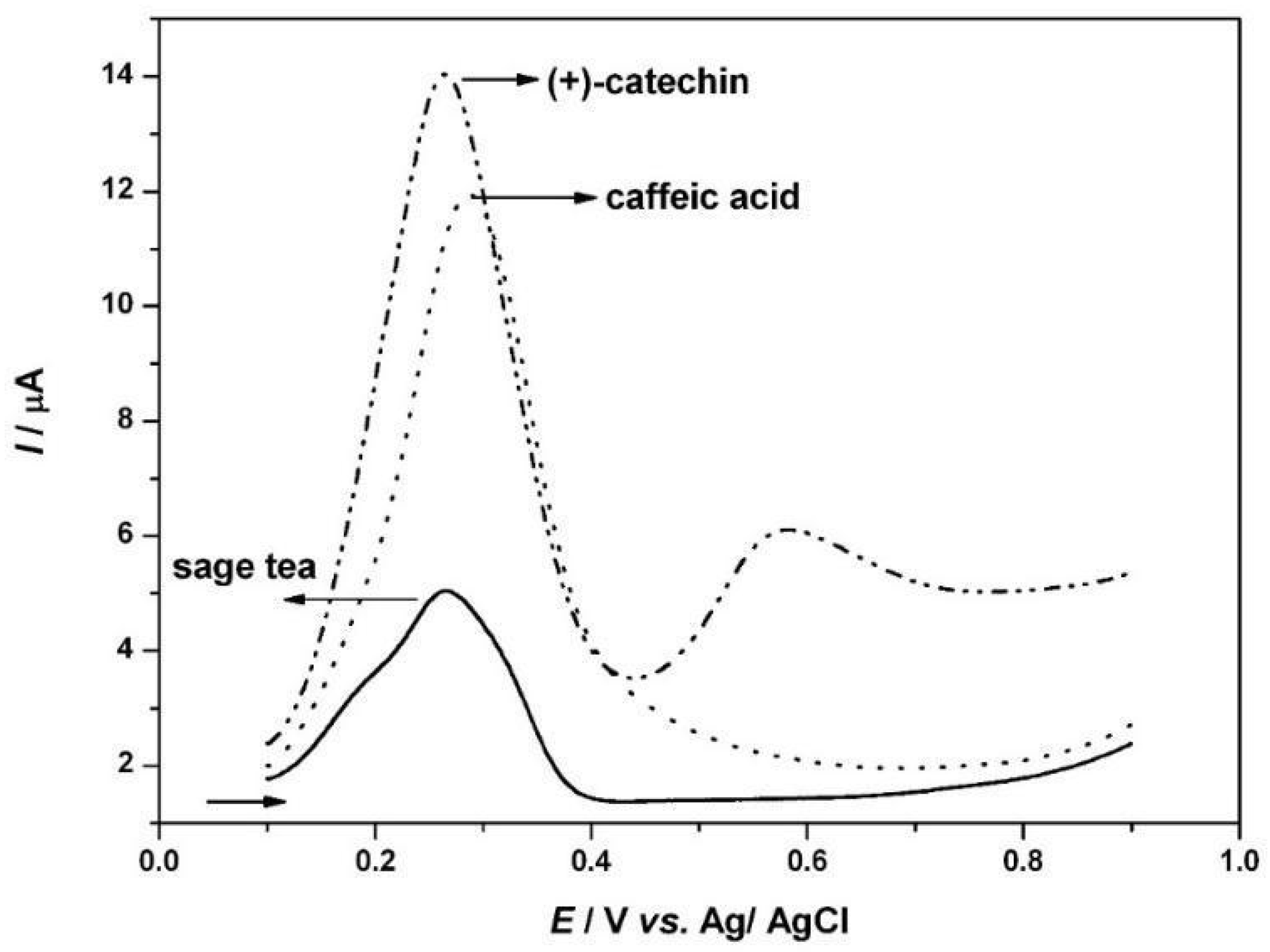
3.1. Voltammetric Study of Polyphenols in Sage Tea

Sage tea is an important source of polyphenols. Most of the polyphenols of Salvia are constructed from caffeic acid through different reactions [5]. The most abundant polyphenols in sage tea are from the groups of caffeic acid metabolites and flavonoids like carnosic and rosmarinic acids [1,27]. In this study, caffeic acid (Figure 1a), as one of the important building blocks of phenolic acids, and (+)-catechin (Figure 1b), as a representative of the flavonoids group in sage tea, were chosen for electrochemical investigation by applying SWV, as this is a fast and sensitive technique in comparison with other instrumental techniques. The advantage of SWV is the possibility of following the reversibility/irreversibility of electron transfer reaction during one scan because the current is simultaneously sampled in the anodic and cathodic pulse. The total current could be separated into forward and backward components (oxidation and reduction peaks). Consequently, SWV is used to investigate mechanisms of electron transfer and analyte concentration. In addition, the potential of the maximum peak can be used to confirm the identity of the analyte in solutions [28]. In this study, the SW voltammogram of 3 × 10⁻⁵ mol L−1 caffeic acid was recorded on the surface of bare GCE in the 0.1 mol L−1 PBS pH 7 (Figure 1a) at room temperature. Figure 1a shows one well-developed peak in caffeic acid total current at around Ep = 0.197 V. The total current (It) of caffeic acid can be separated into forward (oxidative, If) and backward (reductive, Ib) components. Since a reduction peak of CA was present and the potentials of the separated peaks were almost the same, a reversible electrochemical reaction is considered to be present on the surface of the bare GCE. The presence of a reversible electrode process in caffeic acid on the surface of the working electrode was also confirmed by other authors [29,30,31].
The electrooxidation of caffeic acid was studied by many researchers and they all agreed that the product of oxidation is the formation of o-quinone. The chemical structure of CA has one catechol moiety with two hydroxyl groups in the ortho-position following reversible oxidation to the o-quinone structure. In backward (reverse) scans, i.e., in cathodic polarisation, o-quinone reverts to a catechol structure [29,30,31,32]. In our study, one well-shaped peak could be assigned to the formation of the o-quinone, a product of the electrooxidation mechanism of caffeic acid.
Figure 1b shows the SWV of (+)-catechin (c = 3 × 10⁻⁵ mol L−1) oxidation process. SWV measurements of (+)-catechin were performed under the same conditions as the electrochemical oxidation of CA. Two well-defined oxidation peaks could be noticed: one around Ep = 0.232 V and the second one around Ep = 0.6 V. By separating the total current (It) into forward (If) and backward (Ib) components of (+)-catechin, it can be observed that there is no backward (Ib) component of the second peak. The first peak in (+)-catechin showed reversibility, whereas the second one showed irreversibility due to the absence of a backward component. The first peak had the same potential of oxidation and reduction electrode process, which demonstrates that the same potential oxidation and reduction process is taking place at the surface of the bare GCE [33].
The mechanism of the electrochemical oxidation process of (+)-catechin, a representative of flavonoids in sage tea, may be specified by its chemical structure properties. (+)-catechin represents the C-15 skeleton, divided into three units: A, B, and C. Units A and B are phenolic aromatic rings and unit C is an oxygen-containing heterocyclic. The redox behavior of flavonoids is determined by the proton−electron transfer mechanism on the surface of the working electrode, while electroactivity is probably mostly connected with phenolic groups of units of A and B reactivity. As phenolic groups run the electroactivity and determine the antioxidant effect of flavonoids, the oxidation mechanism of flavonoids, i.e., (+)-catechin could be described as an irreversible process of phenol and resorcinol (Figure 1b, peak 2) and a reversible process of the catechol and hydroquinone (Figure 1b, peak 1) [33,34]. According to Gil and Couto (2013) [33] and Janeiro and Brett (2004) [34], the reversible oxidation process takes place at the lower potentials (catechol of unit B) and the irreversible one takes place at the higher potentials. The same electrochemical behaviour of (+)-catechin was observed in this study.
Next, the influence of the caffeic acid and (+)-catechin concentrations was investigated by SWV (Figure 2) and the same experimental conditions of SWV measurements (0.1 mol L−1 PBS pH 7, f = 50 Hz, amplitude 50 mV, potential increment 2 mV) were applied. Figure 2 shows SWVs of caffeic acid (Figure 2a) and (+)-catechin (Figure 2b) obtained by the successive addition of CA and (+)-catechin into 0.1 mol L−1 PBS, pH 7. The concentration range for the caffeic acid was 20–100 μmol L−1 and 5–40 μmol L−1 for a (+)-catechin. The total current of caffeic acid at Ep ≈ 0.197 V increased linearly with concentration (Figure 2a, inset). At concentrations higher than 100 μmol L−1, the peak current of caffeic acid decreased and the function was no longer linear due to the newly formed oxidation products on the surface of the working electrode. A similar situation in the caffeic acid solution was observed for the (+)-catechin. In order to follow the effect of the concentration of (+)-catechin, the first oxidation peak at E ≈ 0.232 V was selected. The linear increase in the first (+)-catechin peak was obtained up to 400 μmol L−1 (Figure 2b, inset). With a further increase in the concentration, the peak current decreased and did not follow a linear function due to the formation of the oxidation product. This means that the electrochemical reaction of caffeic acid and (+)-catechin on the surface of the bare GCE is diffusion-controlled, and the flow of analyte is aggravated due to the formation of oxidation products [20,29,30]. Consequently, the calibration plots of CA and (+)-catechin are formed by linear regression. The equation obtained by the regression of CA was Ip, a (μA) = 0.0246 c (μmol L−1) + 1.5845; R = 0.996, with LOD = 7.4 μmol L−1 and LOQ = 22.4 μmol L−1 and of (+)-catechin Ip, a (μA) = 0.0711 c (μmol L−1) + 2.7563; R = 0.990, with LOD = 4.6 μmol L−1 and LOQ = 14.21 μmol L−1. The obtained results are in accordance with the results of authors who applied voltammetry to test the influence of concentration and quantification of caffeic acid and (+)-catechin contents in real samples [5,8,14,35,36].
Based on the presented results on the electrochemical properties of CA and (+)-catechin, SWV was applied to the investigation (identification and electrochemical oxidation) of polyphenols in sage tea. The same experimental conditions of SWV measurements used for the characterization of CA and (+)-catechin, frequency, pulse amplitude, and potential increment were applied to determine the electrochemical behavior of sage tea.
As sage tea is a rich source of compounds that can easily oxidize, it was expected that phenolic acids and catechins in sage tea would be dominant on voltammograms [14]. Figure 3a shows the SWV of the sage tea (DF = 70) recorded in 0.1 mol L−1 PBS pH 7. Two well-defined peaks are observed: one at around Ep = 0.198 V and the second one at around Ep = 0.268 V. The total current peaks in sage tea are very close to the value of the CA peak (Ep = 0.197 V) and the first (+)-catechin peak (Ep = 0.232 V) (Figure 2). The total current of the sage tea can be separated, and well-developed forward and backward components can be observed (Figure 3a). Hence, it can be noted that the reversible electrode process (the first peak) and quasi-reversible electrode process (the second peak) can be conducted on the surface of GCE (Figure 2).
The SWVs of sage tea in pH 7 0.1 mol L−1 PBS diluted 70-fold in the second and third scans (without cleaning the surface of the working electrode between scans) have an influence on successive polarisation in the sage tea solution (Figure 3b). It can be observed that the total current decreased during the second and third polarisations, which means that an adsorption layer of newly formed oxidation products is probably formed on the surface of GCE. These newly formed oxidation products blocked the electrode surface and the diffusion from the bulk solution is aggravated throughout the formed adsorption layer [33]. As a result, the current in the second and third scan decreases and the total current peaks move to more positive values. A similar influence of successive polarisation was observed for CA and (+)-catechin in this study.
Based on the electrochemical properties of CA and (+)-catechin, the oxidation peaks recorded in sage tea can be ascribed to the oxidation of a mixture of CA and (+)-catechin. The reversible electrode reaction is noted from the SWVs of CA and the first peak in (+)-catechin. For this reason, sample enrichment was performed with 1 mL of the 1 × 10⁻² mol L−1 concentration of individual standards in order to identify and confirm the polyphenol presence ((+)-catechin and caffeic acid) in the sage tea sample (Figure 4). Gomes et al. (2022) [36] stated that the selective determination of catechins had not been reported to date, and the term catechin equivalent (CAE) is used. Figure 4 shows that the total current of the sage tea sample (DF = 70) increased with the addition of caffeic acid (c = 3 × 10⁻⁵ mol L−1) at an Ep around 0.202 V, as well as with the addition of (+)-catechin (c = 3 × 10⁻⁵ mol L−1) at an Ep,1 around 0.232 V, which matched the SWVs caffeic acid and (+)-catechin standards (Figure 2). This enrichment procedure confirmed that the oxidation peak in sage tea recorded by SWV can be ascribed to the electrochemical oxidation of a mixture of CA dimers and catechin in the sage tea sample. Based on SWV measurements, SWV technique was proved to be an applicable technique for the identification of polyphenols in sage tea.

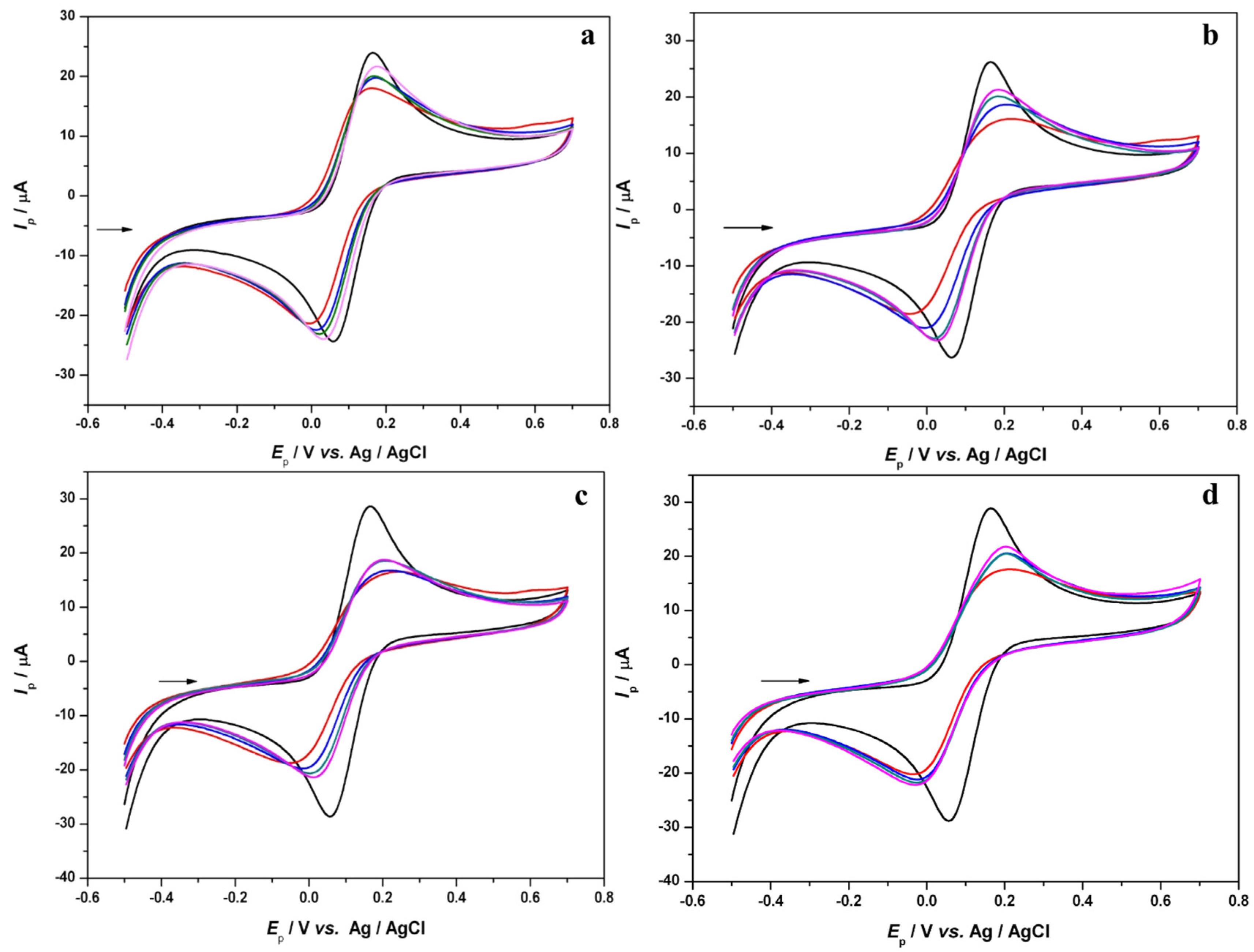
3.2. Determination of the Antioxidant Effect of Sage Tea Using Electrochemical DNA-Based Biosensor

Electrochemical determination of the antioxidant effect of sage tea was performed by cyclic voltammetry using an electrochemical DNA-based biosensor.
The electrochemical DNA-based biosensor was created, as previously stated, by Labuda et al. (2010) [37] and Tomac et al. (2020) [18]. In other words, the dsDNA solution was applied to the surface of a commercial SWCNT-COOH-modified electrode. CV was performed to observe the DNA layer on the surface of the electrode in the range of potential from 0 V to +1.4 V and back to 0 V, with the scan rate of 0.1 V s−1 in PB solution pH 7. As a result, two irreversible anodic peaks in CV response appeared around the potentials +0.83 V and +1.12 V, which can be ascribed to the presence of guanine and adenine residues of DNA [18,37]. The hydroxyl radicals were produced by the reaction between H2O2 and Cu2+ ions (the cleavage mixture) for the deep DNA damage. The DNA damage was estimated by CV measurements.
CVs of 1 mmol L−1 [Fe(CN)6]3−/4− in pH 7 0.1 mol L−1 PB with 0.01 mol L−1 NaCl at the DNA/SWCNT-COOH/SPCE (black curves) were recorded after incubation in the cleavage mixture (Figure 5a), with the addition of (+)-catechin, (1.72 × 10⁻⁴ mol L−1) (Figure 5b), and with the addition of CA (2.77 × 10⁻⁴ mol L−1) (Figure 5c) for 2 min (blue curve), 5 min (green curve) and 15 min (pink curve) of immersion. A gradual degradation of the DNA layer (red curves) was revealed. The longer the immersion time in the cleavage solution, the more progressive the DNA degradation. In Figure 5, the increase in the redox indicator solution current at the DNA/SWCNT-COOH/SPCE can be noticed, as well as the shift in potential to the more positive values, in comparison to the bare SWCNT-COOH/SPCE, which can be ascribed to the effect of hydroxyl radicals’ attack on DNA layer degradation. The normalized signal values of the biosensor responses ΔIa, rel, ΔIc, rel, and Δ(ΔEp, rel) for an individual incubation time of 2, 5, and 15 min were calculated from CVs according to equations mentioned in the work of Hlavatá et al., 2014 [25], Ziyatdinova and Labuda, 2011 [22] and Tomac et al., 2020 [18]. In brief, the equations used to calculate the normalized signals values of the biosensor responses after incubation of the DNA/SWCNT-COOH/SPCE in the cleavage solution are based on the difference in the current response or electrode potential of the surviving DNA at DNA/SWCNT-COOH/SPCE and the bare SWCNT-COOH/SPCE in relation to the difference in the current response or electrode potential of the DNA/SWCNT-COOH/SPCE and the bare SWCNT-COOH/SPCE, expressed in percentages. Table 1 shows summarized values for the normalized biosensor responses after the DNA/SWCNT-COOH/SPCE incubation in a cleavage solution with the addition of (+)-catechin or CA or sage tea, after an immersion time of 15 min. The relative portion of the survived DNA, reported as ΔIa, rel, and ΔIc, rel, is as follows: CA > (+)-catechin > absence of polyphenols. The results presented in Table 1 indicate that the components with the highest relative portion of survived DNA (CA) have the lowest peak potential (0.197 V). As the potential is low, the capacity of the electron donor increases, which leads to a higher antioxidant effect, i.e., stronger antioxidants [20,38]. The current study confirms the premise that CA is a stronger antioxidant than (+)-catechin. Antioxidants are molecules capable of preventing oxidation or inhibiting free-radical formation. The antioxidant effect of such molecules is directly connected to their reducing character and chemical structure. Furthermore, the catechol moieties and the presence of hydroxyl groups, i.e., the presence of electron-donating and electron-withdrawing groups, affect the antioxidant properties of CA and (+)-catechin [20,34,38].
In order to investigate the antioxidant effects of sage tea, the same experimental conditions of CV measurements and the same procedure of constructing biosensors were applied. The antioxidant effects of the sage tea solution were determined with regard to the incubation of the biosensor in the cleavage solution with the addition of sage tea. The appearance of CVs after immersion in the cleavage solution with the addition of sage tea is similar to that of CVs after immersion in the cleavage solution with the addition of CA and (+)-catechin (Figure 5). Based on the values of the normalized CVs responses (Table 1), higher values of the relative portion of survived DNA for sage tea after 15 min of immersion (ΔIa, rel = 62.97%, ΔIc, rel = 67.84%, Δ(ΔEp, rel) = 66.16%) in comparison to that without the addition of sage tea (ΔIa, rel = 39.05%, ΔIc, rel = 39.21%, Δ(ΔEp, rel) = 26.09%) can be noticed. A significant positive correlation (R = 0.9886) between the values of ΔIa, rel and ΔIc, rel was found for individual polyphenols in sage tea, except for Δ(ΔEp, rel), which showed lower values. The lower values of Δ(ΔEp, rel) in comparison to ΔIa, rel and ΔIc, rel were also noticed in other studies [22,25]. The obtained values of the relative portion of surviving DNA reveal that sage tea has a significant antioxidant effect on the DNA layer. The DNA-protective ability of individual polyphenols, CA and (+)-catechin, can contribute to the higher portion of surviving DNA for sage tea. The results of this study indicate the antioxidant effects of sage tea are closely related to the chemical structure and electron donor properties of polyphenols in sage tea (Table 1). According to this, sage tea has strong antioxidant effects, regardless of the fact that the biosensor response expresses a group of antioxidants [18,37].
For comparison purposes, the spectrophotometric Folin−Ciocalteu method was applied for the determination of the total phenolic content in sage tea. Although the spectrophotometric Folin−Ciocalteu assay is a one-electron transfer process, it is an irreplaceable and suitable method for the determination of total phenolic content in natural products [5]. In this study, we slightly modified the procedure proposed by Singleton et al. (1999) [26] and used CA and (+)-catechin in addition to gallic acid as a standard. We believed that adding CA and (+)-catechin would provide a better insight into the agreement of the results. The results obtained by the spectrophotometric Folin−Ciocalteu method are summarized in Table 1. The obtained results of the total phenolic content of sage tea (180 mgCE/g, 102 mgCAE/g, and 107 mgGAE/g) are in agreement with the results of other authors who used the spectrophotometric Folin−Ciocalteu method for the determination of total phenolic content in sage tea (70.5 to 176.5 mgGAE/g) [24]. There are, of course, some slight differences between results, but this is due to the different preparation procedures and different origins of sage tea. Statistical analysis was used to compare the results of the total content of polyphenols in sage tea and confirmed that there is a positive and significant correlation (R = 0.9987) between the antioxidant effects obtained by cyclic voltammetry using biosensor and total phenolic content, determined using the spectrophotometric Folin−Ciocalteu method.

4. Conclusions

In this paper, the characterization of the polyphenol profile of sage tea was investigated using sensitive and fast square-wave voltammetry. Caffeic acid and (+)-catechin were identified and their electrochemical properties were shown to depend on their chemical structure. The SWV measurements show that the electrochemical behavior of sage tea is very similar to that examined in caffeic acid and (+)-catechin. The antioxidant effects of sage tea were determined using cyclic voltammetry by applying the biosensor. The results indicated that sage tea had strong antioxidant effects. Since the biosensor response expresses a group of antioxidants, polyphenols in the composition of sage tea influence the antioxidant and protective effects on DNA damage. Both the total polyphenol concentration and the antioxidant effects of sage tea were determined and found to have a close correlation. The implemented method and applied biosensor can be deemed a good option for the characterization of the profile of herbal tea compounds and their antioxidant effects.

Author Contributions

Conceptualization, I.T., L.B., J.B., L.J. and P.M.; methodology, I.T., L.B., J.B., L.J. and P.M.; formal analysis, I.T., L.B., J.B., L.J. and P.M.; investigation, I.T., L.B., J.B., L.J. and P.M.; data curation, I.T., L.B., J.B., L.J. and P.M.; writing—original draft preparation, I.T., L.B., J.B., L.J. and P.M.; writing—review and editing, I.T., L.B., J.B., L.J. and P.M.; visualization, I.T., L.B., J.B., L.J. and P.M.; supervision, I.T. All authors have read and agreed to the published version of the manuscript.

Funding

This research received no external funding.

Data Availability Statement

All data are available on request from the corresponding author.

Acknowledgments

Special thanks to Jan Labuda, who introduced us to the field of biosensors.

Conflicts of Interest

The authors declare no conflict of interest.

Abbreviations

CA: caffeic acid; CV: cyclic voltammetry; SWV: square-wave voltammetry; GCE: glassy carbon electrode; PB: phosphate-buffer solution; the equations for calculating the normalized signal values of the biosensor responses: ΔIp,rel (%) = (Ip,surv DNAIp,SWCNT-COOH/SPCE)/(Ip,DNAIp,SWCNT-COOH/SPCE) × 100 and Δ(ΔEp,rel) (%) = (ΔEp,surv DNA—ΔEp,SWCNT-COOH/SPCE)/(ΔEp,DNA—ΔEp,SWCNT-COOH/SPCE) × 100; DNA: deoxyribonucleic acid; SWCNT-COOH/SPCE: screen-printed carbon electrode modified with the carboxyl-functionalized single-walled carbon nanotubes; ΔIa, rel: anodic current response; ΔIc, rel: cathodic current response; Δ(ΔEp, rel): anodic to cathodic peak potential separation; Ep: peak potential; It: total current response; If: forward (oxidative) component of current response; Ib: backward (reductive) component of current response; LOD: limit of detection; LOQ: limit of quantification.

References

  1. Bors, W.; Michel, C.; Stettmaier, K.; Lu, Y.; Foo, L.Y. Antioxidant Mechanisms of Polyphenolic Caffeic Acid Oligomers, Constituents of Salvia officinalis. Biol. Res. 2004, 37, 301–311. [Google Scholar] [CrossRef] [PubMed]
  2. Walch, S.G.; Tinzoh, L.N.; Zimmermann, B.F.; Stühlinger, W.; Lachenmeie, D.W. Antioxidant capacity and polyphenolic composition as quality indicators for aqueous infusions of Salvia officinalis L. (sage tea). Front. Pharmacol. 2011, 2, 79. [Google Scholar] [CrossRef]
  3. Zimmermann, B.F.; Walch, S.G.; Tinzoh, L.N.; Stühlinger, W.; Lachenmeier, D.W. Rapid UHPLC determination of polyphenols in aqueous infusions of Salvia officinalis L. (sage tea). J. Chromatogr. B 2011, 879, 2459–2464. [Google Scholar] [CrossRef]
  4. Lu, Y.; Foo, L.Y. Polyphenolics of Salvia—A review. Phytochemistry 2002, 59, 117–140. [Google Scholar] [CrossRef]
  5. David, I.G.; Bizgan, A.-M.C.; Popa, D.E.; Buleandra, M.; Moldovan, Z.; Badea, I.A.; Tekiner, T.A.; Basaga, H.; Ciucu, A.A. Rapid determination of total polyphenolic content in tea samples based on caffeic acid voltammetric behaviour on a disposable graphite electrode. Food Chem. 2015, 173, 1059–1065. [Google Scholar] [CrossRef]
  6. Ziyatdinova, G.K.; Guss, E.V.; Budnikov, H.C. Voltammetric Evaluation of Polyphenol–Protein Interactions and Their Influence on the Antioxidant Capacity of Tea. J. Anal. Chem. 2020, 75, 685–690. [Google Scholar] [CrossRef]
  7. Novak, I.; Šeruga, M.; Komorsky-Lovrić, Š. Characterisation of catechins in green and black teas using square-wave voltammetry and RP-HPLC-ECD. Food Chem. 2010, 122, 1283–1289. [Google Scholar] [CrossRef]
  8. Veljković, J.N.; Stojković, M.; Brcanović, J.M. Evaluation of individual phenolic compounds and antioxidant properties of black, green, herbal and fruit tea infusions consumed in Serbia: Spectrophotometrical and electrochemical approaches. J. Food Nutr. Res. 2013, 52, 12–24. [Google Scholar]
  9. David, I.G.; Buleandră, M.; Popa, D.E.; Bizgan, A.-M.C.; Moldovan, Z.; Badea, I.-A.; Iorgulescu, E.E.; Tekiner, T.A.; Basaga, H. Voltammetric determination of polyphenolic content as rosmarinic acid equivalent in tea samples using pencil graphite electrodes. J. Food Sci. Technol. 2016, 53, 2589–2596. [Google Scholar] [CrossRef]
  10. Sabela, M.I.; Gumede, N.J.; Singh, P.; Bisetty, K. Evaluation of antioxidants in herbal tea with a laccase biosensor. Int. J. Electrochem. Sci. 2012, 7, 4918–4928. [Google Scholar] [CrossRef]
  11. Nehru, R.; Hsu, Y.F.; Wang, S.F. Electrochemical determination of caffeic acid in antioxidant beverages samples via a facile synthesis of carbon/iron-based active electrocatalyst. Anal. Chim. Acta 2020, 1122, 76–88. [Google Scholar] [CrossRef]
  12. Saikrithika, S.; Kumar, A.S. Electrochemical Detections of Tea Polyphenols: A Review. Electroanalysis 2020, 32, 2343–2360. [Google Scholar] [CrossRef]
  13. Shao, J.; Wang, C.; Shen, Y.; Shi, J.; Ding, D. Electrochemical Sensors and Biosensors for the Analysis of Tea Components: A Bibliometric Review. Front. Chem. 2022, 9, 818461. [Google Scholar] [CrossRef]
  14. Suroviec, A.H.; Jones, K.; Sarabia, G. Quantification of Catechins in Tea Using Cyclic Voltammetry. J. Chem. Educ. 2019, 96, 366–371. [Google Scholar] [CrossRef]
  15. Kulikova, T.N.; Porfireva, A.V.; Vorobev, V.V.; Saveliev, A.A.; Ziyatdinova, G.K.; Evtugyn, G.A. Discrimination of Tea by the Electrochemical Determination of its Antioxidant Properties by a Polyaniline–DNA–Polyphenazine Dye Modified Glassy Carbon Electrode. Anal. Lett. 2019, 52, 2562–2582. [Google Scholar] [CrossRef]
  16. Li, Y.; Chen, X.; Lin, Y.; Yang, Y.; Zhang, L.; Zhao, P.; Wang, C.; Fei, J.; Xie, Y. Detection of catechins in tea beverages using a novel electrochemical sensor based on cyclodextrin nanosponges composite. eFood 2023, 4, e64. [Google Scholar] [CrossRef]
  17. Ziyatdinova, G.K.; Nizamova, A.M.; Aytuganova, I.I.; Budnikov, H.C. Voltammetric evaluation of the antioxidant capacity of tea on electrodes modified with multi-walled carbon nanotubes. J. Anal. Chem. 2013, 68, 132–139. [Google Scholar] [CrossRef]
  18. Tomac, I.; Šeruga, M.; Labuda, J. Evaluation of antioxidant activity of chlorogenic acids and coffee extracts by an electrochemical DNA-based biosensor. Food Chem. 2020, 325, 126787. [Google Scholar] [CrossRef]
  19. Ferancová, A.; Heilerová, Ľ.; Korgová, E.; Šilhár, S.; Štěpánek, I.; Labuda, J. Anti/pro-oxidative properties of selected standard chemicals and tea extracts investigated by DNA-based electrochemical biosensor. Eur. Food Res. Technol. 2004, 219, 416–420. [Google Scholar] [CrossRef]
  20. Lima, A.P.; dos Santos, W.T.P.; Nossol, E.; Richter, E.M.; Munoz, R.A.A. Critical evaluation of voltammetric techniques for antioxidant capacity and activity: Presence of alumina on glassy-carbon electrodes alters the results. Electrochim. Acta 2020, 358, 136925. [Google Scholar] [CrossRef]
  21. Kilmartin, P.A.; Hsu, C.F. Characterisation of polyphenols in green, oolong, and black teas, and in coffee, using cyclic voltammetry. Food Chem. 2003, 82, 501–512. [Google Scholar] [CrossRef]
  22. Ziyatdinova, G.; Labuda, J. Complex electrochemical and impedimetric evaluation of DNA damage by using DNA biosensor based on a carbon screen-printed electrode. Anal. Methods 2011, 3, 2777–2782. [Google Scholar] [CrossRef]
  23. Piljac-Žegarac, J.; Valek, L.; Stipčević, T.; Martinez, S. Electrochemical determination of antioxidant capacity of fruit tea infusions. Food Chem. 2010, 121, 820–825. [Google Scholar] [CrossRef]
  24. Khiya, Z.; Oualcadi, Y.; Gamar, A.; Berrekhis, F.; Zair, T.; El Hilali, F. Correlation of Total Polyphenolic Content with Antioxidant Activity of Hydromethanolic Extract and Their Fractions of the Salvia officinalis Leaves from Different Regions of Morocco. J. Chem. 2021, 2021, 8585313. [Google Scholar] [CrossRef]
  25. Hlavatá, L.; Vyskočil, V.; Beníková, K.; Borbélyová, M.; Labuda, J. DNA-based biosensors with external Nafion and chitosan membranes for the evaluation of the antioxidant activity of beer, coffee, and tea. Cent. Eur. J. Chem. 2014, 12, 604–611. [Google Scholar] [CrossRef]
  26. Singleton, V.L.; Orthofer, R.; Lamuela-Raventós, R.M. Analysis of total phenols and other oxidation substrates and antioxidants by means of folin-ciocalteu reagent. Methods Enzymol. 1999, 299, 152–178. [Google Scholar]
  27. Dent, M.; Bursać Kovačević, D.; Bosiljkov, T.; Dragović-Uzelac, V. Polyphenolic Composition and Antioxidant Capacity of Indigenous Wild Dalmatian Sage (Salvia officinalis L.). Croat. Chem. Acta 2017, 90, 451–459. [Google Scholar] [CrossRef]
  28. Mirceski, V.; Gulaboski, R.; Lovric, M.; Bogeski, I.; Kappl, R.; Hoth, M. Square-Wave Voltammetry: A Review on the Recent Progress. Electroanalysis 2013, 25, 2411–2422. [Google Scholar] [CrossRef]
  29. Trabelsi, K.S.; Belhadj Tahar, N.; Abdelhedi, R. Electrochemical behaviour of caffeic acid. Electrochim. Acta 2004, 49, 1647–1654. [Google Scholar] [CrossRef]
  30. Karikalan, N.; Karthik, R.; Chen, S.-M.; Chen, H.-A. A voltammetric determination of caffeic acid in red wines based on the nitrogen doped carbon modified glassy carbon electrode. Sci. Rep. 2017, 7, 45924. [Google Scholar] [CrossRef]
  31. Zare, H.R.; Golabi, S.M. Caffeic acid modified glassy carbon electrode for electrocatalytic oxidation of reduced nicotinamide adenine dinucleotide (NADH). J. Solid State Electrochem. 2000, 4, 87–94. [Google Scholar]
  32. Šeruga, M.; Tomac, I. Electrochemical Behaviour of Some Chlorogenic Acids and Their Characterization in Coffee by Square-Wave Voltammetry. Int. J. Electrochem. Sci. 2014, 9, 6134–6154. [Google Scholar] [CrossRef]
  33. Gil, E.S.; Couto, R.O. Flavonoid electrochemistry: A review on the electrochemical applications. Braz. J. Pharmacol. 2013, 23, 542–558. [Google Scholar] [CrossRef]
  34. Janeiro, P.; Oliveira Brett, A.-M. Catechin electrochemical oxidation mechanisms. Anal. Chim. Acta 2004, 518, 109–115. [Google Scholar] [CrossRef]
  35. Khiya, Z.; Hayani, M.; Hamar, A.; Kharchouf, S.; Amine, S.; Berrekhis, F.; Bouzoubae, A.; Zair, T.; El Hilali, F. Valorization of the Salvia officinalis L. of the Morocco bioactive extracts: Phytochemistry, antioxidant activity and corrosion inhibition. J. King Saud Univ. Sci. 2019, 31, 322–335. [Google Scholar] [CrossRef]
  36. Gomes, J.S.; Costa, E.A.; Munoz, R.A.A.; Oliveira, A.; Sousa, R.M.F. Selective electrochemical detection of catechin compounds in herbal medicines. J. Electrochem. Soc. 2022, 169, 017516. [Google Scholar] [CrossRef]
  37. Labuda, J.; Brett, A.M.O.; Evtigyn, G.; Fojta, M.; Mascini, M.; Ozsoz, M.; Palchetti, I.; Palček, E.; Wang, J. Electrochemical nucleic acid-based biosensors: Concepts terms and methodology (IUPAC Technical Report). Pure Appl. Chem. 2010, 82, 1131–1187. [Google Scholar] [CrossRef]
  38. Pisoschi, A.M.; Cimpeanu, C.; Predoi, G. Electrochemical Methods for Total Antioxidant Capacity and its Main Contributors Determination: A review. Open Chem. 2015, 13, 824–856. [Google Scholar] [CrossRef]
Figure 1. SW voltammograms of (a) caffeic acid and (b) (+)-catechin, c = 3 × 10⁻⁵ mol L−1, in 0.1 mol L−1 PBS pH 7. The total (It), forward (If), and backward (Ib) components are shown. The experimental conditions: frequency 50 Hz, pulse amplitude 50 mV, potential increment 2 mV.
Beverages 09 00076 g001
Figure 2. SWVs (the total response) of (a) caffeic acid and (b) (+)-catechin at GCE in 0.1 mol L−1 PBS pH 7, f = 50 Hz, pulse amplitude 50 mV, potential increment 2 mV. Caffeic acid concentrations: 20 × 10−5 to 100 × 10−5 mol L−1 and (+)-catechin concentrations: 5 × 10⁻⁶ to 40 × 10−5 mol L−1. Inset: calibration plot of CA (a) and calibration plot of (+)-catechin (b).
Beverages 09 00076 g002
Figure 3. Square-wave voltammograms of (a) sage tea (DF = 70) and the successive scans of sage tea (b) recorded in 0.1 mol L−1 PBS pH 7, f = 50 Hz, pulse amplitude 50 mV s−1, potential increment 2 mV.
Beverages 09 00076 g003
Figure 4. SWVs of sage tea (DF = 70) with the addition of 3 × 10⁻⁵ mol L−1 caffeic acid and 3 × 10⁻⁵ mol L−1 (+)-catechin recorded in 0.1 mol L−1 PBS pH 7, f = 50 Hz, pulse amplitude 50 mV s−1, potential increment 2 mV.
Beverages 09 00076 g004
Figure 5. CVs of 1 mmol L−1 [Fe(CN)6]3−/4− in 0.1 mol L−1 PB with 0.01 mol L−1 NaCl pH 7 (black curves) recorded at DNA/SWCNT-COOH/SPCE (red curves) before and after the incubation of the biosensor in the cleavage agent producing hydroxyl radicals (a), with (+)-catechin, c = 1.72 × 10⁻⁴ mol L−1 (b), with caffeic acid, c = 2.77 × 10⁻⁴ mol L−1 (c) and with sage tea, DF = 2 (d) for 2 min (blue curves), 5 min (green curves) and 15 min (pink curves) at 50 mV s−1.
Beverages 09 00076 g005
Table 1. The summarized results obtained by the spectrophotometric Folin−Ciocalteu method, antioxidant effects of (+)-catechin and caffeic acid (c = 50 mg L−1) and sage tea 1:2 (v/v) ratio towards oxidative DNA damage, expressed as a relative portion of survived DNA at the DNA/SWCNT-COOH/SPCE biosensor, and oxidative potential determined by the square-wave potential.
The Relative Portion of Surviving DNA/%Total PolyphenolsOxidative Potential
AntioxidantΔIa, relΔIc, relΔ(ΔEp, rel)mgE/g Sage TeaEp/V vs. Ag/AgCl
Absence of polyphenols39.0539.2126.09
(+)-catechin64.9164.8257.50 0.232
Caffeic acid81.6880.6976.85 0.197
Sample
Sage tea62.9767.8466.16180 mgCE/g
102 mgCAE/g
107 mgGAE/g
The results are the mean values of a minimum of three independent measurements.
Disclaimer/Publisher’s Note: The statements, opinions and data contained in all publications are solely those of the individual author(s) and contributor(s) and not of MDPI and/or the editor(s). MDPI and/or the editor(s) disclaim responsibility for any injury to people or property resulting from any ideas, methods, instructions or products referred to in the content.

