Article

Effect of an In Vitro Digestion on the Antioxidant Capacity of a Microfiltrated Blackberry Juice (Rubus adenotrichos)

1
Departamento de Bioquímica, Escuela de Medicina, Universidad de Costa Rica, 11501-2060 San José, Costa Rica
2
Centro Nacional de Ciencia y Tecnología de Alimentos (CITA), Universidad de Costa Rica, 11501-2060 San José, Costa Rica
3
Centre de Coopération Internationale en Recherche Agronomique pour le Développement (CIRAD), UMR QualiSud, 34967 Montpellier, France
4
UMR QualiSud, Univ Montpellier, CIRAD, Montpellier SupAgro, 34967 Montpellier, France
*
Author to whom correspondence should be addressed.
Beverages 2018, 4(2), 30; https://doi.org/10.3390/beverages4020030
Submission received: 19 December 2017 / Revised: 7 February 2018 / Accepted: 1 March 2018 / Published: 2 April 2018

Abstract

:
The health benefits attributed to berries are largely dependent on their bioavailability. This study evaluated the antioxidant activity for different samples of blackberry juice submitted to an in vitro digestion. The HPLC analysis demonstrated that gastric conditions had only a slight impact on the phenolic composition, but the digestion simulating intestine conditions caused marked changes. The dialyzed fraction, which represents the serum available material, showed 33% and 35% less activity against nitrogen-derived radicals and a significant reduction of 46% in the inhibitory capacity against intracellular ROS. Other models used to evaluate the capacity to inhibit lipid peroxidation did not show significant differences in any of the digestion samples. Our results suggest that blackberry polyphenols could exert their antioxidant capacity after passage through the GIT. However, the dialyzed fraction suffers a partial depletion of its antioxidant ability, this could be attributed to the absence of ellagitannins.

Graphical Abstract

1. Introduction

The biological activity of berry polyphenols has attracted interest of consumers, and different studies have documented in vitro and in vivo activities that could be related with benefits for human health. Specifically, berry polyphenols have been associated with a strong free radical scavenging activity, prevention of cellular lipids oxidation, inhibition of oxidant enzymes, modulation of signal transduction, cancer prevention, anti-inflammatory properties and improvement of cardiovascular risk profiles [1,2,3,4,5]. However, the health effects of polyphenols are largely dependent upon their bioavailability. To validate the beneficial effects of berry polyphenols it is important to consider the levels of absorption and modifications that these compounds suffer during digestion [6].
The bioavailability of each phenolic compound is affected by several factors, as their molecular structure, glycosylation levels, molecular weight, esterification, food matrices or food processing [1,6]. Due to this, it is important to study the bioavailability of each dietary source of polyphenols.
Tropical blackberries, from the Rubus spp. group, contain anthocyanins as their main flavonoid [7,8]. Unlike other flavonoids, anthocyanins can be absorbed in the intact glycoside form or in several other anthocyanin metabolites, such as methylated glycosides, glucuronides of anthocyanidins, sulfoconjugates of cyanidin, and anthocyanidins. In spite of this, the absorption of blackberry anthocyanins has been reported to be low, often between 0.02% and 1.8% of the ingested amounts [8,9,10].
Another important type of polyphenols reported for blackberries are the ellagitannins [7,8]. Some studies have hypothesized that these molecules undergo partial hydrolysis at the pH levels of the small intestine, and the remaining ellagitannins are hydrolyzed by colon microbiota to ellagic acid. Later, the ellagic acid followed a gradual metabolism to produce urolithins, which are molecules more lipophilic and easily absorbed [11,12]. It has been suggested that differences in the colonic microbiota composition can result in variation in the quantity and types of urolithins absorbed and excreted [12,13].
The aim of this study was to evaluate changes in the blackberry juice antioxidant activity of different samples that represent the major stages of the digestion process. These samples were obtained from an in vitro digestion technique, and the comparison between them was done assessing anthocyanin and ellagitannin concentrations, free radical scavenging capacity and evaluating the ability to inhibit lipid peroxidation.

2. Materials and Methods

2.1. Chemicals

All solvents for the polyphenol purification and HPLC were acquired from JT Baker (Griesheim, Germany). Tert-butyl hydroperoxide (TBHP), amberlite XAD-7, 6-hydroxy-2,5,7,8-tetramethylchroman-2-carboxylic acid (Trolox), 2,2 diphenyl-1-picrylhydrazyl (DPPH), thiobarbituric acid (TBA), 2,2-azobis-2-methyl-propion-amidine-dihydrochloride (AAPH), 2,7-dichlorodihydrofluorescein diacetate (DCFDA), quercetin, fluorescein, pepsin, bile salts, and pancreatin were obtained from Sigma Aldrich (St. Louis, MO, USA). Ellagic acid standard was acquired from Fluka (Buchs, Switzerland), and cyanidin-3-glucoside was acquired from Extrasynthese (Lyon, France). The reagents for cell culture: fetal bovine serum, EMEM medium, l-glutamine, trypsin, streptomycin, and penicillin were obtained from Sigma Aldrich (St. Louis, MO, USA). The reagents for the Griess test: sodium nitroprusside (SNP), naphthylethylenediamine dihydrochloride, sulfanilamide, and sodium nitrite were purchased from Merck (Darmstadt, Germany).

2.2. Blackberry Juice Preparation

Full-ripe blackberries (Rubus adenotrichos Schltdl. cv. ‘vino’) were collected from farms in the region of Cartago, Costa Rica (altitude 1864–2517 m, latitude 09°39′57.1′′ N–09°44′40.3′′ N, longitude 83°53′32.1′′ W–84°00′06.3′′ W). Microfiltrated juice was prepared according to a previous publication [14]. Briefly, blackberries were pressed and the juice was treated with a commercial enzymatic preparation, Ultrazym (Novozymes, Bagsvaerd, Denmark) for 1 h at 35 °C using constant agitation. After, the microfiltration was performed in a tubular ceramic membrane (Membralox® 1 P19–40, Pall Exekia, Bazet, France) with a pore size diameter of 0.2 μm.

2.3. In Vitro Digestion

To simulate the physiochemical changes undergone by polyphenols in the upper gastrointestinal tract (GIT), particularly stomach and small intestine, an in vitro digestion method has been adapted from previously described protocols [15,16]. Briefly, 100 mL of microfiltrated blackberry juice (Sample 1, S1) was subjected to a pepsin-HCl digestion, using 31,500 units of pepsin, and the pH was adjusted to 2 by addition of concentrated HCl. The digestion was conducted at 37 °C with continuous agitation for 2 h (Sample 2, S2). Later, 60 mL of the post gastric digestion (Sample 2) were transferred to a beaker which contained two cellulose dialysis tubes (molecular cut-off 12 kDa) filled with sufficient NaHCO3 solution to increase the pH of the gastric digestion to 7 (the required amount of NaHCO3 was previously evaluated). The beaker was sealed with parafilm and incubated at 37 °C with continuous agitation until the pH reached 7 (30 min approximately) and then a mixture of pancreatin (4 g/L)-bile extract (25 g/L) was added and incubated for 2 h. At the end of the incubation, the juice solution outside the dialysis tubes was recovered (Sample 3, S3), representing the compounds that are not absorbed and would reach the colon. The solution inside the dialysis tube was recovered (Sample 4, S4) and represents the absorbed compounds or serum available material. After the digestion Samples 3 and 4 were acidified to pH 2 and centrifuged before the polyphenol purification, in order remove the NaHCO3 and to keep the samples in the optimal pH for the column used in the later purification.

2.4. Polyphenol Purification

The four samples obtained from the in vitro digestion were subjected to a polyphenol purification using an Amberlite XAD-7 column (Sigma Aldrich, St. Louis, MO, USA) (150 mm × 20 mm) packed in water. The different samples were loaded onto the column and then washed with water to remove sugars and other compounds. Finally, the phenolic compounds were eluted with methanol/water (80:20), concentrated under vacuum, freeze-dried and stored at −30 °C for further analysis.

2.5. Phenolic Characterization of the Blackberry Juice Digestion by HPLC

Polyphenol extracts obtained from the digestion samples were analyzed by HPLC for ellagitannins and anthocyanins following a previously-described protocol [7,17]. Briefly, the HPLC quantitative analysis was performed with a Dionex liquid chromatography system equipped with a UVD 340U photodiode array detector (Dionex Corporation, Sunnyvale, CA, USA) and an endcapped reversed-phase Lichrospher ODS-2 column (250 mm × 4.6 mm i.d., 5 μm) (Interchim, Montluçon, France). A previous publication [7], identified the lambertianin C, sanguiin H6, and cyanidin-malonyl-glucoside following an identical HPLC procedure by an additional hyphenation of the diode array detector (DAD) to an electrospray Ion Trap Mass spectrometry detector (ESI-TRAP-MS/MS). Because this paper followed the same method, an identification of the phenolic compounds was done based on a publication by Mertz et al. [7]. The quantification of polyphenols in digestion samples was performed using standards of cyanidin-3-glucoside for anthocyanins and ellagic acid for ellagitannins. The correlation coefficients for this quantification were 0.998 and 0.999, respectively. (Supplementary Material, Figure S1).

2.6. Antioxidant Assays

2.6.1. DPPH Radical-Scavenging Activity

The radical-scavenging activity of the polyphenol extracts obtained from the digestion samples was evaluated by measuring the direct DPPH-scavenging activity as described in a previous publication [18]. DPPH (240 μM) was prepared in methanol, and 0.5 mL of this solution was incubated with 1 mL of various sample dilutions between 1–10 μg/mL. Then, the mixtures were incubated at 25 °C in the dark for 30 min, and the absorbance of DPPH was assessed at 517 nm. Blanks of each sample dilution were prepared. The percentage of the DPPH scavenging activity was plotted against the sample concentration and the IC50 was calculated. The IC50 was defined as the amount of extract necessary to reach the 50% radical scavenging activity. The samples were analyzed in triplicate.

2.6.2. Oxygen Radical Absorbance Capacity (ORAC)

The ORAC assay was performed as described in a previous publication [19]. Oxidation was induced with AAPH and fluorescein was used as a fluorescent probe. Different concentrations of the polyphenol extracts between 0.5–8 μg/mL were incubated with AAPH (1.34 mM) and fluorescein (61 nM). The fluorescence was measured for 45 min (Biotek Instruments, Winooski, VT, USA). ORAC values were calculated as mmol of trolox equivalents per gram of polyphenol extract (mmol TE/g of extract).

2.6.3. Nitric Oxide Scavenging Activity

Nitric oxide (NO) was produced from sodium nitroprusside (SNP) and rapidly transformed into nitrite which is a stable product. The nitrite concentration was determined by the Griess reaction [20].
Different concentrations of the digested polyphenols (25–400 μg/mL) were mixed with SNP (5 mM) and incubated for 60 min in direct light to develop the NO production. After the incubation, the Griess reagent (1% sulfanilamide and 0.1% naphthylethylenediamine dihydrochloride in 2% H3PO4) was added and the absorbance was read at 540 nm. The absorbance of each well were referred to the absorbance of standard solutions of sodium nitrite. Blanks for each sample were prepared.
NO scavenging activity was defined as the amount of extract necessary to reduce 50% of the NO generated by SNP (IC50). The samples were analyzed in triplicate.

2.6.4. Inhibition of Lipid Peroxidation in Liposomes

Liposomes were prepared by dissolving 25 mg of commercial lecithin in 2.15 mL of chloroform and 350 μL of methanol. This mixture was dried under a nitrogen atmosphere, and later the lecithin liposomes were resuspended in 4.5 mL of warm phosphate-buffered saline (PBS) and sonicated for 1 h at 4 °C [21].
To evaluate the capacity to protect lipid peroxidation, 50 μL of various dilutions of the digested polyphenols were mixed with 0.45 mL of liposomes and an oxidative stress was induced with 0.2 mL of AAPH (10 mM final concentration). These solutions were incubated for 2 h at 37 °C in the dark. To assess the malondialdehyde (MDA) concentration, 1 mL of 0.75% TBA (thiobarbituric acid) and 0.2 mL of 5% TCA (trichloroacetic acid) were added to each tube and were heated in boiling water for 30 min. After cooling, 0.25 mL of SDS 3% was added, and the tubes were centrifuged at 2500× g for 15 min. Finally, the supernatant was recovered and the absorbance measured at 532 nm. The thiobarbituric acid reactive substances (TBARS) concentration was calculated using the MDA molar absorption coefficient (1.56 × 105 cm−1·M−1) and theses values were plotted against the sample concentration. The results were reported as the amount of extract that inhibits 50% of lipid peroxidation (IC50). The samples were tested in triplicate.

2.6.5. Inhibition of Lipid Peroxidation in Liver Homogenates

This procedure was validated by the Institutional Committee for Care and Handling of Experimental Animals of the Universidad de Costa Rica (CICUA# 19-06). Sprague-Dawley rats (220 g ± 20 g) were provided by LEBi® (Laboratorio de Ensayos Biológicos, Universidad de Costa Rica, San José, Costa Rica), were anesthetized with CO2 and sacrificed by decapitation. For each rat, the liver was obtained and a 20% tissue suspension was prepared in phosphate-buffered using Ultraturrax T-25 equipment (Ika-Labortechnik, Staufen, Germany). To reduce suspended solids, the suspension was centrifuged at 9000× g for 15 min. Seven hundred fifty microliters of the liver supernatant were mixed with 75 μL of the digested polyphenols in different concentrations and incubated for 30 min at 37 °C. Then, oxidative stress was induced with TBHP (1.7 mM final concentration) and incubated for 1 h at 37 °C. Finally, TBARS were measured as a marker of lipid peroxidation.
TBARS were assessed by a thiobarbituric acid test [22]. For this test, 0.25 mL of liver homogenate, 0.25 mL of Tris-HCl buffer (50 mM, pH 7.4) and 0.25 mL of TCA 35% were mixed and incubated for 10 min at 25 °C. Later, 0.5 mL of TBA 0.75% was added and heated in boiling water for 45 min. Then, the tubes are allowed to cool and 0.5 mL of TCA 70% was added and centrifuged at 2500× g for 15 min. The absorbance of the supernatant was determined at 532 nm. The TBARS concentration was calculated as described previously. The results were reported as nmol MDA/g liver tissue. To calculate the IC50, MDA concentrations were plotted against the sample concentrations.
The assay was performed with liver tissue from 5 rats. To determine the basal lipid peroxidation level, MDA was assessed in each liver homogenate without TBPH. For each experiment, sample blanks were prepared. The samples were tested in triplicate.

2.6.6. Erythrocyte Cellular Antioxidant Activity (ERYCA)

The capacity of polyphenols extract to counteract lipid peroxidation induced in human erythrocytes was performed using ERYCA assay [23]. For this test, human erythrocytes were treated with AAPH (100 µM) in the presence of different concentrations of the extracts from the digestion samples. The erythrocytes hemolysis generates a loss in the light scattering ability (turbidity) which was measured each 5 min for 6 h. The absorbance decay curve (AUC) was plotted for each sample and compared with a quercetin standard calibration curve. ERYCA values were reported in mmol of quercetin equivalents (QE) per gram of polyphenol extract.

2.6.7. Inhibition of Intracellular ROS

Intracellular ROS were quantify using the DCFDA oxidation fluorescent probe. For this test, Vero cells from African green monkey kidney (ATCC, Cell No. CCL-81; Rockville, MD, USA) were grown in EMEM medium supplemented with 4 mM l-glutamine, 10% inactivated fetal bovine serum, streptomycin (20 μg/mL) and penicillin (100 U/mL). The cells were grown in a 24-well plate (5 × 10⁵ cells/well) and were pre-treated for 20 h with different concentrations of the digested polyphenols. After, the cells were washed twice with PBS, and TBHP (0.7 mM) was added for 2 h to induce oxidative stress. Thirty minutes before the conclusion of TBHP incubation, the cell were treated with DCFDA (5 µM). The fluorescent probe is internalized and oxidized by the intracellular ROS to 2′7′-dichlorofluorescein. Finally, the cells were washed again twice with PBS and the intensity of the fluorescence was assessed using a flow cytometer (FACS-Scalibur, Becton-Dickinson). For each concentration, 10,000 events were measured and each sample were tested in three independent experiments.

2.7. Statistical Analysis

The results from each experiment represent the means ± standard error of at least three independent assays. An analysis of variance (ANOVA) followed by a Tukey post hoc test was used to compare the differences between the samples obtained from the in vitro digestion.

3. Results and Discussion

3.1. Phenolic Characterization of the Blackberry Juice Digestion Samples

The HPLC chromatograms of the blackberry juice digestion samples were presented in Figure 1 and showed as main compounds the anthocyanins: cyaniding 3-glucoside and cyaniding-3-malonyl glucoside and the ellagitannins: lambertianin C and sanguiin H-6. This tentative identification of peaks was performed according to the publication by Mertz et al. [7].
The concentration analysis of the blackberry juice digestion samples showed in Table 1 demonstrated that the gastric digestion (S2) did not provoke a statistically significant change in the concentration of anthocyanins and ellagitannins.
Contrarily to the gastric digestion (S2), the non-dialyzed (S3) and the dialyzed fraction (S4) undergone significant changes in the concentrations of the phenolic compounds (Table 1). Assuming that the concentrations of anthocyanins and ellagitannins of the non-digested sample (S1) represent 100% of the total possible metabolites, the non-dialyzed fraction (S3) of this study retained 46% of anthocyanins and 66% of the ellagitannins, whereas the dialyzed fraction (S4) retained 19% of anthocyanins and 6.8% of the ellagitannins. These recovery percentages evidence a different anthocyanin-ellagitannins proportion in the S3 and S4 fraction compared to the original juice (S1); S3 becomes more concentrated in ellagitannins, and sample S4 in anthocyanins. Assuming that the dialysis model used in our study partially reproduces the in vivo conditions, we can suggest that the blackberry anthocyanins could be better absorbed by the cells and the ellagitannins are mainly maintained in the colon.
The anthocyanin concentration after the pancreatic-bile digestion for fruits treated with similar in vitro protocols showed very variable results. Publications assessing blackberry and raspberry, reported an increase in the content of some anthocyanins after an in vitro digestion [24,25]. These authors suggest the phenomena might be due to the release of polyphenols entrapped in the berry matrix. The opposite occurs with other fruits, where the authors showed a decrease in the content of anthocyanins after an in vitro pancreatic-bile digestion: 33% loss in the case of raspberry fruit [26], 43% loss for chokeberry juice [27], and 82% loss for pomegranate juice [28]. The variation in the degradation levels of these molecules depends mainly of the specific chemical composition of the anthocyanins present in each fruit. Different patterns of hydroxylation and mainly the type of sugar moieties determine the sensibility of the anthocyanins to the pancreatic-bile digestion [29,30]. The anthocyanins of the blackberry juice digested in this study showed a higher amount of diffusion through the dialysis tube (S4: 19%) compared with similar in vitro digestions of raspberry (5%) [26] or pomegranate (2.4%) [28]. The better recovery of anthocyanins in the dialyzed fraction could be attributed to the fact that the blackberry presents only one major, but simple, anthocyanin: cyanidin-3-glucoside [7,31,32], whereas raspberries and pomegranates presented two or three main anthocyanins [26,28]. The simple chemical structure and glycoside moiety of the cyanidin-3-glycoside could favor the rates of absorption through the dialysis tube. It has been reported in rats models fed with blackberry that the cyanidin-3-glycoside is absorbed in the intact glycoside form and the absorption is readily and efficient, suggesting that the small molecular size of this compound simplifies the diffusion [31,33,34]. Moreover, it should be considered that under in vivo context, the unabsorbed anthocyanins could be metabolized via intestinal microflora generating simple compounds, such as phenolic acids that might be absorbed at the colon level and contribute to the serum antioxidant capacity [16,35].
The ellagitannins had been less studied in similar in vitro digestion models as the one performed in this study. An in vitro digestion of blackberry fruit, reported a decrease in the recovery rates of ellagitannins due to an hydrolization of ellagic acid and its derivatives to gallic acid [24] and a digestion of raspberries report a recover of less than 20% of the ellagitannins [16]. However, these authors clarify that some specific ellagitannins, as sanguiin H-6, increased six-fold in their concentration because of a breakdown of sanguiin H-10. The blackberry juice digested in our study kept 73% of ellagitannins (S3 + S4); this suggests that ellagitannins are stable to the GIT conditions and only suffer minor re-arrangements, retaining their identity.
For the blackberry juice tested in this study, the absorption of ellagitannins through the dialysis tube (S4) was minimal and most of these molecules were maintained in the non-dialyzed fraction (S3). Lambertanin C and sanguinin H6 are the main ellagitannins detected in Rubus adenotrichos, and these compounds presented large molecular masses of 2804 and 1869 g/mol, respectively [7], that would hinder the diffusion through the dialysis tube. The small amount of ellagitannins detected in the dialyzed fraction (S4) correspond probably to ellagic acid molecules that were released from ellagitannins under mild alkaline pH conditions during the pancreatic-bile digestion [11,36]. The ellagitannins maintained in the non-dialyzed fraction (S3) under physiological conditions will be transformed to ellagic acid and, subsequently, to urolithins by the microbial flora and that will facilitate the intestinal uptake [11,12].

3.2. Antioxidant Activity of the Blackberry Juice Digestion Samples

Even though the biological activities of anthocyanins and ellagitannins are broadly reported in the scientific literature [2,10,36], it is important to evaluate these polyphenol activities after the possible chemical changes caused by the enzymes and conditions of the GIT. Due to this, the samples obtained from the blackberry digestion of this study were lyophilized and then their biological activities evaluated. The main objective was to determine if changes in chemical structures or composition of the different polyphenols in each sample cause an effect in their antioxidant properties.
The free radical-scavenging activity is one of the important functions of antioxidants, because of this, the digestion samples were evaluated against different free radicals and the results are shown in Table 2. Samples S1, S2, and S3 do not show significant differences in their antioxidant capacity against any of the free radicals evaluated. The dialyzed fraction (S4) showed a significant decrease of the scavenging activity against nitrogen derived radicals in the DPPH and NO scavenging techniques, −33% and −35% respectively. Other authors reported greater reductions (−84%) in DPPH activity of a chokeberry juice subjected to a similar in vitro digestion [30].
Sample 4 (S4) presented similar antioxidant capacity than S1, S2 and S3 in the ORAC method. The variation of the antioxidant capacity against nitrogen derived radicals (DPPH and NO methods) or peroxyl radicals (ORAC method), is well-documented in several publications [37,38,39]. Factors regarding the chemical structure of the radical, the kinetic and stoichiometric conditions of the radical-antioxidant interaction, and the use of competition systems in the methods provoke variations in the antioxidant capacity. However, according to Niki [38], it is more appropriate to measure the antioxidant activity against peroxyl radicals, as in ORAC technique, because, in vivo, this radical plays and important role as a chain-carrying radical in lipid peroxidation.
The fact that in our study the dialyzed fraction (S4) partially kept the antioxidant capacity is in agreement with in vivo results in human volunteers, that reported a 5 to 30% increase in plasma antioxidant capacity after the ingestion of different berry products [1,40]. Our data, together with these in vivo reports, suggest that the berry polyphenols could exert their antioxidant capacity even after suffering the effects of GIT enzymes and absorption process.
A possible benefit of the antioxidant capacity of the polyphenols could be the inhibition of lipid peroxidation. Antioxidants could scavenge free radicals and impede the oxidation of lipids, preventing disturbances on the structure, integrity and fluidity of the biomembranes, modifications of low- and high-density lipoproteins and generation of pro-atherogenic and pro-inflammatory forms [41]. In this study, the digested polyphenols inhibited the lipid peroxidation in the three substrates evaluated: artificial lecithin liposomes, rat liver homogenates and human erythrocytes (ERYCA). Table 3 showed that there are no significant differences in the IC50 values of the digestion samples, however there was a tendency to reduce the inhibitory capacity in the non-dialyzed (S3) and dialyzed (S4) fractions. In the case of the liposomes and rat liver homogenate models, which report IC50 values, the S4 showed an IC50 increase of 14% and 20%, respectively. These values compared to the non-digested sample (S1), providing evidence that more extract is required to inhibit the 50% of peroxidation levels.
The results showed in Table 1 and Table 3 demonstrate that even though the non-dialyzed (S3) and the dialyzed fraction (S4) changed the ratio of anthocyanins and ellagitannins, compared to the non-digested sample (S1), the potential to protect against lipid peroxidation suffer a minor decrease. This suggested that both anthocyanins and ellagitannins contribute to the protection against the lipid peroxidation. Some authors consider that the presence of hydroxyl groups on the B-ring of anthocyanins confers stability to the flavonoid phenoxyl radicals, limiting the lipid peroxidation reactions [42]. Other studies confirmed that different tannin components of pomegranate could exert a strong antioxidant activity even after they are metabolized [43,44].
The lipid peroxidation techniques discussed above involve a direct extracellular interaction radical-antioxidant which is particularly important to avoid damage of the cell membranes. Intracellular ROS are also harmful when the levels are increased, which has been implicated in the modification of biological essential molecules and consequently in the progress of various diseases [45]. The assessment of the inhibitory capacity of intracellular ROS involves a more complex scenario than the lipid peroxidation techniques. The incubation of polyphenols in a cellular environment caused additional chemical changes of the bioactive components. Additionally, the cellular transporters and the interaction with internal metabolites could modify the antioxidant capacity of the polyphenols.
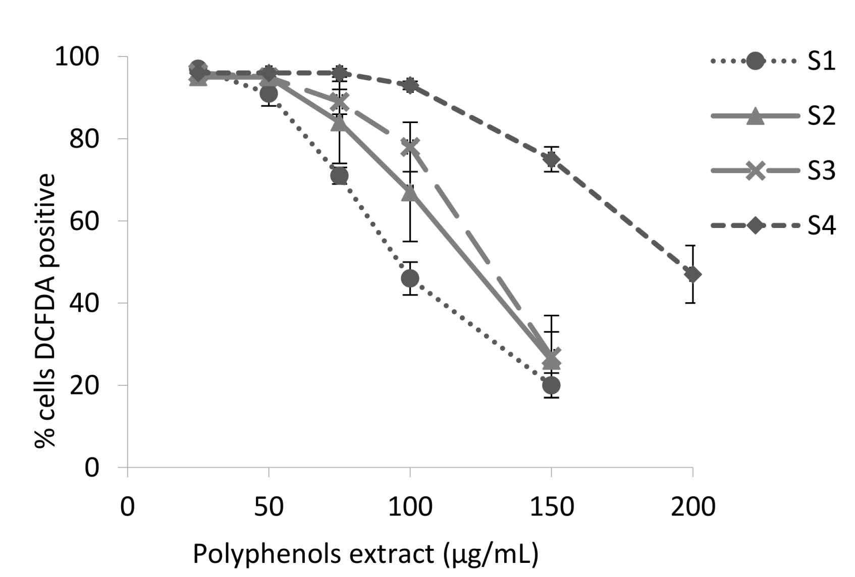
Figure 2 shows that the digested polyphenols reduce the intracellular ROS in a dose-dependent manner. The concentration of polyphenols necessary to scavenge the 50% of the ROS (IC50) was 106 ± 2 μg/mL for sample 1 (S1), 124 ± 2 μg/mL for sample 2 (S2), 132 ± 9 μg/mL for sample 3 (S3) and 196 ± 8 μg/mL for sample 4 (S4). The IC50 of the dialyzed sample (S4) showed a statistical significant decrease of 85% in the inhibitory activity compared with the non-digested sample.
The results obtained with the different methods used in this study indicate that the blackberry polyphenols subjected to the GIT conditions largely retain their antioxidant capacity. Only the fraction of polyphenols dialyzed (S4), which represents the serum available polyphenols, suffer a partial depletion of its antioxidant ability in the different techniques and this could be attributed to the absence of ellagitannins. It is necessary to perform in vivo experiments to confirm the results of our study, mainly because the simulated digestion used did not take in account the effect of microflora of the GIT that degraded polyphenols into smaller phenolic compounds that could be absorbed and contribute to the serum antioxidant capacity. This phenomenon had been demonstrated to play an important role in the case of the ellagitannins [11,46]. Nevertheless, the in vitro procedure performed in this study mimics the physiochemical changes during the digestion and provide a similar composition to that found in vivo, this provide more accurate extracts and reduce the overestimation of biological activities that occur when crude extracts are tested.

Supplementary Materials

The following are available online at https://www.mdpi.com/2306-5710/4/2/30/s1, Figure S1: Calibration curve used for the HPLC quantification of anthocyanins and ellagitanins, Figure S2. Inhibitory capacity of the digested blackberry juice polyphenols against lipid peroxidation in liposomes (A), liver homogenates (B) and erythrocytes (C). Each value is the mean ± S.E. (three independent experiments).

Acknowledgments

We thank Ana Lorena Torres for their excellent technical support. This work was supported by CONARE FS project no. 801-B1-655.

Author Contributions

Fabrice Vaillant and Alain Michel conceived and designed the experiments; Gabriela Azofeifa, Silvia Quesada, and Ana M. Pérez performed the experiments, analyzed the data, and wrote the paper; and Gabriela Azofeifa, Ana M. Pérez, Fabrice Vaillant, and Alain Michel contributed reagents/materials/analysis tools.

Conflicts of Interest

The authors declare no conflict of interest.

References

  1. Paredes-López, O.; Cervantes-Ceja, M.L.; Vigna-Pérez, M.; Hernández-Pérez, T. Berries: Improving Human Health and Healthy Aging, and Promoting Quality Life—A Review. Plant Foods Hum. Nutr. 2010, 65, 299–308. [Google Scholar] [CrossRef] [PubMed]
  2. Bowen-Forbes, C.S.; Zhang, Y.; Nair, M.G. Anthocyanin content, antioxidant, anti-inflammatory and anticancer properties of blackberry and raspberry fruits. J. Food Compos. Anal. 2010, 23, 554–560. [Google Scholar] [CrossRef]
  3. Tjelle, T.E.; Holtung, L.; Bøhn, S.K.; Aaby, K.; Thoresen, M.; Wiik, S.Å.; Paur, I.; Karlsen, A.S.; Retterstøl, K.; Iversen, P.O.; et al. Polyphenol-rich juices reduce blood pressure measures in a randomised controlled trial in high normal and hypertensive volunteers. Br. J. Nutr. 2015, 114, 1054–1063. [Google Scholar] [CrossRef] [PubMed]
  4. Basu, A.; Lyons, T.J. Strawberries, blueberries, and cranberries in the metabolic syndrome: Clinical perspectives. J. Agric. Food Chem. 2012, 60, 5687–5692. [Google Scholar] [CrossRef] [PubMed]
  5. Del Bo’, C.; Martini, D.; Porrini, M.; Klimis-Zacas, D.; Riso, P. Berries and oxidative stress markers: An overview of human intervention studies. Food Funct. 2015, 6, 2890–2917. [Google Scholar] [CrossRef] [PubMed]
  6. D’Archivio, M.; Filesi, C.; Varì, R.; Scazzocchio, B.; Masella, R. Bioavailability of the polyphenols: Status and controversies. Int. J. Mol. Sci. 2010, 11, 1321–1342. [Google Scholar] [CrossRef] [PubMed]
  7. Mertz, C.; Cheynier, V.; Günata, Z.; Brat, P. Analysis of phenolic compounds in two blackberry species (Rubus glaucus and Rubus adenotrichus) by high-performance liquid chromatography with diode array detection and electrospray ion trap mass spectrometry. J. Agric. Food Chem. 2007, 55, 8616–8624. [Google Scholar] [CrossRef] [PubMed]
  8. Kaume, L.; Howard, L.R.; Devareddy, L. The Blackberry Fruit: A Review on Its Composition and Chemistry, Metabolism and Bioavailability, and Health Benefits. J. Agric. Food Chem. 2012, 60, 5716–5727. [Google Scholar] [CrossRef] [PubMed]
  9. Felgines, C.; Talavera, S.; Texier, O.; Gil-Izquierdo, A.; Lamaison, J.L.; Remesy, C. Blackberry anthocyanins are mainly recovered from urine as methylated and glucuronidated conjugates in humans. J. Agric. Food Chem. 2005, 53, 7721–7727. [Google Scholar] [CrossRef] [PubMed]
  10. He, J.; Giusti, M.M. Anthocyanins: Natural Colorants with Health-Promoting Properties. Ann. Rev. Food Sci. Technol. 2010, 1, 163–187. [Google Scholar] [CrossRef] [PubMed]
  11. Espín, J.C.; González-Barrio, R.; Cerdá, B.; López-Bote, C.; Rey, A.I.; Tomás-Barberán, F.A. Iberian pig as a model to clarify obscure points in the bioavailability and metabolism of ellagitannins in humans. J. Agric. Food Chem. 2007, 55, 10476–10485. [Google Scholar] [CrossRef] [PubMed]
  12. García-Muñoz, C.; Hernández, L.; Pérez, A.; Vaillant, F. Diversity of urinary excretion patterns of main ellagitannins’ colonic metabolites after ingestion of tropical highland blackberry (Rubus adenotrichus) juice. Food Res. Int. 2014, 55, 161–169. [Google Scholar] [CrossRef]
  13. Cerdá, B.; Periago, P.; Espín, J.C.; Tomás-Barberán, F.A. Identification of urolithin A as a metabolite produced by human colon microflora from ellagic acid and related compounds. J. Agric. Food Chem. 2005, 53, 5571–5576. [Google Scholar] [CrossRef] [PubMed]
  14. Vaillant, F.; Pérez, A.M.; Acosta, O.; Dornier, M. Turbidity of pulpy fruit juice: A key factor for predicting cross-flow microfiltration performance. J. Membr. Sci. 2008, 325, 404–412. [Google Scholar] [CrossRef]
  15. Gil-Izquierdo, A.; Zafrilla, P.; Tomás-Barberán, F.A. An in vitro method to simulate phenolic compound release from the food matrix in the gastrointestinal tract. Eur. Food Res. Technol. 2002, 214, 155–159. [Google Scholar] [CrossRef]
  16. Coates, E.M.; Popa, G.; Gill, C.I.; McCann, M.J.; McDougall, G.J.; Stewart, D.; Rowland, I. Colon-available raspberry polyphenols exhibit anti-cancer effects on in vitro models of colon cancer. J. Carcinog. 2007, 6, 4. [Google Scholar] [CrossRef] [PubMed]
  17. Acosta-Montoya, Ó.; Vaillant, F.; Cozzano, S.; MertzbAn, C.; Pérez, M.; Castro, M.V. Phenolic content and antioxidant capacity of tropical highland blackberry (Rubus adenotrichus Schltdl.) during three edible maturity stages. Food Chem. 2010, 119, 1497–1501. [Google Scholar] [CrossRef]
  18. Azofeifa, G.; Quesada, S.; Boudard, F.; Morena, M.; Cristol, J.P.; Pérez, A.M.; Vaillant, F.; Michel, A. Antioxidant and anti-inflammatory in vitro activities of phenolic compounds from tropical highland blackberry (rubus adenotrichos). J. Agric. Food Chem. 2013, 61, 5798–5804. [Google Scholar] [CrossRef] [PubMed]
  19. Ou, B.; Hampsch-Woodill, M.; Flanagan, J.; Deemer, E.K.; Prior, R.L.; Huang, D. Novel fluorometric assay for hydroxyl radical prevention capacity using fluorescein as the probe. J. Agric. Food Chem. 2002, 50, 2772–2777. [Google Scholar] [CrossRef] [PubMed]
  20. Balakrishnan, N.; Panda, A.B.; Raj, N.R.; Shrivastava, A.; Prathani, R. The Evaluation of Nitric Oxide Scavenging Activity of Acalypha Indica Linn root. Asian J. Res. Chem. 2009, 2, 148–150. [Google Scholar]
  21. Pérez, R.; Vargas, R.; Martínez, F.; García, E.V. Antioxidant activity of alkaloids from Bocconia arborea. A study on six testing methods. ARS Pharm. 2003, 44, 5–21. [Google Scholar]
  22. Uchiyama, M.; Mihara, M. Determination of malonaldehyde precursor in tissues by thiobarbituric acid test. Anal. Biochem. 1978, 86, 271–278. [Google Scholar] [CrossRef]
  23. González, E.; Vaillant, F.; Rojas, G.; Pérez, A.M. Novel semiautomated method for assessing in vitro cellular antioxidant activity using the light-scattering properties of human erythrocytes. J. Agric. Food Chem. 2010, 58, 1455–1461. [Google Scholar] [CrossRef] [PubMed]
  24. Chen, W.; Xu, Y.; Zhang, L.; Su, H.; Zheng, X. Blackberry subjected to in vitro gastrointestinal digestion affords protection against Ethyl Carbamate-induced cytotoxicity. Food Chem. 2016, 212, 620–627. [Google Scholar] [CrossRef] [PubMed]
  25. Chen, W.; Su, H.; Xu, Y.; Bao, T.; Zheng, X. Protective effect of wild raspberry (Rubus hirsutus Thunb.) extract against acrylamide-induced oxidative damage is potentiated after simulated gastrointestinal digestion. Food Chem. 2016, 196, 943–952. [Google Scholar] [CrossRef] [PubMed]
  26. McDougall, G.J.; Dobson, P.; Smith, P.; Blake, A.; Stewart, D. Assessing potential bioavailability of raspberry anthocyanins using an in vitro digestion system. J. Agric. Food Chem. 2005, 53, 5896–5904. [Google Scholar] [CrossRef] [PubMed]
  27. Bermúdez-Soto, M.J.; Tomás-Barberán, F.A.; García-Conesa, M.T. Stability of polyphenols in chokeberry (Aronia melanocarpa) subjected to in vitro gastric and pancreatic digestion. Food Chem. 2007, 102, 865–874. [Google Scholar] [CrossRef]
  28. Pérez-Vicente, A.; Gil-Izquierdo, A.; García-Viguera, C. In vitro Gastrointestinal Digestion Study of Pomegranate Juice Phenolic Compounds, Anthocyanins, and Vitamin C. J. Agric. Food Chem. 2002, 50, 2308–2312. [Google Scholar] [CrossRef] [PubMed]
  29. McGhie, T.K.; Ainge, G.D.; Barnett, L.E.; Cooney, J.M.; Jensen, D.J. Anthocyanin glycosides from berry fruit are absorbed and excreted unmetabolized by both humans and rats. J. Agric. Food Chem. 2003, 51, 4539–4548. [Google Scholar] [CrossRef] [PubMed]
  30. Stanisavljevic, N.; Samardzic, J.; Jankovic, T.; Šavikin, K.; Mojsin, M.; Topalović, V.; Stevanović, M. Antioxidant and antiproliferative activity of chokeberry juice phenolics during in vitro simulated digestion in the presence of food matrix. Food Chem. 2015, 175, 516–522. [Google Scholar] [CrossRef] [PubMed]
  31. Felgines, C.; Texier, O.; Garcin, P.; Besson, C.; Lamaison, J.L.; Scalbert, A. Tissue distribution of anthocianins in rats fed a blackberry anthocyanin-enriched diet. Mol. Nutr. Food Res. 2009, 53, 1098–1103. [Google Scholar] [CrossRef] [PubMed]
  32. Del Rio, D.; Borges, G.; Crozier, A. Berry flavonoids and phenolics: Bioavailability and evidence of protective effects. Br. J. Nutr. 2010, 104, S67–S90. [Google Scholar] [CrossRef] [PubMed]
  33. Talavéra, S.; Felgines, C.; Texier, O.; Besson, C.; Lamaison, J.L.; Rémésy, C. Anthocyanins are efficiently absorbed from the stomach in anesthetized rats. J. Nutr. 2003, 133, 4178–4182. [Google Scholar] [CrossRef] [PubMed]
  34. Talavéra, S.; Felgines, C.; Texier, O.; Besson, C.; Manach, C.; Lamaison, J.L.; Rémésy, C. Anthocyanins are efficiently absorbed from the small intestine in rats. J. Nutr. 2004, 134, 2275–2279. [Google Scholar] [CrossRef] [PubMed]
  35. Aura, A.-M.; Martin-Lopez, P.; O’Leary, K.A.; Williamson, G.; Oksman-Caldentey, K.M.; Poutanen, K.; Santos-Buelga, C. In vitro metabolism of anthocyanins by human gut microflora. Eur. J. Nutr. 2005, 44, 133–142. [Google Scholar] [CrossRef] [PubMed]
  36. Larrosa, M.; Tomás-Barberán, F.A.; Espín, J.C. The dietary hydrolysable tannin punicalagin releases ellagic acid that induces apoptosis in human colon adenocarcinoma Caco-2 cells by using the mitochondrial pathway. J. Nutr. Biochem. 2006, 17, 611–625. [Google Scholar] [CrossRef] [PubMed]
  37. Huang, D.; Boxin, O.U.; Prior, R.L. The chemistry behind antioxidant capacity assays. J. Agric. Food Chem. 2005, 53, 1841–1856. [Google Scholar] [CrossRef] [PubMed]
  38. Niki, E. Antioxidant capacity: Which capacity and how to assess it? J. Berry Res. 2011, 1, 169–176. [Google Scholar] [CrossRef]
  39. Niki, E. Assessment of antioxidant capacity in vitro and in vivo. Free Radic. Biol. Med. 2010, 49, 503–515. [Google Scholar] [CrossRef] [PubMed]
  40. Basu, A.; Rhone, M.; Lyons, T.J. Berries: Emerging impact on cardiovascular health. Nutr. Rev. 2010, 68, 168–177. [Google Scholar] [CrossRef] [PubMed]
  41. Niki, E. Do antioxidants impair signaling by reactive oxygen species and lipid oxidation products? FEBS Lett. 2012, 586, 3767–3770. [Google Scholar] [CrossRef] [PubMed]
  42. Serram, N.P.; Nair, M. Inhibition of Lipid Peroxidation and Structura-Activity-Related Studied of the dietary constituents Anthoyanins, Anthocyanidins, and Actechins. J. Agric. Food Chem. 2002, 50, 5308–5312. [Google Scholar] [CrossRef]
  43. Reddy, M.K.; Gupta, S.K.; Jacob, M.R.; Khan, S.I.; Ferreira, D. Antioxidant, antimalarial and antimicrobial activities of tannin-rich fractions, ellagitannins and phenolic acids from Punica granatum L. Planta Med. 2007, 73, 461–467. [Google Scholar] [CrossRef] [PubMed]
  44. Dobroslawa, B.; Kasimsetty, S.G.; Khan, S.I.; Daneel, F. Urolithins, intestinal microbial metabolites of pomegranate ellagitannins, exhibit potent antioxidant activity in a cell-based assay. J. Agric. Food Chem. 2009, 57, 10181–10186. [Google Scholar] [CrossRef]
  45. Reuter, S.; Gupta, S.; Chaturvedi, M.; Aggarwal, B. Oxidative stress, inflammation, and cancer: How are they linked? Free Radic. Biol. Med. 2011, 49, 1603–1616. [Google Scholar] [CrossRef] [PubMed]
  46. Garcia-Muñoz, C.; Vaillant, F. Metabolic fate of ellagitannins: Implications for health, and research perspectives for innovative functional foods. Crit. Rev. Food Sci. Nutr. 2014, 54, 1584–1598. [Google Scholar] [CrossRef] [PubMed]
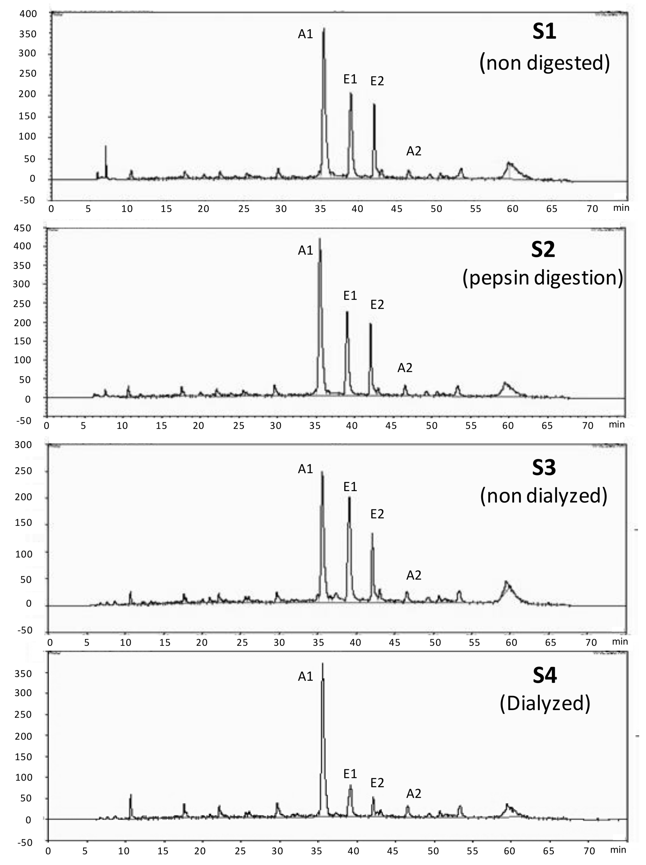
Figure 1. HPLC Chromatogram of the digested blackberry juice polyphenols. A1: cyanidin-3-glucoside, A2: cyanidin-3-malonyl glucoside, E1: lambertianin C, E2: sanguiin H-6. (Corresponding tentative identification of peaks according to Mertz et al. [7]).
Beverages 04 00030 g001
Figure 2. Inhibitory capacity of the digested blackberry juice polyphenols against intracellular ROS. Each value is the mean ± S.E. (three independent experiments).
Beverages 04 00030 g002
Table 1. Anthocyanins and ellagitannins concentrations in the blackberry juice digestion samples.
Blackberry Digestion SamplesHPLC-Anthocyanins
mg C3G/L
HPLC-Ellagitannins
mg EA/L
S1 (Non-digested)480 ± 37 a353 ± 13 a
S2 (Pepsin digestion)580 ± 15 a394 ± 24 a
S3 (Non-dialyzed)220 ± 19 b233 ± 6 b
S4 (Dialyzed)91 ± 4 c24 ± 3 c
Each value is the mean ± SE of three replicate experiments. Means in columns followed by different letters differed significantly (p < 0.05). C3G: cyanidin-3-glucoside, EA: ellagic acid.
Table 2. Free radical-scavenging of the digested blackberry juice polyphenols.
Blackberry Digestion SamplesDPPH
IC50 (μg/mL)
ORAC
mmol TE/g of Extract
NO Scavenging
IC50 (μg/mL)
S1 (Non-digested)5.10 ± 0.20 a5.94 ± 0.41 a197 ± 19 a
S2 (Pepsin digestion)4.75 ± 0.03 a6.30 ± 0.42 a183 ± 14 a
S3 (Non-dialyzed)5.30 ± 0.15 a5.86 ± 0.27 a217 ± 13 ab
S4 (Dialyzed)6.79 ± 0.06 b6.56 ± 0.18 a267 ± 21 b
Each value is the mean ± SE of three replicate experiments. Means in columns followed by different letters differed significantly (p < 0.05).
Table 3. Lipid peroxidation inhibitory capacity of the digested blackberry juice polyphenols.
Blackberry Digestion SamplesLiposomesRat Liver HomogenateERYCA
IC50 (µg/mL)IC50 (µg/mL)mmol QE/g of Extract
S1 (Non-digested)5.03 ± 0.49 a32.7 ± 4.8 a3.87 ± 0.19 a
S2 (Pepsin digestion)4.27 ± 0.27 a29.2 ± 5.0 a3.88 ± 0.41 a
S3 (Non-dialyzed)5.36 ± 0.62 a37.0 ± 7.1 a3.80 ± 0.25 a
S4 (Dialyzed)5.84 ± 0.41 a39.1 ± 6.3 a3.54 ± 0.25 a
Each value is the mean ± SE of three replicate experiments. Means in columns followed by different letters differed significantly (p < 0.05). (See dose-response curves, in Supplementary Materials, Figure S2).