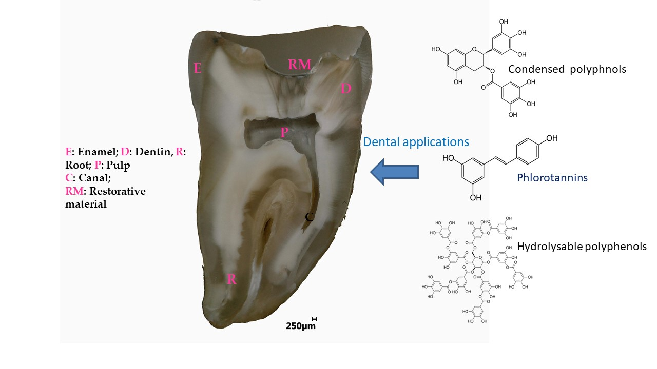
Polyphenols in Dental Applications


Abstract

1. Introduction
2. Condensed Polyphenols
2.1. Dentin Modifier, Dentin Pretreatment, Collagen Cross-Linking, and Resin-Dentin Stability
2.2. Remineralization, Cell Viability, and Differentiation
2.3. Antibacterial Activity
2.4. Anti-Inflammatory and Antioxidant Activity
3. Hydrolysable Tannins and Gallic Acid
3.1. Dentin Modifier, Dentin Pretreatment, Collagen Cross-Linking, and Resin-Dentin Stability
3.2. Remineralization, Cell Viability, and Differentiation
3.3. Antibacterial Activity
3.4. Anti-Inflammatory and Antioxidant Activity
4. Polyphenol Mixtures
4.1. Dentin Modifier, Dentin Pretreatment, Collagen Cross-Linking, and Resin-Dentin Stability
4.2. Remineralization, Cell Viability, and Differentiation
4.3. Antibacterial Activity
4.4. Anti-Inflammatory and Antioxidant Activity
5. Summary of the Major Findings Reported in This Review
6. Future Perspectives and Concluding Remarks
Author Contributions
Funding
Conflicts of Interest
References
- Pérez-Jiménez, J.; Neveu, V.; Vos, F.; Scalbert, A. Identification of the 100 richest dietary sources of polyphenols: An application of the Phenol-Explorer database. Eur. J. Clin. Nutr. 2010, 64 (Suppl. 3), S112–S120. [Google Scholar]
- Handique, J.G.; Baruah, J.B. Polyphenolic compounds: An overview. React. Funct. Polym. 2002, 52, 163–188. [Google Scholar] [CrossRef]
- Reitzer, F.; Allais, M.; Ball, V.; Meyer, F. Polyphenols at interfaces. Adv. Colloid Interface Sci. 2018, 257, 31–41. [Google Scholar] [CrossRef]
- Petti, S.; Scully, C. Polyphenols, oral health and disease: A review. J. Dent. 2009, 37, 413–423. [Google Scholar] [CrossRef] [PubMed]
- Sánchez, M.C.; Ribeiro-Vidal, H.; Esteban-Fernández, A.; Bartolomé, B.; Figuero, E.; Moreno-Arribas, M.V.; Sanz, M.; Herrera, D. Antimicrobial activity of red wine and oenological extracts against periodontal pathogens in a validated oral biofilm model. BMC Complement. Altern. Med. 2019, 19, 145. [Google Scholar] [CrossRef] [PubMed]
- Fibach, E.; Ginsburg, I. The Antioxidant Effect of Fermented Papaya Preparation in the Oral Cavity. Phytother. Res. 2015, 29, 1317–1322. [Google Scholar] [CrossRef] [PubMed]
- Shavandi, A.; Bekhit, A.E.-D.A.; Saeedi, P.; Izadifar, Z.; Bekhit, A.A.; Khademhosseini, A. Polyphenol uses in biomaterials engineering. Biomaterials 2018, 167, 91–106. [Google Scholar] [CrossRef]
- Catapano-Martinez, D.; Boyce, M.; Garland, M. The Protective Role of Polyphenols in Oral Health. Decis. Dent. 2018, 33–36. [Google Scholar]
- Sileika, T.S.; Barrett, D.G.; Zhang, R.; Lau, K.H.A.; Messersmith, P.B. Colorless multifunctional coatings inspired by polyphenols found in tea, chocolate, and wine. Angew. Chem. Int. Ed. Engl. 2013, 52, 10766–10770. [Google Scholar] [CrossRef]
- Quideau, S.; Deffieux, D.; Douat-Casassus, C.; Pouységu, L. Plant polyphenols: Chemical properties, biological activities, and synthesis. Angew. Chem. Int. Ed. Engl. 2011, 50, 586–621. [Google Scholar] [CrossRef]
- Le Bourvellec, C.; Renard, C.M.G.C. Interactions between polyphenols and macromolecules: Quantification methods and mechanisms. Crit. Rev. Food Sci. Nutr. 2012, 52, 213–248. [Google Scholar] [CrossRef]
- Marshall, G.W.; Marshall, S.J.; Kinney, J.H.; Balooch, M. The dentin substrate: Structure and properties related to bonding. J. Dent. 1997, 25, 441–458. [Google Scholar] [CrossRef]
- Kharouf, N.; Rapp, G.; Mancino, D.; Hemmerlé, J.; Haikel, Y.; Reitzer, F. Effect of etching the coronal dentin with the rubbing technique on the microtensile bond strength of a universal adhesive system. Dent. Med. Probl. 2019, 56, 343–348. [Google Scholar] [CrossRef]
- Osorio, R.; Yamauti, M.; Osorio, E.; Ruiz-Requena, M.E.; Pashley, D.; Tay, F.; Toledano, M. Effect of dentin etching and chlorhexidine application on metalloproteinase-mediated collagen degradation. Eur. J. Oral Sci. 2011, 119, 79–85. [Google Scholar] [CrossRef] [PubMed]
- Toledano, M.; Osorio, R.; Osorio, E.; Aguilera, F.S.; Yamauti, M.; Pashley, D.H.; Tay, F. Effect of bacterial collagenase on resin-dentin bonds degradation. J. Mater. Sci. Mater. Med. 2007, 18, 2355–2361. [Google Scholar] [CrossRef] [PubMed]
- Mazzoni, A.; Nascimento, F.D.; Carrilho, M.; Tersariol, I.; Papa, V.; Tjäderhane, L.; Di Lenarda, R.; Tay, F.R.; Pashley, D.H.; Breschi, L. MMP activity in the hybrid layer detected with in situ zymography. J. Dent. Res. 2012, 91, 467–472. [Google Scholar] [CrossRef]
- Aguiar, T.R.; Vidal, C.M.P.; Phansalkar, R.S.; Todorova, I.; Napolitano, J.G.; McAlpine, J.B.; Chen, S.N.; Pauli, G.F.; Bedran-Russo, A.K. Dentin biomodification potential depends on polyphenol source. J. Dent. Res. 2014, 93, 417–422. [Google Scholar] [CrossRef]
- Vidal, C.M.P.; Leme, A.A.; Aguiar, T.R.; Phansalkar, R.; Nam, J.-W.; Bisson, J.; McAlpine, J.B.; Chen, S.-N.; Pauli, G.F.; Bedran-Russo, A. Mimicking the hierarchical functions of dentin collagen cross-links with plant derived phenols and phenolic acids. Langmuir 2014, 30, 14887–14893. [Google Scholar] [CrossRef]
- Leme-Kraus, A.A.; Aydin, B.; Vidal, C.M.P.; Phansalkar, R.M.; Nam, J.W.; McAlpine, J.; Pauli, G.F.; Chen, S.; Bedran-Russo, A.K. Biostability of the Proanthocyanidins-Dentin Complex and Adhesion Studies. J. Dent. Res. 2017, 96, 406–412. [Google Scholar] [CrossRef]
- Tang, C.; Fang, M.; Liu, R.; Dou, Q.; Chai, Z.; Xiao, Y.; Chen, J. The role of grape seed extract in the remineralization of demineralized dentine: Micromorphological and physical analyses. Arch. Oral Biol. 2013, 58, 1769–1776. [Google Scholar] [CrossRef]
- Zheng, K.; Wu, S.; Chen, B.; Liao, W.; Li, Y. [Effect of baicalein and quercetin on enzymatic resistance of dentin collagen]. Zhonghua Kou Qiang Yi Xue Za Zhi 2014, 49, 667–671. [Google Scholar] [PubMed]
- Bedran-Russo, A.K.B.; Yoo, K.J.; Ema, K.C.; Pashley, D.H. Mechanical properties of tannic-acid-treated dentin matrix. J. Dent. Res. 2009, 88, 807–811. [Google Scholar] [CrossRef] [PubMed]
- Liu, Y.; Bai, X.; Li, S.; Liu, Y.; Keightley, A.; Wang, Y. Molecular weight and galloylation affect grape seed extract constituents’ ability to cross-link dentin collagen in clinically relevant time. Dent. Mater. 2015, 31, 814–821. [Google Scholar] [CrossRef] [PubMed][Green Version]
- Nam, J.-W.; Phansalkar, R.S.; Lankin, D.C.; Bisson, J.; McAlpine, J.B.; Leme, A.A.; Vidal, C.M.P.; Ramirez, B.; Niemitz, M.; Bedran-Russo, A.; et al. Subtle Chemical Shifts Explain the NMR Fingerprints of Oligomeric Proanthocyanidins with High Dentin Biomodification Potency. J. Org. Chem. 2015, 80, 7495–7507. [Google Scholar] [CrossRef]
- Phansalkar, R.S.; Nam, J.-W.; Chen, S.-N.; McAlpine, J.B.; Napolitano, J.G.; Leme, A.; Vidal, C.M.P.; Aguiar, T.; Bedran-Russo, A.K.; Pauli, G.F. A galloylated dimeric proanthocyanidin from grape seed exhibits dentin biomodification potential. Fitoterapia 2015, 101, 169–178. [Google Scholar] [CrossRef] [PubMed]
- Lucas, P.W.; Wagner, M.; Al-Fadhalah, K.; Almusallam, A.S.; Michael, S.; Thai, L.A.; Strait, D.S.; Swain, M.V.; van Casteren, A.; Renno, W.M.; et al. Dental abrasion as a cutting process. Interface Focus 2016, 6, 20160008. [Google Scholar] [CrossRef]
- Zhang, L.; Huang, L.; Xiong, Y.; Fang, M.; Chen, J.-H.; Ferrari, M. Effect of post-space treatment on retention of fiber posts in different root regions using two self-etching systems. Eur. J. Oral Sci. 2008, 116, 280–286. [Google Scholar] [CrossRef]
- Kharouf, N.; Mancino, D.; Naji-Amrani, A.; Eid, A.; Haikel, Y.; Hemmerle, J. Effectiveness of Etching by Three Acids on the Morphological and Chemical Features of Dentin Tissue. J. Contemp. Dent. Pract. 2019, 20, 915–919. [Google Scholar] [CrossRef]
- Gu, X.-H.; Mao, C.-Y.; Liang, C.; Wang, H.-M.; Kern, M. Does endodontic post space irrigation affect smear layer removal and bonding effectiveness? Eur. J. Oral Sci. 2009, 117, 597–603. [Google Scholar] [CrossRef]
- Santiago, S.L.; Osorio, R.; Neri, J.R.; Carvalho, R.M.; Toledano, M. Effect of the flavonoid epigallocatechin-3-gallate on resin-dentin bond strength. J. Adhes. Dent. 2013, 15, 535–540. [Google Scholar]
- Singh, P.; Nagpal, R.; Singh, U.P. Effect of dentin biomodifiers on the immediate and long-term bond strengths of a simplified etch and rinse adhesive to dentin. Restor. Dent. Endod. 2017, 42, 188–199. [Google Scholar] [CrossRef] [PubMed]
- Fialho, M.P.N.; Hass, V.; Nogueira, R.P.; França, F.M.G.; Turssi, C.P.; Basting, R.T.; Amaral, F.L.B. Effect of epigallocatechin-3- gallate solutions on bond durability at the adhesive interface in caries-affected dentin. J. Mech. Behav. Biomed. Mater. 2019, 91, 398–405. [Google Scholar] [CrossRef]
- De Costa, C.A.G.; Passos, V.F.; Neri, J.R.; Mendonça, J.S.; Santiago, S.L. Effect of Metalloproteinase Inhibitors on Bond Strength of a Self-etching Adhesive on Erosively Demineralized Dentin. J. Adhes. Dent. 2019, 21, 337–344. [Google Scholar]
- Kalaiselvam, R.; Ganesh, A.; Rajan, M.; Kandaswamy, D. Evaluation of bioflavonoids on the immediate and delayed microtensile bond strength of self-etch and total-etch adhesive systems to sound dentin. Indian J. Dent. Res. 2018, 29, 133–136. [Google Scholar] [CrossRef] [PubMed]
- Yu, H.-H.; Zhang, L.; Xu, S.; Li, F.; Yu, F.; Liu, Z.-Y.; Huang, L.; Chen, J.-H. Effects of Epigallocatechin-3-gallate (EGCG) on the bond strength of fiber posts to Sodium hypochlorite (NaOCl) treated intraradicular dentin. Sci. Rep. 2017, 7, 4235. [Google Scholar] [CrossRef] [PubMed]
- Pheenithicharoenkul, S.; Panichuttra, A. Epigallocatechin-3-gallate increased the push out bond strength of an epoxy resin sealer to root dentin. Dent. Mater. J. 2016, 35, 888–892. [Google Scholar] [CrossRef]
- Frassetto, A.; Breschi, L.; Turco, G.; Marchesi, G.; Di Lenarda, R.; Tay, F.R.; Pashley, D.H.; Cadenaro, M. Mechanisms of degradation of the hybrid layer in adhesive dentistry and therapeutic agents to improve bond durability—A literature review. Dent. Mater. 2016, 32, e41–e53. [Google Scholar] [CrossRef] [PubMed]
- Dutra-Correa, M.; Leite, A.A.B.V.; de Cara, S.P.H.M.; Diniz, I.M.A.; Marques, M.M.; Suffredini, I.B.; Fernandes, M.S.; Toma, S.H.; Araki, K.; Medeiros, I.S. Antibacterial effects and cytotoxicity of an adhesive containing low concentration of silver nanoparticles. J. Dent. 2018, 77, 66–71. [Google Scholar] [CrossRef]
- Mishra, P.; Jaiswal, S.; Nikhil, V.; Gupta, S.; Jha, P.; Raj, S. Evaluation of marginal sealing ability of self-adhesive flowable composite resin in Class II composite restoration: An in vitro study. J. Conserv. Dent. 2018, 21, 363–368. [Google Scholar] [CrossRef]
- Hong, J.-Y.; Yon, J.; Lee, J.-S.; Lee, I.-K.; Yang, C.; Kim, M.-S.; Choi, S.-H.; Jung, U.-W. Effects of epigallocatechin-3-gallate on the healing of extraction sockets with a periapical lesion: A pilot study in dogs. J. Biomed. Mater. Res. Part B Appl. Biomater. 2015, 103, 727–734. [Google Scholar] [CrossRef]
- De Assis, J.S.; Lima, R.A.; Marques Lima, J.P.; Azevedo Rodrigues, L.K.; Santiago, S.L. Effect of epigallocatechin-3-gallate application for remaining carious dentin disinfection. J. Conserv. Dent. 2015, 18, 51–55. [Google Scholar] [CrossRef] [PubMed]
- Neri, J.R.; Yamauti, M.; Feitosa, V.P.; Pires, A.P.M.; Araújo, R.D.S.; Santiago, S.L. Physicochemical properties of a methacrylate-based dental adhesive incorporated with epigallocatechin-3-gallate. Braz. Dent. J. 2014, 25, 528–531. [Google Scholar] [CrossRef] [PubMed]
- Fonseca, B.M.; Barcellos, D.C.; da Silva, T.M.; Borges, A.L.S.; das Neves Cavalcanti, B.; Prakki, A.; de Oliveira, H.P.M.; de Paiva Gonçalves, S.E. Mechanical-physicochemical properties and biocompatibility of catechin-incorporated adhesive resins. J. Appl. Oral Sci. 2019, 27, e20180111. [Google Scholar] [CrossRef]
- De Macedo, F.A.A.; Souza, N.O.; Lemos, M.V.S.; De-Paula, D.M.; Santiago, S.L.; Feitosa, V.P. Dentin bonding and physicochemical properties of adhesives incorporated with epigallocatechin-3-gallate. Odontology 2019, 107, 23–28. [Google Scholar] [CrossRef]
- Albuquerque, N.; Neri, J.R.; Lemos, M.; Yamauti, M.; de Sousa, F.; Santiago, S.L. Effect of Polymeric Microparticles Loaded with Catechin on the Physicochemical Properties of an Adhesive System. Oper. Dent. 2019, 44, E202–E211. [Google Scholar] [CrossRef] [PubMed]
- Yu, H.H.; Zhang, L.; Yu, F.; Zhou, H.; Shen, L.J.; Chen, J.H. [Effects of epigallocatechin-3-gallate modification on the bonding stability of an etch-and-rinse adhesive to intraradicular dentin]. Zhonghua Kou Qiang Yi Xue Za Zhi 2017, 52, 114–119. [Google Scholar]
- Hanks, C.T.; Strawn, S.E.; Wataha, J.C.; Craig, R.G. Cytotoxic effects of resin components on cultured mammalian fibroblasts. J. Dent. Res. 1991, 70, 1450–1455. [Google Scholar] [CrossRef]
- Hu, J.; Du, X.; Huang, C.; Fu, D.; Ouyang, X.; Wang, Y. Antibacterial and physical properties of EGCG-containing glass ionomer cements. J. Dent. 2013, 41, 927–934. [Google Scholar] [CrossRef]
- Yang, H.; Li, K.; Yan, H.; Liu, S.; Wang, Y.; Huang, C. High-performance therapeutic quercetin-doped adhesive for adhesive-dentin interfaces. Sci. Rep. 2017, 7, 8189. [Google Scholar] [CrossRef]
- Gotti, V.B.; Feitosa, V.P.; Sauro, S.; Correr-Sobrinho, L.; Leal, F.B.; Stansbury, J.W.; Correr, A.B. Effect of antioxidants on the dentin interface bond stability of adhesives exposed to hydrolytic degradation. J. Adhes. Dent. 2015, 17, 35–44. [Google Scholar]
- Wang, F.; Han, Y.; Xi, S.; Lu, Y. Catechins reduce inflammation in lipopolysaccharide-stimulated dental pulp cells by inhibiting activation of the NF-κB pathway. Oral Dis. 2020, 26, 815–821. [Google Scholar] [CrossRef]
- Lim, E.; Lim, M.-J.; Min, K.-S.; Kwon, Y.-S.; Hwang, Y.-C.; Yu, M.-K.; Hong, C.-U.; Lee, K.-W. Effects of epicatechin, a crosslinking agent, on human dental pulp cells cultured in collagen scaffolds. J. Appl. Oral Sci. 2016, 24, 76–84. [Google Scholar] [CrossRef] [PubMed]
- Kim, J.-G.; Son, K.M.; Park, H.C.; Zhu, T.; Kwon, J.H.; Yang, H.-C. Stimulating effects of quercetin and phenamil on differentiation of human dental pulp cells. Eur. J. Oral Sci. 2013, 121, 559–565. [Google Scholar] [CrossRef] [PubMed]
- Liu, X.-C.; Wang, X.-X.; Zhang, L.-N.; Yang, F.; Nie, F.-J.; Zhang, J. Inhibitory effects of resveratrol on orthodontic tooth movement and associated root resorption in rats. Arch. Oral Biol. 2020, 111, 104642. [Google Scholar] [CrossRef] [PubMed]
- De Rezende Barbosa, G.L.; Pimenta, L.A.; de Almeida, S.M. Micro-CT evaluation of the radioprotective effect of resveratrol on the mandibular incisors of irradiated rats. Braz. Oral Res. 2016, 30. [Google Scholar] [CrossRef] [PubMed]
- Xu, X.; Zhou, X.D.; Wu, C.D. The tea catechin epigallocatechin gallate suppresses cariogenic virulence factors of Streptococcus mutans. Antimicrob. Agents Chemother. 2011, 55, 1229–1236. [Google Scholar] [CrossRef]
- Hara, K.; Ohara, M.; Hayashi, I.; Hino, T.; Nishimura, R.; Iwasaki, Y.; Ogawa, T.; Ohyama, Y.; Sugiyama, M.; Amano, H. The green tea polyphenol (-)-epigallocatechin gallate precipitates salivary proteins including alpha-amylase: Biochemical implications for oral health. Eur. J. Oral Sci. 2012, 120, 132–139. [Google Scholar] [CrossRef] [PubMed]
- Feng, L.; Yan, Q.; Zhang, B.; Tian, X.; Wang, C.; Yu, Z.; Cui, J.; Guo, D.; Ma, X.; James, T.D. Ratiometric fluorescent probe for sensing Streptococcus mutans glucosyltransferase, a key factor in the formation of dental caries. Chem. Commun. 2019, 55, 3548–3551. [Google Scholar] [CrossRef] [PubMed]
- Melok, A.L.; Lee, L.H.; Mohamed Yussof, S.A.; Chu, T. Green Tea Polyphenol Epigallocatechin-3-Gallate-Stearate Inhibits the Growth of Streptococcus mutans: A Promising New Approach in Caries Prevention. Dent. J. 2018, 6, 38. [Google Scholar] [CrossRef] [PubMed]
- Lee, P.; Tan, K.S. Effects of Epigallocatechin gallate against Enterococcus faecalis biofilm and virulence. Arch. Oral Biol. 2015, 60, 393–399. [Google Scholar] [CrossRef] [PubMed]
- Mankovskaia, A.; Lévesque, C.M.; Prakki, A. Catechin-incorporated dental copolymers inhibit growth of Streptococcus mutans. J. Appl. Oral Sci. 2013, 21, 203–207. [Google Scholar] [CrossRef]
- Kwon, Y.-S.; Kim, H.-J.; Hwang, Y.-C.; Rosa, V.; Yu, M.-K.; Min, K.-S. Effects of Epigallocatechin Gallate, an Antibacterial Cross-linking Agent, on Proliferation and Differentiation of Human Dental Pulp Cells Cultured in Collagen Scaffolds. J. Endod. 2017, 43, 289–296. [Google Scholar] [CrossRef] [PubMed]
- Du, X.; Huang, X.; Huang, C.; Wang, Y.; Zhang, Y. Epigallocatechin-3-gallate (EGCG) enhances the therapeutic activity of a dental adhesive. J. Dent. 2012, 40, 485–492. [Google Scholar] [CrossRef] [PubMed]
- Yang, W.-H.; Deng, Y.-T.; Kuo, M.Y.-P.; Liu, C.-M.; Chang, H.-H.; Chang, J.Z.-C. Epigallocatechin-3-gallate blocks triethylene glycol dimethacrylate-induced cyclooxygenase-2 expression by suppressing extracellular signal-regulated kinase in human dental pulp and embryonic palatal mesenchymal cells. J. Endod. 2013, 39, 1407–1412. [Google Scholar] [CrossRef] [PubMed]
- Hirao, K.; Yumoto, H.; Nakanishi, T.; Mukai, K.; Takahashi, K.; Takegawa, D.; Matsuo, T. Tea catechins reduce inflammatory reactions via mitogen-activated protein kinase pathways in toll-like receptor 2 ligand-stimulated dental pulp cells. Life Sci. 2010, 86, 654–660. [Google Scholar] [CrossRef]
- Nakanishi, T.; Mukai, K.; Yumoto, H.; Hirao, K.; Hosokawa, Y.; Matsuo, T. Anti-inflammatory effect of catechin on cultured human dental pulp cells affected by bacteria-derived factors. Eur. J. Oral Sci. 2010, 118, 145–150. [Google Scholar] [CrossRef]
- Nakanishi, T.; Mukai, K.; Hosokawa, Y.; Takegawa, D.; Matsuo, T. Catechins inhibit vascular endothelial growth factor production and cyclooxygenase-2 expression in human dental pulp cells. Int. Endod. J. 2015, 48, 277–282. [Google Scholar] [CrossRef]
- Lee, D.-S.; Li, B.; Kim, K.-S.; Jeong, G.-S.; Kim, E.-C.; Kim, Y.-C. Butein protects human dental pulp cells from hydrogen peroxide-induced oxidative toxicity via Nrf2 pathway-dependent heme oxygenase-1 expressions. Toxicol In Vitro 2013, 27, 874–881. [Google Scholar] [CrossRef]
- Park, S.Y.; Jeong, Y.J.; Kim, S.H.; Jung, J.Y.; Kim, W.J. Epigallocatechin gallate protects against nitric oxide-induced apoptosis via scavenging ROS and modulating the Bcl-2 family in human dental pulp cells. J. Toxicol Sci. 2013, 38, 371–378. [Google Scholar] [CrossRef]
- Mahmoud Hashemi, A.; Solahaye Kahnamouii, S.; Aghajani, H.; Frozannia, K.; Pournasrollah, A.; Sadegh, R.; Esmaeeli, H.; Ghadimi, Y.; Razmpa, E. Quercetin Decreases Th17 Production by Down-Regulation of MAPK- TLR4 Signaling Pathway on T Cells in Dental Pulpitis. J. Dent. 2018, 19, 259–264. [Google Scholar]
- Yonehiro, J.; Yoshida, Y.; Yamashita, A.; Yoshizawa, S.; Ohta, K.; Kamata, N.; Okihara, T.; Nishimura, F. Flavonol-containing phosphorylated pullulan may attenuate pulp inflammation. Int. Endod. J. 2013, 46, 119–127. [Google Scholar] [CrossRef] [PubMed]
- Wang, F.-M.; Hu, Z.; Liu, X.; Feng, J.Q.; Augsburger, R.A.; Gutmann, J.L.; Glickman, G.N. Resveratrol represses tumor necrosis factor α/c-Jun N-terminal kinase signaling via autophagy in human dental pulp stem cells. Arch. Oral Biol. 2019, 97, 116–121. [Google Scholar] [CrossRef] [PubMed]
- Geng, Y.-W.; Zhang, Z.; Liu, M.-Y.; Hu, W.-P. Differentiation of human dental pulp stem cells into neuronal by resveratrol. Cell Biol. Int. 2017, 41, 1391–1398. [Google Scholar] [CrossRef] [PubMed]
- Lee, S.-I.; Min, K.-S.; Bae, W.-J.; Lee, Y.-M.; Lee, S.-Y.; Lee, E.-S.; Kim, E.-C. Role of SIRT1 in heat stress- and lipopolysaccharide-induced immune and defense gene expression in human dental pulp cells. J. Endod. 2011, 37, 1525–1530. [Google Scholar] [CrossRef]
- Feng, G.; Zheng, K.; Song, D.; Xu, K.; Huang, D.; Zhang, Y.; Cao, P.; Shen, S.; Zhang, J.; Feng, X.; et al. SIRT1 was involved in TNF-α-promoted osteogenic differentiation of human DPSCs through Wnt/β-catenin signal. In Vitro Cell. Dev. Biol. Anim. 2016, 52, 1001–1011. [Google Scholar] [CrossRef]
- Atalayin, C.; Armagan, G.; Konyalioglu, S.; Kemaloglu, H.; Tezel, H.; Ergucu, Z.; Keser, A.; Dagci, T.; Onal, B. The protective effect of resveratrol against dentin bonding agents-induced cytotoxicity. Dent. Mater. J. 2015, 34, 766–773. [Google Scholar] [CrossRef]
- Atalayin, C.; Tezel, H.; Ergucu, Z.; Unlu, N.; Armagan, G.; Dagci, T.; Kose, T. The improvement of biocompatibility of adhesives: The effects of resveratrol on biocompatibility and dentin micro-tensile bond strengths of self-etch adhesives. Clin. Oral Investig. 2019, 23, 3213–3218. [Google Scholar] [CrossRef]
- Moreira, M.A.; Souza, N.O.; Sousa, R.S.; Freitas, D.Q.; Lemos, M.V.; De Paula, D.M.; Maia, F.J.N.; Lomonaco, D.; Mazzetto, S.E.; Feitosa, V.P. Efficacy of new natural biomodification agents from Anacardiaceae extracts on dentin collagen cross-linking. Dent. Mater. 2017, 33, 1103–1109. [Google Scholar] [CrossRef]
- Mukai, Y.; Tomiyama, K.; Okada, S.; Mukai, K.; Negishi, H.; Fujihara, T.; Teranaka, T. Dentinal tubule occlusion with lanthanum fluoride and powdered apatite glass ceramics in vitro. Dent. Mater. J. 1998, 17, 253–263. [Google Scholar] [CrossRef]
- Mancino, D.; Kharouf, N.; Hemmerlé, J.; Haïkel, Y. Microscopic and Chemical Assessments of the Filling Ability in Oval-Shaped Root Canals Using Two Different Carrier-Based Filling Techniques. Eur. J. Dent. 2019, 13, 166–171. [Google Scholar] [CrossRef]
- Haapasalo, M.; Shen, Y.; Wang, Z.; Gao, Y. Irrigation in endodontics. Br. Dent. J. 2014, 216, 299–303. [Google Scholar] [CrossRef] [PubMed]
- Bitter, N.C. A 25% tannic acid solution as a root canal irrigant cleanser: A scanning electron microscope study. Oral Surg. Oral Med. Oral Pathol. 1989, 67, 333–337. [Google Scholar] [CrossRef]
- Bitter, N.C. Tannic acid for smear layer removal: Pilot study with scanning electron microscope. J. Prosthet. Dent. 1989, 61, 503–507. [Google Scholar] [CrossRef]
- Bitter, N.C. The effect of 25% tannic acid on prepared dentin: A scanning electron microscope-methylene blue dye study. J. Prosthet. Dent. 1990, 64, 12–16. [Google Scholar] [CrossRef]
- Sabbak, S.A.; Hassanin, M.B. A scanning electron microscopic study of tooth surface changes induced by tannic acid. J. Prosthet. Dent. 1998, 79, 169–174. [Google Scholar] [CrossRef]
- Takahashi, H.; Okamoto, Y.; Fujinaka, S.; Shintani, H. A pilot study of exposure of the smear layer to tannic acid solutions. J. Prosthet. Dent. 1993, 70, 261–263. [Google Scholar] [CrossRef]
- Yamaga, M.; Koide, T.; Hieda, T. Adhesiveness of glass ionomer cement containing tannin-fluoride preparation (HY agent) to dentin—An evaluation of adding various ratios of HY agent and combination with application diammine silver fluoride. Dent. Mater. J. 1993, 12, 36–44. [Google Scholar] [CrossRef][Green Version]
- Okamoto, Y.; Shintani, H.; Yamaki, M. A medicated polycarboxylate cement to prevent complications in composite resin therapy. J. Prosthet. Dent. 1990, 63, 37–40. [Google Scholar] [CrossRef]
- Oh, S.; Gu, Y.; Perinpanayagam, H.; Yoo, Y.-J.; Lee, Y.; Kim, R.K.; Chang, S.W.; Lee, J.; Zhu, Q.; Kum, K.Y. Dentinal tubule sealing effects of 532-nm diode-pumped solid-state laser, gallic acid/Fe3+ complex, and three commercial dentin desensitizers. Lasers Med. Sci. 2018, 33, 1237–1244. [Google Scholar] [CrossRef]
- Ejima, H.; Richardson, J.J.; Liang, K.; Best, J.P.; van Koeverden, M.P.; Such, G.K.; Cui, J.; Caruso, F. One-step assembly of coordination complexes for versatile film and particle engineering. Science 2013, 341, 154–157. [Google Scholar] [CrossRef]
- Christopher, S.R.; Mathai, V.; Nair, R.S.; Angelo, J.M.C. The effect of three different antioxidants on the dentinal tubular penetration of Resilon and Real Seal SE on sodium hypochlorite-treated root canal dentin: An in vitro study. J. Conserv. Dent. 2016, 19, 161–165. [Google Scholar]
- Oguz Ahmet, B.S.; Sayin Ozel, G.; Mutluay, M.M.; Tezvergil Mutluay, A. Effect of gallic acid addition on some mechanical properties of self-adhesive resin cements. Braz. Oral Res. 2019, 33, e053. [Google Scholar] [CrossRef]
- Hu, J.C.-C.; Chun, Y.-H.P.; Al Hazzazzi, T.; Simmer, J.P. Enamel formation and amelogenesis imperfecta. Cells Tissues Organs (Print) 2007, 186, 78–85. [Google Scholar] [CrossRef] [PubMed]
- Lacruz, R.S.; Habelitz, S.; Wright, J.T.; Paine, M.L. Dental enamel formation and implications for oral health and diseases. Physiol. Rev. 2017, 97, 939–993. [Google Scholar] [CrossRef]
- Hannig, M.; Hannig, C. The pellicle and erosion. Monogr. Oral Sci. 2014, 25, 206–214. [Google Scholar] [PubMed]
- Babaeekhou, L.; Ghane, M. Antimicrobial activity of ginger on cariogenic bacteria: Molecular networking and molecular docking analyses. J. Biomol. Struct. Dyn. 2020, 1–12. [Google Scholar] [CrossRef]
- Kolahi, J.; Fazilati, M.; Kadivar, M. Towards tooth friendly soft drinks. Med. Hypotheses 2009, 73, 524–525. [Google Scholar] [CrossRef] [PubMed]
- Veloz, J.J.; Alvear, M.; Salazar, L.A. Antimicrobial and Antibiofilm Activity against Streptococcus mutans of Individual and Mixtures of the Main Polyphenolic Compounds Found in Chilean Propolis. BioMed Res. Int. 2019, 2019, 7602343. [Google Scholar] [CrossRef]
- Zhang, J.; Huang, X.; Huang, S.; Deng, M.; Xie, X.; Liu, M.; Liu, H.; Zhou, X.; Li, J.; Ten Cate, J.M. Changes in composition and enamel demineralization inhibition activities of gallic acid at different pH values. Acta Odontol. Scand. 2015, 73, 595–601. [Google Scholar] [CrossRef] [PubMed]
- Huang, X.-L.; Liu, M.-D.; Li, J.-Y.; Zhou, X.-D.; ten Cate, J.M. Chemical composition of Galla chinensis extract and the effect of its main component(s) on the prevention of enamel demineralization in vitro. Int. J. Oral Sci. 2012, 4, 146–151. [Google Scholar] [CrossRef] [PubMed]
- Gao, S.S.; Qian, L.M.; Huang, S.B.; Yu, H.Y. Effect of gallic acid on the wear behavior of early carious enamel. Biomed. Mater. 2009, 4, 034101. [Google Scholar] [CrossRef] [PubMed]
- Tang, B.; Yuan, H.; Cheng, L.; Zhou, X.; Huang, X.; Li, J. Effects of gallic acid on the morphology and growth of hydroxyapatite crystals. Arch. Oral Biol. 2015, 60, 167–173. [Google Scholar] [CrossRef] [PubMed]
- Tang, B.; Yuan, H.; Cheng, L.; Zhou, X.; Huang, X.; Li, J. Control of hydroxyapatite crystal growth by gallic acid. Dent. Mater. J. 2015, 34, 108–113. [Google Scholar] [CrossRef] [PubMed]
- Juntavee, A.; Peerapattana, J.; Ratanathongkam, A.; Nualkaew, N.; Chatchiwiwattana, S.; Treesuwan, P. The Antibacterial Effects of Apacaries Gel on Streptococcus mutans: An in vitro Study. Int. J. Clin. Pediatr. Dent. 2014, 7, 77–81. [Google Scholar] [CrossRef]
- Porto, I.C.C.M.; Nascimento, T.G.; Oliveira, J.M.S.; Freitas, P.H.; Haimeur, A.; França, R. Use of polyphenols as a strategy to prevent bond degradation in the dentin-resin interface. Eur. J. Oral Sci. 2018, 126, 146–158. [Google Scholar] [CrossRef] [PubMed]
- He, L.; Deng, D.; Zhou, X.; Cheng, L.; ten Cate, J.M.; Li, J.; Li, X.; Crielaard, W. Novel tea polyphenol-modified calcium phosphate nanoparticle and its remineralization potential. J. Biomed. Mater. Res. Part B Appl. Biomater. 2015, 103, 1525–1531. [Google Scholar] [CrossRef] [PubMed]
- Guo, B.; Que, K.-H.; Yang, J.; Wang, B.; Liang, Q.-Q.; Xie, H.-H. Effect of Galla chinensis on the remineralization of two bovine root lesions morphous in vitro. Int. J. Oral Sci. 2012, 4, 152–156. [Google Scholar] [CrossRef]
- Li, Y.; Jiang, X.; Hao, J.; Zhang, Y.; Huang, R. Tea polyphenols: Application in the control of oral microorganism infectious diseases. Arch. Oral Biol. 2019, 102, 74–82. [Google Scholar] [CrossRef]
- Cheng, L.; Li, J.; He, L.; Zhou, X. Natural products and caries prevention. Caries Res. 2015, 49 (Suppl. 1), 38–45. [Google Scholar] [CrossRef]
- Slobodníková, L.; Fialová, S.; Rendeková, K.; Kováč, J.; Mučaji, P. Antibiofilm Activity of Plant Polyphenols. Molecules 2016, 21, 1717. [Google Scholar] [CrossRef]
- Daglia, M.; Papetti, A.; Mascherpa, D.; Grisoli, P.; Giusto, G.; Lingström, P.; Pratten, J.; Signoretto, C.; Spratt, D.A.; Wilson, M.; et al. Plant and fungal food components with potential activity on the development of microbial oral diseases. J. Biomed. Biotechnol. 2011, 2011, 274578. [Google Scholar] [CrossRef] [PubMed]
- Friedman, M. Overview of antibacterial, antitoxin, antiviral, and antifungal activities of tea flavonoids and teas. Mol. Nutr. Food Res. 2007, 51, 116–134. [Google Scholar] [CrossRef] [PubMed]
- Okubo, S.; Toda, M.; Hara, Y.; Shimamura, T. Antifungal and fungicidal activities of tea extract and catechin against Trichophyton. Nippon Saikingaku Zasshi 1991, 46, 509–514. [Google Scholar] [CrossRef]
- Signoretto, C.; Burlacchini, G.; Bianchi, F.; Cavalleri, G.; Canepari, P. Differences in microbiological composition of saliva and dental plaque in subjects with different drinking habits. New Microbiol. 2006, 29, 293–302. [Google Scholar] [PubMed]
- Ferrazzano, G.F.; Amato, I.; Ingenito, A.; De Natale, A.; Pollio, A. Anti-cariogenic effects of polyphenols from plant stimulant beverages (cocoa, coffee, tea). Fitoterapia 2009, 80, 255–262. [Google Scholar] [CrossRef] [PubMed]
- Jazaeri, M.; Pakdek, F.; Rezaei-Soufi, L.; Abdolsamadi, H.; Rafieian, N. Cariostatic effect of green tea in comparison with common anticariogenic agents: An in vitro study. J. Dent. Res. Dent. Clin. Dent. Prospect. 2015, 9, 44–48. [Google Scholar] [CrossRef] [PubMed]
- Ferrazzano, G.F.; Roberto, L.; Amato, I.; Cantile, T.; Sangianantoni, G.; Ingenito, A. Antimicrobial properties of green tea extract against cariogenic microflora: An in vivo study. J. Med. Food 2011, 14, 907–911. [Google Scholar] [CrossRef]
- Hambire, C.U.; Jawade, R.; Patil, A.; Wani, V.R.; Kulkarni, A.A.; Nehete, P.B. Comparing the antiplaque efficacy of 0.5% Camellia sinensis extract, 0.05% sodium fluoride, and 0.2% chlorhexidine gluconate mouthwash in children. J. Int. Soc. Prev. Community Dent. 2015, 5, 218–226. [Google Scholar] [CrossRef]
- Lee, M.-J.; Lambert, J.D.; Prabhu, S.; Meng, X.; Lu, H.; Maliakal, P.; Ho, C.-T.; Yang, C.S. Delivery of tea polyphenols to the oral cavity by green tea leaves and black tea extract. Cancer Epidemiol. Biomark. Prev. 2004, 13, 132–137. [Google Scholar] [CrossRef]
- Hattori, M.; Kusumoto, I.T.; Namba, T.; Ishigami, T.; Hara, Y. Effect of tea polyphenols on glucan synthesis by glucosyltransferase from Streptococcus mutans. Chem. Pharm. Bull. 1990, 38, 717–720. [Google Scholar] [CrossRef]
- Sakanaka, S.; Shimura, N.; Aizawa, M.; Kim, M.; Yamamoto, T. Preventive Effect of Green Tea Polyphenols against Dental Caries in Conventional Rats. Biosci. Biotechnol. Biochem. 1992, 56, 592–594. [Google Scholar] [CrossRef] [PubMed]
- Goenka, P.; Sarawgi, A.; Karun, V.; Nigam, A.G.; Dutta, S.; Marwah, N. Camellia sinensis (Tea): Implications and role in preventing dental decay. Pharm. Rev. 2013, 7, 152–156. [Google Scholar] [CrossRef] [PubMed]
- Matsumoto, M.; Hamada, S.; Ooshima, T. Molecular analysis of the inhibitory effects of oolong tea polyphenols on glucan-binding domain of recombinant glucosyltransferases from Streptococcus mutans MT8148. FEMS Microbiol. Lett. 2003, 228, 73–80. [Google Scholar] [CrossRef]
- Ooshima, T.; Minami, T.; Matsumoto, M.; Fujiwara, T.; Sobue, S.; Hamada, S. Comparison of the cariostatic effects between regimens to administer oolong tea polyphenols in SPF rats. Caries Res. 1998, 32, 75–80. [Google Scholar] [CrossRef] [PubMed]
- Ooshima, T.; Minami, T.; Aono, W.; Izumitani, A.; Sobue, S.; Fujiwara, T.; Kawabata, S.; Hamada, S. Oolong tea polyphenols inhibit experimental dental caries in SPF rats infected with mutans streptococci. Caries Res. 1993, 27, 124–129. [Google Scholar] [CrossRef] [PubMed]
- Ooshima, T.; Minami, T.; Aono, W.; Tamura, Y.; Hamada, S. Reduction of dental plaque deposition in humans by oolong tea extract. Caries Res. 1994, 28, 146–149. [Google Scholar] [CrossRef] [PubMed]
- Signoretto, C.; Bianchi, F.; Burlacchini, G.; Sivieri, F.; Spratt, D.; Canepari, P. Drinking habits are associated with changes in the dental plaque microbial community. J. Clin. Microbiol. 2010, 48, 347–356. [Google Scholar] [CrossRef]
- Gaur, S.; Agnihotri, R. Green tea: A novel functional food for the oral health of older adults. Geriatr Gerontol Int 2014, 14, 238–250. [Google Scholar] [CrossRef]
- Papetti, A.; Pruzzo, C.; Daglia, M.; Grisoli, P.; Bacciaglia, A.; Repetto, B.; Dacarro, C.; Gazzani, G. Effect of barley coffee on the adhesive properties of oral streptococci. J. Agric. Food Chem. 2007, 55, 278–284. [Google Scholar] [CrossRef]
- Stauder, M.; Papetti, A.; Daglia, M.; Vezzulli, L.; Gazzani, G.; Varaldo, P.E.; Pruzzo, C. Inhibitory activity by barley coffee components towards Streptococcus mutans biofilm. Curr. Microbiol. 2010, 61, 417–421. [Google Scholar] [CrossRef]
- Daglia, M.; Papetti, A.; Grisoli, P.; Aceti, C.; Dacarro, C.; Gazzani, G. Antibacterial activity of red and white wine against oral streptococci. J. Agric. Food Chem. 2007, 55, 5038–5042. [Google Scholar] [CrossRef] [PubMed]
- Chikte, U.M.E.; Naidoo, S.; Kolze, T.J.; Grobler, S.R. Patterns of tooth surface loss among winemakers. SADJ 2005, 60, 370–374. [Google Scholar] [PubMed]
- Borges, A.; Caneppele, T.; Luz, M.; Pucci, C.; Torres, C. Color stability of resin used for caries infiltration after exposure to different staining solutions. Oper. Dent. 2014, 39, 433–440. [Google Scholar] [CrossRef] [PubMed]
- Di Lorenzo, A.; Bloise, N.; Meneghini, S.; Sureda, A.; Tenore, G.C.; Visai, L.; Arciola, C.R.; Daglia, M. Effect of Winemaking on the Composition of Red Wine as a Source of Polyphenols for Anti-Infective Biomaterials. Materials 2016, 9, 316. [Google Scholar] [CrossRef] [PubMed]
- Tagashira, M.; Uchiyama, K.; Yoshimura, T.; Shirota, M.; Uemitsu, N. Inhibition by hop bract polyphenols of cellular adherence and water-insoluble glucan synthesis of mutans streptococci. Biosci. Biotechnol. Biochem. 1997, 61, 332–335. [Google Scholar] [CrossRef]
- Shinada, K.; Tagashira, M.; Watanabe, H.; Sopapornamorn, P.; Kanayama, A.; Kanda, T.; Ikeda, M.; Kawaguchi, Y. Hop bract polyphenols reduced three-day dental plaque regrowth. J. Dent. Res. 2007, 86, 848–851. [Google Scholar] [CrossRef] [PubMed]
- Kholkhal, W.; Ilias, F.; Traore, B.; Bekkara, F.A.; Bekhechi, C. Salvadora persica: A rich medicinal plant of polyphenols and alkaloids with biological activity. Nat. Prod. Indian J. 2010, 6, 136–142. [Google Scholar]
- Khalil, M.A.; El-Sabbagh, M.S.; El Naggar, E.B.; El-Erian, R.H. Antibacterial activity of Salvadora persica against oral pathogenic bacterial isolates. Niger. J. Clin. Pract. 2019, 22, 1378–1387. [Google Scholar] [CrossRef]
- Schött, G.; Liesegang, S.; Gaunitz, F.; Gleß, A.; Basche, S.; Hannig, C.; Speer, K. The chemical composition of the pharmacologically active Thymus species, its antibacterial activity against Streptococcus mutans and the antiadherent effects of T. vulgaris on the bacterial colonization of the in situ pellicle. Fitoterapia 2017, 121, 118–128. [Google Scholar] [CrossRef]
- Yamamoto, H.; Ogawa, T. Antimicrobial activity of perilla seed polyphenols against oral pathogenic bacteria. Biosci. Biotechnol. Biochem. 2002, 66, 921–924. [Google Scholar] [CrossRef]
- Yamaguti-Sasaki, E.; Ito, L.A.; Canteli, V.C.D.; Ushirobira, T.M.A.; Ueda-Nakamura, T.; Dias Filho, B.P.; Nakamura, C.V.; de Mello, J.C.P. Antioxidant capacity and in vitro prevention of dental plaque formation by extracts and condensed tannins of Paullinia cupana. Molecules 2007, 12, 1950–1963. [Google Scholar] [CrossRef] [PubMed]
- Matsumoto-Nakano, M.; Nagayama, K.; Kitagori, H.; Fujita, K.; Inagaki, S.; Takashima, Y.; Tamesada, M.; Kawabata, S.; Ooshima, T. Inhibitory effects of Oenothera biennis (evening primrose) seed extract on Streptococcus mutans and S. mutans-induced dental caries in rats. Caries Res. 2011, 45, 56–63. [Google Scholar] [CrossRef] [PubMed]
- Konaté, K.; Zerbo, P.; Ouédraogo, M.; Dibala, C.I.; Adama, H.; Sytar, O.; Brestic, M.; Barro, N. Anti-nociceptive properties in rodents and the possibility of using polyphenol-rich fractions from Sida urens L. (Malvaceae) against of dental caries bacteria. Ann. Clin. Microbiol. Antimicrob. 2013, 12, 14. [Google Scholar] [CrossRef] [PubMed]
- Wittpahl, G.; Kölling-Speer, I.; Basche, S.; Herrmann, E.; Hannig, M.; Speer, K.; Hannig, C. The Polyphenolic Composition of Cistus incanus Herbal Tea and Its Antibacterial and Anti-adherent Activity against Streptococcus mutans. Planta Med. 2015, 81, 1727–1735. [Google Scholar] [CrossRef]
- Damiano, S.; Forino, M.; De, A.; Vitali, L.A.; Lupidi, G.; Taglialatela-Scafati, O. Antioxidant and antibiofilm activities of secondary metabolites from Ziziphus jujuba leaves used for infusion preparation. Food Chem. 2017, 230, 24–29. [Google Scholar] [CrossRef]
- Dadpe, M.V.; Dhore, S.V.; Dahake, P.T.; Kale, Y.J.; Kendre, S.B.; Siddiqui, A.G. Evaluation of antimicrobial efficacy of Trachyspermum ammi (Ajwain) oil and chlorhexidine against oral bacteria: An in vitro study. J. Indian Soc. Pedod. Prev. Dent. 2018, 36, 357–363. [Google Scholar]
- Klinke, T.; Kneist, S.; de Soet, J.J.; Kuhlisch, E.; Mauersberger, S.; Forster, A.; Klimm, W. Acid production by oral strains of Candida albicans and lactobacilli. Caries Res. 2009, 43, 83–91. [Google Scholar] [CrossRef]
- Farkash, Y.; Feldman, M.; Ginsburg, I.; Steinberg, D.; Shalish, M. Green Tea Polyphenols and Padma Hepaten Inhibit Candida albicans Biofilm Formation. Evid. Based Complement. Altern. Med. 2018, 2018, 1690747. [Google Scholar] [CrossRef]
- Farkash, Y.; Feldman, M.; Ginsburg, I.; Steinberg, D.; Shalish, M. Polyphenols Inhibit Candida albicans and Streptococcus mutans Biofilm Formation. Dent. J. 2019, 7, 42. [Google Scholar] [CrossRef]
- Pinna, R.; Filigheddu, E.; Juliano, C.; Palmieri, A.; Manconi, M.; D’hallewin, G.; Petretto, G.; Maioli, M.; Caddeo, C.; Manca, M.L.; et al. Antimicrobial Effect of Thymuscapitatus and Citruslimon var. pompia as Raw Extracts and Nanovesicles. Pharmaceutics 2019, 11, 234. [Google Scholar] [CrossRef]
- Philip, N.; Leishman, S.J.; Bandara, H.; Walsh, L.J. Polyphenol-Rich Cranberry Extracts Modulate Virulence of Streptococcus mutans-Candida albicans Biofilms Implicated in the Pathogenesis of Early Childhood Caries. Pediatr. Dent. 2019, 41, 56–62. [Google Scholar]
- Alalwan, H.; Rajendran, R.; Lappin, D.F.; Combet, E.; Shahzad, M.; Robertson, D.; Nile, C.J.; Williams, C.; Ramage, G. The Anti-Adhesive Effect of Curcumin on Candida albicans Biofilms on Denture Materials. Front. Microbiol. 2017, 8, 659. [Google Scholar] [CrossRef] [PubMed]
- Yu, H.; Oho, T.; Xu, L.X. Effects of several tea components on acid resistance of human tooth enamel. J. Dent. 1995, 23, 101–105. [Google Scholar] [CrossRef]
- Giacaman, R.A.; Contzen, M.P.; Yuri, J.A.; Muñoz-Sandoval, C. Anticaries effect of an antioxidant-rich apple concentrate on enamel in an experimental biofilm-demineralization model. J. Appl. Microbiol. 2014, 117, 846–853. [Google Scholar] [CrossRef] [PubMed]
- Hannig, C.; Spitzmüller, B.; Knausenberger, S.; Hoth-Hannig, W.; Hellwig, E.; Hannig, M. Detection and activity of peroxidase in the in situ formed enamel pellicle. Arch. Oral Biol. 2008, 53, 849–858. [Google Scholar] [CrossRef] [PubMed]
- Hannig, C.; Spitzmüller, B.; Hoth-Hannig, W.; Hannig, M. Targeted immobilisation of lysozyme in the enamel pellicle from different solutions. Clin. Oral Investig. 2011, 15, 65–73. [Google Scholar] [CrossRef]
- Hannig, C.; Sorg, J.; Spitzmüller, B.; Hannig, M.; Al-Ahmad, A. Polyphenolic beverages reduce initial bacterial adherence to enamel in situ. J. Dent. 2009, 37, 560–566. [Google Scholar] [CrossRef]
- Balata, G.F.; Abdelhady, M.I.S.; Mahmoud, G.M.; Matar, M.A.; Abd El-Latif, A.N. Formulation of Saudi Propolis into Biodegradable Chitosan Chips for Vital Pulpotomy. Curr. Drug Deliv. 2018, 15, 97–109. [Google Scholar] [CrossRef]
- Guo, J.; Suàstegui, M.; Sakimoto, K.K.; Moody, V.M.; Xiao, G.; Nocera, D.G.; Joshi, N.S. Light driven fine chemical production in yeast biohybrids. Science 2018, 362, 813–816. [Google Scholar] [CrossRef]


| Condensed Polyphenols | |||
| Dentin Modifier, Dentin Pretreatment, Collagen Cross-Linking and Resin-Dentin Stability | |||
| Author | Polyphenol Used | Concentration | Effect |
| Leme-Kraus [19] | Proanthocyanidins | Enriched grape seed extract, 6.5, 15, 30% (w/v) | Stable interaction for resin—dentin with low collagen digestion |
| Santiago [30] | Epigallocatechin-3-gallate | 0.02, 0.1% (w/v) | Preserve bond strength |
| Singh [31] | Epigallocatechin-3-gallate | 0.1% (w/v) | Preserve bond strength |
| Kalaiselvam [34] | Epigallocatechin gallate, catechin | 0.2 M, 0.2 M, respectively | Epigallocatechin gallate promotes higher bond strength |
| Yu [35] | Epigallocatechin gallate | 400 μg/mL | Increase the bond strength of fiber post |
| Kwon [62] | Epigallocatechin gallate | 0.1, 1, 10, 100 μmol/L | Collagen cross-linked produced in short setting time and high compressive strength |
| de Macedo [44] | Epigallocatechin gallate | 0.5, 0.1% (w/v) | Increase and preserve the bond strength |
| Albuquerque [45] | Epigallocatechin gallate | 0.5, 0.1% (w/v) | Increase and preserve the bond strength |
| Zheng [21] | Flavonols: baicalein, quercetin | 50 g/L | Protection of dentin against collagenase digestion |
| Yang [49] | Flavonols: quercetin | 500 μg/mL | Inhibition the collagenase activity |
| Gotti [50] | Flavonols: quercetin | 5% (w/w) | Preserve the durability of bond strength |
| Atalayin [77] | Phlorotannins: resveratrol | 0.5 μM | Promote biocompatibility of adhesive material without alteration in bond strength |
| Remineralization, Cell Viability, and Differentiation | |||
| Tang [20] | Proanthocyanidins | Enriched grape seed extract, 15% (w/v) | Protection for the collagen matrix and promote dentin remineralization |
| Lucas [26] | Epigallocatechin gallate | 0.1 M | Enamel resistance against abrasion |
| Lim [52] | Epicatechin | 0.01, 0.05, 0.1 mM | Positive effects on the proliferation of pulp cells |
| Kim [53] | Flavonols: quercetin, genistein, baicalin | 1–25 μM | Osteogenic differentiation |
| Feng [75] | Phlorotannins: resveratrol | 5 μmol/L | Osteogenic differentiation |
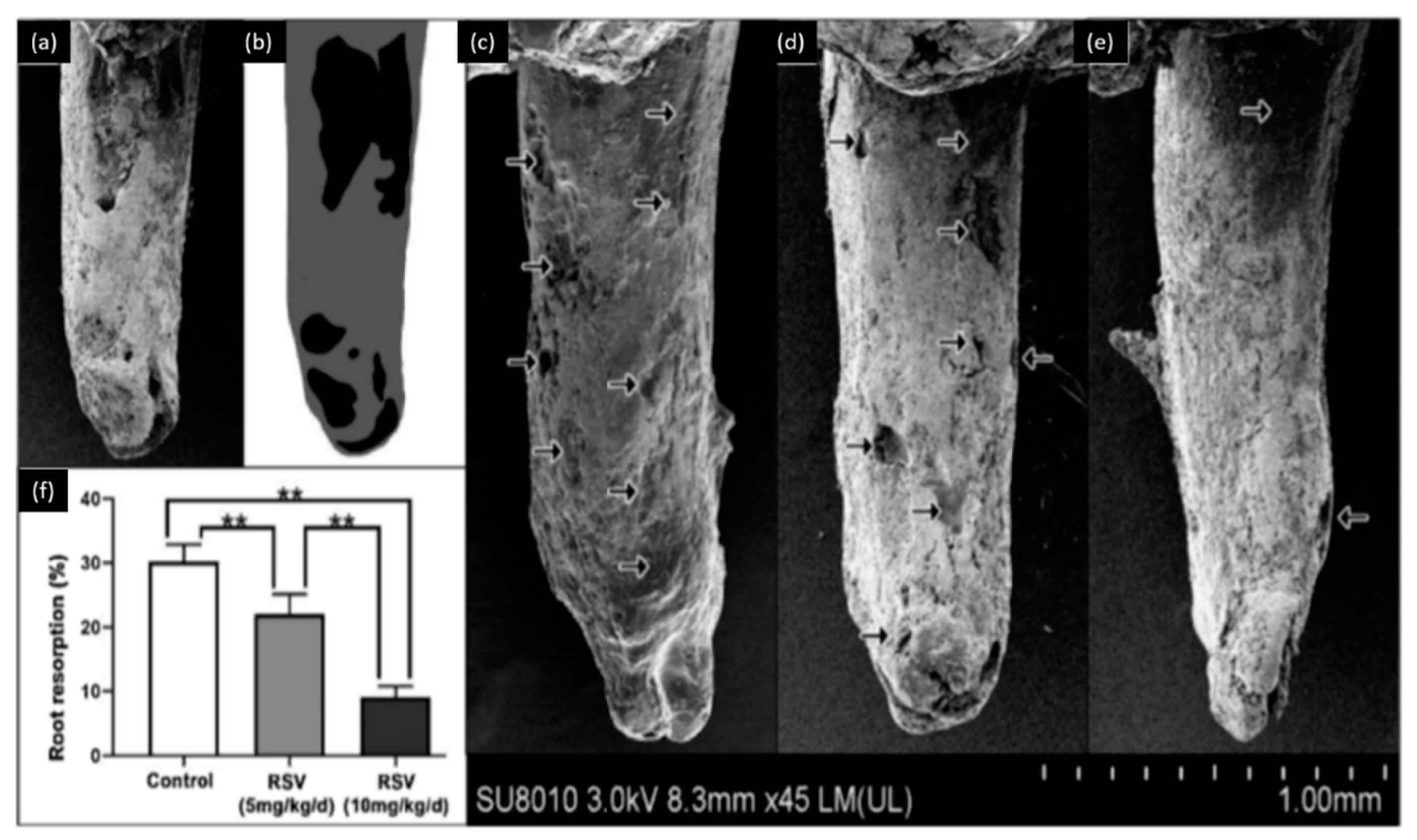
| Liu [54] | Phlorotannins: resveratrol | 5 mg/kg/d, 10 mg/kg/d Rat model | Promote osteoblastic activity |
| Antibacterial Activity | |||
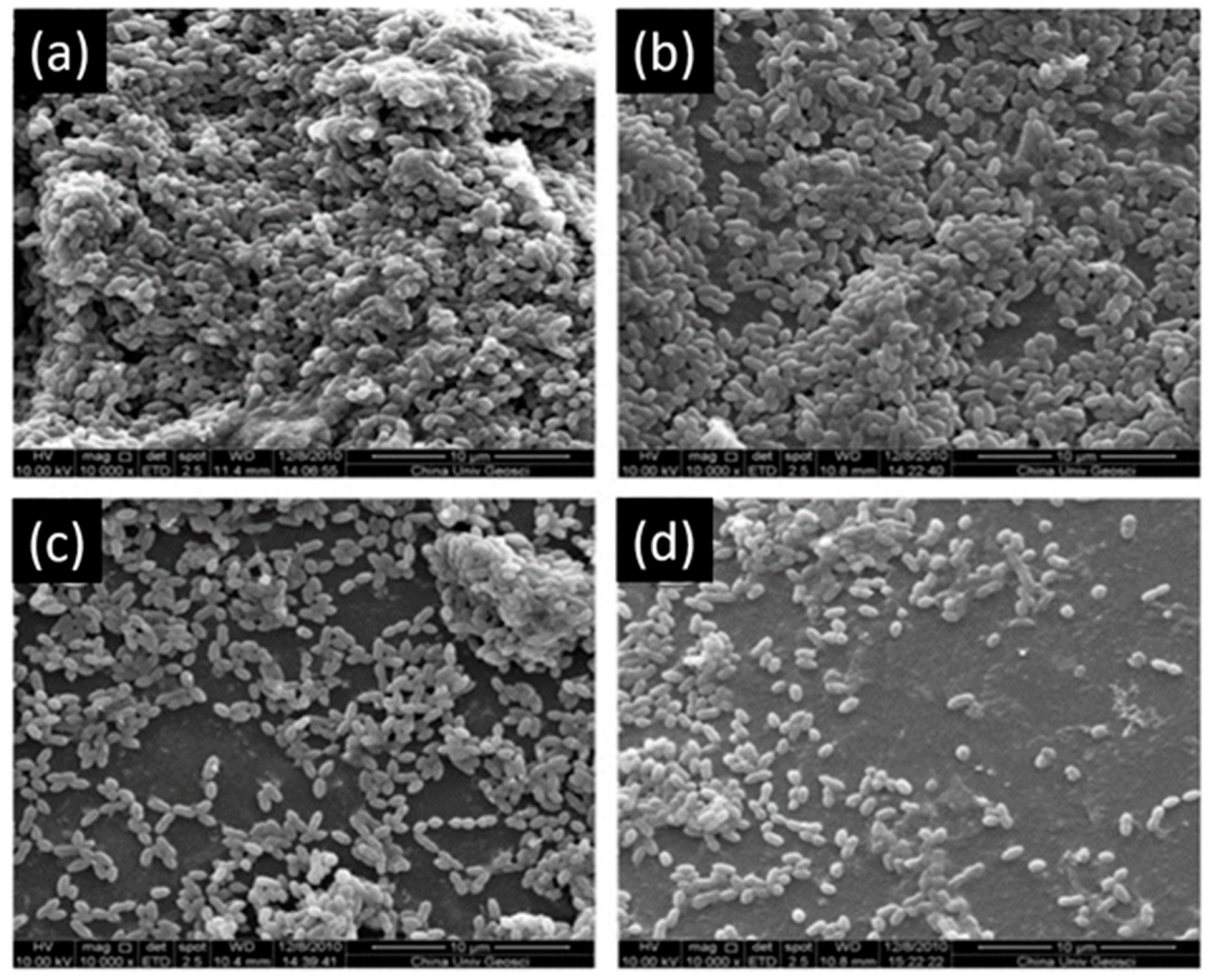
| Xu [56] | Epigallocatechin gallate | 15.6 μg/mL | Inhibition of S. mutans biofilm activity |
| Hara [57] | Epigallocatechin gallate | More than 0.5 mg/mL | Inhibition of alpha-amylase |
| Feng [58] | Gallocatechin gallate, epigallocatechin gallate | 0.32 mM, 0.31 mM, respectively | Inhibition of S. mutans glucosyltransferases |
| Melok [59] | Epigallocatechin-3-gallate | 250 μg/mL | Inhibition of S. mutans growth |
| Lee [60] | Epigallocatechin gallate | 500 μg/mL | Eradication of Enterococcus faecalis after 7 days |
| Du [63] | Epigallocatechin gallate | 200, 300 μg/mL | Inhibition of S. mutans growth and preserve the bond strength of resin-adhesive |
| Anti-Inflammatory and Antioxidant Activity | |||
| Hirao [65] | Catechins | 10 and 50 μg/mL | Inhibition of cytokines and chemokines |
| Nakanishi [66] | Epigallocatechin gallate, epicatechin gallate | 20 and 50 μg/mL | Reduce of pro-inflammatory cytokines |
| Yang [64] | Epigallocatechin gallate | 10–15 μmol/L | Suppression of the cyclooxydenase-2 |
| Mahmoud Hashemi [70] | Flavonols: quercetin | 0.5 mg/mL | Reduce of pro-inflammatory cytokines |
| Yonehiro [71] | Flavonols: luteolin | 35 μmol/L | Reduce of pro-inflammatory cytokines |
| Lee [68] | Flavonols: butein | 2.5–20 μM | Protective agent in dental pulp diseases |
| Wang [72] | Phlorotannins: resveratrol | 50, 100 μM | Decrease pulpal damage |
| Hydrolysable Tannins and Gallic Acid | |||
| Dentin Modifier, Dentin Pretreatment, Collagen Cross-Linking, and Resin-Dentin Stability | |||
| Author | Polyphenol | Concentration | Effect |
| Bedran-Russo [22] | Tannic acid | 10%, 20% (w/v) | Increase dentin stiffness and reduce enzymatic degradation |
| Bitter [82] | Tannic acid | 25% (w/v) | Smooth and clean pulp chamber |
| Bitter [83] | Tannic acid | 25% (w/v) for 60s | Remove the smear layer and partially the organic material |
| Oh [89] | Gallic acid+FeCl3 | 0.47 × 10⁻³ M, 1.2 × 10⁻³ M, respectively | Reduce the dentin hypersensitivity |
| Mukai [79] | Fluoride-tannin acid-lanthanum-apatite | Containing 5% (w/v) of tannic acid | Reduce the dentin hypersensitivity |
| Christopher [91] | Tannic acid, gallic acid | 10%, 10%, respectively | Gallic acid shows more infiltration of resin in dental tubules |
| Remineralization, Cell Viability, and Differentiation | |||
| Zhang [99] | Gallic acid | 4 g/L | Inhibition of enamel demineralization |
| Huang [100] | Gallic acid | 4 g/L | Inhibition of enamel demineralization |
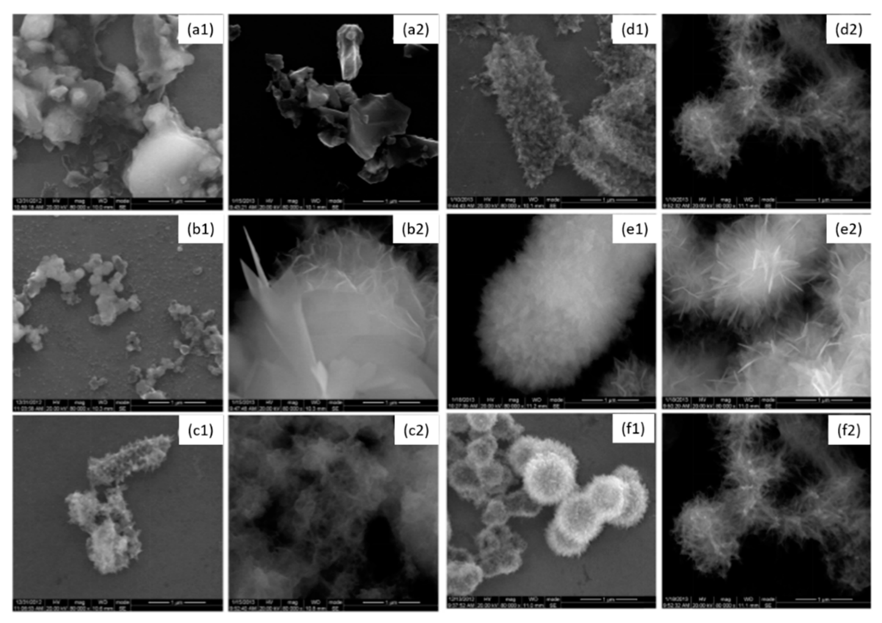
| Tang [102,103] | Gallic acid | 4 g/L | Change the hydroxyapatite size and morphology |
| Polyphenol Mixtures | |||
| Dentin Modifier, Dentin Pretreatment, Collagen Cross-Linking, and Resin-Dentin Stability | |||
| Author | Polyphenol | Concentration | Effect |
| Porto [105] | Quercetin and resveratrol mixture | 100–1000 μg/mL | Preserve bond strength and protect dentin matrix |
| Remineralization, Cell Viability, and Differentiation | |||
| Guo [107] | Galla chinensis | 4000 mg/mL | Enhance the remineralization of root lesions, protect the collagen fibers |
| Antibacterial Activity | |||
| Jazaeri [116] | Tea polyphenol | 10% (w/v) | Anticariogenic effects |
| Ferrazzano [117] | Tea polyphenol | 1.6 g in 40 mL of water | Using as mouthwash reduce S. mutans |
| Hattori [120] | Tea polyphenol | 1–10 mM | Reduce the effect of glucosyltransferase of S. mutans |
| Sakanaka [121] | Green tea polyphenols | 0.1–0.5% (w/v) Rat model | Reduce caries activity |
| Ooshima [126] | Oolong tea extracts | 0.5 mg/mL | Reduce the plaque deposition on teeth |
| Signoretto [127] | Coffee, wine | 30–40 mL/d, 400mL/d, respectively | Effective on supra- and subgingival plaque |
| Shinada [136] | Hop Bract polyphenol | 0.1% Mouth rinse (5 times/d) | Reduce 25.4% of the dental plaque |
| Khalil [138] | Salvadora Persica | 20 mh/mL | high activity against S. aureus and Streptococcus sp. |
| Farkash [148] | Green tea polyphenols and Padma Hepaten | 1.25 μg/mL, 0.16 μg/mL, respectively | Inhibition of C. albicans growth (88%) |
| He [106] | Green tea polyphenols mixed with nano-sized calcium phosphate particles | 12–27 mg/mL | Antibacterial activity and increase the remineralization process |
© 2020 by the authors. Licensee MDPI, Basel, Switzerland. This article is an open access article distributed under the terms and conditions of the Creative Commons Attribution (CC BY) license (http://creativecommons.org/licenses/by/4.0/).
Share and Cite
Kharouf, N.; Haikel, Y.; Ball, V. Polyphenols in Dental Applications. Bioengineering 2020, 7, 72. https://doi.org/10.3390/bioengineering7030072
Kharouf N, Haikel Y, Ball V. Polyphenols in Dental Applications. Bioengineering. 2020; 7(3):72. https://doi.org/10.3390/bioengineering7030072
Chicago/Turabian StyleKharouf, Naji, Youssef Haikel, and Vincent Ball. 2020. "Polyphenols in Dental Applications" Bioengineering 7, no. 3: 72. https://doi.org/10.3390/bioengineering7030072
APA StyleKharouf, N., Haikel, Y., & Ball, V. (2020). Polyphenols in Dental Applications. Bioengineering, 7(3), 72. https://doi.org/10.3390/bioengineering7030072