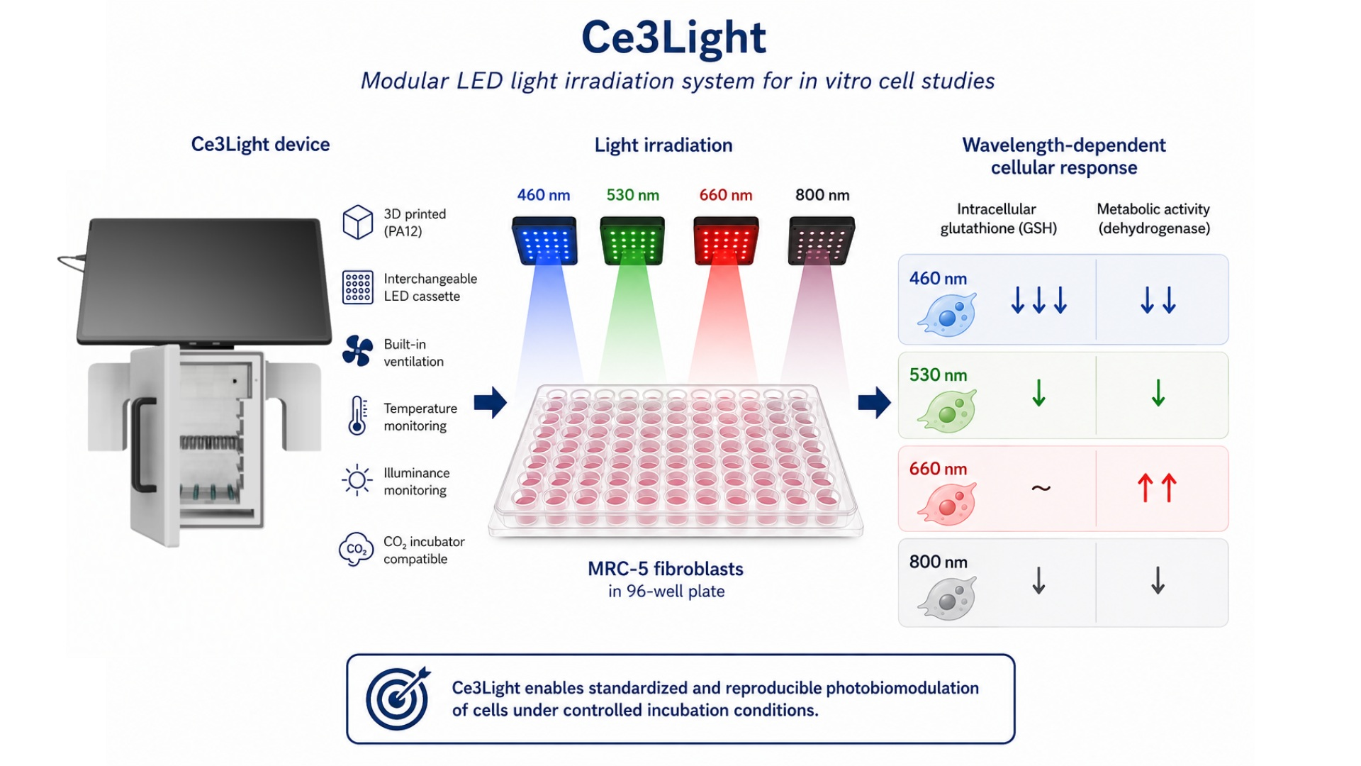
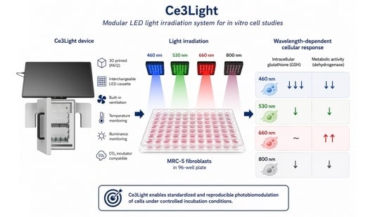
Ce3Light: Design, Construction, and Testing of a Light-Irradiation System for In Vitro Cell Studies

 , , ,
, , ,  , and
, and 
Abstract
1. Introduction
2. Materials and Methods
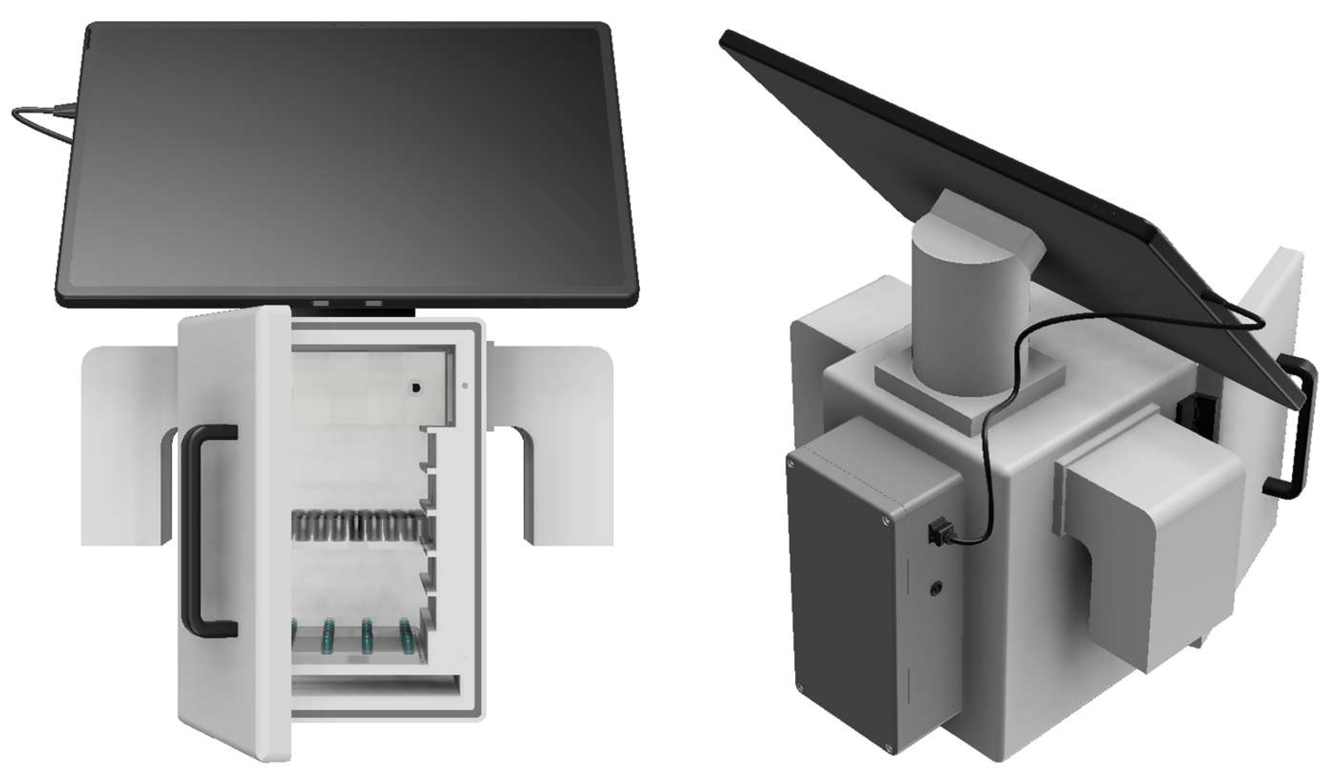
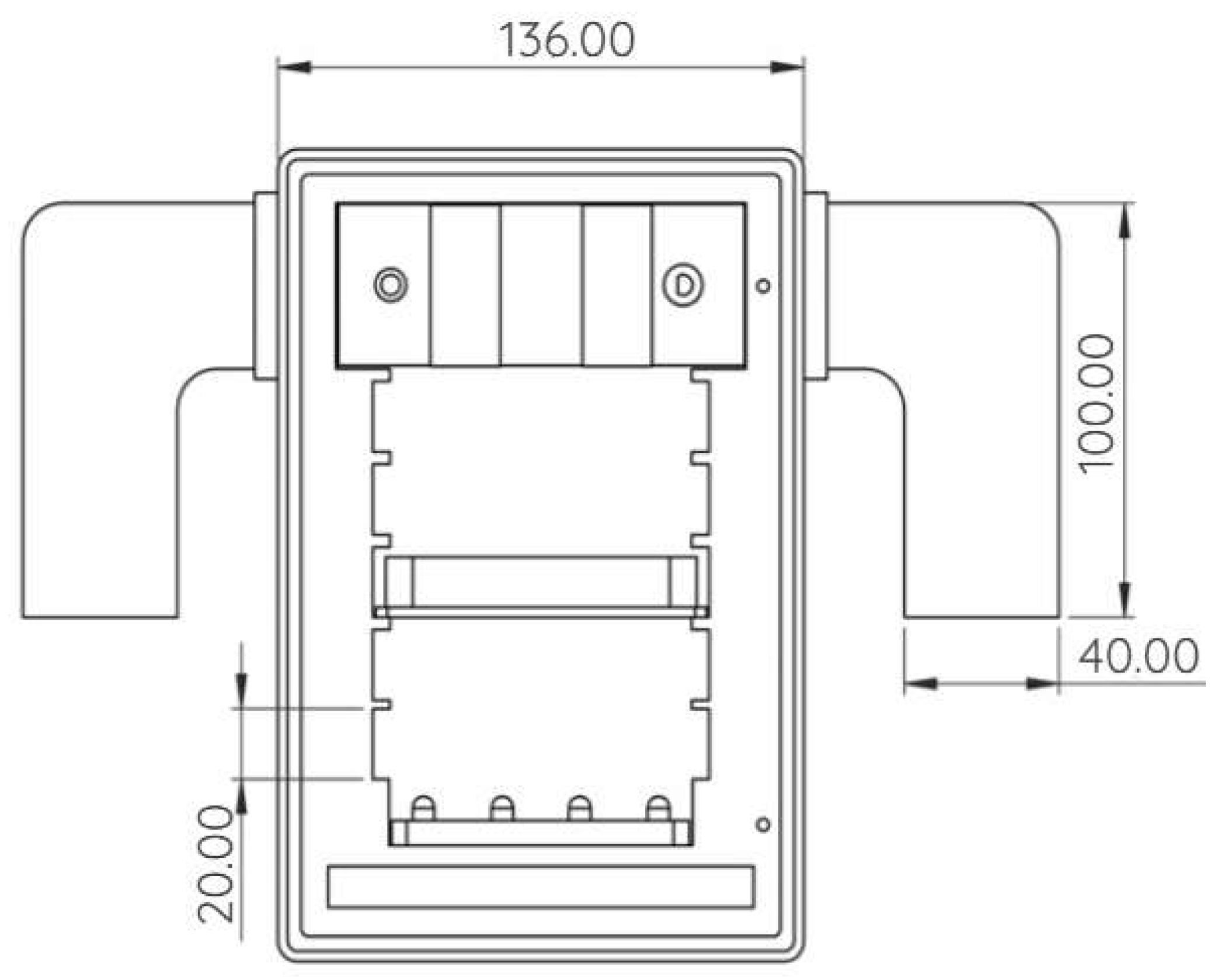
2.1. Design of Ce3Light
2.2. Construction of Ce3Light
- Model preparation in software: The process begins with importing a 3D model in STL format into the appropriate software, where the model is placed in the workspace (build volume). In this phase, the orientation of the model and its placement in the print chamber are optimized to maximize production efficiency. At the same time, printing parameters can be adjusted, which affect the quality of the final product.
- Preparing the printing unit: Before starting printing, it is necessary to prepare a container with powder material. Usually, a mixture of recycled and new powder is used.
- Printing process: The production itself takes place by applying thin layers of powder, which are then selectively sintered with a laser beam according to the geometry of the model. After one layer is sintered, another layer of powder is applied and the process is repeated until the entire part is created.
- Cooling the prints: After printing is finished, it is necessary to let the build chamber with the prints cool down (at least 24 h after completion). This step is key to minimizing internal stresses and deformations of the printed parts.
- Removing excess powder: After cooling, the prints are removed from the powder bed and cleaned of unsintered material. Excess powder is collected and can be reused.
- Final processing: The final step is the additional processing of the product, which mainly includes mechanical cleaning of the surface, for example by sandblasting. This process removes powder residues and improves the surface properties of the product. If necessary, additional treatments, can also be applied.
2.2.1. Material PA12
2.2.2. Construction of LED Cassettes
2.3. Software
2.4. Validation of Ce3Light
- Cell culture and treatment
- Glutathione assay
- Dehydrogenase activity assay
- Fluorescence Microscopy
- Statistical Analysis
3. Results
4. Discussion
5. Conclusions
Author Contributions
Funding
Institutional Review Board Statement
Informed Consent Statement
Data Availability Statement
Acknowledgments
Conflicts of Interest
References
- Dompe, C.; Roana, G.; Rossi, G.; Pasinetti, M.; Panetta, V. Photobiomodulation—Underlying Mechanism and Clinical Applications. J. Clin. Med. 2020, 9, 1724. [Google Scholar] [CrossRef] [Scilit] [PubMed]
- Bikmulina, P.; Shpichka, A.; Timashev, P. Photobiomodulation in 3D tissue engineering. J. Biomed. Opt. 2022, 27, 090901. [Google Scholar] [CrossRef] [Scilit] [PubMed]
- Heiskanen, V.; Hamblin, M.R. Photobiomodulation: Lasers vs. light emitting diodes? Photochem. Photobiol. Sci. 2018, 17, 1003–1017. [Google Scholar] [CrossRef] [Scilit] [PubMed]
- Anders, J.J.; Lanzafame, R.J.; Arany, P.R. Low–level light/laser therapy versus photobiomodulation therapy. Photomed. Laser Surg. 2015, 33, 183–184. [Google Scholar] [CrossRef] [Scilit] [PubMed]
- Su, C.T.; Wu, J.H. Photobiomodulation in biological tissues: Light penetration, dosimetry, and potential applications. Opt. Lasers Eng. 2025, 186, 108852. [Google Scholar] [CrossRef] [Scilit]
- De Freitas, L.F.; Hamblin, M.R. Proposed Mechanisms of Photobiomodulation or Low–Level Light Therapy. IEEE J. Sel. Top. Quantum Electron. 2016, 22, 7000417. [Google Scholar] [CrossRef] [Scilit] [PubMed]
- Esteves-Pereira, T.C.; Santos-Silva, A.R. Understanding Dosimetry in Photobiomodulation Therapy. In Photobiomodulation Therapy in Oral Medicine, 1st ed.; Santos-Silva, A.R., Ed.; Springer: Cham, Switzerland, 2025; pp. 11–32. [Google Scholar]
- Chaudary, S.; Holnthoner, W. Stimulation by Light. In Vascularization for Tissue Engineering and Regenerative Medicine, 1st ed.; Holnthoner, W., Ed.; Springer: Cham, Switzerland, 2021; pp. 55–72. [Google Scholar]
- Van Tran, V.; Park, B.; Cho, S.W.; Lee, J.S.; Moon, J.H. Light emitting diodes technology–based photobiomodulation therapy (PBMT) for dermatology and aesthetics: Recent applications, challenges, and perspectives. Opt. Laser Technol. 2021, 135, 106698. [Google Scholar] [CrossRef] [Scilit]
- Hamblin, M.R. Mechanisms and Mitochondrial Redox Signaling in Photobiomodulation. Photochem. Photobiol. 2018, 94, 199–212. [Google Scholar] [CrossRef] [Scilit] [PubMed]
- Glass, G.E. Photobiomodulation: A review of the molecular evidence for low level light therapy. J. Plast. Reconstr. Aesthet. Surg. 2021, 74, 1050–1060. [Google Scholar] [CrossRef] [Scilit] [PubMed]
- Selestin Raja, I.; Fathima, A.; Lee, J.M.; Choi, S.M.; Han, D.W. Tailoring photobiomodulation to enhance tissue regeneration. Biomaterials 2024, 309, 122623. [Google Scholar] [CrossRef] [Scilit] [PubMed]
- Kreisler, M.; Christoffers, A.B.; Willershausen, B.; d’Hoedt, B. Low level 809-nm diode laser-induced in vitro stimulation of the proliferation of human gingival fibroblasts. Lasers Surg. Med. 2002, 30, 365–369. [Google Scholar] [CrossRef] [Scilit] [PubMed]
- Khadra, M.; Kasem, N.; Haanaes, H.R.; Nilsen, S.T.; Hjørting-Hansen, E.; Ronold, H.J. Effect of laser therapy on attachment, proliferation and differentiation of human osteoblast–like cells cultured on titanium implant material. Biomaterials 2005, 26, 3503–3509. [Google Scholar] [CrossRef] [Scilit] [PubMed]
- Almeida-Lopes, L.; Rigau, J.; Zângaro, R.A.; Brucone, J.D.; Jaeger, M.M. Comparison of the low level laser therapy effects on cultured human gingival fibroblasts proliferation using different irradiance and same fluence. Lasers Surg. Med. 2001, 29, 179–184. [Google Scholar] [CrossRef] [Scilit] [PubMed]
- Alghamdi, K.M.; Al-Zahrani, A.F.; Al-Ghamdi, S.S. Influence of Low-Level Red Laser Irradiation on the Proliferation, Viability, and Differentiation of Human Embryonic Stem Cell-Derived Mesenchymal Stem Cells. Life 2025, 15, 1125. [Google Scholar] [CrossRef] [Scilit] [PubMed]
- Andreeva, N.V.; Golovynsky, G.P.; Turchin, I.V. Cytotoxic Effect of Low-Intensity Infrared Laser Irradiation on Human Melanoma Cells. Mol. Biol. 2018, 52, 1024–1031. [Google Scholar] [CrossRef] [Scilit]
- Wang, Y.; Huang, Y.Y.; Wang, Y.; Lyu, P.; Hamblin, M.R. Red (660 nm) or near-infrared (810 nm) photobiomodulation stimulates, while blue (415 nm), green (540 nm) light inhibits proliferation in human adipose-derived stem cells. Sci. Rep. 2017, 7, 7781. [Google Scholar] [CrossRef] [Scilit] [PubMed]
- Wang, Y.; Huang, Y.Y.; Wang, Y.; Lyu, P.; Hamblin, M.R. Photobiomodulation (blue and green light) encourages osteoblastic-differentiation of human adipose-derived stem cells: Role of intracellular calcium and light-gated ion channels. Sci. Rep. 2016, 6, 33719. [Google Scholar] [CrossRef] [Scilit] [PubMed]
- Segismondi, L.C.; Soares, L.E.S.; Pacheco-Soares, C. In vitro evaluation of red and near infrared LED photobiomodulation on L929 fibroblasts metabolic activity and morphology. Res. Biomed. Eng. 2025, 41, 1–10. [Google Scholar] [CrossRef] [Scilit]
- Baldassarro, V.A.; Ciali, A.; Sivilia, S.; Calzà, L.; Giardino, L. Photobiomodulation at Defined Wavelengths Regulates Mitochondrial Membrane Potential and Redox Balance in Skin Fibroblasts. Oxid. Med. Cell. Longev. 2023, 2023, 7638223. [Google Scholar] [CrossRef] [Scilit] [PubMed]
- Yang, Y.; Ding, T.; Zhou, F.; Qin, W. Irradiation with blue light-emitting diode enhances osteogenic differentiation of stem cells from the apical papilla. Lasers Med. Sci. 2020, 35, 1515–1522. [Google Scholar] [CrossRef] [Scilit]
- Shao, J.; Zhang, L.; Wang, X.; Chen, H.; Li, Y. Photobiomodulation of 450 nm Blue Light on Human Keratinocytes, Fibroblasts, and Endothelial Cells: An In Vitro and Transcriptomic Study on Cells Involved in Wound Healing and Angiogenesis. Biomedicines 2025, 13, 1876. [Google Scholar] [CrossRef] [Scilit] [PubMed]
- Vlasceanu, A.M.; Baconi, D.L.; Galateanu, B.; Stan, M. Comparative Cytotoxicity Study of Nicotine and Cotinine on MRC-5 Cell Line. J. Mind Med. Sci. 2018, 5, 117–122. [Google Scholar] [CrossRef] [Scilit]
- Jones, P.; Benghuzzi, H.; Tucci, M.; Tardy, F. Morphometric analysis of MRC-5 fibroblast like cells exposed to intermittent UV radiation. Biomed. Sci. Instrum. 2003, 39, 415–420. [Google Scholar] [PubMed]
- Socci, M.C.; Rodríguez-Rodríguez, R.; García-Carvajal, Z.Y.; Jiménez-Palomar, I.; Solis-Correa, S.M.; Alvarez-Perez, M.A. Polymeric Materials, Advances and Applications in Tissue Engineering: A Review. Bioengineering 2023, 10, 218. [Google Scholar] [CrossRef] [Scilit] [PubMed]
- Kalyan, B.P.; Kumar, L. 3D Printing: Applications in Tissue Engineering, Medical Devices, and Drug Delivery. AAPS PharmSciTech 2022, 23, 92. [Google Scholar] [CrossRef] [Scilit]
- Poomathi, N.; Singh, S.; Prakash, C.; Subramanian, A.; Sahay, R. 3D printing in tissue engineering: A state of the art review of technologies and biomaterials. Rapid Prototyp. J. 2020, 26, 1313–1334. [Google Scholar] [CrossRef] [Scilit]
- Mani, M.P.; Jaganathan, S.K.; Ismail, A.F.; Chander, S.C. A review on 3D printing in tissue engineering applications. J. Polym. Eng. 2022, 42, 243–265. [Google Scholar] [CrossRef] [Scilit]
- Fina, F.; Goyanes, A.; Gaisford, S.; Basit, A.W. Selective laser sintering (SLS) 3D printing of medicines. Int. J. Pharm. 2017, 529, 285–293. [Google Scholar] [CrossRef] [Scilit] [PubMed]
- Kafle, A.; Luis, E.; Silwal, R.; Pan, H.M.; Shrestha, P.L.; Bastola, A.K. 3D/4D Printing of Polymers: Fused Deposition Modelling (FDM), Selective Laser Sintering (SLS), and Stereolithography (SLA). Polymers 2021, 13, 3101. [Google Scholar] [CrossRef] [Scilit] [PubMed]
- Charoo, N.A.; Barakh Ali, S.F.; Mohamed, E.M.; Kuttolamadom, M.A.; Ozkan, T.; Khan, M.A.; Rahman, Z. Selective laser sintering 3D printing—An overview of the technology and pharmaceutical applications. Drug Dev. Ind. Pharm. 2020, 46, 869–877. [Google Scholar] [CrossRef] [Scilit] [PubMed]
- EOS GmbH. EOSINT P 390: System for Additive Manufacturing of Plastic Parts. Available online: https://www.eos.info (accessed on 2 June 2026).
- Ferencik, N.; Danko, M.; Nadova, Z.; Kolembusova, P.; Steingartner, W. PA12 Surface Treatment and Its Effect on Compatibility with Nutritional Culture Medium to Maintain Cell Vitality and Proliferation. Bioengineering 2024, 11, 442. [Google Scholar] [CrossRef] [Scilit] [PubMed]
- Bazan, A.; Gnatowski, A.; Kijo-Kleckowska, A.; Grabowski, M. Influence of Antibacterial Coating and Mechanical and Chemical Treatment on the Surface Properties of PA12 Parts Manufactured with SLS and MJF Techniques in the Context of Medical Applications. Materials 2023, 16, 2405. [Google Scholar] [CrossRef] [Scilit] [PubMed]
- HP Inc. Biocompatibility Certificate: HP 3D High Reusability PA 12. Available online: https://cdn2.hubspot.net/hubfs/5154612/Material%20documentation/MJF%20PA12/Biocompatibility%20Certificate%20-%203200%204200.pdf (accessed on 19 May 2026).
- HP Inc. HP 3D Printing Materials: Materials Brochure–4AA7–1533ENA. Available online: https://h20195.www2.hp.com/v2/getpdf.aspx/4AA7-1533ENA.pdf (accessed on 2 June 2026).
- Materialise. PA 12 (SLS). Available online: https://www.materialise.com/en/industrial/3d-printing-materials/pa12-sls (accessed on 27 May 2026).
- Standard Practices for Testing Polymeric Powders and Powder Coatings. Available online: https://www.astm.org/d3451-92.html (accessed on 9 March 2024).
- Standard Test Method for Transition Temperatures and Enthalpies of Fusion and Crystallization of Polymers by Differential Scanning Calorimetry. Available online: https://www.astm.org/d3418-21.html (accessed on 9 March 2024).
- Standard Test Methods for Density and Specific Gravity (Relative Density) of Plastics by Displacement. Available online: https://www.astm.org/d0792-20.html (accessed on 9 March 2024).
- Standard Test Methods for Apparent Density, Bulk Factor, and Pourability of Plastic Materials. Available online: https://www.astm.org/d1895-17.html (accessed on 9 March 2024).
- Čapek, J.; Hauschke, M.; Brůčková, L.; Roušar, T. Comparison of glutathione levels measured using optimized monochlorobimane assay with those from ortho-phthalaldehyde assay in intact cells. J. Pharmacol. Toxicol. Methods 2017, 88, 40–45. [Google Scholar] [CrossRef] [Scilit] [PubMed]
- Ticha, L.; Capek, J.; Vlcek, A.; Roušar, T. Evaluation of a human 3D multi-spheroid model derived from SH-SY5Y cells for cytotoxicity testing. Sci. Rep. 2026, 16, 56896. [Google Scholar] [CrossRef]
- Rousar, T.; Handl, J.; Capek, J.; Kučera, O.; Červinková, Z. Cysteine conjugates of acetaminophen and p-aminophenol are potent inducers of cellular impairment in human proximal tubular kidney HK-2 cells. Arch. Toxicol. 2023, 97, 2943–2954. [Google Scholar] [CrossRef] [Scilit] [PubMed]
- Tonolli, P.N.; Vera Palomino, C.M.; Junqueira, H.C.; Baptista, M.S. The phototoxicity action spectra of visible light in HaCaT keratinocytes. J. Photochem. Photobiol. 2023, 14, 112703. [Google Scholar] [CrossRef] [Scilit]
- Ramakrishnan, P.; Maclean, M.; MacGregor, S.J.; Anderson, J.G.; Grant, M.H. Cytotoxic responses to 405nm light exposure in mammalian and bacterial cells: Involvement of reactive oxygen species. Toxicol. Vitro 2016, 33, 54–62. [Google Scholar] [CrossRef] [Scilit]
- Kim, J.S.; Lim, S.S. LED Light-Induced ROS Differentially Regulates Focal Adenosine Kinase Activity in HaCaT Cell Viability. Curr. Issues Mol. Biol. 2022, 44, 1235–1246. [Google Scholar] [CrossRef] [Scilit] [PubMed]
- Kao, W.Z.; Chuang, Y.C.; Chen, K.Y.; Wang, S.T.; Lee, O.K. Enhancing human-induced pluripotent stem cell proliferation and cardiac differentiation through 810-nm photobiomodulation. Lasers Med. Sci. 2026, 41, 125. [Google Scholar] [CrossRef] [Scilit]
- Shimizu, S.; Aoki, A.; Takahashi, T.; Harano, F. Infrared-A Irradiation-induced Inhibition of Human Keratinocyte Proliferation and Potential Mechanisms. Photochem. Photobiol. 2020, 96, 1105–1115. [Google Scholar] [CrossRef] [Scilit] [PubMed]
- Rosenberg, N.; Gendelman, R.; Noofi, N. Photobiomodulation of human osteoblast-like cells in vitro by low-intensity-pulsed LED light. FEBS Open Bio 2020, 10, 1276–1287. [Google Scholar] [CrossRef] [Scilit] [PubMed]
- Rossi, F.; Magni, G.; Tatini, F.; Banchelli, M.; Cherchi, F.; Rossi, G.; Pini, R. Photobiomodulation of Human Fibroblasts and Keratinocytes with Blue Light: Implications in Wound Healing. Biomedicines 2021, 9, 41. [Google Scholar] [CrossRef] [Scilit] [PubMed]
- Kocherova, I.; Bryja, A.; Błochowiak, K.; Kaczmarek, M.; Dompe, C.; Zabel, M.; Dyszkiewicz-Konwińska, M. Photobiomodulation with Red and Near-Infrared Light Improves Viability and Modulates Expression of Mesenchymal and Apoptotic-Related Markers in Human Gingival Fibroblasts. Materials 2021, 14, 3427. [Google Scholar] [CrossRef] [Scilit] [PubMed]




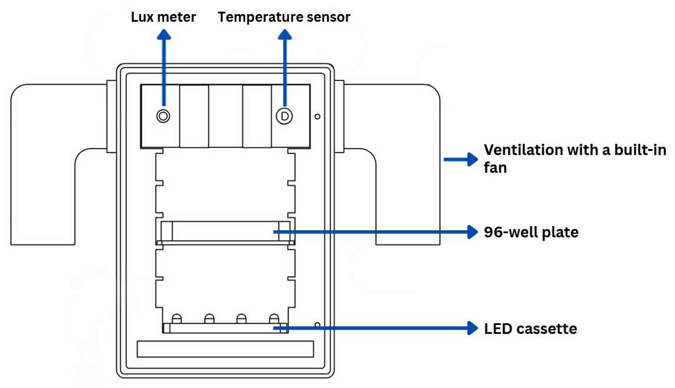
| Parameter | Value |
|---|---|
| Width | 136.00 mm |
| Height | 196.00 mm |
| Depth | 163.00 mm |
| Air inlet and outlet | Through vents (65 × 35 mm) on both sides |
| Thermometer | LM35 (0–100 °C) |
| Lux meter | LUX DFR0026 (1–6000 lux) |
| Number of microtiter plate positions | 5 positions |
| Number of cards in the device at one time | 1 card |
| Model material | PA12 |
| Card material | PETG |
| Property | Value | Standard/Conditions | Reference |
|---|---|---|---|
| Powder melting point (DSC) | 187 °C | ASTM D3418 | [40] |
| Melting temperature | 180 °C | - | |
| Particle density | 1.01 g/cm3 | ASTM D792 | [41] |
| Bulk density | 0.425 g/cm3 | ASTM D1895 | [42] |
| Average particle size | 60 µm | ASTM D3451 | [39] |
| Tensile strength | 50 MPa | At 50 mm/min | |
| Tensile modulus | 1800 MPa | At 1 mm/min | |
| Flexural modulus | 1700 MPa | At 2 mm/min, 10 N |
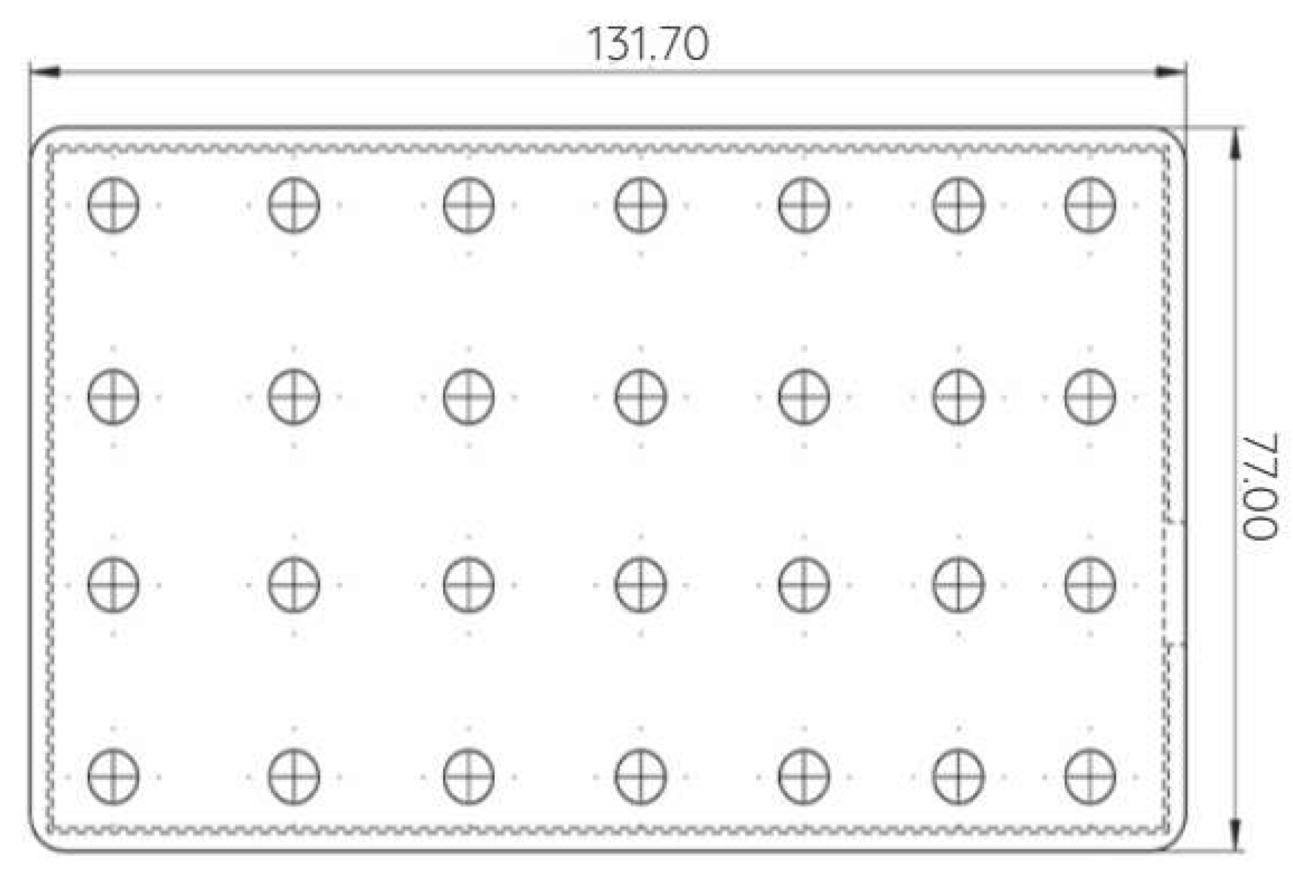
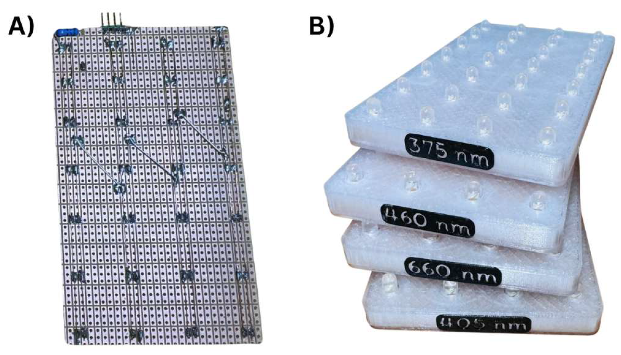
| Parameter | 460 nm | 530 nm | 660 nm | 800 nm |
|---|---|---|---|---|
| LED manufacturer | OptoSupply | OptoSupply | OptoSupply | OptoSupply |
| Number of LEDs | 28 | 28 | 28 | 28 |
| LED arrangement | 7 × 4 | 7 × 4 | 7 × 4 | 7 × 4 |
| Peak wavelength [nm] | 455 | 525 | 660 | 805 |
| Forward current, IF [mA] | 19 | 20 | 20 | 45 |
| Radiant power per LED [mW] | 22 | 35 | 25 | 36 |
| Total radiant power of LED [mW] | 616 | 980 | 700 | 1008 |
| Beam angle [2θ½, °] | 15 | 30 | 15 | 30 |
| Illuminated area [cm2] | 109 | 109 | 109 | 109 |
| Estimated irradiance [mW/cm2] | 5.65 | 8.99 | 6.42 | 9.25 |
| Exposure time [h] | 24 | 24 | 24 | 24 |
| Estimated fluence [24 h, J/cm2] | 488.6 | 776.7 | 554.7 | 799.2 |
| Estimated fluence [2 h, J/cm2] | 40.7 | 64.7 | 46.2 | 66.6 |
| Wavelength [nm] | Illuminance [lx] | TAvg [°C] | TMax [°C] | Evaporation of Medium |
|---|---|---|---|---|
| 460 | 690 ± 10 | 38 ± 1 | 39.0 | <5% |
| 530 | 700 ± 10 | 38 ± 1 | 39.5 | <5% |
| 660 | 700 ± 10 | 38 ± 1 | 39.1 | <3% |
| 800 | 90 ± 5 | 38 ± 1 | 39.2 | <3% |
Disclaimer/Publisher’s Note: The statements, opinions and data contained in all publications are solely those of the individual author(s) and contributor(s) and not of MDPI and/or the editor(s). MDPI and/or the editor(s) disclaim responsibility for any injury to people or property resulting from any ideas, methods, instructions or products referred to in the content. |
© 2026 by the authors. Licensee MDPI, Basel, Switzerland. This article is an open access article distributed under the terms and conditions of the Creative Commons Attribution (CC BY) license.
Share and Cite
Handl, J.; Radochlibová, H.; Čapek, J.; Roušar, T.; Babicová, P.; Ferenčík, N.; Danko, M.; Hudák, R. Ce3Light: Design, Construction, and Testing of a Light-Irradiation System for In Vitro Cell Studies. Bioengineering 2026, 13, 841. https://doi.org/10.3390/bioengineering13070841
Handl J, Radochlibová H, Čapek J, Roušar T, Babicová P, Ferenčík N, Danko M, Hudák R. Ce3Light: Design, Construction, and Testing of a Light-Irradiation System for In Vitro Cell Studies. Bioengineering. 2026; 13(7):841. https://doi.org/10.3390/bioengineering13070841
Chicago/Turabian StyleHandl, Jiří, Helena Radochlibová, Jan Čapek, Tomáš Roušar, Petra Babicová, Norbert Ferenčík, Mária Danko, and Radovan Hudák. 2026. "Ce3Light: Design, Construction, and Testing of a Light-Irradiation System for In Vitro Cell Studies" Bioengineering 13, no. 7: 841. https://doi.org/10.3390/bioengineering13070841
APA StyleHandl, J., Radochlibová, H., Čapek, J., Roušar, T., Babicová, P., Ferenčík, N., Danko, M., & Hudák, R. (2026). Ce3Light: Design, Construction, and Testing of a Light-Irradiation System for In Vitro Cell Studies. Bioengineering, 13(7), 841. https://doi.org/10.3390/bioengineering13070841