Tissue Engineering for Massive Bone Defects: The Volumetric Scaling Problem and Strategies to Solve It

Abstract
1. The Clinical Problem of Massive Bone Defects
1.1. Aim and Objectives of the Review
1.2. Review Methodology
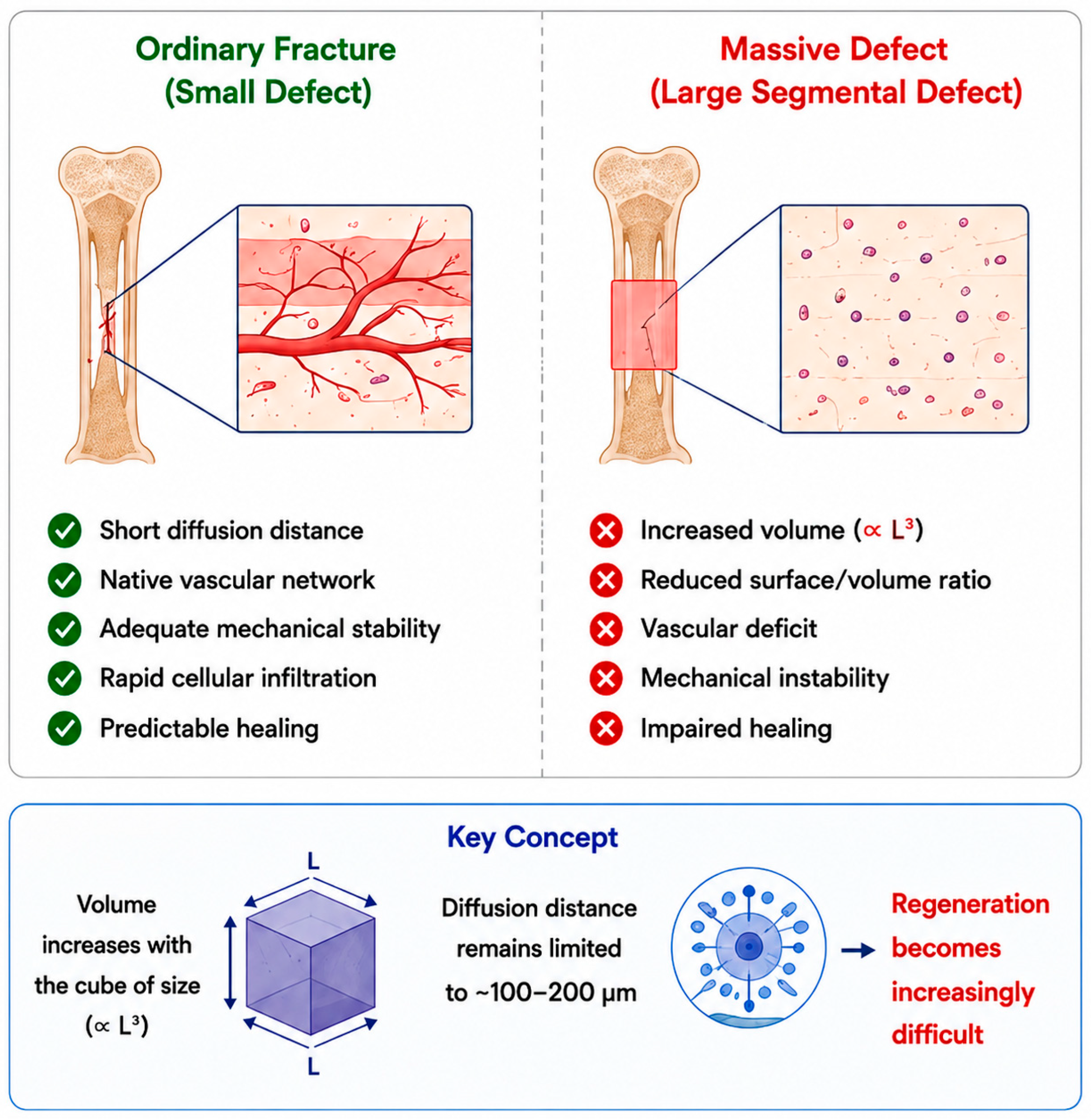
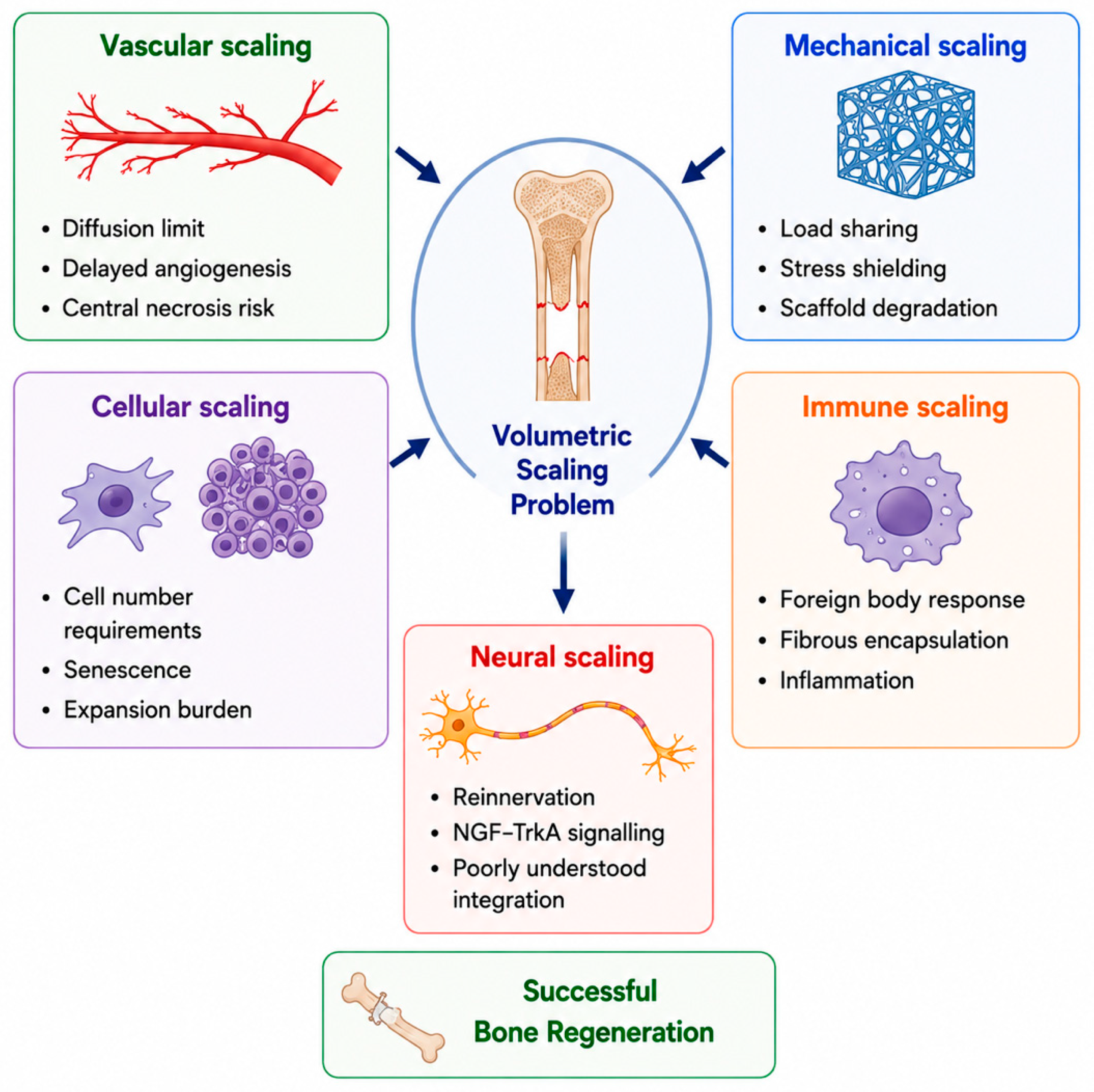
2. Why Massive Defects Are Biologically Different: The Volumetric Scaling Problem
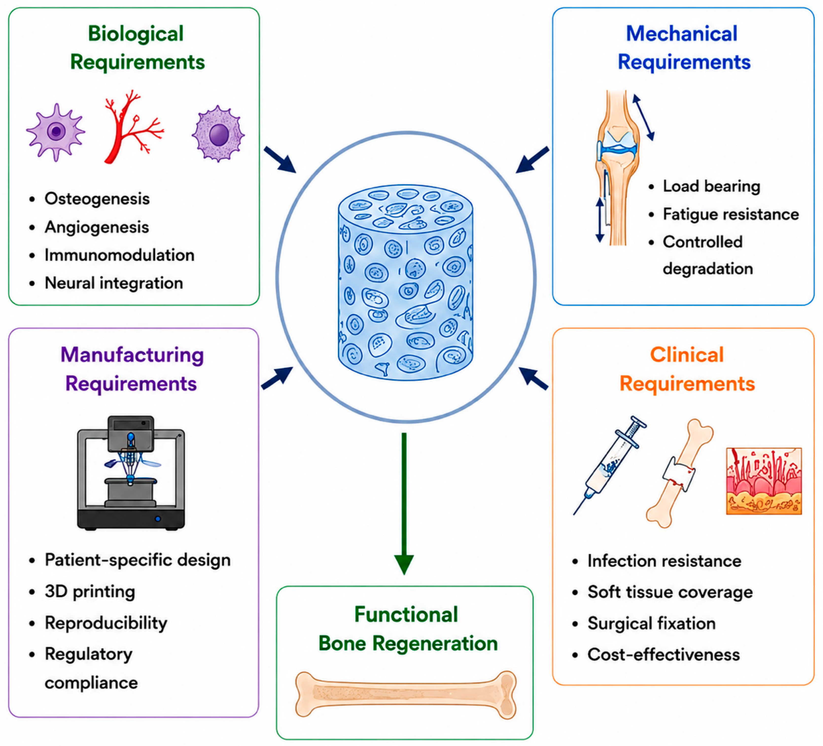
3. Scaffolds for Centimetre-Scale Reconstruction

| Material Class | Compressive Performance at cm-Scale (Qualitative) | Stiffness (Relative to Cortical Bone) | In Vivo Resorption Window | Principal Limitation at the cm-Scale | Ref. |
|---|---|---|---|---|---|
| Stoichiometric hydroxyapatite (HA) | Very high in dense form; brittle; greatly reduced in porous scaffolds | Very high in dense form; substantially reduced with porosity | Slowly resorbing or relatively persistent (years) | Brittle; limited remodelling; ceramic strut fatigue | [31,40] |
| β-Tricalcium phosphate (β-TCP) | Moderate in dense form; highly porosity-dependent | Moderate to high in dense form; substantially lower in porous scaffolds | 6–18 months (porosity-dependent) | Loss of mechanical competence during resorption | [31] |
| Biphasic CaP (HA:β-TCP 60:40 to 80:20) | Moderately dense; tunable via HA:β-TCP ratio and porosity | Variable; depends on phase ratio, porosity, and processing | Tunable (months–years) via HA:β-TCP ratio | Optimal HA:β-TCP ratio contested across anatomical sites and indications | [31] |
| 45S5/13-93 bioactive glass | High in dense form; greatly reduced in porous scaffolds | High in dense form (comparable to cortical bone); markedly lower in porous form | Weeks to months | Intrinsic brittleness; processing constraints for load-bearing cm-scale constructs | [32,41] |
| Polycaprolactone (PCL) | Low; insufficient for unsupported load-bearing | Very low; well below cortical bone | ~2–4 years | Bioinert; low strength without ceramic phase | [26] |
| PLA/PLGA copolymers | Low to moderate; architecture-dependent | Low; decreases as degradation proceeds | 1–12 months (composition-dependent) | Bulk-hydrolytic acidic by-products in large volumes | [26] |
| mPCL/β-TCP composite (80:20 wt%) | Low; supplementary fixation required | Very low; well below cortical bone; fixation-dependent construct | 2–4 years (PCL-rate-limited) | Extensively evaluated in cm-scale ovine studies; modulus is still well below the cortical bone | [34,35] |
| Selective-laser-melted Ti alloy TPMS lattice | Moderate to high; highly architecture-dependent * | Tunable by porosity and unit-cell design; can approach the cortical bone range * | Non-resorbing | Permanent implant; modulus tunable, but does not remodel | [27,28] |
| Native cortical bone | 100–230 MPa (design benchmark) | 7–30 GPa (design benchmark) | — | (benchmark for diaphyseal load-bearing design) | [40] |
| Native trabecular bone | 2–12 MPa (design benchmark) | 0.1–5 GPa (design benchmark) | — | (benchmark for metaphyseal/cancellous design) | [40] |
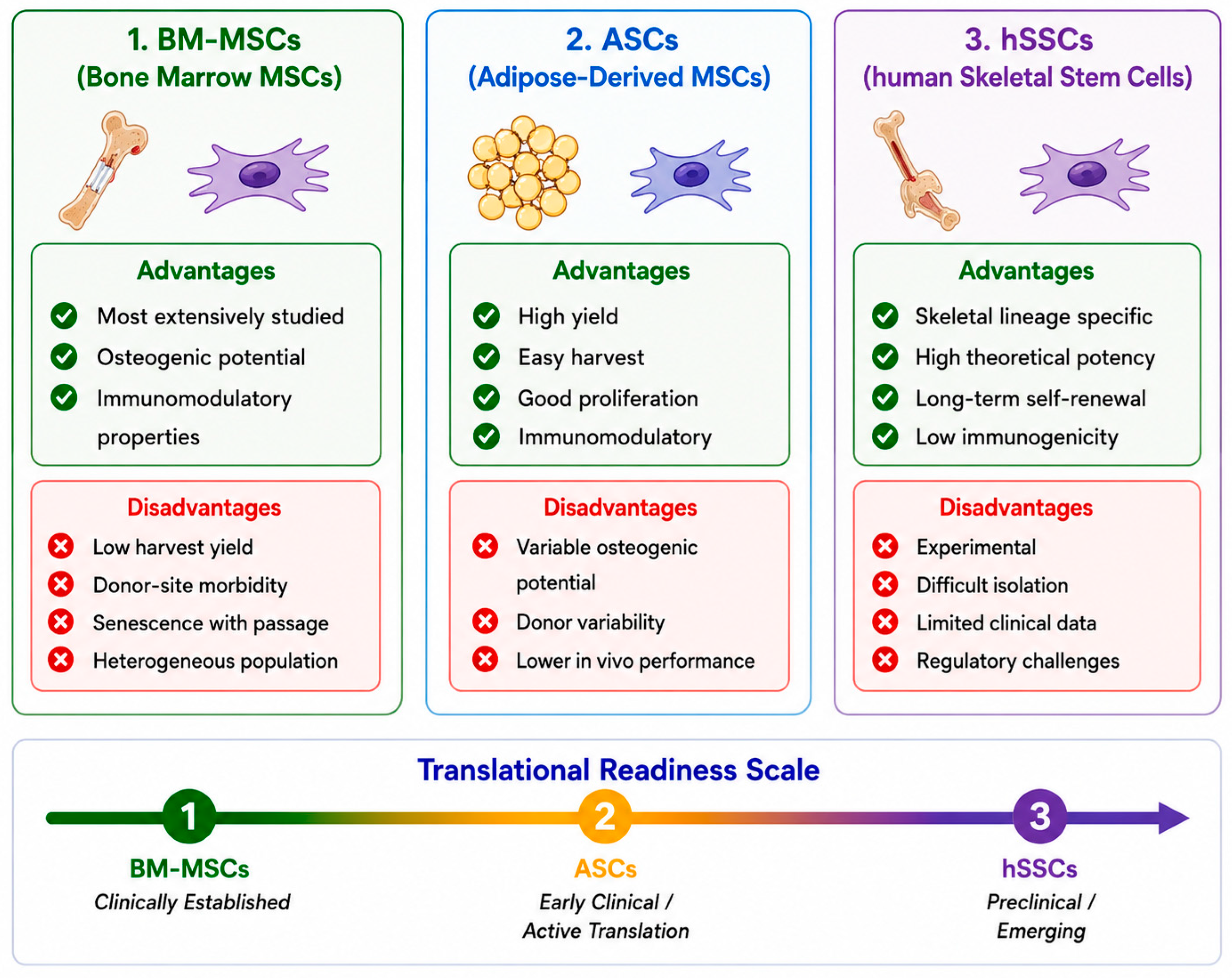
4. Cellular Components at a Clinically Relevant Scale
5. Vascularisation as a Central Rate-Limiting Barrier in Massive Defect Reconstruction
6. Large-Animal Validation and Clinical Translation
7. Persistent Barriers Unique to Large-Scale Defects
8. Knowledge Gaps, Contradictions, and Evidence Strength
9. Conclusions
Funding
Data Availability Statement
Conflicts of Interest
Abbreviations
References
- Nauth, A.; Schemitsch, E.; Norris, B.; Nollin, Z.; Watson, J.T. Critical-size bone defects: Is there a consensus for diagnosis and treatment? J. Orthop. Trauma 2018, 32, S7–S11. [Google Scholar] [CrossRef] [Scilit] [PubMed]
- Roddy, E.; DeBaun, M.R.; Daoud-Gray, A.; Yang, Y.P.; Gardner, M.J. Treatment of critical-sized bone defects: Clinical and tissue engineering perspectives. Eur. J. Orthop. Surg. Traumatol. 2018, 28, 351–362. [Google Scholar] [CrossRef] [Scilit] [PubMed]
- Foster, R.D.; Anthony, J.P.; Sharma, A.; Pogrel, M.A. Vascularized bone flaps versus nonvascularised bone grafts for mandibular reconstruction: An outcome analysis of primary bony union and endosseous implant success. Head. Neck 1999, 21, 66–71. [Google Scholar] [CrossRef]
- Pogrel, M.A.; Podlesh, S.; Anthony, J.P.; Alexander, J. A comparison of vascularised and nonvascularised bone grafts for reconstruction of mandibular continuity defects. J. Oral Maxillofac. Surg. 1997, 55, 1200–1206. [Google Scholar] [CrossRef] [Scilit] [PubMed]
- Myeroff, C.; Archdeacon, M. Autogenous bone graft: Donor sites and techniques. J. Bone Jt. Surg. Am. 2011, 93, 2227–2236. [Google Scholar] [CrossRef] [Scilit] [PubMed]
- Haugen, H.J.; Lyngstadaas, S.P.; Rossi, F.; Perale, G. Bone grafts: Which is the ideal biomaterial? J. Clin. Periodontol. 2019, 46, 92–102. [Google Scholar] [CrossRef] [Scilit] [PubMed]
- Wheeler, D.L.; Enneking, W.F. Allograft bone decreases in strength in vivo over time. Clin. Orthop. Relat. Res. 2005, 435, 36–42. [Google Scholar] [CrossRef] [Scilit] [PubMed]
- Mankin, H.J.; Gebhardt, M.C.; Jennings, L.C.; Springfield, D.S.; Tomford, W.W. Long-term results of allograft replacement in the management of bone tumors. Clin. Orthop. Relat. Res. 1996, 324, 86–97. [Google Scholar] [CrossRef] [Scilit] [PubMed]
- Enneking, W.F.; Campanacci, D.A. Retrieved human allografts: A clinicopathological study. J. Bone Jt. Surg. Am. 2001, 83, 971–986. [Google Scholar] [CrossRef] [Scilit] [PubMed]
- Aktuglu, K.; Erol, K.; Vahabi, A. Ilizarov bone transport and treatment of critical-sized tibial bone defects: A narrative review. J. Orthop. Traumatol. 2019, 20, 22. [Google Scholar] [CrossRef] [Scilit] [PubMed]
- Falder, S.; Sinclair, J.S.; Rogers, C.A.; Townsend, P.L.G. Long-term behaviour of the free vascularised fibula following reconstruction of large bony defects. Br. J. Plast. Surg. 2003, 56, 571–584. [Google Scholar] [CrossRef] [Scilit] [PubMed]
- Feltri, P.; Solaro, L.; Errani, C.; Schiavon, G.; Candrian, C.; Filardo, G. Vascularized fibular grafts for the treatment of long bone defects: Pros and cons. A systematic review and meta-analysis. Arch. Orthop. Trauma Surg. 2023, 143, 29–48. [Google Scholar] [CrossRef] [Scilit] [PubMed]
- Benevenia, J.; Kirchner, R.; Patterson, F.; Beebe, K.; Wirtz, D.C.; Rivero, S.; Palma, M.; Friedrich, M.J. Outcomes of a modular intercalary endoprosthesis as treatment for segmental defects of the femur, tibia, and humerus. Clin. Orthop. Relat. Res. 2016, 474, 539–548. [Google Scholar] [CrossRef] [Scilit] [PubMed]
- Alford, A.I.; Nicolaou, D.; Hake, M.; McBride-Gagyi, S. Masquelet’s induced membrane technique: Review of current concepts and future directions. J. Orthop. Res. 2021, 39, 707–718. [Google Scholar] [CrossRef] [Scilit] [PubMed]
- Novosel, E.C.; Kleinhans, C.; Kluger, P.J. Vascularization is the key challenge in tissue engineering. Adv. Drug Deliv. Rev. 2011, 63, 300–311. [Google Scholar] [CrossRef] [Scilit] [PubMed]
- Rouwkema, J.; Rivron, N.C.; van Blitterswijk, C.A. Vascularization in tissue engineering. Trends Biotechnol. 2008, 26, 434–441. [Google Scholar] [CrossRef] [Scilit] [PubMed]
- Kusumbe, A.P.; Ramasamy, S.K.; Adams, R.H. Coupling of angiogenesis and osteogenesis by a specific vessel subtype in bone. Nature 2014, 507, 323–328, Erratum in Nature 2014, 513, 574. [Google Scholar] [CrossRef] [Scilit] [PubMed]
- Ramasamy, S.K.; Kusumbe, A.P.; Wang, L.; Adams, R.H. Endothelial Notch activity promotes angiogenesis and osteogenesis in bone. Nature 2014, 507, 376–380. [Google Scholar] [CrossRef] [Scilit] [PubMed]
- Xu, R.; Yallowitz, A.; Qin, A.; Wu, Z.; Shin, D.Y.; Kim, J.-M.; Debnath, S.; Ji, G.; Bostrom, M.P.; Yang, X.; et al. Targeting skeletal endothelium to ameliorate bone loss. Nat. Med. 2018, 24, 823–833. [Google Scholar] [CrossRef] [Scilit] [PubMed]
- Xie, H.; Cui, Z.; Wang, L.; Xia, Z.; Hu, Y.; Xian, L.; Li, C.; Xie, L.; Crane, J.; Wan, M.; et al. PDGF-BB secreted by preosteoclasts induces angiogenesis during coupling with osteogenesis. Nat. Med. 2014, 20, 1270–1278. [Google Scholar] [CrossRef] [Scilit] [PubMed]
- Chan, C.K.F.; Gulati, G.S.; Sinha, R.; Tompkins, J.V.; Lopez, M.; Carter, A.C.; Ransom, R.C.; Reinisch, A.; Wearda, T.; Murphy, M.; et al. Identification of the human skeletal stem cell. Cell 2018, 175, 43–56.e21. [Google Scholar] [CrossRef] [Scilit] [PubMed]
- Brazill, J.M.; Beeve, A.T.; Craft, C.S.; Ivanusic, J.J.; Scheller, E.L. Nerves in bone: Evolving concepts in pain and anabolism. J. Bone Miner. Res. 2019, 34, 1393–1406. [Google Scholar] [CrossRef] [Scilit] [PubMed]
- Li, Z.; Meyers, C.A.; Chang, L.; Lee, S.; Li, Z.; Tomlinson, R.; Hoke, A.; Clemens, T.L.; James, A.W. Fracture repair requires TrkA signaling by skeletal sensory nerves. J. Clin. Investig. 2019, 129, 5137–5150. [Google Scholar] [CrossRef] [Scilit] [PubMed]
- Tandon, B.; Blaker, J.J.; Cartmell, S.H. Piezoelectric materials as stimulatory biomedical materials and scaffolds for bone repair. Acta Biomater. 2018, 73, 1–20. [Google Scholar] [CrossRef] [Scilit] [PubMed]
- Frost, H.M. Bone’s mechanostat: A 2003 update. Anat. Rec. A Discov. Mol. Cell. Evol. Biol. 2003, 275A, 1081–1101. [Google Scholar] [CrossRef] [Scilit] [PubMed]
- Hutmacher, D.W. Scaffolds in tissue engineering bone and cartilage. Biomaterials 2000, 21, 2529–2543. [Google Scholar] [CrossRef] [Scilit] [PubMed]
- Bobbert, F.S.L.; Lietaert, K.; Eftekhari, A.A.; Pouran, B.; Ahmadi, S.M.; Weinans, H.; Zadpoor, A.A. Additively manufactured metallic porous biomaterials based on minimal surfaces: A unique combination of topological, mechanical, and mass transport properties. Acta Biomater. 2017, 53, 572–584. [Google Scholar] [CrossRef] [Scilit] [PubMed]
- Pobloth, A.M.; Checa, S.; Razi, H.; Petersen, A.; Weaver, J.C.; Schmidt-Bleek, K.; Windolf, M.; Tatai, A.Á.; Roth, C.P.; Schaser, K.-D.; et al. Mechanobiologically optimised 3D titanium-mesh scaffolds enhance bone regeneration in critical segmental defects in sheep. Sci. Transl. Med. 2018, 10, eaam8828. [Google Scholar] [CrossRef] [Scilit] [PubMed]
- Anderson, J.M.; Rodriguez, A.; Chang, D.T. Foreign body reaction to biomaterials. Semin. Immunol. 2008, 20, 86–100. [Google Scholar] [CrossRef] [Scilit] [PubMed]
- Brown, B.N.; Ratner, B.D.; Goodman, S.B.; Amar, S.; Badylak, S.F. Macrophage polarization: An opportunity for improved outcomes in biomaterials and regenerative medicine. Biomaterials 2012, 33, 3792–3802. [Google Scholar] [CrossRef] [Scilit] [PubMed]
- Bohner, M.; Santoni, B.L.G.; Döbelin, N. β-tricalcium phosphate for bone substitution: Synthesis and properties. Acta Biomater. 2020, 113, 23–41. [Google Scholar] [CrossRef] [Scilit] [PubMed]
- Hench, L.L. The story of Bioglass. J. Mater. Sci. Mater. Med. 2006, 17, 967–978. [Google Scholar] [CrossRef] [Scilit] [PubMed]
- Bose, S.; Roy, M.; Bandyopadhyay, A. Recent advances in bone tissue engineering scaffolds. Trends Biotechnol. 2012, 30, 546–554. [Google Scholar] [CrossRef] [Scilit] [PubMed]
- Reichert, J.C.; Cipitria, A.; Epari, D.R.; Saifzadeh, S.; Krishnakanth, P.; Berner, A.; Woodruff, M.A.; Schell, H.; Mehta, M.; Schuetz, M.A.; et al. A tissue engineering solution for segmental defect regeneration in load-bearing long bones. Sci. Transl. Med. 2012, 4, 141ra93. [Google Scholar] [CrossRef] [Scilit] [PubMed]
- Cipitria, A.; Reichert, J.C.; Epari, D.R.; Saifzadeh, S.; Berner, A.; Schell, H.; Mehta, M.; Schuetz, M.A.; Duda, G.N.; Hutmacher, D.W. Polycaprolactone scaffold and reduced rhBMP-7 dose for the regeneration of critical-sized defects in sheep tibiae. Biomaterials 2013, 34, 9960–9968. [Google Scholar] [CrossRef] [Scilit] [PubMed]
- Karageorgiou, V.; Kaplan, D. Porosity of 3D biomaterial scaffolds and osteogenesis. Biomaterials 2005, 26, 5474–5491. [Google Scholar] [CrossRef] [Scilit] [PubMed]
- Hollister, S.J. Porous scaffold design for tissue engineering. Nat. Mater. 2005, 4, 518–524, Erratum in Nat. Mater. 2006, 5, 590. [Google Scholar] [CrossRef] [Scilit] [PubMed]
- Murphy, C.M.; Haugh, M.G.; O’Brien, F.J. The effect of mean pore size on cell attachment, proliferation and migration in collagen-glycosaminoglycan scaffolds. Biomaterials 2010, 31, 461–466. [Google Scholar] [CrossRef] [Scilit] [PubMed]
- Zhang, M.; Lin, R.; Wang, X.; Xue, J.; Deng, C.; Feng, C.; Zhuang, H.; Ma, J.; Qin, C.; Wan, L.; et al. 3D printing of Haversian bone-mimicking scaffolds for multicellular delivery in bone regeneration. Sci. Adv. 2020, 6, eaaz6725. [Google Scholar] [CrossRef] [Scilit] [PubMed]
- Rho, J.Y.; Kuhn-Spearing, L.; Zioupos, P. Mechanical properties and the hierarchical structure of bone. Med. Eng. Phys. 1998, 20, 92–102. [Google Scholar] [CrossRef] [Scilit] [PubMed]
- Rahaman, M.N.; Day, D.E.; Bal, B.S.; Fu, Q.; Jung, S.B.; Bonewald, L.F.; Tomsia, A.P. Bioactive glass in tissue engineering. Acta Biomater. 2011, 7, 2355–2373. [Google Scholar] [CrossRef] [Scilit] [PubMed]
- Quarto, R.; Mastrogiacomo, M.; Cancedda, R.; Kutepov, S.M.; Mukhachev, V.; Lavroukov, A.; Kon, E.; Marcacci, M. Repair of large bone defects with the use of autologous bone marrow stromal cells. N. Engl. J. Med. 2001, 344, 385–386. [Google Scholar] [CrossRef] [Scilit] [PubMed]
- Kern, S.; Eichler, H.; Stoeve, J.; Klüter, H.; Bieback, K. Comparative analysis of mesenchymal stem cells from bone marrow, umbilical cord blood, or adipose tissue. Stem Cells 2006, 24, 1294–1301. [Google Scholar] [CrossRef] [Scilit] [PubMed]
- Banfi, A.; Muraglia, A.; Dozin, B.; Mastrogiacomo, M.; Cancedda, R.; Quarto, R. Proliferation kinetics and differentiation potential of ex vivo expanded human bone marrow stromal cells: Implications for their use in cell therapy. Exp. Hematol. 2000, 28, 707–715. [Google Scholar] [CrossRef] [Scilit] [PubMed]
- Gimble, J.M.; Katz, A.J.; Bunnell, B.A. Adipose-derived stem cells for regenerative medicine. Circ. Res. 2007, 100, 1249–1260. [Google Scholar] [CrossRef] [Scilit] [PubMed]
- Rossi, N.; Hadad, H.; Bejar-Chapa, M.; Peretti, G.M.; Randolph, M.A.; Redmond, R.W.; Guastaldi, F.P.S. Bone marrow stem cells with tissue-engineered scaffolds for large bone segmental defects: A systematic review. Tissue Eng. Part B Rev. 2023, 29, 457–472. [Google Scholar] [CrossRef] [Scilit] [PubMed]
- Takahashi, K.; Yamanaka, S. Induction of pluripotent stem cells from mouse embryonic and adult fibroblast cultures by defined factors. Cell 2006, 126, 663–676. [Google Scholar] [CrossRef] [Scilit] [PubMed]
- de Peppo, G.M.; Marcos-Campos, I.; Kahler, D.J.; Alsalman, D.; Shang, L.; Vunjak-Novakovic, G.; Marolt, D. Engineering bone tissue substitutes from human induced pluripotent stem cells. Proc. Natl. Acad. Sci. USA 2013, 110, 8680–8685. [Google Scholar] [CrossRef] [Scilit] [PubMed]
- Ben-David, U.; Benvenisty, N. The tumorigenicity of human embryonic and induced pluripotent stem cells. Nat. Rev. Cancer 2011, 11, 268–277. [Google Scholar] [CrossRef] [Scilit] [PubMed]
- Kempen, D.H.R.; Lu, L.; Heijink, A.; Hefferan, T.E.; Creemers, L.B.; Maran, A.; Yaszemski, M.J.; Dhert, W.J.A. Effect of local sequential VEGF and BMP-2 delivery on ectopic and orthotopic bone regeneration. Biomaterials 2009, 30, 2816–2825. [Google Scholar] [CrossRef] [Scilit] [PubMed]
- Martino, M.M.; Tortelli, F.; Mochizuki, M.; Traub, S.; Ben-David, D.; Kuhn, G.A.; Müller, R.; Livne, E.; Eming, S.A.; Hubbell, J.A. Engineering the growth factor microenvironment with fibronectin domains to promote wound and bone tissue healing. Sci. Transl. Med. 2011, 3, 100ra89. [Google Scholar] [CrossRef] [Scilit] [PubMed]
- Carragee, E.J.; Hurwitz, E.L.; Weiner, B.K. A critical review of recombinant human bone morphogenetic protein-2 trials in spinal surgery: Emerging safety concerns and lessons learned. Spine J. 2011, 11, 471–491. [Google Scholar] [CrossRef] [Scilit] [PubMed]
- Kneser, U.; Polykandriotis, E.; Ohnolz, J.; Heidner, K.; Grabinger, L.; Euler, S.; Amann, K.U.; Hess, A.; Brune, K.; Greil, P.; et al. Engineering of vascularised transplantable bone tissues: Induction of axial vascularisation in an osteoconductive matrix using an arteriovenous loop. Tissue Eng. 2006, 12, 1721–1731. [Google Scholar] [CrossRef] [Scilit] [PubMed]
- Warnke, P.H.; Springer, I.N.G.; Wiltfang, J.; Acil, Y.; Eufinger, H.; Wehmöller, M.; Russo, P.; Bolte, H.; Sherry, E.; Behrens, E.; et al. Growth and transplantation of a custom vascularised bone graft in a man. Lancet 2004, 364, 766–770. [Google Scholar] [CrossRef] [Scilit] [PubMed]
- Mesimäki, K.; Lindroos, B.; Törnwall, J.; Mauno, J.; Lindqvist, C.; Kontio, R.; Miettinen, S.; Suuronen, R. Novel maxillary reconstruction with ectopic bone formation by GMP adipose stem cells. Int. J. Oral Maxillofac. Surg. 2009, 38, 201–209. [Google Scholar] [CrossRef] [Scilit] [PubMed]
- Kang, H.W.; Lee, S.J.; Ko, I.K.; Kengla, C.; Yoo, J.J.; Atala, A. A 3D bioprinting system to produce human-scale tissue constructs with structural integrity. Nat. Biotechnol. 2016, 34, 312–319. [Google Scholar] [CrossRef] [Scilit] [PubMed]
- Petite, H.; Viateau, V.; Bensaïd, W.; Meunier, A.; de Pollak, C.; Bourguignon, M.; Oudina, K.; Sedel, L.; Guillemin, G. Tissue-engineered bone regeneration. Nat. Biotechnol. 2000, 18, 959–963. [Google Scholar] [CrossRef] [Scilit] [PubMed]
- Sparks, D.S.; Savi, F.M.; Dlaska, C.E.; Saifzadeh, S.; Brierly, G.; Ren, E.; Cipitria, A.; Reichert, J.C.; Wille, M.-L.; Schuetz, M.A.; et al. Convergence of scaffold-guided bone regeneration principles and microvascular tissue transfer surgery. Sci. Adv. 2023, 9, eadd6071. [Google Scholar] [CrossRef] [Scilit] [PubMed]
- Castrisos, G.; Gonzalez Matheus, I.; Sparks, D.; Lowe, M.; Ward, N.; Sehu, M.; Wille, M.-L.; Phua, Y.; Savi, F.M.; Hutmacher, D.; et al. Regenerative matching axial vascularisation of absorbable 3D-printed scaffold for large bone defects: A first-in-human series. J. Plast. Reconstr. Aesthet. Surg. 2022, 75, 2108–2118. [Google Scholar] [CrossRef] [Scilit] [PubMed]
- Laubach, M.; Hildebrand, F.; Suresh, S.; Wagels, M.; Kobbe, P.; Gilbert, F.; Kneser, U.; Holzapfel, B.M.; Hutmacher, D.W. The concept of scaffold-guided bone regeneration for the treatment of long bone defects: Current clinical application and future perspective. J. Funct. Biomater. 2023, 14, 341. [Google Scholar] [CrossRef] [Scilit] [PubMed]
- Lodewijks, A.J.L.; Warin, M.M.R.; van der Broeck, L.C.A.; Fois, M.G.; Lopez-Iglesias, C.; Blokhuis, T.J.; van Griensven, M.; Poeze, M. Long-term follow-up of patients with large segmental bone defects treated with 3D-printed polycaprolactone/tricalcium phosphate scaffolds. Eur. J. Trauma Emerg. Surg. 2025, 51, 326. [Google Scholar] [CrossRef] [Scilit] [PubMed]
- Henkel, J.; Woodruff, M.A.; Epari, D.R.; Steck, R.; Glatt, V.; Dickinson, I.C.; Choong, P.; Schuetz, M.A.; Hutmacher, D.W. Bone regeneration based on tissue engineering conceptions—A 21st century perspective. Bone Res. 2013, 1, 216–248. [Google Scholar] [CrossRef] [Scilit] [PubMed]
- Iglesias-Lopez, C.; Obach, M.; Vallano, A.; Agustí, A. Comparison of regulatory pathways for the approval of advanced therapies in the European Union and the United States. Cytotherapy 2021, 23, 261–274. [Google Scholar] [CrossRef] [Scilit] [PubMed]
- Akiva, A.; Melke, J.; Ansari, S.; Liv, N.; van der Meijden, R.; van Erp, M.; Zhao, F.; Stout, M.; Nijhuis, W.H.; de Heus, C.; et al. An organoid for woven bone. Adv. Funct. Mater. 2021, 31, 2010524. [Google Scholar] [CrossRef] [Scilit]
- European Medicines Agency, Committee for Advanced Therapies (CAT). Reflection Paper on Classification of Advanced Therapy Medicinal Products; EMA/CAT/600280/2010 Rev.1; European Medicines Agency: Amsterdam, The Netherlands, 2015; Available online: https://www.ema.europa.eu/en/documents/scientific-guideline/reflection-paper-classification-advanced-therapy-medicinal-products_en.pdf (accessed on 1 June 2026).
- Hasenboehler, E.A.; Choudhry, I.K.; Newman, J.T.; Smith, W.R.; Ziran, B.H.; Stahel, P.F. Bias towards publishing positive results in orthopedic and general surgery: A patient safety issue? Patient Saf. Surg. 2007, 1, 4. [Google Scholar] [CrossRef] [Scilit] [PubMed]
- Wang, L.; Zhou, F.; Zhang, P.; Wang, H.; Qu, Z.; Jia, P.; Yao, Z.; Shen, G.; Li, G.; Zhao, G.; et al. Human type H vessels are a sensitive biomarker of bone mass. Cell Death Dis. 2017, 8, e2760. [Google Scholar] [CrossRef] [Scilit] [PubMed]


| Approach | Mechanism | Practical Volumetric Limit | Principal Limitations |
|---|---|---|---|
| Iliac crest autograft | Osteogenic cells + osteoinductive factors + osteoconductive matrix [2] | Finite, patient-dependent graft volume (commonly tens of millilitres per donor site) | Donor-site morbidity; finite supply; lacks structural strength as an unsupported graft in load-bearing diaphyses |
| Structural cortical allograft | Cortical strut or segment; slow and often incomplete creeping substitution [6,7] | Longer segments available; practical limit depends on site, fixation, and host biology | Host–graft non-union; late fatigue fracture; mechanical deterioration [7]; infection; immunogenicity [8,9] |
| Distraction osteogenesis (Ilizarov) | Gradual mechanical lengthening drives endogenous bone formation in the regenerate | Large defects, commonly 6–10 cm; longer defects are possible in selected cases | Prolonged fixation; pin-site complications; regenerate consolidation problems; docking-site delayed union or non-union; high patient burden |
| Vascularised fibular flap | Microsurgical transfer of living bone with intrinsic blood supply | Long vascularised cortical segment; length depends on patient anatomy and donor-site constraints | Donor-site morbidity; tubular geometry; limited initial mechanical strength until union, and adaptive hypertrophy |
| Masquelet-induced membrane | PMMA spacer induces vascular, growth factor-rich biological membrane containing subsequent cancellous autograft [14] | Reported in very large defects (>15 cm in case reports/series) | Two-stage procedure; complication risk; requires sufficient graft volume, stable fixation, host biology, and soft-tissue coverage |
| Custom patient-specific metallic implant/tumour endoprosthesis | 3D-printed titanium cage, modular tumour prosthesis, or reconstruction nail with cage; space-filling rather than regenerative; permanent implant [2] | Oncological or post-traumatic diaphyseal and periarticular defects; large segments possible with patient-specific CAD/CAM design | No bone regeneration; aseptic loosening; periprosthetic fracture; infection; revision complexity; lacks biological remodelling capacity |
| Defect Context | Loading Environment | Vascular Bed | Soft-Tissue Envelope | Infection Risk | Typical Fixation | Likely Regulatory Pathway |
|---|---|---|---|---|---|---|
| Long-bone diaphysis (post-traumatic) | Axial, bending, torsion | Periosteal + endosteal | Often compromised | Moderate–high | IM nail or plate | Device or combination product |
| Long-bone diaphysis (oncological) | Full weight-bearing | Usually preserved | Usually intact | Low–moderate | IM nail or plate | Combination product/ATMP |
| Mandible (oncological/post-traumatic) | Masticatory; non-weight-bearing | Terminal, radiation-sensitive | Frequently compromised | Moderate (oral flora) | Titanium reconstruction plate | Combination product/ATMP |
| Infected/osteomyelitis-related | Full weight-bearing | Compromised | Scarred/fistulated | Very high | Staged; external fixation common | ATMP + infection-control pathway |
| Study | Defect Model and Size | Construct | Follow-Up | Comparator and Principal Finding |
|---|---|---|---|---|
| Reichert 2012 [34] (Preclinical; large-animal model) | Sheep tibia, 3 cm mid-diaphyseal | mPCL-TCP + rhBMP-7 (3.5 mg) vs. mPCL-TCP + BM-MSCs vs. mPCL-TCP alone | 12 months | rhBMP-7 + scaffold performed at least comparably with autograft; scaffold alone and BM-MSC/scaffold groups were inferior |
| Cipitria 2013 [35] (Preclinical; large-animal model) | Sheep tibia, 3 cm mid-diaphyseal | mPCL-TCP + rhBMP-7 at low (1.75 mg) vs. high (3.5 mg) dose | 12 months | Reduced-dose rhBMP-7 produced outcomes comparable with the higher dose in that model |
| Pobloth 2018 [28] (Preclinical; large-animal model) | Sheep tibia, approximately 4 cm segmental | Mechanobiologically optimised additively manufactured Ti-mesh, two strut thicknesses (high vs. low effective stiffness) | 24 weeks | Lower-stiffness, less stress-shielded scaffold produced earlier defect bridging and greater endochondral bone formation |
| Petite 2000 [57] (Preclinical; large-animal model) | Sheep metatarsus segmental defect | Coralline scaffold (calcium carbonate) + BM-MSCs vs. scaffold alone vs. fresh marrow | 16 weeks | Scaffold + expanded BM-MSCs achieved superior bone formation compared with scaffold alone or fresh marrow in that model |
| Sparks 2023 (preclinical) [58] (Preclinical; large-animal model) | Sheep tibia, 3 cm M-size (9.5 cm3); XL pilot 6 cm (19 cm3) | mPCL-TCP + autologous corticoperiosteal flap (RMAV; no exogenous rhBMP—biological component provided by autologous corticoperiosteal tissue) | 12 months | RMAV comparable to autograft and superior to scaffold alone in the reported study; XL pilot provided preliminary scalability data |
| Sparks 2023/Castrisos 2022 [58,59] (First-in-human series; case-report level) | Human tibia, 36 cm intercalary defect after osteomyelitis in a 27-year-old male | mPCL-TCP + corticoperiosteal flap (RMAV; no exogenous rhBMP) | Multi-year follow-up reported | Weight-bearing radiographic consolidation reported by the authors; case-report level evidence |
Disclaimer/Publisher’s Note: The statements, opinions and data contained in all publications are solely those of the individual author(s) and contributor(s) and not of MDPI and/or the editor(s). MDPI and/or the editor(s) disclaim responsibility for any injury to people or property resulting from any ideas, methods, instructions or products referred to in the content. |
© 2026 by the authors. Licensee MDPI, Basel, Switzerland. This article is an open access article distributed under the terms and conditions of the Creative Commons Attribution (CC BY) license.
Share and Cite
Mosaid, S.; Jihad, Y.; Jihad, M.; Marudanayagam, A.; Lee, P. Tissue Engineering for Massive Bone Defects: The Volumetric Scaling Problem and Strategies to Solve It. Bioengineering 2026, 13, 814. https://doi.org/10.3390/bioengineering13070814
Mosaid S, Jihad Y, Jihad M, Marudanayagam A, Lee P. Tissue Engineering for Massive Bone Defects: The Volumetric Scaling Problem and Strategies to Solve It. Bioengineering. 2026; 13(7):814. https://doi.org/10.3390/bioengineering13070814
Chicago/Turabian StyleMosaid, Sedeek, Yousif Jihad, Mostafa Jihad, Ashok Marudanayagam, and Paul Lee. 2026. "Tissue Engineering for Massive Bone Defects: The Volumetric Scaling Problem and Strategies to Solve It" Bioengineering 13, no. 7: 814. https://doi.org/10.3390/bioengineering13070814
APA StyleMosaid, S., Jihad, Y., Jihad, M., Marudanayagam, A., & Lee, P. (2026). Tissue Engineering for Massive Bone Defects: The Volumetric Scaling Problem and Strategies to Solve It. Bioengineering, 13(7), 814. https://doi.org/10.3390/bioengineering13070814