Tissue Structure as a Primary Factor Influencing Vascular Sealing: Results of an Ex Vivo Study on Porcine Carotid Arteries

Abstract
1. Introduction
2. Materials and Methods
3. Results
4. Discussion
5. Conclusions
Author Contributions
Funding
Institutional Review Board Statement
Data Availability Statement
Acknowledgments
Conflicts of Interest
References
- Cuva, D.; Park, J.; Chui, P.; Lipman, J.; Einersen, P.; Saunders, J.K.; Parikh, M. Comparison of Postoperative Bleed Rates and Location of Bleed Between Vessel Sealing Devices after Laparoscopic Sleeve Gastrectomy. J. Laparoendosc. Adv. Surg. Tech. 2025, 35, 1–5. [Google Scholar] [CrossRef] [Scilit]
- d’Ovidio, D.; Adami, C. Comparison of two haemostasis techniques on procedural duration, postoperative pain and gastrointestinal function in pet rabbits undergoing ovariohysterectomy. Top. Companion Anim. Med. 2024, 62, 100906. [Google Scholar] [CrossRef] [Scilit] [PubMed]
- Arslan, T.; Heuts, S.; Balkhy, H.H.; Jacobs, S.; Oosterlinck, W. How to Robotically Harvest the Internal Mammary Artery in a Skeletonized, Clipless Fashion Using Bipolar Micro Forceps. Available online: https://mmcts.org/tutorial/2062 (accessed on 20 April 2026).
- Dupre, A.; Buiron, C.; De Crignis, L.; Bouroche, G.; Coutzac, C.; Mouton, N. Laparoscopic Extended Segmentectomy VIII Guided by Three-Dimensional Reconstruction and Hepatic Veins with a Cranio-Caudal Approach. Ann. Surg. Oncol. 2024, 31, 6567–6568. [Google Scholar] [PubMed]
- Gonzalez-Rivas, D. VATS lobectomy: Surgical evolution from conventional VATS to uniportal approach. Sci. World J. 2012, 2012, 780842. [Google Scholar] [CrossRef] [Scilit]
- Kondrup, J.D.; Anderson, F.; Quick, B. Use of the ENSEAL® G2 tissue sealer in gynecologic surgery. Surg. Technol. Int. 2013, 23, 191–195. [Google Scholar] [PubMed]
- Takahashi, M.; Wakabayashi, G.; Nitta, H.; Takeda, D.; Hasegawa, Y.; Takahara, T.; Ito, N. Pure laparoscopic right hepatectomy by anterior approach with hanging maneuver for large intrahepatic cholangiocarcinoma. Surg. Endosc. 2013, 27, 4732–4733. [Google Scholar] [CrossRef] [Scilit] [PubMed]
- Alexiou, V.G.; Salazar-Salvia, M.S.; Jervis, P.N.; Falagas, M.E. Modern technology-assisted vs conventional tonsillectomy: A meta-analysis of randomized controlled trials. Arch. Otolaryngol. Head. Neck Surg. 2011, 137, 558–570. [Google Scholar] [CrossRef] [Scilit] [PubMed]
- Ikeda, N.; Akahori, T.; Yokotani, T.; Fujii, T.; Sho, M. Total Sealing Technique (TST) with a bipolar vessel sealing system reduces lymphorrhea and seroma formation for axillary lymph node dissection in primary breast cancer. Surg. Open Sci. 2024, 19, 1–7. [Google Scholar] [CrossRef] [Scilit] [PubMed]
- Lyons, S.D.; Law, K.S. Laparoscopic vessel sealing technologies. J. Minim. Invasive Gynecol. 2013, 20, 301–307. [Google Scholar] [CrossRef] [Scilit] [PubMed]
- Sutton, C.; Abbott, J. History of power sources in endoscopic surgery. J. Minim. Invasive Gynecol. 2013, 20, 271–278. [Google Scholar] [CrossRef] [Scilit] [PubMed]
- Tremp, M.; Hefermehl, L.; Largo, R.; Knönagel, H.; Sulser, T.; Eberli, D. Electrosurgery in urology: Recent advances. Expert Rev. Med. Devices 2011, 8, 597–605. [Google Scholar] [CrossRef] [Scilit] [PubMed]
- Butenas, S.; Orfeo, T.; Mann, K.G. Tissue factor in coagulation: Which? Where? When? Arterioscler. Thromb. Vasc. Biol. 2009, 29, 1989–1996. [Google Scholar] [CrossRef] [Scilit]
- Jozsa, F.; Munakomi, S. Anatomy, Head and Neck: Carotid Arteries; StatPearls: Treasure Island, FL, USA, 2025. [Google Scholar]
- Jozsa, F.; Varacallo, M.A. Anatomy, Shoulder and Upper Limb, Hand Volar Arch Arteries; StatPearls: Treasure Island, FL, USA, 2025. [Google Scholar]
- Kimura, W.; Hirai, I.; Yamaguchi, H.; Wakiguchi, S.; Murakami, G.; Kimura, Y. Surgical anatomy of arteries running transversely in the pancreas, with special reference to the superior transverse pancreatic artery. Hepatogastroenterology 2004, 51, 973–979. [Google Scholar] [PubMed]
- Lin, C.-J.; Staiculescu, M.C.; Hawes, J.Z.; Cocciolone, A.J.; Hunkins, B.M.; Roth, R.A.; Lin, C.-Y.; Mecham, R.P.; Wagenseil, J.E. Heterogeneous cellular contributions to elastic laminae formation in arterial wall development. Circ. Res. 2019, 125, 1006–1018. [Google Scholar] [CrossRef] [Scilit] [PubMed]
- Inoue, H.; Takahashi, L.; Tomiyama, H.; Araki, A.; Nagao, T.; Chikamori, T.; Usuki, T.; Yokoyama, U. Circulating elastin crosslinking desmosines are associated with arterial wall degradation in older adults with atherosclerosis. Physiol. Rep. 2026, 14, e70854. [Google Scholar] [CrossRef] [Scilit] [PubMed]
- Wang, R.; Yu, X.; Zhang, Y. Mechanical and structural contributions of elastin and collagen fibers to interlamellar bonding in the arterial wall. Biomech. Model. Mechanobiol. 2021, 20, 93–106. [Google Scholar] [PubMed]
- Giudici, A.; Wilkinson, I.B.; Khir, A.W. Review of the Techniques Used for Investigating the Role Elastin and Collagen Play in Arterial Wall Mechanics. IEEE Rev. Biomed. Eng. 2021, 14, 256–269. [Google Scholar] [CrossRef] [Scilit] [PubMed]
- Wyatt, H.L.; Richards, R.; Pullin, R.; Yang, T.J.; Blain, E.J.; Evans, S.L. Variation in electrosurgical vessel seal quality along the length of a porcine carotid artery. Proc. Inst. Mech. Eng. Part H J. Eng. Med. 2016, 230, 169–174. [Google Scholar] [CrossRef] [Scilit]
- Latimer, C.A.; Nelson, M.; Moore, C.M.; Martin, K.E. Effect of collagen and elastin content on the burst pressure of human blood vessel seals formed with a bipolar tissue sealing system. J. Surg. Res. 2014, 186, 73–80. [Google Scholar] [CrossRef] [Scilit] [PubMed]
- Martin, K.; Krugman, K.; Latimer, C.; Moore, C. The impact of atherosclerosis and vascular collagen on energy-based vessel sealing. J. Surg. Res. 2013, 185, 485–492. [Google Scholar] [CrossRef] [Scilit] [PubMed]
- Sindram, D.; Martin, K.; Meadows, J.P.; Prabhu, A.S.; Heath, J.J.; McKillop, I.H.; Iannitti, D.A. Collagen-elastin ratio predicts burst pressure of arterial seals created using a bipolar vessel sealing device in a porcine model. Surg. Endosc. 2011, 25, 2604–2612. [Google Scholar] [CrossRef] [Scilit] [PubMed]
- Ryan, A.J.; O’Brien, F.J. Insoluble elastin reduces collagen scaffold stiffness, improves viscoelastic properties, and induces a contractile phenotype in smooth muscle cells. Biomaterials 2015, 73, 296–307. [Google Scholar] [CrossRef] [Scilit] [PubMed]
- Ikeda, N.; Nagata, T.; Umemura, T.; Watanabe, M. Total Sealing Technique: A Preliminary Study on a Novel Surgical Approach That Significantly Reduces the Incidence of Upper Extremity Lymphedema Following Axillary Dissection in Patients with Breast Cancer. Cancers 2025, 17, 1285. [Google Scholar] [CrossRef] [Scilit] [PubMed]
- Bianchi, F.; Giacomelli, L.; Rossi, A.; Conti, M.; Mariani, F. Impact of extracellular matrix composition on vessel sealing performance. Ann. Biomed. Eng. 2023, 51, 1123–1135. [Google Scholar]
- García-Herrera, C.; Celentano, D.J.; Martínez, M.A.; Peña, E.; Calvo, B. Mechanical and structural characterization of porcine carotid arteries for vascular research. J. Mech. Behav. Biomed. Mater. 2021, 115, 104254. [Google Scholar]
- Lathrop, R.; Ryan, T.; Gaspredes, J.; Woloszko, J.; Coad, J.E. Histological Evaluation and Optimization of Surgical Vessel Sealing Systems. In Energy-Based Treatment of Tissue and Assessment IX (Proc. SPIE Vol. 10066, 100660T); SPIE: Bellingham, WA, USA, 2017. [Google Scholar] [CrossRef] [Scilit]
- Halper, B.; Fulcher, R. Use of a bipolar vessel-sealing device for removal of caudal rectal masses in the dog has a similar complication rate to mucosal eversion suture technique (2019–2023). J. Am. Vet. Med. Assoc. 2025, 263, 1–6. [Google Scholar] [CrossRef] [Scilit] [PubMed]
- Lovasova, V.; Bem, R.; Chlupac, J.; Dubsky, M.; Husakova, J.; Nemcova, A.; Fronek, J. Animal experimental models of ischemic limbs—A systematic review. Vasc. Pharmacol. 2023, 153, 107237. [Google Scholar] [CrossRef] [Scilit]
- O’Connell, M.K.; Murfee, W.L.; Stepp, D.W. Elastin architecture and arterial mechanics: A review of structure–function relationships. J. Biomech. 2020, 102, 109587. [Google Scholar]




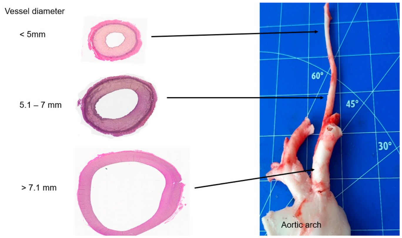
| Groups (n = 8 Specimen Each) | External Diameter (mm) | Wall Thickness (mm) |
|---|---|---|
| 1 (<5 mm) | 4.82 ± 0.01 | 0.5 ± 0.01 |
| 2 (5.1–7 mm) | 5.8 ±0.05 | 0.6 ± 0.02 |
| 3 (>7.1 mm) | 10.7 ± 0.04 | 0.75 ± 0.03 |
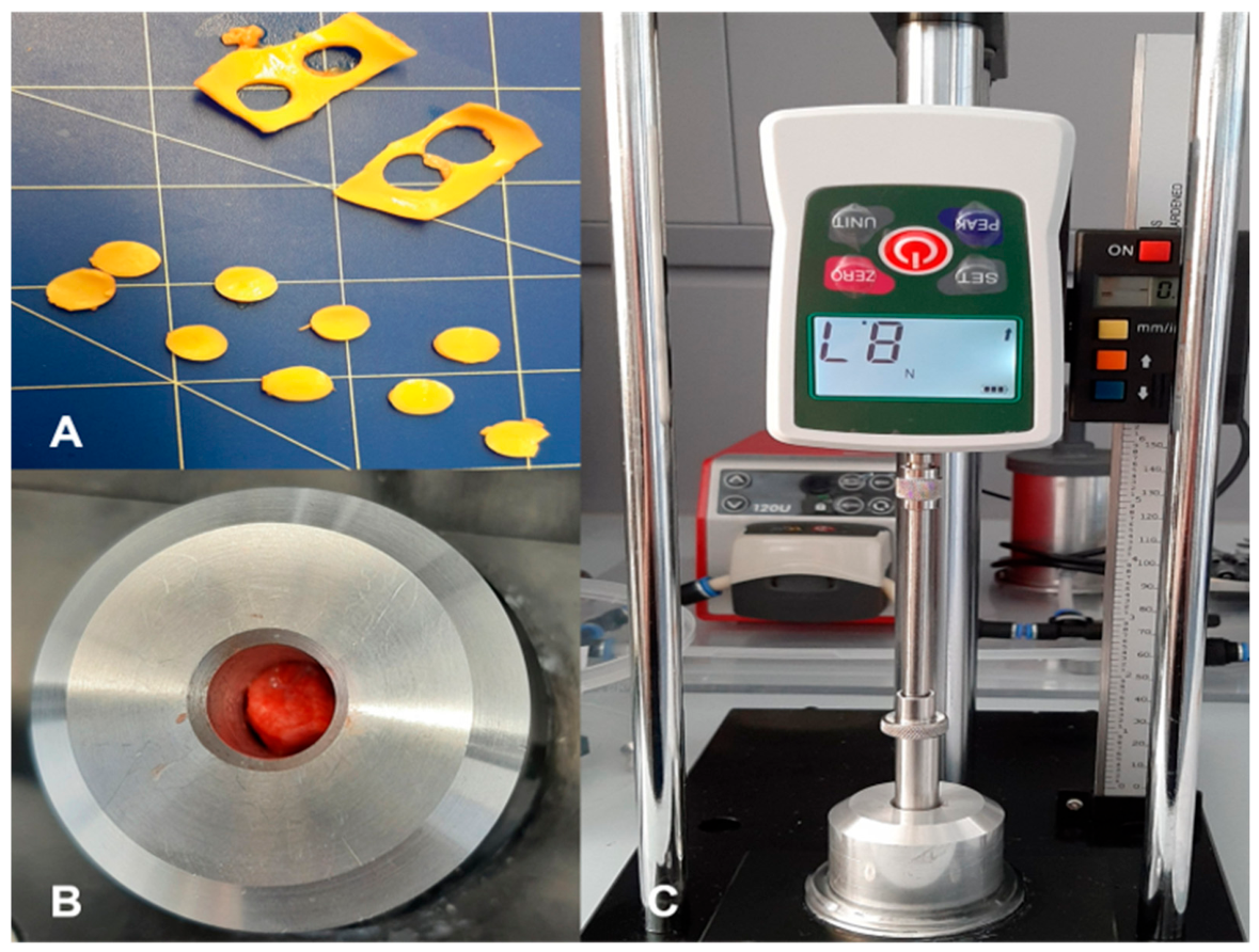
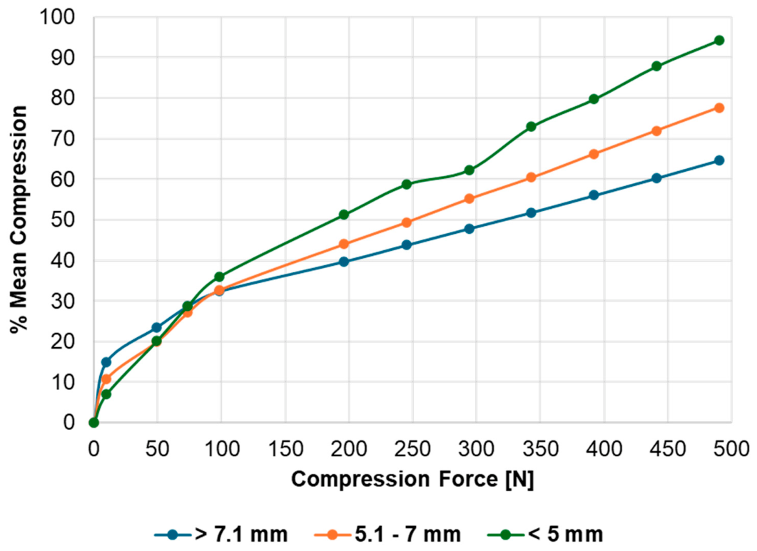
| Compression Force [N] | 0 | 9.5 | 49 | 73.5 | 98 | 196 | 245 | 294 | 343 | 392 | 441 | 490 |
|---|---|---|---|---|---|---|---|---|---|---|---|---|
| Group 1 % mean compression SD± | 0 | 6.99 ± 2.74 | 20.16 ± 2.99 | 28.83 ± 3.3 | 36.04 ± 3.35 | 51.31 ± 3.81 | 58.75 ± 3.37 | 62.83 ± 6.24 | 72.97 ± 3.84 | 79.75 ± 3.74 | 87.86 ± 4.33 | 94.25 ± 3.14 |
| Group 2 % mean compression SD± | 0 | 10.73 ± 4.74 | 19.95 ± 7.44 | 27.15 ± 7.68 | 32.67 ± 8.55 | 43.99 ± 7.18 | 49.39 ± 7.18 | 55.16 ± 7.53 | 60.4 ± 7.53 | 66.26 ± 7.92 | 72.01 ± 7.51 | 77.68 ± 7.36 |
| Group 3 % mean compression SD± | 0 | 15.09 ± 5.26 | 23.56 ± 4.99 | 28.78 ± 5.41 | 32.41 ± 5.68 | 39.76 ± 7.21 | 43.89 ± 7.51 | 47.94 ± 7.12 | 51.85 ± 7.45 | 56.04 ± 7.86 | 60.39 ± 8.27 | 64.66 ± 7.76 |
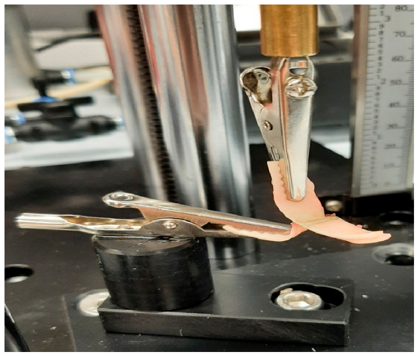
| Groups (n = 8 Specimen Each) | Mean Compression [N] | SD± | Min | Max |
|---|---|---|---|---|
| 1 (<5 mm) | 1.75 | 0.07 | 1.64 | 1.86 |
| 2 (5.1–7 mm) | 0.65 | 0.03 | 0.62 | 0.7 |
| 3 (>7.1 mm) | 0.26 | 0.11 | 0.14 | 0.45 |
Disclaimer/Publisher’s Note: The statements, opinions and data contained in all publications are solely those of the individual author(s) and contributor(s) and not of MDPI and/or the editor(s). MDPI and/or the editor(s) disclaim responsibility for any injury to people or property resulting from any ideas, methods, instructions or products referred to in the content. |
© 2026 by the authors. Licensee MDPI, Basel, Switzerland. This article is an open access article distributed under the terms and conditions of the Creative Commons Attribution (CC BY) license.
Share and Cite
Kirschbaum, A.; Raico, D.; Kirschbaum, F.; Jesinghaus, M.; Mirow, N. Tissue Structure as a Primary Factor Influencing Vascular Sealing: Results of an Ex Vivo Study on Porcine Carotid Arteries. Bioengineering 2026, 13, 719. https://doi.org/10.3390/bioengineering13070719
Kirschbaum A, Raico D, Kirschbaum F, Jesinghaus M, Mirow N. Tissue Structure as a Primary Factor Influencing Vascular Sealing: Results of an Ex Vivo Study on Porcine Carotid Arteries. Bioengineering. 2026; 13(7):719. https://doi.org/10.3390/bioengineering13070719
Chicago/Turabian StyleKirschbaum, Andreas, Dimitri Raico, Florian Kirschbaum, Moritz Jesinghaus, and Nikolas Mirow. 2026. "Tissue Structure as a Primary Factor Influencing Vascular Sealing: Results of an Ex Vivo Study on Porcine Carotid Arteries" Bioengineering 13, no. 7: 719. https://doi.org/10.3390/bioengineering13070719
APA StyleKirschbaum, A., Raico, D., Kirschbaum, F., Jesinghaus, M., & Mirow, N. (2026). Tissue Structure as a Primary Factor Influencing Vascular Sealing: Results of an Ex Vivo Study on Porcine Carotid Arteries. Bioengineering, 13(7), 719. https://doi.org/10.3390/bioengineering13070719