
Article

Study on the Kinetics of Vitamin U Release from a Cosmetic Formulation
by
Małgorzata Kucia
1,*,
Agnieszka Leśniak
1 and
Elżbieta Sikora
2
1
Department of General Chemistry, Institute of Quality and Product Management Science, Cracow University of Economics, 31-510 Cracow, Poland
2
Department of Organic Chemistry and Technology, Faculty of Chemical Engineering and Technology, Cracow University of Technology, 31-155 Cracow, Poland
*
Author to whom correspondence should be addressed.
Standards 2026, 6(3), 27; https://doi.org/10.3390/standards6030027
Submission received: 29 May 2026 / Revised: 10 July 2026 / Accepted: 14 July 2026 / Published: 16 July 2026

Abstract

S-Methylmethionine (SMM), also called vitamin U, shows antihistamine, anti-inflammatory, radioprotective and anti-irritant activity, as well as enhanced wound healing. In addition, vitamin U affects the regeneration and renewal of the skin hydrolipid mantle and offers some UVB-protective effects on the skin. Since there are currently no reports in the scientific literature concerning the release of vitamin U from cosmetic formulations, the present study was undertaken as a preliminary and exploratory pilot investigation. The research focused on evaluating the release behavior of SMM (synthetic methylmethionine), a compound recognized for its potential skin-regenerating, soothing, and protective properties, from several commonly used topical delivery systems. Therefore, the various topical formulations, including oil-in-water (O/W) and water-in-oil (W/O) emulsions, as well as hydrogel formulations, were evaluated as potential, effective vitamin U skin delivery systems. Vitamin U-loaded emulsions (O/W and W/O) differing in droplet size in the internal phase and a hydrogel were prepared. The physicochemical properties of the formulations, such as emulsion type, stability, viscosity, pH and droplet size, were evaluated. The study of vitamin U release was performed in thermostatic diffusion chambers at a temperature of T = 32 °C using the Spectra/Por Standard Regenerated Cellulose dialysis membrane. A phosphate buffer (PBS) with pH 7.4 was used as the receptor solution. The concentration of the released S-Methylmethionine was analyzed with ninhydrin-based spectrophotometric assays. The obtained results showed that the type of the formulation significantly influenced the SMM release. The highest release of SMM was observed from hydrogel and O/W emulsions.

1. Introduction

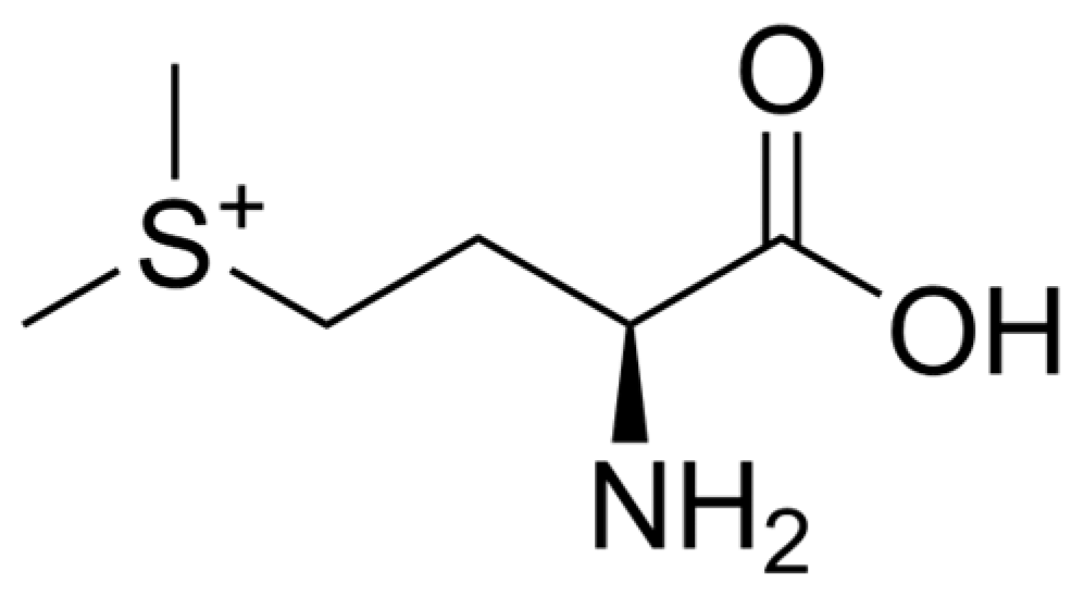
S-Methylmethionine (SMM), also called vitamin U (Figure 1), is a derivative of the amino acid methionine and can be synthesized from methionine and S-adenosylmethionine or isolated from plant materials [1]. The term “vitamin U” originates from the Latin word ulcus, meaning ulcer, and was introduced by Garnett Cheney in 1950 following observations of the anti-ulcer effects of raw cabbage juice. He reported that the systematic consumption of the juice promoted the healing of peptic ulcers. However, its classification as a vitamin has not been formally accepted [1,2].
Vitamin U was first isolated from leaves of raw cabbage in 1966. Other natural sources of SMM include commonly consumed vegetables such as asparagus [3,4], onion, kale, celery, spinach [4], maize, turnip and Brassica vegetables (e.g., broccoli, Chinese cabbage, pak choi) [4,5,6,7], as well as wheat [8] and tea [9,10]. It also occurs in juices obtained from parsley root, horseradish leaves, gooseberry [11], and soybean [12]. Cabbage (Brassica oleracea L. var. capitata) contains various biologically active compounds, including polyphenols, minerals, ascorbic acid, and amino acids such as glutamine, which exhibit anti-inflammatory properties. In addition, it contains S-methylmethionine, which has been associated with beneficial effects in the treatment and prevention of peptic ulcer disease. The content of SMM in selected plant sources is summarized in Table 1 [13,14,15,16,17,18,19,20,21].
Vitamin U exhibits a broad spectrum of biological activities. SMM demonstrates gastroprotective, cytoprotective, antioxidant, anti-inflammatory, wound-healing, radioprotective, and hypolipidemic properties [22,23].
Ji Hoon Song et al., in an animal model, demonstrated that the administration of vitamin U in the range of 10–200 mg/g body weight exhibited beneficial effects in digestive system disorders and liver injury models. As the authors showed, higher doses of SMM were required to achieve significant biological activity following oral administration [24].
In recent years, increasing attention has been paid to the potential application of S-methylmethionine in cosmetic and dermal cosmetic formulations owing to its antioxidant, anti-inflammatory, and skin-protective properties. Although several studies have demonstrated its beneficial effects on skin regeneration, oxidative stress reduction, and cellular protection, research concerning its incorporation into cosmetic formulations remains limited. Furthermore, information regarding the stability and release behavior of SMM is scarce. Previous investigations have primarily focused on pharmaceutical or microencapsulated systems intended for oral administration, while studies evaluating the release of vitamin U from conventional cosmetic formulations have not been reported.
Despite its well-documented biological activity, the application of SMM (S-methylmethionine) in cosmetic formulations remains poorly explored. Most published studies have focused on its anti-ulcer, antioxidant, and cytoprotective properties, whereas relatively few have investigated its potential as an active ingredient in topical cosmetic products. The available evidence suggests that SMM may support skin regeneration, reduce oxidative stress, and improve skin barrier function, indicating its promise for dermal cosmetic applications. However, despite this growing interest, its performance in topical formulations has not been systematically investigated. In particular, data on the release of vitamin U from commonly used cosmetic dosage forms are scarce. The most recent review by Ananian et al. highlights that the clinical translation of S-methylmethionine (vitamin U) remains limited not only due to the insufficient number of high-quality clinical studies but also because of challenges associated with the development of appropriate pharmaceutical formulations. The authors indicate that semisolid dosage forms, such as gels, films, and other topical formulations, demonstrate the greatest potential. Furthermore, they emphasize that the development of controlled-release and targeted delivery systems represents an important direction for future pharmaceutical research, with the potential to enhance the skin bioavailability of SMM (S-methylmethionine) and improve its therapeutic efficacy [25].
To address this gap, the present study was conducted as a preliminary pilot investigation to evaluate the formulation-dependent release characteristics of SMM.
The research focused on evaluating the release behavior of SMM (synthetic methylmethionine), a compound recognized for its potential skin-regenerating, soothing, and protective properties, from several commonly used topical delivery systems. Synthetic vitamin U was selected for the release studies due to its high purity, well-defined composition, and excellent batch-to-batch reproducibility. The use of a synthetic compound eliminates the influence of accompanying substances present in natural extracts, such as polyphenols, sugars, proteins, and other metabolites, which could affect the release process or interfere with analytical measurements. Consequently, the obtained results reflect the properties of the delivery system and vitamin U itself rather than the variable composition of the natural source. Furthermore, the use of synthetic vitamin U enables precise determination of the amount of the active compound in each sample and ensures high experimental reproducibility.
Since there are currently no reports in the scientific literature concerning the release of vitamin U from cosmetic formulations, the present study was undertaken as a preliminary and exploratory pilot investigation. The research focused on evaluating the release behavior of SMM (synthetic methylmethionine), a compound recognized for its potential skin-regenerating, soothing, and protective properties, from several commonly used topical delivery systems.
In contrast to advanced delivery systems (e.g., liposomes or nanoemulsions), this study intentionally focuses on conventional cosmetic vehicles, including oil-in-water (O/W) emulsions, water-in-oil (W/O) emulsions, and hydrogel systems. These formulations were selected because of their widespread application in cosmetic products and their distinct physicochemical properties, such as polarity and viscosity, which are known to influence the release and availability of active compounds.
By evaluating these basic formulation types, the study aims to establish a comparative baseline for SMM release, which may serve as a foundation for the future development of more advanced delivery systems.
The results are intended to provide initial insight into the relationship between formulation type and SMM release behavior, rather than to describe transdermal permeation or biological efficacy.
The obtained results contribute to a better understanding of the influence of formulation type on the release characteristics of SMM and may support the future optimization of cosmetic formulations designed to improve the bioavailability, stability, and overall efficacy of SMM in topical applications. However, further studies, including skin permeation and biological activity assessments, are required to confirm these findings.

Review of the Scientific Literature

In the study by Abouzed et al. [26], SMM, also known as vitamin U, was investigated for its antitumor and antioxidant effects in a rat model of liver cancer. The administration of MMSC reduced oxidative stress, improved liver function, and inhibited tumor-related changes in liver tissue. These results suggest a potential protective role of vitamin U in liver diseases.
Research on the combined use of vitamin B5 and vitamin U indicates potential benefits in treating inflammatory and erosive gastrointestinal diseases, attributed to gastroprotective and regenerative mechanisms. However, the authors emphasize the need for further clinical trials to confirm efficacy, establish optimal dosages, and assess the safety of such therapy. Combining these vitamins may serve as a valuable adjunctive treatment for gastric and duodenal mucosal damage [27].
Stoliarov et al. presented that S-methylmethionine exhibits antidepressant properties irrespectively of the duration of the depression phase or preceding treatment [28]. Sokmen B. et al. reported that vitamin U restores the GSH (glutathione levels) and consequently protects the body against growth oxidative stress. GSH plays a significant role in critical cell processes and is important in maintaining and regulating the redox status of thiol cells. In addition, the authors showed the protective effects of vitamin U against valproic acid (VPA), recommended for several therapeutic purposes in induced liver damage in rats. Vitamin U was shown to protect against VPA-induced hepatotoxicity [29].
It is worth noting that S-methylmethionine may have a beneficial effect in preventing kidney damage that occurs in patients with epilepsy. It was proven that this ingredient has a protective effect on valproic acid-induced drug used to treat kidney damage due to its antioxidant, anti-inflammatory and anti-fibrotic properties [30]. Tunali and others showed that vitamin U revealed antioxidant properties and may prevent lens damage caused by valporic acid, which occurs in antiepileptic drugs [31].
Choi et al. studied the bioavailability and intestinal cellular uptake of bioactive components from the steamed extract of graviola leaves (SGV) and graviola leaves enriched with kale extract (SGK). They evaluated the ability of SGV and SGK extracts as excipient ingredients to detoxify nicotine. They reported that graviola leaf extract enriched with kale could improve vitamin U absorption and provide a natural therapy for detoxifying nicotine [32].
Gezginci-Oktayoglu et al. investigated the effect of vitamin U on oxidative stress, such as kidney inflammation induced by valproic acid. A control study was conducted on rats. The results indicate that the vitamin may be a potential drug used in preventing and inhibiting kidney damage induced by valproic acid (VPA), as a drug used in patients with epilepsy [33].
S-methylmethionine (SMM, vitamin U) demonstrated neuroprotective effects in rat models of brain injury induced by pentylenetetrazole-induced seizures and amiodarone toxicity by reducing oxidative stress and inflammation. SMM administration restored antioxidant balance, improved the activity of protective enzymes, and reduced oxidative damage in brain tissue, indicating its potential protective role under neurotoxic conditions [34,35].
The literature also describes the results of the studies aimed at demonstrating the effect of the methionine derivative on the production of collagen and its direct effect on the multiplication of fibroblasts [34]. Kim et al. also showed that the SMM and its derivatives accelerate wound healing through increased proliferation and migration of fibroblasts through the ERK1 pathway because the pathophysiology of wounds is similar to the processes of skin aging (wrinkles, photoaging). Moreover, vitamin U affects the regeneration and restoration of the skin hydrolipidic coat [36].
Min and Kim, in the patent revealed in Ref. [37], describe the use of vitamin U (SMM) as an active ingredient in cosmetic and dermatological preparations intended for skin regeneration, wound healing, and wrinkle reduction. The formulations contained vitamin U (synthetic SMM) at a concentration of approximately 1% in creams, lotions, and O/W emulsions. It was demonstrated that vitamin U formulations significantly accelerated wound healing and improved the appearance of wrinkles (clinical evaluation of cream/lotion applied twice daily for two months). The stability of a cream containing 1% SMM was also examined after storage for four weeks under different thermal conditions (RT, 4 °C, 45 °C). The formulations showed good stability, with only minor changes in viscosity (≤10%), pH (≤0.1–0.2), color and odor [38].
Min and Kim [39] showed that the composition of SMM has an excellent effect on healing wounds and repairing skin wounds. They highlight the cytoprotective and anti-inflammatory properties of SMM, emphasizing that its stability within emulsions or gels allows for prolonged contact with the skin, which is essential for therapeutic efficacy. While no quantitative release data are reported, the description implies that the formulations were specifically designed to achieve a gradual release profile and sustained availability of SMM at the site of application.
The photoprotective effects of SMM were also demonstrated by reducing skin damage caused by UV radiation. Studies provided by Kim et al. evidence the photoprotective effect of SMM on KPC (keratinocyte progenitor cells) and hDF (human skin fibroblasts) from UVB irradiation through the activation of the MAPK pathway (mitogen-activated kinases) [37]. In addition, many studies have already reported that the combination is able to increase SMM penetration and deposition in the skin through the addition of oleic acid and ethanol at various concentrations [40,41]. The effectiveness of topical preparations depends on the ability of the active substance to penetrate the epidermal barrier and accumulate in specific layers of the skin. Modern transdermal delivery systems, including patches and other topical carriers, enable the controlled release of active ingredients and enhance their bioavailability in the skin through appropriate formulation design and optimization of the physicochemical properties of the carrier [42]. The effectiveness of cosmetic preparations is closely related to the deposition (concentration) of SMM in the epidermis/dermis. The amount of compound present in the skin after twelve hours was tested with appropriate assumptions in a hairless mouse model. It is known that permeability through human skin is at least two times lower than that of hairless mouse skin [43,44]. S-Methylmethionine and plant extracts, which are the sources of vitamin U, show potential as cosmetic active ingredients with anti-aging properties. However, there has been no systematic study concerning SMM skin penetration/deposition. Taking into consideration the fact that SMM is a substance with high hydrophilicity, its skin permeation is expected to be limited. It is generally known that apart from the active properties, such as molecule size, solubility and degree of dissociation, in addition to the possibility of creating chemical bonds with components of the skin’s structure, the physiochemical form of the cosmetic product (the cosmetic vesicle) is also of great importance. Furthermore, the use of a skin permeation enhancer may improve the therapeutic efficacy of the actives. In order to increase the permeability of this substance, it would be worth using liposomes as carriers of the active substance. In medicine, they effectively deliver drugs into the bloodstream, slowly releasing the active substance and thus protecting the body from adverse effects. Their potential benefits have been particularly appreciated by the cosmetics and pharmaceutical industries, as well as in the food industry [45,46].

2. Materials and Methods

2.1. Materials

In this work, SMM-loaded (0.1 w/%) oil-in-water (O/W) and water-in-oil (W/O) emulsions and hydrogel (HG) were prepared. Due to the low stability of vitamin U (it degrades when exposed to heat or high pH), resulting in a low yield while obtaining and isolating it, its extraction from plant extracts is not economically viable [47]. For this reason, we used chemically synthesized SMM in our work. All of the raw materials used in the formulations were cosmetic or pharmaceutical grade. SMM (Table 2) was purchased from Sigma-Aldrich (St. Louis, MO, USA). Versaflex™ V-175, caprylic/capric triglyceride (MCT), and lanolin were kindly supplied by Croda Poland Sp. z o.o. (Cracow, Poland). Cera alba, Cocos nucifera oil, and vitamin E were supplied by Alfa Sagittarius Sp. z o.o. (Cracow, Poland). Sodium benzoate was purchased from Warchem Sp. z o.o. (Zakręt, Poland), and carbomer (TEGO® Carbomer 750 HD) was purchased from Evonik Industries AG (Essen, Germany). Table 3 shows the composition of the prepared formulations.

2.2. Preparation of Formulations and Their Characteristics

The emulsions (E1O/W, E2W/O) were prepared using a classic emulsification method. Homogeneous internal phase was slowly added to the external ones (phase A into phase B in the case of O/W, and phase B into phase A in the case of W/O), and then the systems were mixed at temperature T = 60 °C using a mechanical stirrer (IKA®) with n = 500 s−1 rate for t = 30 min. To obtain emulsions differing in droplet size, additional homogenization steps (Ultra Turex homogenizer (IKA-Werke GmbH & Co. KG, Staufen, Germany), n = 12,000 s−1, t = 2 min, T = 25 °C) were performed in the cases of E3O/W and E4W/OIn all cases, vitamin U was dissolved in water before being added in both of phases.
The vitamin U-loaded hydrogel was prepared using an IKA® mechanical stirrer (IKA-Werke GmbH & Co. KG, Staufen, Germany) at n = 500 s−1 for t = 30 min and T = 60 °C. Vitamin U was added after pre-mixing the remaining formulation ingredients. All prepared formulations were evaluated for physicochemical properties. Their stability was tested using the centrifuge method (Andreas Hettich GmbH & Co. KG, Tuttlingen, Germany) at a speed of 3000 revolutions per minute for 15 min. In the temperature-variable tests, emulsion samples were placed alternately at T = 40 °C and at T = −20 °C for a period of 24 h, and the cycles were repeated three times. Both the type of emulsion (by measuring electrical conductivity) and the pH values were evaluated using a Mettler Toledo SevenMulti™ pH/conductivity meter (Mettler-Toledo AG, Greifensee, Switzerland). An optical microscope with a computer-controlled video track (Motic images Plus 2.0 ML) was used to measure the average drop size of the emulsions. The rheological properties of the vitamin U-loaded formulations were performed using a Brookfield model R/S Plus rheometer (Brookfield Engineering Laboratories, Inc., Middleboro, MA, USA) equipped with a cone-plate measuring system at temperatures of 25 °C and 32 °C in the range of shear rate from 1 to 100 s−1. Measurements were made before and after the release tests. Each determination was performed in triplicate. The microbiological stability of SMM-loaded systems was checked using the Schülke Mikrocount® Combi kit (Sigma-Aldrich, St. Louis, MO, USA) [49]. The test vials were placed in an incubator for an incubation time ranging from 24 to 48 h. No bacteria, mold or yeast was detected.

2.3. Study of Properties of Vitamin U-Loaded Formulations

All prepared formulations were evaluated for physicochemical properties. The evaluation of stability in a cosmetic formulation is related to determining whether any undesirable changes occurred in the formulation (such as phase separation, change in color, odor, formation of lumps, etc.). Their stability was tested using the centrifuge method (Andreas Hettich GmbH & Co. KG, Tuttlingen, Germany) at the speed of 3500 revolutions per minute for 15 min, and for the temperature-variable tests, emulsion samples were placed alternately at T = 40 °C and at T = −20 °C for a period of 24 h; the cycles were repeated three times. Both the type of emulsion (by measuring electrical conductivity) and the pH values were evaluated using a Mettler Toledo SevenMulti™ pH/conductivity meter (Mettler-Toledo AG, Greifensee, Switzerland). An optical microscope (Motic LED BA210, Xiamen, China) equipped with a CCD camera and connected to digital image processing software (Motic Images Plus 2.0 ML Xiamen, China) was used to observe the emulsions’ morphology and measure droplet size. Automatic calculations were performed on the microscope images, and the average diameter of the droplets was calculated from the average circumference of the object using the formula for the circumference of a circle. For non-homogenized emulsion samples, the average number of objects was 650, and for homogenized emulsions, it was 960. In contrast, the average diameter of the object for non-homogenized emulsion was between 9.5 and 10.9 micrometers, and for homogenized emulsion it was between 6.3 and 7.8 micrometers. The rheological properties of the vitamin U-loaded formulations were performed by use of a Brookfield model R/S Plus rheometer equipped with a cone-plate measuring system at temperatures of 25 °C and 32 °C in the range of shear rate from 1 to 100 s−1. Measurements were made before and after the release tests. The microbiological stability of systems containing SMM was tested using the Schülke Mikrocount® Combi kit [50]. The tests were performed on fresh samples and after 14 and 28 days. At the time of testing, the test vials were placed in an incubator for 24 to 48 h and then checked for the presence of pathogenic microorganisms.

2.4. Vitamin U Release Studies

The release study was carried out at temperature T = 32 °C in thermostatic diffusion cell system using Spectra/Por (Spectrum Laboratories, Inc., Rancho Dominguez, CA, USA) standard regenerated cellulose (RC) dialysis membrane (MWCO; 6–8 kDa). Phosphoric buffer (PBS of pH = 7.4) was used as a receptor solution because it is the most commonly recommended receptor medium for in vitro release testing, providing sink conditions and analytical stability of SMM (S-methylmethionine). The dialysis bags were filled with prepared formulation samples of 3 g and placed in thermostatic chambers filled with 200 mL of PBS to maintain sink conditions. At specified intervals during 24 h, 1 mL samples were withdrawn from the receptor compartment, and an equivalent amount of solvent was added in order to maintain a constant volume. The number of replicates per formulation was n = 3. The concentration of vitamin U in the receptor solution was determined based on the modified conventional ninhydrin method [51,52,53] by use of a Macherey-Nagel UV–Vis spectrophotometer (Macherey-Nagel GmbH & Co. KG, Düren, Germany). To each of the analyzed samples, ethanolic solution of ninhydrin (2 w/%) was added in a 1:1 ratio and heated for 15 min. Then, the absorbance at a wavelength of 568 nm was measured. The concentration of SMM was calculated on the basis of the pre-determined calibration curve of absorbance, (A), as a function of SMM concentration (CSMM) (A = 0.0023 · CSMM + 0.0186; R2 > 0.999).

2.5. Evaluation of Release Kinetics

The analysis of vitamin U release was evaluated by linear regression for four mathematical models: zero-order (1), first-order (2), Higuchi (3) and Korsmeyer–Peppas (4) [54,55]:
Qt = Q0 + K0 · t
log Qt = log Q0Kt/2.303
Qt = Q0 + KH · t1/2
Qt = Q0 + KHP · tn
where
Qt—the amount of drug dissolved in time t;
Q0—the initial amount of drug (most times, Q0 = 0);
K0—zero-order release constant;
K—first-order rate constant;
KH—Higuchi kinetic constant;
KHP—Korsmeyer–Peppas release constant;
n—diffusional release exponent;
t—time.

3. Results and Discussion

Characteristic of the Formulations

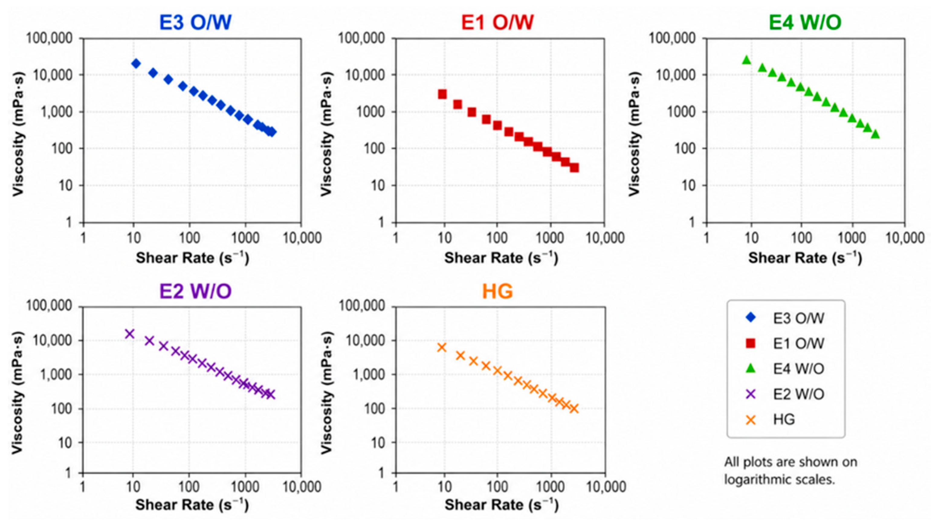
Kinetically stable vitamin U-loaded emulsions with droplet sizes ranging from 7 to 12.5 µm and SMM-loaded hydrogels (HG) were obtained. Table 4 presents the properties of the obtained formulations. According to the assumptions, depending on the type of emulsifier used, two different types of stable emulsions (W/O or O/W) were obtained. The use of the homogenization process, as an additional step of the emulsification procedure, allowed for emulsions (E3O/W, E4W/O) with a higher degree of internal phase dispersion to be obtained (Table 4, Figure 2), with mean droplet diameters approximately 3 µm lower than in non-homogenized emulsions (E1O/W, E2W/O). All obtained preparations exhibited pH values slightly above physiological skin pH (5.9–6.1), ranging between 6.6 and 7.1. Although these conditions do not fully reflect the physiological skin surface environment, they were selected to ensure formulation stability and compatibility with in vitro release testing conditions.
No phase separation was observed in the centrifugal test or during accelerated aging. Microbiological stability tests did not reveal contamination by bacteria, molds, or pathogens.
Based on microscopic analysis, it was confirmed that homogenization enhanced the degree of internal phase dispersion. Droplet diameters ranged from approximately 7.5 to 8.1 µm and were smaller than those observed in non-homogenized emulsions. This reduction in droplet size is particularly relevant, as it increases the interfacial surface area available for mass transfer, which may significantly influence release kinetics. It can also be noted that the homogenization process resulted in more homogeneous systems.
Viscosity changes during storage were small (≤10% relative to initial values). The structure of O/W emulsions remained stable, and SMM did not destabilize the systems. It should be emphasized that these findings refer to short-term stability evaluation only and do not allow for conclusions regarding long-term shelf-life performance.
Figure 2 presents the viscosity curves of the prepared SMM-loaded formulations. All samples exhibited shear-thinning behavior, with viscosity decreasing as shear rate increased, indicating non-Newtonian, pseudoplastic flow. O/W emulsions showed lower viscosity compared to hydrogels and W/O emulsions.
Viscosity is a key parameter in cosmetic and pharmaceutical formulations, affecting spread ability, sensory properties, and diffusion behavior [56].
In the present study, the analysis of release kinetics was limited to physicochemical characterization and aimed at evaluating formulation-dependent differences, rather than predicting in vivo performance or therapeutic efficacy.
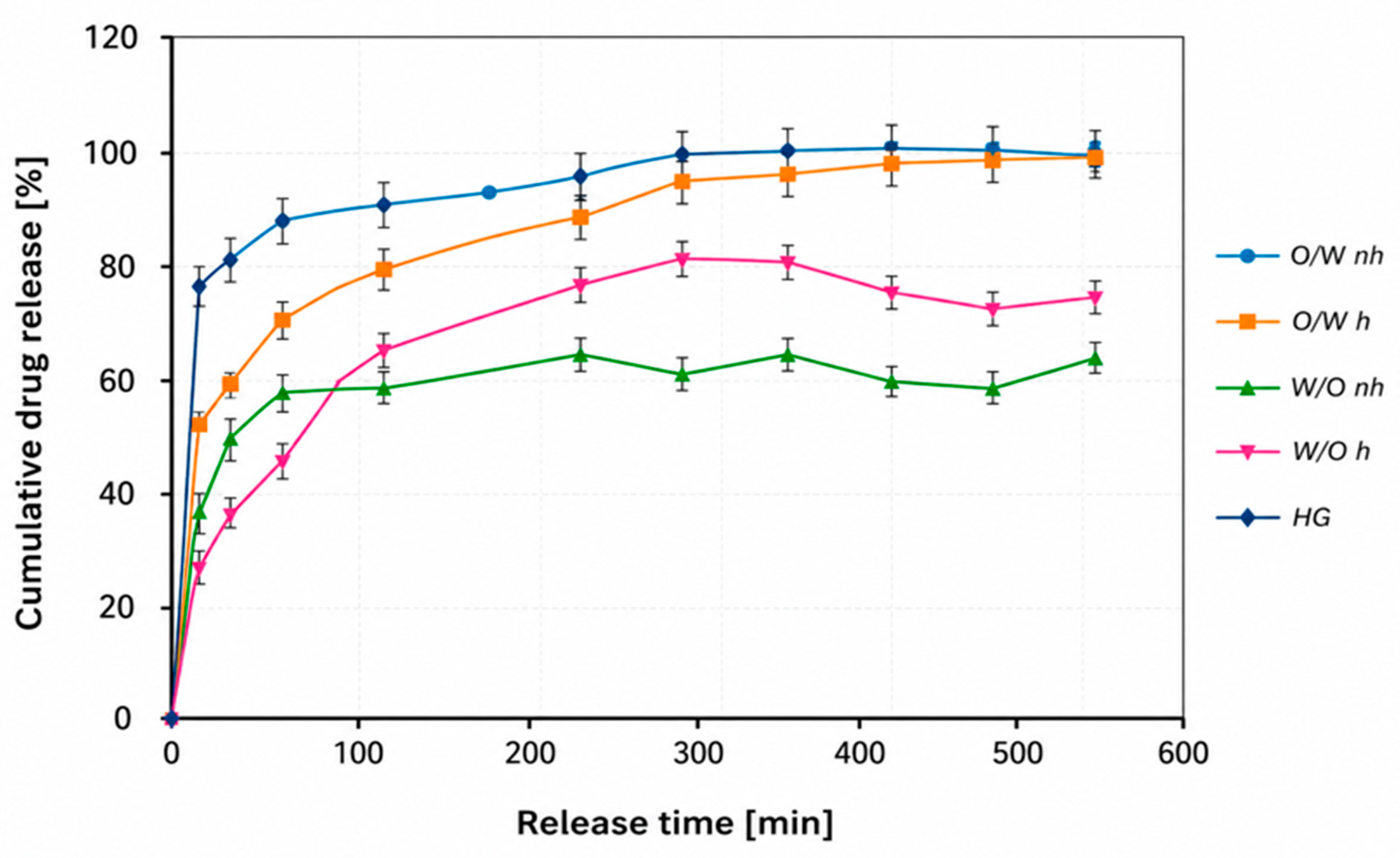
The amount of SMM released was expressed as the fraction of the total incorporated substance as a function of time. Figure 3 shows the release profiles of vitamin U from the investigated formulations.
For hydrogels and O/W emulsions (E1O/W, E3O/W), a high percentage of SMM release was observed, reaching a plateau (~100%) after approximately 300, 400, and 480 min, respectively. In contrast, W/O emulsions (E4W/O and E2W/O) showed lower maximum release values (above 87% and 63%, respectively).
The observed differences are related to the hydrophilic nature of SMM (logP = −3.3). Hydrophilic compounds are preferentially released from hydrophilic matrices such as hydrogels and O/W emulsions (containing 76.7% and 98.9% water, respectively), whereas release from lipophilic bases such as W/O emulsions is limited.
Additionally, droplet size plays a critical role in the release process. Smaller droplets increase the interfacial surface area and reduce diffusion path length, facilitating the mass transfer of the active substance into the release medium. This is consistent with the higher release rates observed for homogenized systems.
A relationship between viscosity and release behavior was also observed (Table 5): lower viscosity systems exhibited higher SMM release rates, likely due to reduced diffusional resistance.
After 7 h, the percentage of released SMM reached 99.8% for non-homogenized O/W emulsion (E1O/W) and 93.6% for homogenized emulsion (E3O/W). The extension of the study duration to 24 h allowed for confirmation of complete release profiles and enabled a comparison between formulations with both rapid and sustained release characteristics.
Viscosity appears to significantly influence active ingredient transport. O/W emulsions, characterized by lower viscosity, exhibited higher release rates.
The release data were fitted to several kinetic models (zero-order, first-order, Higuchi, and Korsmeyer–Peppas) to better understand the release mechanism. The coefficient of determination (R2) was used to identify the best-fitting model, and the corresponding kinetic parameters were calculated (Table 6).
The release kinetics of vitamin U from the cosmetic formulations were evaluated using zero-order, first-order, Higuchi, and Korsmeyer–Peppas models. The goodness of fit was assessed based on the coefficient of determination (R2).
Among the tested kinetic models, the Korsmeyer–Peppas model provided the best fit for all formulations, with R2 values ranging from 0.7931 to 0.9703. In comparison, the zero-order, first-order, and Higuchi models exhibited considerably lower coefficients of determination (R2 = 0.5226–0.7602), indicating that these models did not adequately describe the release behavior of vitamin U.
The highest goodness of fit was observed for formulation E4W/O (R2 = 0.9703), followed by E2W/O (R2 = 0.9563) and E1O/W (R2 = 0.9404), suggesting that the release of vitamin U from these emulsions is predominantly governed by mechanisms described by the Korsmeyer–Peppas model. Formulations E3O/W (R2 = 0.8250) and the hydrogel (HG) (R2 = 0.7931) also followed this model, although with a lower degree of correlation, indicating a more complex release behavior.
The release exponent (n), determined using the Korsmeyer–Peppas model, provided additional information regarding the release mechanism. Formulations E2W/O (n = 0.59) and E4W/O (n = 0.60) exhibited higher n-values, suggesting a slight deviation from ideal Fickian diffusion [57,58] and potentially indicating anomalous (non-Fickian) transport, in which both vitamin U diffusion and relaxation (swelling) of the polymeric chain contribute to the release process. In contrast, formulation E3O/W (n = 0.10) and the hydrogel (HG, n = 0.20) were characterized by significantly lower n-values, suggesting that vitamin U release was primarily controlled by Fickian diffusion.
Overall, these findings indicate that the Korsmeyer–Peppas model most accurately describes vitamin U release from all investigated cosmetic formulations. Moreover, the emulsion systems, particularly the W/O formulations, exhibited more complex release mechanisms involving both diffusion and matrix relaxation, whereas the hydrogel and formulations E3O/W and E1O/W showed diffusion-dominated release.
To the best of our knowledge, no published studies have described the release of SMM (vitamin U) from conventional cosmetic formulations such as emulsions or hydrogels. Previous work by Koksal et al. investigated microencapsulated cabbage extract intended for oral delivery. Release was evaluated under simulated gastrointestinal conditions over 48 h and showed pronounced pH-dependent behavior. Under simulated gastric conditions (pH 1.2), only approximately 27% of the encapsulated extract was released, indicating good stability of the microcapsules in an acidic environment. In contrast, under simulated intestinal conditions (pH 7.4), cumulative release reached 93.6% for gelatin/gum Arabic microcapsules and 82.9% for gelatin/sodium alginate microcapsules. More than 50% of the encapsulated extract was released within the first 4 h, followed by a slower, sustained release phase, consistent with an initial burst release from the microcapsule surface and subsequent diffusion-controlled release from the capsule core [13].
In contrast, the present study provides the first baseline release data for conventional cosmetic vehicles, which are directly relevant to topical formulation development.
Kinetic analysis indicated that the release of vitamin U from the emulsions was predominantly diffusion-controlled. These findings suggest that oil-in-water emulsions may represent promising carriers for the topical delivery of SMM owing to their favorable in vitro release characteristics.
However, it should be emphasized that the dialysis membrane model employed in this study evaluates formulation-controlled release rather than skin permeation. Therefore, the obtained results cannot be directly extrapolated to in vivo conditions. Further studies using biologically relevant models, such as Franz diffusion cells with excised human or porcine skin, are required to evaluate skin penetration and confirm the suitability of these formulations for dermal delivery.

4. Conclusions

The obtained results confirmed the high physicochemical and microbiological stability of the prepared vitamin U-loaded formulations. It should be noted, however, that this stability evaluation was limited to short-term observations and does not allow for conclusions regarding long-term shelf-life performance.
The results of the release study demonstrated that the type of cosmetic base significantly influences the release of SMM. The greatest amount of the active substance was released from the hydrogel and O/W emulsions, where diffusion into the acceptor solution reached approximately 100% after 7 h. In contrast, for water-in-oil emulsions, the release reached 56.3% and 73.2% of the initial amount for non-homogenized (E2W/O) and homogenized (E4W/O) systems, respectively. These differences can be attributed to the hydrophilic nature of SMM, as well as formulation parameters such as droplet size and viscosity, which affect the interfacial surface area and mass transfer kinetics. The droplet size of the internal phase influenced the rate of SMM release, particularly in W/O emulsions, where homogenization led to improved release profiles.
In conclusion, O/W emulsions demonstrated more favorable release characteristics compared to W/O systems and may be considered promising carriers for SMM in topical formulations. However, it should be emphasized that the present study was limited to in vitro physicochemical release using a dialysis membrane model. Therefore, the results reflect formulation-controlled release rather than skin permeation and cannot be directly extrapolated to in vivo conditions or biological efficacy. Additionally, the exploratory nature of the study, including the lack of batch-to-batch reproducibility and absence of dedicated control formulations, represents a limitation that should be addressed in future work.

Directions for Future Research

Vitamin U (SMM), apart from its well-known gastroprotective effects, has been reported to exhibit properties that may support skin regeneration, including the stimulation of cell proliferation and inhibition of collagen degradation. The available patent literature suggests its potential as a dermal cosmetic ingredient with regenerative and anti-aging applications, as well as its compatibility with various formulation types. Due to the limited availability of peer-reviewed data, a more detailed discussion is currently not possible. In future studies, plant-derived (natural) vitamin U will be obtained, and its release behavior may be comparatively evaluated against synthetic S-methylmethionine (SMM). In vitro permeation studies using Franz diffusion cells and biologically relevant membranes will also be considered, together with the assessment of skin retention and the potential biological activity of SMM-containing formulations. Advanced delivery systems, such as nanoemulsions and liposomes, may be further investigated. In addition, batch-to-batch reproducibility and the long-term stability of the developed formulations will be evaluated. In the future, investigations should include additional characterization parameters, such as droplet size distribution and zeta potential measurements, to further evaluate the long-term stability and structural properties of the developed formulations.

Author Contributions

Conceptualization: E.S., M.K. and A.L.; methodology: E.S., M.K. and A.L.; software: M.K., A.L. and E.S.; validation: M.K., A.L. and E.S.; formal analysis: M.K., A.L. and E.S.; investigation: M.K., A.L. and E.S.; writing—original draft preparation: M.K., A.L. and E.S.; writing—review and editing: A.L., M.K. and E.S.; visualization: M.K. and A.L.; supervision: M.K., A.L. and E.S. All authors have read and agreed to the published version of the manuscript.

Funding

This research received no external funding.

Institutional Review Board Statement

Not applicable.

Informed Consent Statement

Not applicable.

Data Availability Statement

The original contributions presented in this study are included in the article. Further inquiries can be directed to the corresponding author.

Conflicts of Interest

The authors declare no conflicts of interest.

Abbreviations

The following abbreviations are used in this manuscript:
SMMS-methylmethionine
PBSPhosphate buffer
VPAValproic acid
MCTMedium-Chain Triglycerides

References

  1. Green, R.C.; Davis, N.B. Biosynthesis of S-methylmethionine in the jack bean. Biochim. Biophys. Acta 1960, 43, 360–362. [Google Scholar] [CrossRef] [Scilit]
  2. Cheney, G. Anti-peptic ulcer dietary factor (vitamin “U”) in the treatment of peptic ulcer. J. Am. Diet. Assoc. 1950, 26, 668–672. [Google Scholar] [CrossRef] [Scilit]
  3. Challenger, F.; Hayward, B. The occurrence of a methylsulphonium derivative of methionine in asparagus. Biochem. J. 1954, 58, iv. [Google Scholar] [PubMed]
  4. Kim, G.-H. Determination of vitamin U in food plants. Food Sci. Technol. Res. 2003, 9, 316–319. [Google Scholar] [CrossRef] [Scilit]
  5. Larina, T.V.; Gessler, N.N.; Bezzubov, A.A.; Elizarova, L.G. Vitamin U (S-methylmethionine) in Brassica vegetables. Izv. Vyss. Uchebnykh Zaved. Pishchevaya Tekhnologiya 1991, 1–3, 99–101. [Google Scholar]
  6. Kim, G.-H. Changes in vitamin U and amino acids in Korean Chinese cabbages under various drying methods. J. Food Hyg. Saf. 2003, 18, 243–250. [Google Scholar]
  7. Hong, E.; Kim, G.-H. Changes in vitamin U, amino acid and sugar levels in Chinese cabbages during storage. Korean J. Food Preserv. 2006, 13, 589–595. [Google Scholar]
  8. Bourgis, F. S-methylmethionine plays a major role in phloem sulfur transport and is synthesized by a novel type of methyltransferase. Plant Cell 1999, 11, 1485–1498. [Google Scholar] [CrossRef] [Scilit] [PubMed]
  9. Kiribuchi, T.; Yamanishi, T. Studies on the flavor of green tea. Part IV. Dimethyl sulfide and its precursor. Agric. Biol. Chem. 1963, 27, 56–59. [Google Scholar] [CrossRef] [Scilit]
  10. Ohtsuki, K.; Kawabata, M.; Taguchi, K.; Kokura, H.; Kawamura, S. Determination of S-methylmethionine (vitamin U) in various teas. Agric. Biol. Chem. 1984, 48, 2471–2475. [Google Scholar] [CrossRef] [Scilit]
  11. Bezzubov, A.A.; Gessler, N.N. Plant sources of S-methylmethionine. Appl. Biochem. Microbiol. 1992, 28, 423–429. [Google Scholar]
  12. Morisaki, A.; Yanada, N.; Yamanaka, S.; Matsui, K. Dimethyl sulfide as a source of seaweed-like aroma in cooked soybeans and correlation with its precursor S-methylmethionine. J. Agric. Food Chem. 2014, 62, 8289–8294. [Google Scholar] [CrossRef] [Scilit] [PubMed]
  13. Koksal, E.; Gode, F.; Ozaltin, K.; Karakurt, I.; Suly, P.; Saha, P. Controlled release of vitamin U from microencapsulated Brassica oleracea L. var. capitata extract for peptic ulcer treatment. Food Bioprocess Technol. 2023, 16, 677–689. [Google Scholar] [CrossRef] [Scilit]
  14. Carvalho, C.A.; Fernandes, K.M.; Matta, S.L.P.; Silva, M.B.; Oliveira, L.L.; Fonseca, C.C. Evaluation of antiulcerogenic activity of aqueous extract of Brassica oleracea var. capitata on gastric ulceration. Arq. Gastroenterol. 2011, 48, 276–282. [Google Scholar] [CrossRef] [Scilit] [PubMed]
  15. Lee, Y.; Kim, S.; Yang, B.; Lim, C.; Kim, J.-H.; Kim, H.; Cho, S. Anti-inflammatory effects of Brassica oleracea var. capitata methanol extract. Pharmacogn. Mag. 2018, 14, 174–179. [Google Scholar] [CrossRef] [Scilit] [PubMed]
  16. Nosek, M.; Surówka, E.; Cebula, S.; Libik, A.; Goraj, S.; Kornaś, A.; Miszalski, Z. Distribution pattern of antioxidants in white cabbage heads. Acta Physiol. Plant. 2011, 33, 2125–2134. [Google Scholar] [CrossRef] [Scilit]
  17. Rokayya, S.; Chun-Juan, L.; Zhao, Y.; Li, Y.; Sun, C.-H. Cabbage phytochemicals with antioxidant and anti-inflammatory potential. Asian Pac. J. Cancer Prev. 2014, 14, 6657–6662. [Google Scholar] [CrossRef] [Scilit] [PubMed]
  18. Samec, D.; Pavlović, I.; Salopek-Sondi, B. White cabbage (Brassica oleracea var. capitata f. alba): Botanical, phytochemical and pharmacological overview. Phytochem. Rev. 2017, 16, 117–135. [Google Scholar] [CrossRef] [Scilit]
  19. Poschner, S.; Maier-Salamon, A.; Thalhammer, T.; Jäger, W. Resveratrol and other dietary polyphenols are inhibitors of estrogen metabolism. J. Steroid Biochem. Mol. Biol. 2019, 190, 11–18. [Google Scholar] [CrossRef] [Scilit] [PubMed]
  20. Cvetković, B.R.; Pezo, L.L.; Mišan, A.; Mastilović, J.; Kevrešan, Ž.; Ilić, N.; Filipčev, B. Effects of osmotic dehydration of white cabbage on polyphenols and mineral content. LWT 2019, 110, 332–337. [Google Scholar] [CrossRef] [Scilit]
  21. Gruszecki, R.; Walasek-Janusza, M.; Caruso, G.; Zawiślak, G.; Golubkina, N.; Tallarita, A.; Zalewska, E.; Sękara, A. Cabbage in Polish folk and veterinary medicine. S. Afr. J. Bot. 2022, 149, 435–445. [Google Scholar] [CrossRef] [Scilit]
  22. Doba, S.; Buzlama, A.; Aleksenko, E.; Sviridova, O. Pharmacological Effects of S-Methylmethionine Sulfonium Chloride (Vitamin U). Res. J. Pharm. Technol. 2025, 12, 5911. [Google Scholar] [CrossRef] [Scilit]
  23. Urazaeva, L.G. Anti-inflammatory effect of methylmethionine sulfonium chloride (vitamin U). Farmakol. Toksikol. 1976, 39, 216–319. [Google Scholar]
  24. Song, J.-H.; Lee, H.-R.; Shim, S.-M. Determination of S-methyl-L-methionine from Brassicaceae vegetables and intestinal transport. J. Food Sci. 2017, 82, 36–43. [Google Scholar] [CrossRef] [Scilit] [PubMed]
  25. Ananian, A.A.; Zelenina, T.Z.; Kosenkova, S.I.; Stepanova, O.I.; Popova, A.A.; Bagatelia, Z.T.; Evzikov, G.Y.; Sysuev, B.B.; Vasil’ev, Y.L.; Bakhrushina, E.O. S-Methylmethionine (Vitamin U): A Critical Narrative Review of Pharmacological Mechanisms, Evidence Levels, and Translational Barriers. Pharmaceuticals 2026, 19, 743. [Google Scholar] [CrossRef] [Scilit]
  26. Abouzed, T.K.; Althobaiti, F.; Abdelkhlek, N.A.; Eldomany, E.B.; Nasr, N.E.; Sadek, K.M.; El-Shazly, S.A.; Kahilo, K.A.; Dorghamm, D.A. Antitumor and antioxidant activity of S-methyl methionine sulfonium chloride against liver cancer induced in wistar albino rats by diethyl nitrosamine and carbon tertrachloride. Int. J. Envirion. Res. Public Health 2021, 18, 9726. [Google Scholar] [CrossRef] [Scilit] [PubMed]
  27. Shichkin, V.P. Vitamin B5 and vitamin U review: Justification of combined use for the treatment of mucosa-associated gastrointestinal pathologies. Front. Pharmacol. 2025, 16, 1587627. [Google Scholar] [CrossRef] [Scilit] [PubMed]
  28. Stoliarov, G.V.; Mys’ko. Treatment of depressive conditions with S-methylmethionine. Zhurnal Nevrol. I Psikhiatrii Im. S. S. Korsakova 1981, 81, 1209–1212. [Google Scholar]
  29. Sokmen, B.B.; Tunali, S.; Yanardag, R. Effects of vitamin U on valproic acid-induced liver damage. Food Chem. Toxicol. 2012, 50, 3562–3566. [Google Scholar] [CrossRef] [Scilit] [PubMed]
  30. Tong, V.; Teng, X.W.; Chang, T.K.H.; Abbott, F.S. Valproic acid: Time course of lipid peroxidation biomarkers and liver toxicity. Toxicol. Sci. 2005, 86, 427–435. [Google Scholar] [CrossRef] [Scilit] [PubMed]
  31. Tunali, S.; Kahraman, S.; Yanardag, R. Vitamin U prevents lens injury in rats administered with valproic acid. Hum. Exp. Toxicol. 2015, 34, 904–910. [Google Scholar] [CrossRef] [Scilit] [PubMed]
  32. Choi, E.-H.; Lee, S.-B.; Lee, D.-Y.; Kim, G.-T.; Shim, S.-M.; Park, T.-S. Increased intestinal absorption of vitamin U in steamed extract activates nicotine detoxification. Nutrients 2019, 11, 1334. [Google Scholar] [CrossRef] [Scilit] [PubMed]
  33. Gezginci-Oktayoglu, S.; Turkyilmaz, I.B.; Ercin, M.; Yanardag, R.; Bolkent, S. Vitamin U protects against renal damage. Protoplasma 2016, 253, 127–135. [Google Scholar] [CrossRef] [Scilit] [PubMed]
  34. Bayrak, G.; Turkyilmaz, I.B.; Yanardag, R. The Protective Effect of Vitamin U on Pentylenetetrazole-Induced Brain Damage in Rats. J. Biochem. Mol. Toxicol. 2022, 36, e23169. [Google Scholar] [CrossRef] [Scilit] [PubMed]
  35. Turkyilmaz, I.B. Oxidative Brain Injury Induced by Amiodarone in Rats: Protective Effect of S-Methyl Methionine Sulfonium Chloride. Acta Chim. Slov. 2023, 70, 131–138. [Google Scholar] [CrossRef] [Scilit]
  36. Kim, W.-S.; Yang, Y.J.; Min, H.G.; Song, M.G.; Lee, J.S.; Park, K.Y.; Kim, J.J.; Sung, J.H.; Choi, J.S.; Cha, H.J. Accelerated wound healing by S-methylmethionine sulfonium. Pharmacology 2010, 85, 68–76. [Google Scholar] [CrossRef] [Scilit] [PubMed]
  37. Min, G.H.; Kim, W.-S. Cosmetic Composition Comprising Vitamin U as Active Ingredient for Healing Wounds and Reducing Wrinkles. U.S. Patent US20110237671A1, 29 September 2011. [Google Scholar]
  38. Min, G.H.; Kim, W.-S. Composition Comprising Vitamin U as Active Ingredient for Healing Skin Wound. U.S. Patent US20120172447A1, 5 July 2012. [Google Scholar]
  39. Kim, W.-S.; Seo, H.-M.; Kim, W.K.; Choi, J.S.; Kim, I.Y.; Sung, J.H. The photoprotective effect of S-methylmethionine sulfonium. Int. J. Mol. Sci. 2015, 16, 17088–17100. [Google Scholar] [CrossRef] [Scilit] [PubMed]
  40. Jung, E.; Kang, Y.P.; Yoon, I.S.; Kim, J.S.; Kwon, S.W.; Chung, S.J.; Shim, C.K.; Kim, D.D. Effect of permeation enhancers on transdermal delivery of fluoxetine. Int. J. Pharm. 2013, 456, 362–369. [Google Scholar] [CrossRef] [Scilit] [PubMed]
  41. Kim, D.D.; Chien, Y.W. Transdermal delivery of zalcitabine: In vitro skin permeation study. AIDS 1995, 9, 1331–1336. [Google Scholar] [CrossRef] [Scilit] [PubMed]
  42. Ashfaq, A.; Riaz, T.; Waqar, M.A.; Zaman, M.; Majeed, I. A comprehensive review on transdermal patches as an efficient approach for the delivery of drug. Polym.-Plast. Technol. Mater. 2024, 63, 1045–1069. [Google Scholar] [CrossRef] [Scilit]
  43. Koyama, Y.; Bando, H.; Yamashita, F.; Takakura, Y.; Sezaki, H.; Hashida, M. Comparative analysis of percutaneous absorption enhancement. Pharm. Res. 1994, 11, 377–383. [Google Scholar] [CrossRef] [Scilit] [PubMed]
  44. Jung, E.J.; Lee, E.Y.; Choi, H.K.; Ban, S.J.; Choi, S.H.; Kim, J.S.; Yoon, I.S.; Kim, D.D. Development of drug-in-adhesive patch formulation. Int. J. Pharm. 2015, 487, 49–55. [Google Scholar] [CrossRef] [Scilit] [PubMed]
  45. Choy, Y.B.; Prausnitz, M.R. The rule of five for non-oral drug delivery. Pharm. Res. 2011, 28, 943–948. [Google Scholar] [CrossRef] [Scilit] [PubMed]
  46. Sikora, E. Cosmetic Emulsions; Politechnika Krakowska: Kraków, Poland, 2019. [Google Scholar]
  47. Sikora, E.; Miastkowska, M.; Lasoń, E. Selected Skin Delivery Systems; Politechnika Krakowska: Kraków, Poland, 2020. [Google Scholar]
  48. Toyo Shinyaku Co Ltd. Method for Increasing Vitamin U Content in Plants. JP3366910B1, 27 March 2002. [Google Scholar]
  49. Sigma-Aldrich. S-Methylmethionine. Available online: https://www.sigmaaldrich.com (accessed on 6 May 2026).
  50. Schülke. Microcount Duo. Available online: https://www.schuelke.com (accessed on 6 May 2026).
  51. Moore, S.; Stein, W.H. Photometric ninhydrin method for use in chromatography of amino acids. J. Biol. Chem. 1954, 176, 367–388. [Google Scholar] [CrossRef] [Scilit]
  52. Doi, E.; Shibata, D.; Matoba, T. Modified colorimetric ninhydrin methods for peptidase assay. Anal. Biochem. 1981, 118, 173–184. [Google Scholar] [CrossRef] [Scilit] [PubMed]
  53. Dubin, A. Wprowadzenie do Chemii Białek; Uniwersytet Jagielloński: Kraków, Poland, 2003. [Google Scholar]
  54. Costa, P.; Sousa Lobo, J.M. Modeling and comparison of dissolution profiles. Eur. J. Pharm. Sci. 2001, 13, 123–133. [Google Scholar] [CrossRef] [Scilit] [PubMed]
  55. Dash, S.; Murthy, P.N.; Nath, L.; Chowdhury, P. Kinetic modeling on drug release. Acta Pol. Pharm. 2010, 67, 217–223. [Google Scholar] [PubMed]
  56. Gallegos, C.; Franco, J.M. Rheology of food, cosmetics and pharmaceuticals. Curr. Opin. Colloid Interface Sci. 1999, 4, 288–293. [Google Scholar] [CrossRef] [Scilit]
  57. Vigata, M.; Meinert, C.; Hutmacher, D.W.; Bock, N. Hydrogels as Drug Delivery Systems: A Review of Current Characterization and Evaluation Techniques. Pharmaceutics 2020, 12, 1188. [Google Scholar] [CrossRef] [Scilit] [PubMed]
  58. Zengin, A.; Castro, J.P.O.; Habibovic, P.; van Rijt, S.H. Injectable, Self-Healing Mesoporous Silica Nanocomposite Hydrogels with Improved Mechanical Properties. Nanoscale 2021, 13, 1144–1154. [Google Scholar] [CrossRef] [Scilit] [PubMed]
Figure 1. Chemical structure of SMM.
Standards 06 00027 g001
Figure 2. Viscosity curves of the SMM-loaded formulations (T = 32 °C).
Standards 06 00027 g002
Figure 3. Profiles of SMM release from emulsions differing in type and droplet size and from hydrogel (as reference samples).
Standards 06 00027 g003
Table 1. Level of vitamin U in various food plant resources [4].
Name of ResourcesContent of Vitamin U (mg/100 g)
Asparagus18.7
Broccoli18.9
Bud of aralia19.3
Celery8.3
Crown daisy11.1
Garlic2.8
Green onion2.6
Kale23.4
Kimchi4.7
Onion2.7
Pak choi34.0
Sanmanul14.4
Shepherd’s purse3.4
Spinach45.2
Wasabi18.7
White radish4.7
Table 2. Physicochemical properties of SMM [48].
Name of PropertySpecifications
Appearance (color)Colorless or white
Appearance (form)Powder
Molar mass199.7 g/mol
pH4.0 to 5.5
Clarity and color of solutionColorless and clear
Loss on drying≤3.0%
Miscellaneous testsHeavy metals ≤ 0.002%
Residue on ignition≤0.1%
Argentometer titration97.0–103.0%
Solubility (method)H2O: 100 mg/mL
Solubility (turbidity)Clear
Table 3. Formulation composition.
PhaseComponents (INCI Name)Content [w/%]
O/WW/OHG
ACaprylic/Capric Triglyceride (MCT)1723.4-
Cocos Nucifera oil313-
Cera alba-10-
Lanolin-15-
Vitamin E0.20.2-
BSucrose Palmitate, Glyceryl Stearate, Glyceryl Stearate Citrate, Sucrose, Mannam, and Xanthan Gum (Versaflex™ V-175)2.5--
Carbomer--0.5
Sodium benzoate0.50.50.5
Vitamin U (SMM)0.10.10.1
Aqua76.737.898.9
Table 4. Physical properties of emulsions.
Name of FormulationsStabilitypH
(±SD)
Conductometric Method, [mS/cm]
(±SD)
The Droplet Size d [µm]
(±SD)
Centrifugal MethodMethods of Thermal Shocks
0°C40°C
E1O/W++6.99 ± 0.1149.2 ± 0.110.4 ± 0.5
E2W/O++7.01 ± 0.520.085 ± 0.111.4 ± 0.1
E3O/W++6.89 ± 0.1 147.1 ± 0.17.3 ± 0.5
E4W/O++6.61 ± 0.190.085 ± 0.17.5 ± 0.3
HG++6.77 ± 0.42123.5 ± 0.1-
+ stable formulation; E1O/W, E2W/O—non-homogenized emulsions; E3O/W, E4W/O—homogenized emulsions; HG—hydrogel.
Table 5. The influence of products’ viscosity on vitamin U release process.
Type of EmulsionViscosity [mPa·s]
(γ = 50 s−1, T = 32 °C)
(±SD)
Release Amount for 7 h [%]
(±SD)
E1O/W190 ± 6099.8 ± 0.2
E2W/O6680 ± 74056.3 ± 1.7
E3O/W800 ± 11093.6 ± 2.3
E4W/O769± 22073.2 ± 1.4
HG1820 ± 10099.3 ± 0.5
Table 6. The kinetic model parameters fitting the release results.
Model
FormulationZero-OrderFirst-OrderHiguchiKorsmeyer-Peppas
R2Ko
[μg/h]
R2Kt
[h−1]
R2KH
[μg/h1/2]
KHP
[h−n]
R2n
E1O/W0.68970.00020.57260.03550.68970.00010.00160.94040.51
E3O/W0.57900.19560.52670.13470.57900.097811.16210.82500.1
E2W/O0.71880.06700.54760.11880.71880.03358.82030.95630.59
E4W/O0.76020.06940.54200.11850.76020.03470.40890.97030.6
HG0.53920.95420.52260.15740.53920.477128.00790.79310.2
Disclaimer/Publisher’s Note: The statements, opinions and data contained in all publications are solely those of the individual author(s) and contributor(s) and not of MDPI and/or the editor(s). MDPI and/or the editor(s) disclaim responsibility for any injury to people or property resulting from any ideas, methods, instructions or products referred to in the content.