Review

The Role of Zwitterionic Materials in the Fight against Proteins and Bacteria

by
Montserrat Colilla
1,2,
Isabel Izquierdo-Barba
1,2 and
María Vallet-Regí
1,2,*
1
Departamento de Química en Ciencias Farmacéuticas, Facultad de Farmacia, Universidad Complutense de Madrid, Instituto de Investigación Sanitaria Hospital 12 de Octubre i + 12, Plaza Ramón y Cajal s/n, 28040 Madrid, Spain
2
Center on Bioengineering, Biomaterials and Nanomedicine (CIBER-BBN), 28040 Madrid, Spain
*
Author to whom correspondence should be addressed.
Medicines 2018, 5(4), 125; https://doi.org/10.3390/medicines5040125
Submission received: 31 October 2018 / Revised: 17 November 2018 / Accepted: 19 November 2018 / Published: 22 November 2018

Abstract

:
Zwitterionization of biomaterials has been heightened to a potent tool to develop biocompatible materials that are able to inhibit bacterial and non-specific proteins adhesion. This constitutes a major progress in the biomedical field. This manuscript overviews the main functionalization strategies that have been reported up to date to design and develop these advanced biomaterials. On this regard, the recent research efforts that were dedicated to provide their surface of zwitterionic nature are summarized by classifying biomaterials in two main groups. First, we centre on biomaterials in clinical use, concretely bioceramics, and metallic implants. Finally, we revise emerging nanostructured biomaterials, which are receiving growing attention due to their multifunctionality and versatility mainly in the local drug delivery and bone tissue regeneration scenarios.

1. Introduction

The increased usage of implantable medical devices is likely to result in a rise in the number of infections that are associated to these cases. Bacterial contamination during biomaterial implantation is often inevitable, provoking a battle between host cells and bacteria that may eventually cause the infection. This is a devastating complication that presents a heterogeneous clinical profile being considered as the most difficult infection disease to treat with serious clinical and socio-economic implications [1,2]. Biomaterials-associated infections generally include bacterial adhesion, colonization, and biofilm formation on the biomaterial surfaces. In general, these infections are mainly caused by different pathogens as Staphylococcus epidermidis and Staphylococcus aureus [3], although Escherichia coli and Pseudomonas aeruginosa are also present [4]. These bacterial colonizations result in an inflammatory reaction and are accompanied by significant morbidity and mortality rate. For all these reasons, there is an urgent need to develop biomaterials with improved properties that are able to provide solution to this serious clinical complication [4]. In this sense, preventive strategies that are aimed at inhibiting the first stages of any infective process constitute a powerful and promising alternative to tackle this issue [5].
Much research effort is being dedicated to develop new approaches to modify biomaterial surface to inhibit the bacterial adhesion. This could be an attractive alternative to antibiotics, which are associated with severe side effects, that moreover could create bacterial resistance. In this sense, it has recently been recognized that both nanotopography and chemical surfaces show an essential role in bacterial adhesion and biofilm formation [6]. In this sense, nanostructured surfaces currently represent a good alternative as bacterial-repelling surfaces [7,8]. These surfaces comprise a variety of nanotubes- or nanoparticle-based surfaces and nanostructured coatings [9], which create a superhydrophobic surface (also called leaves lotus effect) repelling the bacteria adhesion and compromising in most of the cases to the host cell-tissue integration [10].
Concerning chemical modification to create bacterial-repelling surfaces, zwitterionization has emerged as a revolutionary approach to provide biomaterials of high resistance to nonspecific protein adsorption, bacterial adhesion, and/or biofilm formation [11]. This strategy also allows for the preservation of the biomaterial biocompatibility in terms of host cell adhesion and colonization, cytotoxicity, and differentiation.
The aim of this review manuscript is to describe the different strategies developed so far for the zwitterionization of biomaterials. First, we focus on biomaterials in clinical use, concretely bioceramics and metallic implants. Later on, we revise the recent advances on nanostructured biomaterials, which have gained much attention by the scientific community, owing to their great potential and versatility in local drug delivery and bone tissue regeneration. To understand the processes underlying the different behaviors of zwitterionic surfaces by repelling bacteria, meanwhile allowing host cell colonization, an overview of the significance of the “race for the surface” concept between bacteria and eukaryotic cells is also given.

2. Tuning the Surface Properties of Biomaterials

The biocompatibility of the biomaterials is tightly related to the performance of cells when they came in contact and adhere to its surface. In this regard, the surface features of these materials, such as their topography chemistry or surface energy, become essential pillars in their biocompatibility [6]. Moreover, depending of the bioceramic functionality, the specific requirements in terms of cells/proteins adhesion are totally different. Therefore, in bone tissue regeneration applications, tuning the biomaterials surface to trigger bone bonding at the same time that inhibiting bacterial colonization constitutes an exciting challenge to achieve better clinical outcomes.
Bacteria exist in nature under two states: planktonic (free-floating bacteria) and biofilm (sessile microorganism communities). From all bacteria, just a small fraction (≈1%) exists in the free floating form, while around the 99% appear forming biofilms [12]. It is important to denote that the primary and harder barrier in treating S. aureus infections is often attributed to the formation of bacterial biofilm. In this sense, the biofilms are mainly built by big sessile bacteria communities that are embedded in a self-produced extracellular polymeric matrix or glycocalyx [13]. Within the biofilm, bacteria grow protected from environmental stress and resist attack by antibiotics, disinfectants, and the immune system. [14]. Biofilm formation involves a sequence of four steps, including: (i) adhesion, initial attachment of the bacteria to the surface of the tissue implant; (ii) growth, bacterial aggregation and accumulation in multiple bacterial layers; (iii) biofilm maturation; and, (iv) dispersion, detachment of bacteria from the biofilm and spreading to other places (asepsis state) [15].
In 1987, the orthopedic surgeon Anthony G. Gristina described the concept of “race for the surface” to predict the evolution of an implant in the specific relation to an infection process [16]. This concept contemplates “a race” between the eukaryotic cells (host cells) and the bacteria towards the implant surface, arguing that when the host cells colonize its surface, the probability of bacterial colonization is very low. However, when the implant is first colonized by the bacteria, it is irreversibly infected, not allowing the eukaryotic cell to colonize it. Thus, such a concept has stimulated technological and biomaterial progress while emphasizing the role of implant biocompatibility and tissue-integration. Thus, the great challenge is to design implants that make the race being won by eukaryotic cells, covering all biomaterial surfaces at the same time that inhibit bacterial colonization. However, the “race for the surface” concept has been criticized for its simplicity (simple rules) and the static conditions in which it is assessed, being notably useful for specific purposes in determining surface affinity to different species and the effects of coexistence [17]. Concerning this issue, the most destabilizing factor is the great survival rate of the bacteria, since they are able to adhere and survive on any surface [18,19].
The bacterial adhesion process can be divided into two main phases as reversible and irreversible stages, with the first phase being mechanically and biologically less stable than the second one [20,21]. The first phase, encompassing the bacteria adhesion and micro-colonies formation, is mainly governed by the electrostatic attraction forces between bacteria and surfaces (mediated by certain proteins like adhesins) and where the electrochemical nature of the biomaterial plays a major role [22,23]. On the contrary, the second phase is governed by molecular and cellular interactions that are closely related with the expression of specific gene clusters of the biofilm. They initiate the secretion of protective slime formed by mucopolysaccharide layer, which becomes extremely resistant to both host immune system and antibiotic diffusion [24]. It is important to remark that bacteria cannot initiate the biofilm-related phenotype before they firmly attach to the implant. Thus, the transition phase between reversible and irreversible processes of biofilm formation constitutes the last “window of opportunity” for clinically reasonable preventive treatments.
On the other hand, concerning the host site, the way that eukaryotic cells interact with the implant surface is through an interface that consists of discrete attachment protein points (integrins). These integrins interact with specific moieties of the extracellular matrix, such as RGD motifs [25], which contribute to bone regeneration and remodeling processes, being protected against bacterial colonization. However, neither osseointegration nor fibrous tissue encapsulation of an implant can eliminate the long-term survival of bacterial micro-colonies, which also contribute to a possible delayed infection. As a result, there is a strong need to design an intrinsic implant with antibacterial functionality that can overcome and kill these remnant bacteria [26].
Figure 1 shows the process of biomaterial surface colonization by bacteria starting from a reversible phase where individual floating microorganisms settle down by low stable adhesin-mediated non-specific interactions with the biomaterial. These first steps delimit a “window of opportunity” for almost all antibiofilm strategies, in which it is possible to inhibit the final biofilm formation and to reverse the final destiny of biomaterial for cell colonization. In this sense, if the host cell win the “race for the surface”, which attains irreversible attachments on the biomaterial surface, first, the presence of a continual cell layer makes it complicated for bacteria adhesion and biofilm formation.

3. Significance of Zwitterionization of Biomaterials

The development of zwitterionic bioceramics to inhibit unspecific protein adsorption was reported for the first time in 2010 by Colilla et al. [27]. Previously, Jiang and co-workers had described the zwitterionization to develop polymeric and metallic biomaterials fulfilling the ultralow-fouling criterion (<5 ng/cm2) [28,29]. Zwitterionic surfaces possess an equal number of both negatively and positively charged groups maintaining overall electrical neutrality, which depends on the pH of the environment [30]. Their non-fouling properties, as in the case of hydrophilic materials, are associated to the formation of a hydration layer on the surface of biomaterial, forming a physical and energetic barrier that hinders unspecific proteins adhesion. Recently, zwitterionization of bioceramics has emerged as a cutting-edge technology to confer surfaces not only of high resistance to non-specific protein adsorption, but also to bacterial adhesion and/or biofilm formation (Figure 2) [11,31]. However the main requisite of any biomaterial, biocompatibility, must be kept in mind, i.e., zwitterionization must prevent bacterial adhesion but allow for adequate host cell colonization. In vitro studies using osteoblastic-like cells revealed that they are able to appropriately adhere, colonize, and spread onto the surface of these zwitterionic bioceramics [31]. This different behaviour between prokaryotic and eukaryotic cells are caused by two main reasons [32]: i) the wall of the bacteria is formed by phospholipids layer, as eukaryotic cells, being much more rigid due to an external layer of peptidoglycan and ii) bacteria are much smaller in size (ca. 1 µm) than eukaryotic cells (ca. 50 µm). It has been proven that these both bacteria characteristics could be responsible of their capacity to discriminate differences in the biomaterials surfaces at the nanoscale level. As it has been above discussed in Section 2, since eukaryotic cells adhere to surfaces via integrins-mediated mechanisms, bacteria adhesion is mainly driven by electrostatic attractive forces that are mediated by adhesions. In this last case, the electrochemistry of the biomaterial surface is an essential factor governing their adhesion [22,23], which explains the different behaviour as compared to eukaryotic cells.

3.1. Chemical Strategies for the Zwitterionization of Biomaterials

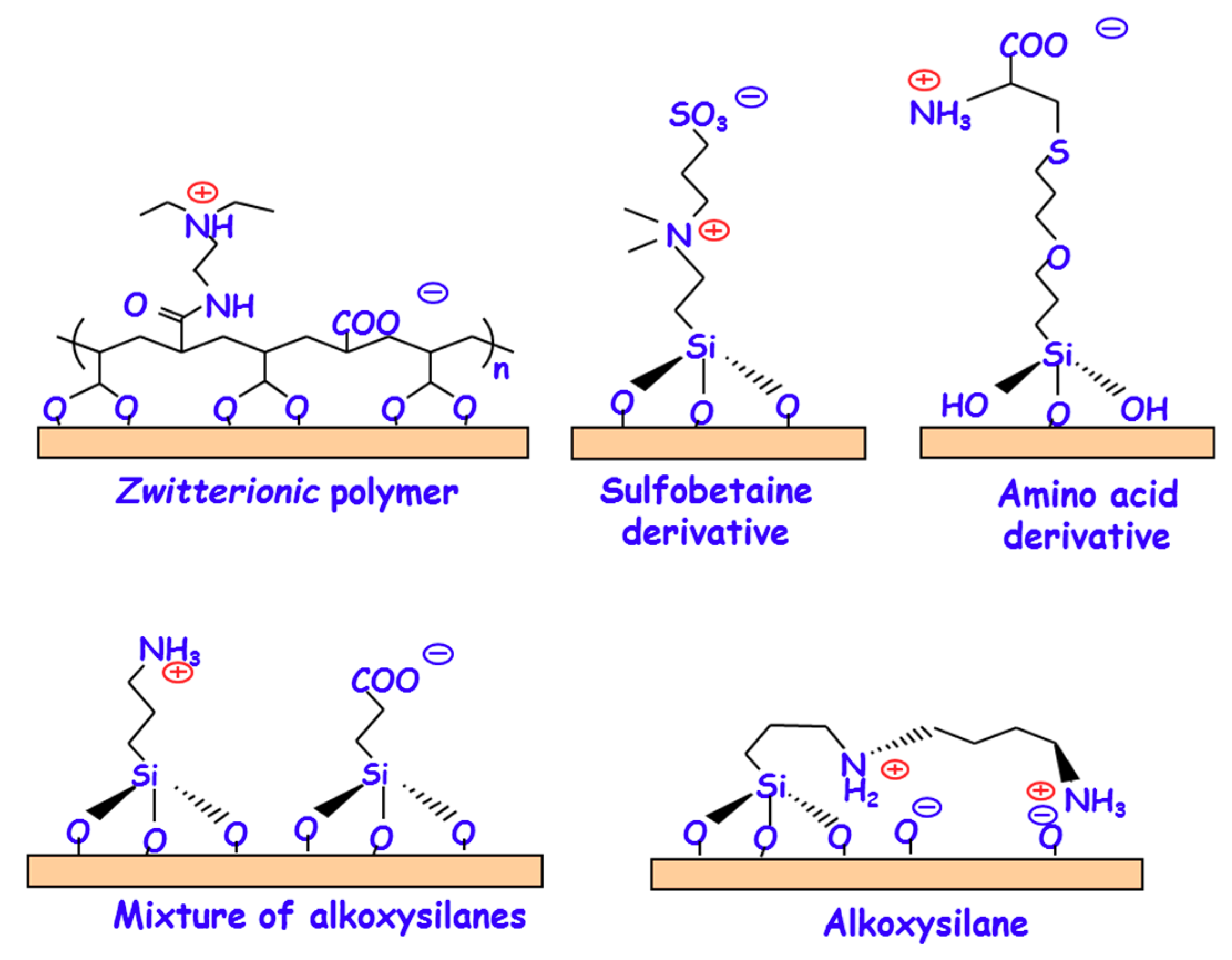
In general terms, the zwitterionization of biomaterials involved in the functionalization of their surfaces at the atomic level [11] In the beginning, research efforts were focused on functionalizing with zwitterionic polymers bearing mixed positively and negatively charged moieties within the same chain and overall charge neutrality (Figure 3) [28,29,33,34,35]. There are three main methodologies to graft these polymers to the surface of biomaterials: (i) for functionalizing with poly(sulfobetaine) and polycarboxybetaine derivatives by surface-initiated atom transfer radical polymerization (SI-ATRP) [36,37,38,39]; (ii) for grafting sulfobetaine copolymers by surface reversible addition-fragmentation chain transfer (RAFT) through the polymerization method denoted as graft-from-surface”; and, (iii) more simple procedures through the polymerization method “graft-to-surface” [40]. In this method, polymers carrying adhesive moieties with strong surface affinity are synthesized and then grafted onto the surface through their adhesive moieties [41,42]. Furthermore, it is possible to confer biomaterials of zwitterionic nature by decorating their surface with low-molecular weight moieties bearing the same number of negative and positive charges (Figure 3). For instance, it is possible to functionalize with different amino acids, such as cysteine and lysine [43,44,45], sulfobetaine derivatives [46,47,48], or dopamine [49,50,51], exhibiting zwitterionic characteristics depending on the pH. Although the reported methods usually require several synthetic steps involving different intermediate products, they offer distinct advantages when compared to zwitterionic polymers, since they are usually associated to relatively more simple methods and lead to more biocompatible surfaces. A significant advance in the design and development of zwitterionic surfaces has consisted on the use of more direct and simple grafting methods by functionalization with organosilanes. In this case, the presence of hydroxyl (–OH) on the biomaterial surface helps to simultaneously attach two organosilanes bearing positive and negative charges, respectively. This strategy allows for tailoring the zwitterionic properties by adjusting the molar ratio of the two organosilanes that are used during the synthesis. This process can be accomplished using two different alternatives, the co-condensation and the post-synthesis route. In the case of co-condensation method, functionalization takes place at the same time that the biomaterial is being synthetized. For instance, zwitterionic mesoporous SBA-15 material containing NH3+/–COO groups was synthesized by adding two alkoxysilanes, aminopropyltrimethoxysilane (APTES) and carboxyethyl silanetriol sodium salt (CES) (Figure 3), together with the tetraethylorthosilicate (TEOS), as silica precursor, during the synthesis step [27]. Moreover, our research group has also reported the synthesis of zwitterionic SBA-15 by functionalization with (N-(2-aminoethyl)-3-aminopropyl-trimethoxysilane) (DAMO) alkoxysilane, which contains primary and secondary amine groups [52]. As it can be observed in Figure 3, its zwitterionic nature is provided by the presence of –NH3+/–SiO and >NH2+/–SiO zwitterionic pairs. On the other hand, the post-synthesis route relies on grafting the organosilanes to the biomaterials surface once they have been synthesized. Following this methodology, zwitterionic hydroxyapatite has been also been prepared by linking APTES and CES to the P–OH groups that are present on the surface of this biomaterial [53].

3.2. Zwitterionization of Biomaterials to Prevent Bacterial Infection

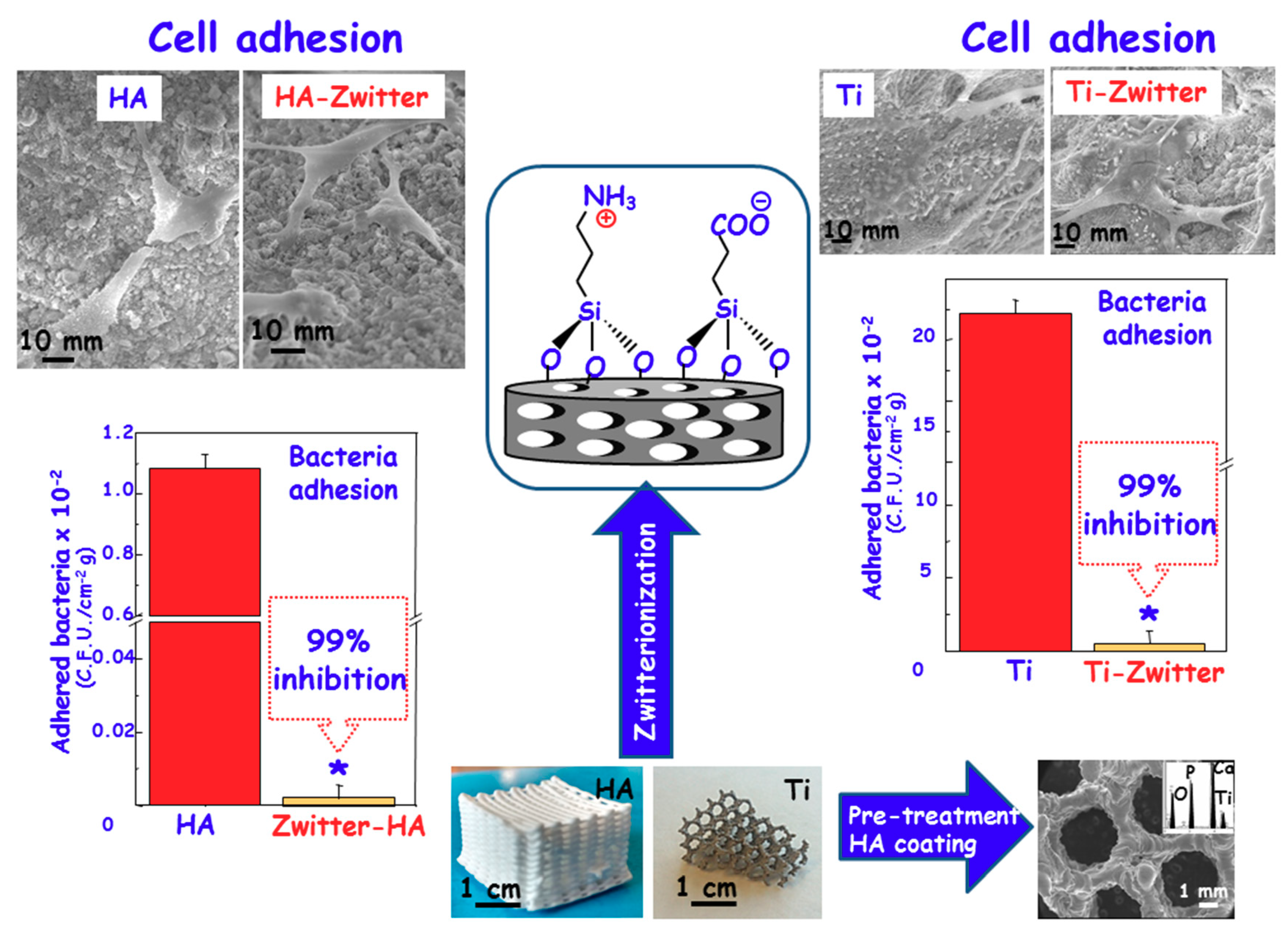
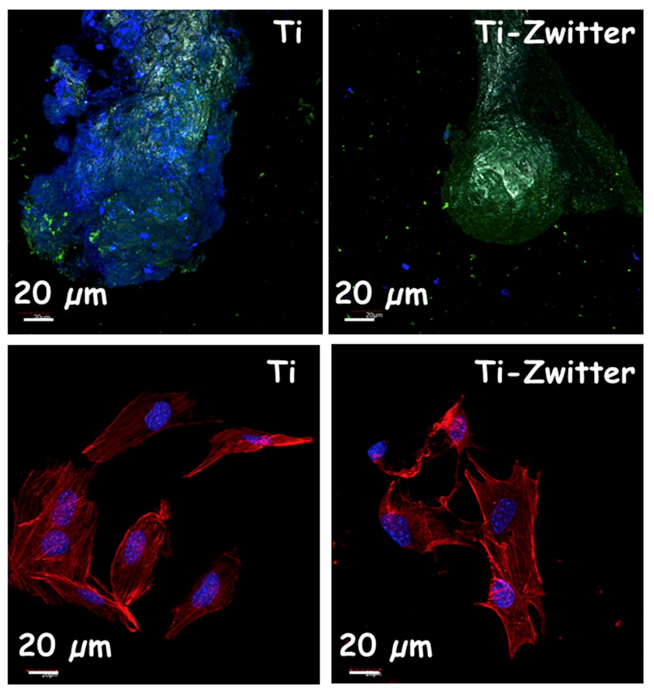
Currently, one of the major clinical challenges regarding the use of biomaterials is their custom-made design depending on the biomedical application [54,55]. Bioceramic implants for bone tissue regeneration, such as those that are based on calcium phosphates or bioactive glasses, can be manufactured as three dimensional (3D) scaffolds using rapid prototyping (RP) methods [54]. These scaffolds exhibit a high percentage of porosity and interconnectivity, and they are easily modulated with improved mechanical properties as compared to scaffolds that are fabricated by conventional methods [56]. Moreover, it is possible to combine nanostructural characteristics with micro-macro architecture for a fine-tuning of cellular behavior [57,58]. However, when facing the regeneration of large and critical bone defects, bioceramic implants are not suitable due to their intrinsic brittles [54]. It is feasible to manufacture metallic alloys using these RP methods, providing strong scaffolding to the bone regenerations purposes with porosities higher than 50% in volume and the rest being engaged by a metal skeleton [59,60]. In this regard, the milestone in bone tissue regeneration is to design these 3D scaffolds with surfaces that are capable of inhibiting and/or abolishing bacterial infection meanwhile allowing for osteoblast cells colonization. This surface would constitute a great technological advance to achieve better clinical outcomes. Currently, the scientific community is focussed on the design of 3D scaffolds that dynamically contribute to the regeneration process, stimulating the osteoconduction and angiogenesis at the same time than evade the bacterial infection [60,61,62,63]. However, the challenge is to provide 3D scaffolds of zwitterionic character. In this sense, 3D scaffolds based on pure nanocrystalline HA have been successfully constructed with a zwitterionic nature by the post-synthesis grafting of APTES and CES [53], which incorporates both –NH3+ and –COO groups on the surface, respectively (Figure 4, Left). To attain this goal, HA 3D scaffolds were first prepared by using the RP technique and then the resulting 3D-HA scaffolds were bifunctionalized by grafting both alkoxysilanes. Microbiological assays regarding bacterial adhesion using Escherichia coli (E. coli) showed a noticeable inhibition of 99% with respect to unmodified 3D-HA. The coexistence of –NH3+/–COO pairs onto 3D-HA scaffold avails its bacterial-repelling properties. At the same time, in vitro assays using HOS osteoblastic-like cells cultures demonstrated excellent biocompatibility as the cells were able to spread and colonize the entire scaffold surface. Scanning Electron Microscopy (SEM) micrographs show viable osteoblastic-like cells, exhibiting polygonal shapes with filopodia-like projections that are attached to the surfaces (Figure 4, Left). Moreover, the cell migration within the overall 3D-HA structure was demonstrated, showing the total colonization of 3D scaffold at different levels. This zwitterionization approach has been also applied onto Ti6Al4V 3D-scaffolds that were also fabricated by RP techniques [64]. In this case, to improve the functionalization capability of the metallic surface, it was previously coated by a HA layer using the dip-coating method. Once the HA coating was formed onto the Ti6Al4V 3D-scaffold, its surface was zwitterionized by the direct grafting of APTES and CES, following the same procedure as that reported for pure 3D-HA scaffolds [53]. Again, the presence of zwitterionic pairs inhibits S. aureus adhesion and biofilm formation, while permiting the osseointegration of this metallic implant, showing MC3T3-E1 preosteoblasts colonizing the entire scaffold surface. The obtained results indicate that the zwitterionization process does not affect the biocompatible properties of the metallic Ti6Al4V 3D-scaffolds, showing neither noticeable differences regarding cytotoxicity nor less proliferation as compared to bare 3D scaffold (Figure 4, right). Regarding the biofilm formation capability of these surfaces, we carried out confocal microscopy to study the biofilm formation after 24 h of incubation with S. aureus by using directly simultaneously acridine orange (green) and calcofluor (blue) fluorescent dyes, which label live bacteria and extracellular matrix of biofilms, respectively (Figure 4, Top). The obtained results clearly display the biofilm formation by the blue staining of a typical extracellular matrix covering the bacterial colonies with a thickness of 15 ± 3.3 μm on the Ti6Al4V scaffolds, while blue staining is absent in the Ti-Zwitter scaffolds, revealing the non-formation of the biofilm after 24 h of assay.
Concerning the regeneration process, we carried out a simple assay by confocal microscopy using preosteoblast MC3T3-E1 seeded on the surface of these scaffolds and incubated during 7 days. Both colonization and cell-morphology were studied by staining the cytoskeleton with Atto 565-conjugated phalloidin (red) and nuclei with DAPI (blue), as it can be observed in Figure 5 (bottom). In both casses, the cells display a high spreading grade with a well-built actin cytoskeleton and high level of colonization in all entire surface of both scaffolds. These results revealed that the zwitterionization process does not affect the healing process, showing the same behavior that unmodified Ti [64]. In the case of zwitterionic biomaterials currently under research, nanostructured bioceramics are receiving growing attention by the scientific community. Among these nanostructured biomaterials, silica-based mesoporous bioceramics are in the crest of the wave because of their exceptional features, such as high surface areas and pore volumes, tunable and narrow pore size distributions, and easy-to-functionalize surfaces [65,66,67]. Therefore they become excellent candidates to be provided of zwitterionic nature but also to host a great variety of antibiotics, allowing the combination of bacterial repellent and killing capabilities [68,69,70,71].
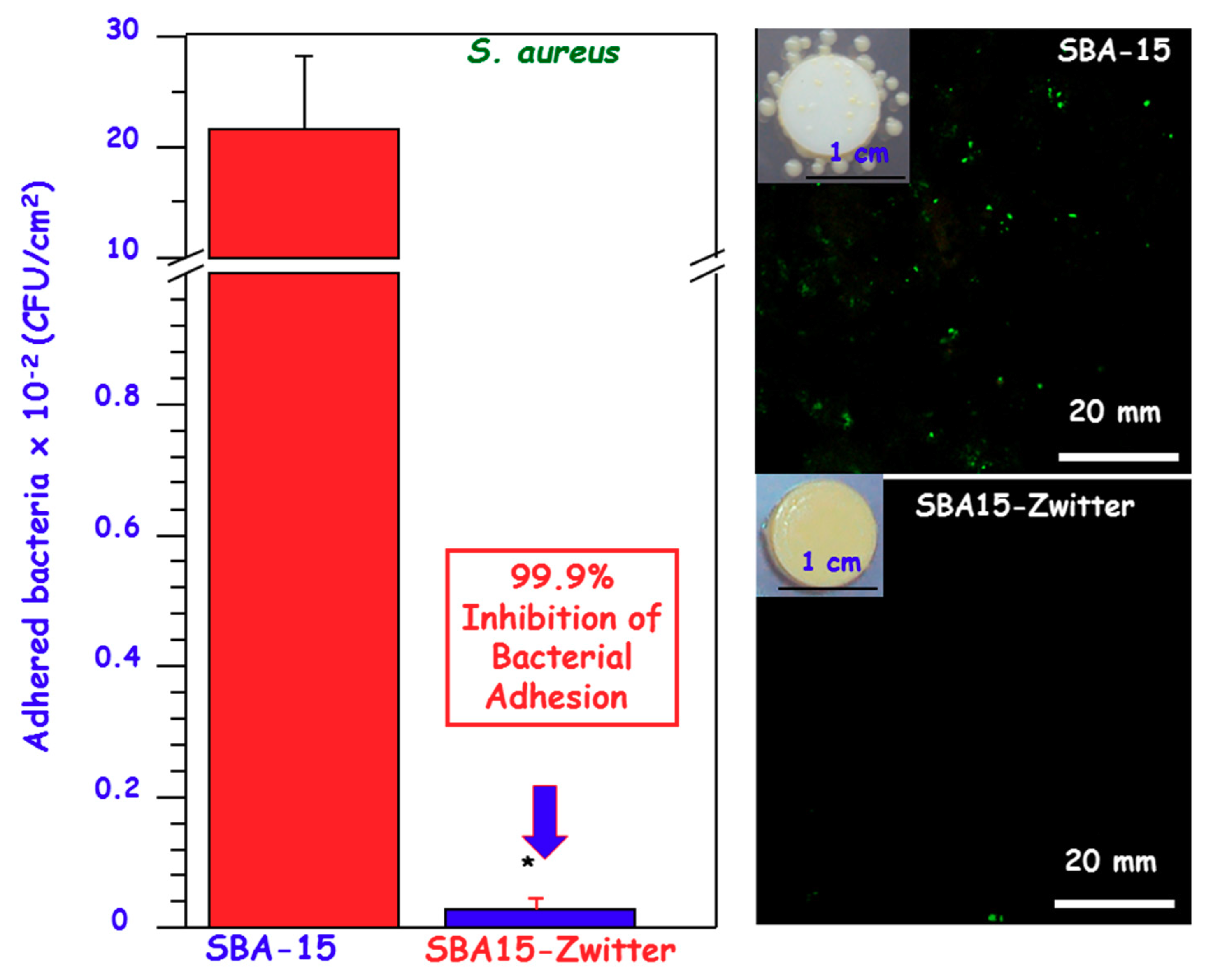
Figure 6 shows the performance of zwitterionic SBA-15 (SBA15-Zwitter) nanostructured bioceramic owning –NH3+/–SiO and >NH2+/–SiO pairs [52], provided by the co-condensation functionalization with the diamine alkoxysilane (DAMO) (Figure 3). This bioceramic exhibits the zwitterionic character at the physiological pH of 7.4, which constitutes a significant advance in this kind of materials for biomedical applications. In vitro bacterial adhesion tests using S. aureus strains reveal a great reduction of 99.9% with respect to unmodified SBA-15 (Figure 6). The intrinsic features of this nanostructured bioceramic permits the loading of the broad-spectrum antibiotic, cephalexin, showing a loading capability of around 13 mg∙g−1, together with a sustained drug release during more than 15 days. The synergistic combination of zwitterionic nature and antibiotic hosting capability opens up a new insight in the management of bone-associated infections. Zwitterionization has been also implemented on mesoporous bioactive glasses (MBG) having the SiO2–CaO–P2O5 composition [44]. These MBG are a type are nanostructured bioceramics analogous in composition to conventional bioglasses but exhibiting outstanding bioactive and cell response behaviors [69,72]. Thus, Sánchez-Salcedo et al. have recently reported the zwitterionization of MBG by tethering lysine (MBG-Lys). In vitro bacterial adhesion assays with S. aureus proved a reduction up to 99.9% as compared to unmodified MBG. Moreover, MBG-Lys are cytocompatible, as demonstrated by in vitro studies carried out with MC3T3-E1 preosteoblasts cultures, which increases the potential application of these nanostructured bioceramics in bone tissue regeneration.

4. Conclusions

Zwitterionization is emerging as a powerful strategy to design advanced biomaterials for the management of infection that is envisioned to result in better clinical outcomes. The possibility to easily functionalize biomaterials surface at the atomic and nanoscale levels to prevent the non-specific protein and bacterial adhesion opens up many paths to tackle severe clinical concerns. Indeed, zwitterionic biomaterials have shown an opposite behaviour by inhibiting bacterial adhesion, while allowing host cells adhesion and colonization of the surface. This fact constitutes the cornerstone in their potential clinical application and much research effort is committed to translate these significant advances from bench to bedside. However, there are certain challenges that these biomaterials have to face for their clinical stage of development. These challenges include: (i) preservation of biocompatibility, (ii) adequate pharmacokinetics through the local administration of antimicrobial agents, and (iii) bone healing capacity, which guarantee success in bone regeneration.

Author Contributions

All the authors have participated equally in the elaboration of this review manuscript. Conceptualization, M.V.-R., M.C. and I.I.-B.; Writing-Original Draft Preparation, M.C. and I.I.-B.; Writing-Review & Editing, M.V.-R., M.C. and I.I.-B.; Supervision, M.V.-R.; Project Administration and Funding Acquisition, M.V.-R.

Funding

This research was funded by European Research Council through ERC-2015-AdG-694160 (VERDI) grant.

Acknowledgments

The authors acknowledge financial support from European Research Council through ERC-2015-AdG-694160 (VERDI) project.

Conflicts of Interest

The authors declare no conflict of interest.

References

  1. Ofori-Asenso, R. “When the Bug Cannot Be Killed”—The Rising Challenge of Antimicrobial Resistance. Medicines 2017, 4, 40. [Google Scholar] [CrossRef] [PubMed]
  2. Taubes, G. The bacteria fight back. Science 2008, 321, 356–361. [Google Scholar] [CrossRef] [PubMed]
  3. Campoccia, D.; Montanaro, L.; Arciola, C.R. The significance of infection related to orthopedic devices and issues of antibiotic resistance. Biomaterials 2006, 27, 2331–2339. [Google Scholar] [CrossRef] [PubMed]
  4. Vardakas, K.Z.; Kontopidis, I.; Gkegkes, I.D.; Rafailidis, P.I.; Falagas, M.E. Incidence, characteristics, and outcomes of patients with bone and joint infections due to community-associated methicillin-resistant Staphylococcus aureus: A systematic review. Eur. J. Clin. Microbiol. Infect. Dis. 2013, 32, 711–721. [Google Scholar] [CrossRef] [PubMed]
  5. Campoccia, D.; Montanaro, L.; Arciola, C.R. A review of the biomaterials technologies for infection-resistant surfaces. Biomaterials 2013, 34, 8533–8554. [Google Scholar] [CrossRef] [PubMed]
  6. Anselme, K.; Davidson, P.; Popa, A.M.; Giazzon, M.; Liley, M.; Ploux, L. The interaction of cells and bacteria with surfaces structured at the nanometre scale. Acta Biomater. 2010, 6, 3824–3846. [Google Scholar] [CrossRef] [PubMed]
  7. Izquierdo-Barba, I.; García-Martín, J.M.; Álvarez, R.; Palmero, A.; Esteban, J.; Pérez-Jorge, C.; Arcos, D.; Vallet-Regí, M. Nanocolumnar coatings with selective behavior towards osteoblast and Staphylococcus aureus proliferation. Acta Biomater. 2015, 15, 20–28. [Google Scholar] [CrossRef] [PubMed] [Green Version]
  8. Sánchez-Salcedo, S.; Colilla, M.; Izquierdo-Barba, I.; Vallet-Regí, M. Preventing bacterial adhesion on scaffolds for bone tissue engineering. Int. J. Bioprinting 2015, 2, 20. [Google Scholar] [CrossRef]
  9. Sengstock, C.; Lopian, M.; Motemani, Y.; Borgmann, A.; Khare, C.; Buenconsejo, P.J.; Schildhauer, T.A.; Ludwig, A.; Köller, M. Structure-related antibacterial activity of a titanium nanostructured surface fabricated by glancing angle sputter deposition. Nanotechnology 2014, 25, 195101. [Google Scholar] [CrossRef] [PubMed]
  10. Truonga, V.K.; Lapovok, R.; Estrin, Y.S.; Rundell, S.; Wang, J.Y.; Fluke, C.J.; Crawford, R.J.; Ivanova, E.P. The influence of nano-scale surface roughness on bacterial adhesion to ultrafine-grained titanium. Biomaterials 2010, 31, 3674–3684. [Google Scholar] [CrossRef] [PubMed]
  11. Izquierdo-Barba, I.; Colilla, M.; Vallet-Regí, M. Zwitterionic ceramics for biomedical applications. Acta Biomater. 2016, 40, 201–211. [Google Scholar] [CrossRef] [PubMed] [Green Version]
  12. Donlan, R.M. Biofilms: Microbial life on surfaces. Emerg. Infect. Dis. 2002, 8, 881–890. [Google Scholar] [CrossRef] [PubMed]
  13. Davies, D. Understanding biofilm resistance to antibacterial agents. Nat. Rev. Drug Discov. 2003, 2, 114–122. [Google Scholar] [CrossRef] [PubMed]
  14. Stewart, P.S.; Costerton, J.W. Antibiotic resistance of bacteria in biofilms. Lancet (Lond. Engl.) 2001, 358, 135–138. [Google Scholar] [CrossRef]
  15. Costerton, J.W.; Montanaro, L.; Arciola, C.R. Biofilm in implant infections: Its production and regulation. Int. J. Artif. Organs 2005, 28, 1062–1068. [Google Scholar] [CrossRef] [PubMed]
  16. Gristina, A.G. Biomaterial-centered infection: Microbial adhesion versus tissue integration. Science 1987, 237, 1588–1595. [Google Scholar] [CrossRef] [PubMed]
  17. Gallo, J.; Holinka, M.; Moucha, C.S. Antibacterial Surface Treatment for Orthopaedic Implants. Int. J. Mol. Sci. 2014, 15, 13849–13880. [Google Scholar] [CrossRef] [PubMed] [Green Version]
  18. Busscher, H.J.; van der Mei, H.C. How do bacteria know they are on a surface and regulate their response to an adhering state? PLoS Pathog. 2012, 8, e1002440. [Google Scholar] [CrossRef] [PubMed]
  19. Costerton, W.; Veeh, R.; Shirtliff, M.; Pasmore, M.; Post, C.; Ehrlich, G. The application of biofilm science to the study and control of chronic bacterial infections. J. Clin. Investig. 2003, 112, 1466–1477. [Google Scholar] [CrossRef] [PubMed] [Green Version]
  20. Stoodley, P.; Ehrlich, G.D.; Sedghizadeh, P.P.; Hall-Stoodley, L.; Baratz, M.E.; Altman, D.T.; Sotereanos, N.G.; Costerton, J.W.; Demeo, P. Orthopaedic biofilm infections. Curr. Orthop. Pract. 2011, 22, 558–563. [Google Scholar] [CrossRef] [PubMed] [Green Version]
  21. Chagnot, C.; Zorgani, M.A.; Astruc, T. Proteinaceous determinants of surface colonization in bacteria: Bacterial adhesion and biofilm formation from a protein secretion perspective. Front. Microbiol. 2013, 4, 303. [Google Scholar] [CrossRef] [PubMed]
  22. Ploux, L.; Ponche, A.; Anselme, K. Bacteria/material interfaces: Role of the material and cell wall properties. J. Adhes. Sci. Technol. 2010, 24, 2165–2201. [Google Scholar] [CrossRef]
  23. Bohmler, J.; Ponche, A.; Anselme, K.; Ploux, L. Self-assembled molecular platforms for bacteria/material biointerface studies: Importance to control functional group accessibility. ACS Appl. Mater. Interfaces 2013, 5, 10478–10488. [Google Scholar] [CrossRef] [PubMed]
  24. Laverty, G.; Gorman, S.P.; Gilmore, B.F. Biomolecular mechanisms of Staphylococcal biofilm formation. Future Microbiol. 2013, 8, 509–524. [Google Scholar] [CrossRef] [PubMed] [Green Version]
  25. D’Souza, S.E.; Ginsberg, M.H.; Plow, E.F. Arginyl-glycyl-aspartic acid (RGD): A cell adhesion motif. Trends Biochem. Sci. 1991, 16, 246–250. [Google Scholar] [CrossRef]
  26. Subbiahdoss, G.; Kuijer, R.; Grijpma, D.W.; Van der Mei, H.C.; Busscher, H.J. Microbial biofilm growth vs. tissue integration: ‘The race for the surface’ experimentally studied. Acta Biomater. 2009, 5, 1399–1404. [Google Scholar] [CrossRef] [PubMed]
  27. Colilla, M.; Izquierdo-Barba, I.; Sánchez-Salcedo, S.; Fierro, J.L.G.; Hueso, J.L.; Vallet-Regí, M. Synthesis and characterization of zwitterionic SBA-15 nanostructured materials. Chem. Mater. 2010, 23, 6459–6466. [Google Scholar] [CrossRef]
  28. Cheng, G.; Zhang, Z.; Chen, S.; Bryers, J.D.; Jiang, S. Inhibition of bacterial adhesion and biofilm formation on zwitterionic surfaces. Biomaterials 2007, 28, 4192–4199. [Google Scholar] [CrossRef] [PubMed] [Green Version]
  29. Jiang, S.; Cao, Z. Ultralow-fouling, functionalizable, and hydrolysable zwitterionic materials and their derivatives for biological applications. Adv. Mater. 2010, 22, 920–932. [Google Scholar] [CrossRef] [PubMed]
  30. Chen, S.; Li, L.; Zhao, C.; Zheng, J. Surface hydration: Principles and applications toward low-fouling/nonfouling biomaterials. Polymer 2010, 51, 5283–5293. [Google Scholar] [CrossRef]
  31. Izquierdo-Barba, I.; Sánchez-Salcedo, S.; Colilla, M.; Feito, M.J.; Ramírez-Santillán, C.; Portolés, M.T.; Vallet-Regí, M. Inhibition of bacterial adhesion on biocompatible zwitterionic SBA-15 mesoporous materials. Acta Biomater. 2011, 7, 2977–2985. [Google Scholar] [CrossRef] [PubMed]
  32. Srivastava, S.; Srivastava, P.S. Understanding Bacteria; Kluwer Academic: Dordrecht, The Netherland, 2003. [Google Scholar]
  33. Cheng, G.; Xue, H.; Zhang, Z.; Chen, S.; Jiang, S. A switchable biocompatible polymer surface with self-sterilizing and nonfouling capabilities. Angew. Chem. Int. Ed. 2008, 47, 8831–8834. [Google Scholar] [CrossRef] [PubMed]
  34. Cheng, G.; Li, G.; Xue, H.; Chena, S.; Bryers, J.D.; Jiang, S. Zwitterionic carboxybetaine polymer surfaces and their resistance to long-term biofilm formation. Biomaterials 2009, 30, 5234–5240. [Google Scholar] [CrossRef] [PubMed] [Green Version]
  35. Lalani, R.; Liu, L. Electrospun zwitterionic poly(sulfobetaine methacrylate) for nonadherent, superabsorbent, and antimicrobial wound dressing applications. Biomacromolecules 2012, 13, 1853–1863. [Google Scholar] [CrossRef] [PubMed]
  36. Zhang, Z.; Chen, S.; Chang, Y.; Jiang, S. Surface grafted sulfobetaine polymers via atom transfer radical polymerization as superlow fouling coatings. J. Phys. Chem. B 2006, 110, 10799–10804. [Google Scholar] [CrossRef] [PubMed]
  37. Dong, Z.; Mao, J.; Yang, M.; Wang, D.; Bo, S.; Ji, X. Phase Behavior of Poly(sulfobetaine methacrylate)-Grafted Silica Nanoparticles and Their Stability in Protein Solutions. Langmuir 2011, 27, 15282–15291. [Google Scholar] [CrossRef] [PubMed]
  38. Suzuki, H.; Murou, M.; Kitano, H.; Ohno, K.; Saruwatari, Y. Silica particles coated with zwitterionic polymer brush: Formation of coloidal crystals and anti-biofouling properties in aqueous medium. Colloids Surf. B Biointerfaces 2011, 84, 111–116. [Google Scholar] [CrossRef] [PubMed]
  39. Jia, G.; Cao, Z.; Xue, H.; Xu, Y.; Jiang, S. Novel zwitterionic-polymer-coated silica nanoparticles. Langmuir 2009, 25, 3196–3199. [Google Scholar] [CrossRef] [PubMed]
  40. Sun, J.T.; Yu, Z.Q.; Hong, C.Y.; Pan, C.Y. Biocompatible zwitterionic sulfobetaine copolymer-coated mesoporous silica nanoparticles for temperature-responsive drug release. Macromol. Rapid Commun. 2012, 14, 811–818. [Google Scholar] [CrossRef] [PubMed]
  41. Zhang, L.; Xue, H.; Gao, C.; Carr, L.; Wang, J.; Chu, B.; Jiang, S. Imaging and cell targeting characteristics of magnetic nanoparticles modified by a functionalizable zwitterionic polymer with adhesive 3,4-dihydroxyphenyl-l-alanine linkages. Biomaterials 2010, 31, 6582–6588. [Google Scholar] [CrossRef] [PubMed]
  42. Xiao, W.; Lin, J.; Li, M.; Ma, Y.; Chen, Y.; Zhang, C.; Li, D.; Gu, H. Prolonged in vivo circulation time by zwitterionic modification of magnetite nanoparticles for blood pool contrast agents. Contrast Media Mol. Imaging 2012, 7, 320–327. [Google Scholar] [CrossRef] [PubMed]
  43. Rosen, J.E.; Gu, F.X. Surface functionalization of silica nanoparticles with cysteine: A low-fouling zwitterionic surface. Langmuir 2011, 27, 10507–10513. [Google Scholar] [CrossRef] [PubMed]
  44. Villegas, F.; Garcia-Uriostegui, L.; Rodríguez, O.; Izquierdo-Barba, I.; Salinas, A.J.; Toriz, G.; Vallet-Regí, M.; Delgado, E. Lysine-Grafted MCM-41 Silica as An Antibacterial Biomaterial. Bioengineering 2017, 4, 80. [Google Scholar] [CrossRef] [PubMed]
  45. Sánchez-Salcedo, S.; García, A.; Vallet-Regí, M. Prevention of bacterial adhesion to zwitterionic biocompatible mesoporous glasses. Acta Biomater. 2017, 57, 472–486. [Google Scholar] [CrossRef] [PubMed]
  46. Estephan, Z.G.; Jaber, J.A.; Schlenoff, J.B. Zwitterion-stabilized silica nanoparticles: Toward nonstick nano. Langmuir 2010, 26, 16884–16889. [Google Scholar] [CrossRef] [PubMed]
  47. Estephan, Z.G.; Schlenoff, P.S.; Schlenoff, J.B. Zwitteration as an alternative to PEGylation. Langmuir 2011, 27, 6794–6800. [Google Scholar] [CrossRef] [PubMed]
  48. Estephan, Z.G.; Hariri, H.H.; Schlenoff, J.B. One-pot, exchange-free, room-temperature synthesis of sub-10 nm aqueous, noninteracting, and stable zwitterated iron oxide nanoparticles. Langmuir 2008, 29, 2572–2579. [Google Scholar] [CrossRef] [PubMed]
  49. Wei, H.; Lee, N.I.J.; Han, H.-S.; Cordero, J.M.; Liu, W.; Bawendi, M.G. Compact zwitterion-coated iron oxide nanoparticles for biological applications. Nano Lett. 2012, 12, 22–25. [Google Scholar] [CrossRef] [PubMed]
  50. Wei, H.; Bruns, O.T.; Chen, O.; Bawendi, M.G. Compact zwitterion-coated iron oxide nanoparticles for in vitro and in vivo imaging. Integr. Biol. 2013, 5, 108–114. [Google Scholar] [CrossRef] [PubMed] [Green Version]
  51. Zhou, Z.; Wang, L.; Chi, X.; Bao, J.; Yang, L.; Zhao, W.; Chen, Z.; Wang, X.; Chen, X.; Gao, J. Engineered iron-oxide-based nanoparticles as enhanced T1 contrast agents for efficient tumor imaging. ACS Nano 2013, 7, 3287–3296. [Google Scholar] [CrossRef] [PubMed]
  52. Colilla, M.; Martínez-Carmona, M.; Sánchez-Salcedo, S.; Ruiz-González, M.L.; González-Calbet, J.M.; Vallet-Regí, M. A novel zwitterionic bioceramic with dual antibacterial capability. J. Mater. Chem. B 2014, 2, 5639–5651. [Google Scholar] [CrossRef]
  53. Sánchez-Salcedo, S.; Colilla, M.; Izquierdo-Barba, I.; Vallet-Regí, M. Design and preparation of biocompatible zwitterionic hydroxyapatite. J. Mater. Chem. B 2013, 1, 1595–1606. [Google Scholar] [CrossRef]
  54. Vallet-Regí, M. Bio-Ceramics with Clinical Applications; John Wiley & Sons Ltd.: Chichester, UK, 2014. [Google Scholar]
  55. Arcos, D.; Boccaccini, A.R.; Bohner, M.; Díez-Pérez, A.; Epple, M.; Gómez-Barrena, E.; Herrera, A.; Planell, J.A.; Rodríguez-Mañas, L.; Vallet-Regí, M. The relevance of biomaterials to the prevention and treatment of osteoporosis. Acta Biomater. 2014, 10, 1793–1805. [Google Scholar] [CrossRef] [PubMed] [Green Version]
  56. Hutmacher, D.W. Scaffolds in tissue engineering bone and cartilage. Biomaterials 2000, 21, 2529–2543. [Google Scholar] [CrossRef]
  57. García, A.; Izquierdo-Barba, I.; Colilla, M.; López de Laorden, C.; Vallet-Regí, M. Preparation of 3D scaffolds in the SiO2–P2O5 system with tailored hierarchical mesomacroporosity. Acta Biomater. 2011, 7, 1265–1273. [Google Scholar] [CrossRef] [PubMed]
  58. Cicuéndez, M.; Malstem, M.; Doadrio, J.C.; Portolés, M.T.; Izquierdo-Barba, I.; Vallet-Regí, M. Tailoring hierarchical meso-macroporous 3D scaffolds: From nano to macro. J. Mater. Chem. B 2014, 2, 49–58. [Google Scholar] [CrossRef]
  59. Quan, Y.J.; Drescher, P.; Zhang, F.M.; Burkel, E.; Seitz, H. Cellular Ti6Al4V with carbon nanotube-like structures fabricated by selective electron beam melting. Rapid Prototyp. J. 2014, 20, 541. [Google Scholar] [CrossRef]
  60. Heinl, P.; Müller, L.; Körner, C.; Singer, R.F.; Müller, F.A. Cellular Ti-6Al-4V structures with interconnected macro porosity for bone implants fabricated by selective electron beam melting. Acta Biomater. 2008, 4, 1536–1544. [Google Scholar] [CrossRef] [PubMed]
  61. Dvir, T.; Timko, B.P.; Kohane, D.S.; Langer, R. Nanotechnological strategies forengineering complex tissues. Nat. Nanotechnol. 2011, 6, 13–22. [Google Scholar] [CrossRef] [PubMed]
  62. Charlotte, L.H.; Wei, L.L.; Joachim, S.C.L. Drug-eluting scaffolds for bone and cartilage regeneration. Drug Discov. Today 2014, 19, 714–724. [Google Scholar]
  63. Cicuéndez, M.; Doadrio, J.C.; Hernández, A.; Portolés, M.T.; Izquierdo-Barba, I.; Vallet-Regí, M. Multifunctional pH sensitive 3D scaffolds for treatment and prevention of bone infection. Acta Biomater. 2018, 65, 450–461. [Google Scholar]
  64. Rodríguez-Palomo, A.; Monopoli, D.; Afonso, H.; Izquierdo-Barba, I.; Vallet-Regí, M. Surface zwitterionization of customized 3D Ti6Al4V scaffolds: A promising alternative to eradicate bone infection. J. Mater. Chem. B 2016, 4, 4356–4365. [Google Scholar] [CrossRef]
  65. Lanone, S.; Boczkowski, J. Biomedical applications and potential health risks of nanomaterials: Molecular mechanisms. Curr. Mol. Med. 2006, 6, 651–663. [Google Scholar] [CrossRef] [PubMed]
  66. Wang, Y.; Zhao, Q.; Han, N.; Bai, L.; Li, J.; Liu, J.; Che, E.; Hu, L.; Zhang, Q.; Jiang, T.; et al. Mesoporous silica nanoparticles in drug delivery and biomedical applications. Nanomed. Nanotech. Biol. Med. 2015, 11, 313–327. [Google Scholar] [CrossRef] [PubMed]
  67. Vallet-Regí, M. Ordered mesoporous materials in the context of drug delivery systems and bone tissue engineering. Chem. A Eur. J. 2006, 12, 5934–5943. [Google Scholar] [CrossRef] [PubMed]
  68. Vallet-Regí, M.; Colilla, M.; González, B. Medical applications of organic-inorganic hybrid materials within the field of silica-based bioceramics. Chem. Soc. Rev. 2011, 40, 596–607. [Google Scholar] [CrossRef] [PubMed]
  69. Vallet-Regí, M.; Izquierdo-Barba, I.; Colilla, M. Structure and functionalization of mesoporous bioceramics for bone tissue regeneration and local drug delivery. Philos. Trans. R. Soc. A Math. Phys. Eng. Sci. 2012, 370, 1400–1421. [Google Scholar] [CrossRef] [PubMed] [Green Version]
  70. Vallet-Regí, M.; Rámila, A.; del Real, R.P.; Pérez-Pariente, J. A new property of MCM-41: Drug delivery system. J. Chem. Mater. 2001, 13, 308–311. [Google Scholar] [CrossRef]
  71. Vallet-Regí, M.; Balas, F.; Arcos, D. Mesoporous materials for drug delivery. Angew. Chem. Int. Ed. 2007, 46, 7548–7558. [Google Scholar] [CrossRef] [PubMed]
  72. Izquierdo-Barba, I.; Vallet-Regí, M. Fascinating properties of bioactive templated glasses: A new generation of nanostructured bioceramics. Solid State Sci. 2011, 13, 773–783. [Google Scholar] [CrossRef]
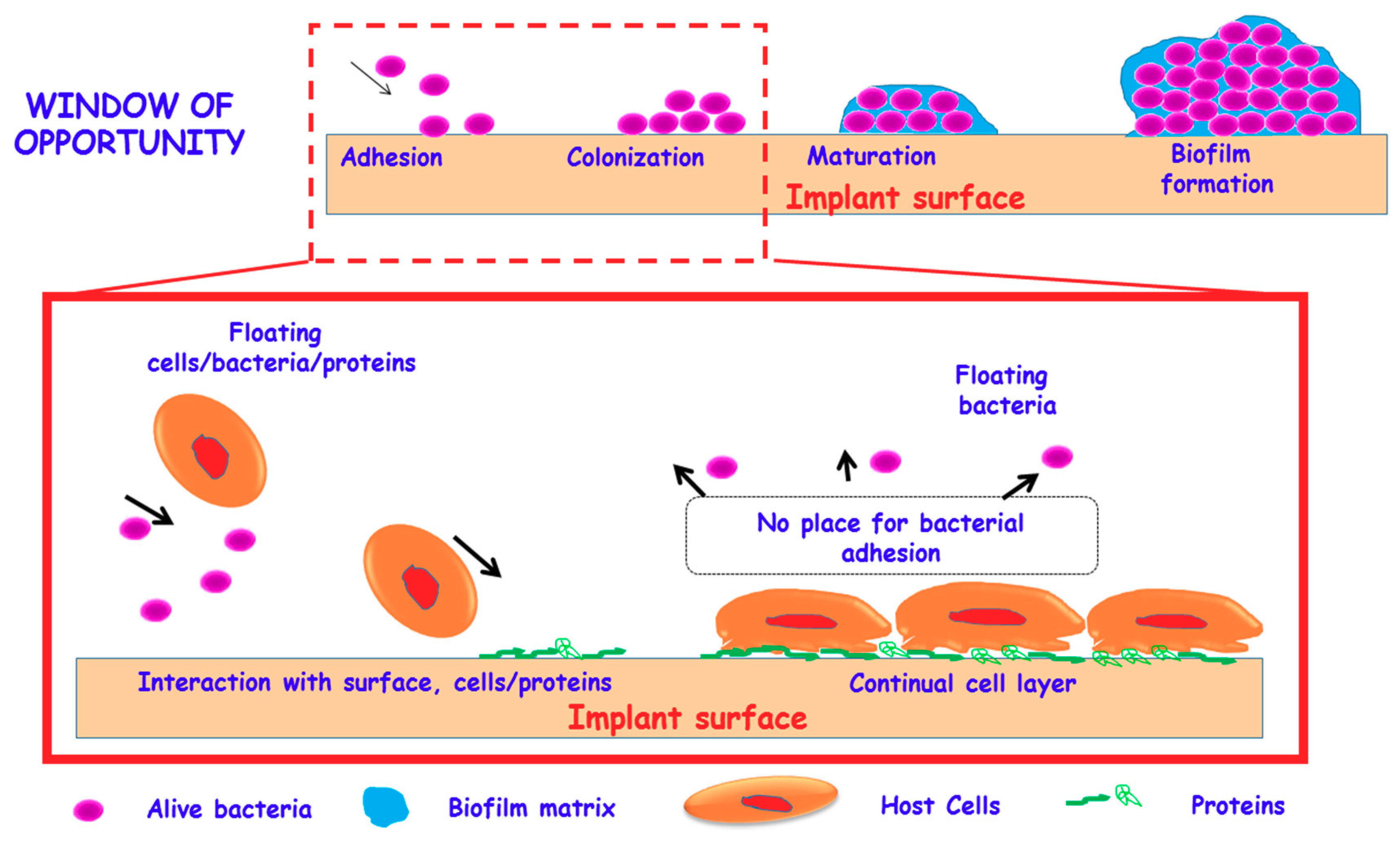
Figure 1. Concept of “race for the surface” and significance of “window of opportunity” in the development of implants capable to be colonized by host cells, while impede bacteria adhesion and the formation of the biofilm.
Medicines 05 00125 g001
Figure 2. Schematic illustration of the different behavior of conventional biomaterial surfaces vs biomaterials zwitterionic surfaces against bacterial colonization. As bacteria get close to the surface of conventional biomaterials, they are able to adhere, colonize and forming a biofilm, which is one of the major concerns in biomaterials associated infections. Oppositely, zwitterionic surfaces provide biomaterials of bacterial-repelling properties, thus inhibiting the subsequent biofilm formation, which constitutes a promising alternative in the biomaterials scenario to prevent bacterial infection.
Medicines 05 00125 g002
Figure 3. Representative examples of the different chemical strategies developed so far for the zwitterionization of biomaterials: Grafting of zwitterionic polymers [e.g., 3-(diethylamino)propylamine (DEAPA) coupled to poly(acrylic acid) (PAA)]; Sulfobetaine siloxane derivatives; Amino acid derivatives (e.g., cysteine); Mixture of alkoxysilanes [e.g., 3-aminopropyltrimethoxysilane (APTES) and carboxyethylsilanetriol sodium salt (CES)]; Alkoxysilane [N-(2-aminoethyl)-3-aminopropyl-trimethoxysilane) (DAMO)].
Medicines 05 00125 g003
Figure 4. Zwitterionization of different biomaterials in clinical use by grafting of carboxyethylsilanotriol sodium salt (CES) and aminopropyltriethoxysilane (APTES) onto the surface of these biomaterials. Left: Pure HA three-dimensional (3D) Rapid Prototyping (RP) scaffolds. Right: Electron Beam Melting (EBM) Ti6Al4V 3D scaffolds.
Medicines 05 00125 g004
Figure 5. Confocal microscopy images showing of EBM-Ti6Al4V 3D scaffolds before (Ti) and after being zwitterionized (Ti-Zwitter). In vitro behavior of the biomaterials after being incubated during 24 h in S. aureus bacteria (Top) and during 4 days in MC3T3-E1 preosteoblast cells (Bottom). The results reveal the biofilm formation on the Ti surface, appearing the blue coating corresponding to the polysaccharide matrix (calcofluor), whereas this coating is not observed on the surface of Ti-zwitter surface, which confirms the antibiofilm formation preventing capability of this material. Both Ti and Ti-zwitter surfaces undergo an appropriate preosteoblastic colonization and spreading in the entire surface, with the nuclei stained in blue (DAPI) and the cytoskeleton stained in red (phalloidin).
Medicines 05 00125 g005
Figure 6. Schematic depiction of pure silica SBA-15 and zwitterionic SBA-15 (SBA-Zwitter) nanostructured materials. SBA-Zwitter was prepared following the co-condensation route in the presence of DAMO. Counting of colony forming units of S. aureus after 90 min of culture onto SBA-15 and SBA15-Zwitter surfaces. Statistical significance: * p < 0.01. Confocal microscopy images of S. aureus adhered onto SBA-15 and SBA15-Zwitter surfaces after staining with Baclight® KitTM.
Medicines 05 00125 g006