Correction: Lenarczyk et al. T Cells Contribute to Pathological Responses in the Non-Targeted Rat Heart following Irradiation of the Kidneys. Toxics 2022, 10, 797

 , , ,
, , , 
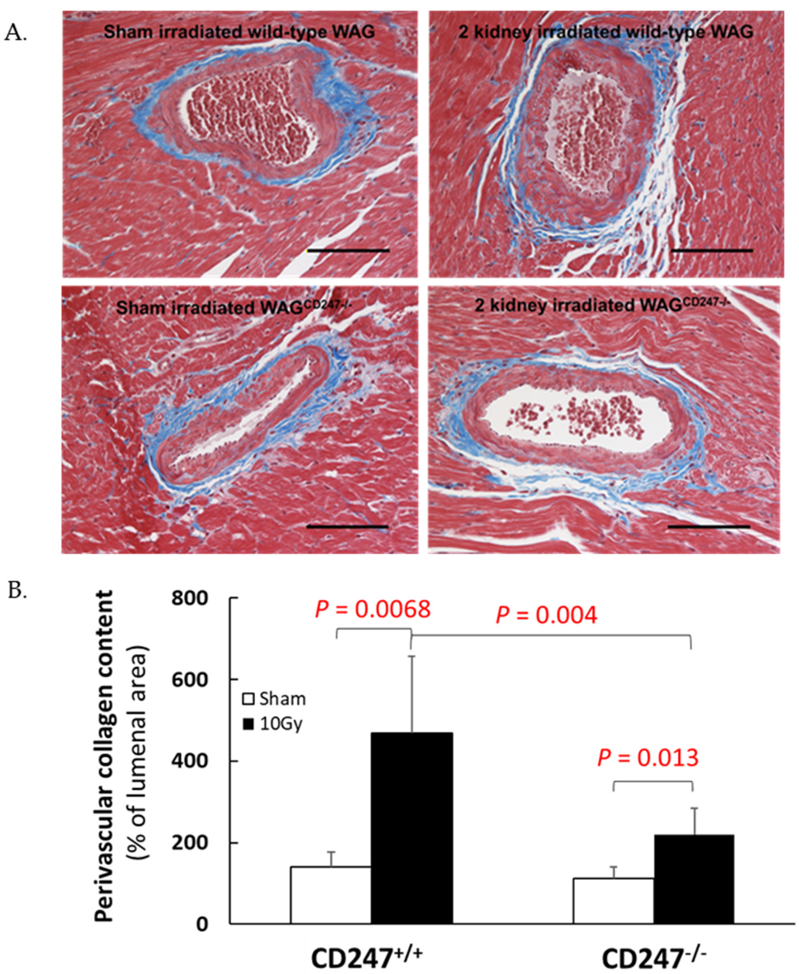
1. Error in Figure
2. Figure Legend

Reference
- Lenarczyk, M.; Alsheikh, A.J.; Cohen, E.P.; Schaue, D.; Kronenberg, A.; Geurts, A.; Klawikowski, S.; Mattson, D.; Baker, J.E. T Cells Contribute to Pathological Responses in the Non-Targeted Rat Heart Following Irradiation of the Kidneys. Toxics 2022, 10, 797. [Google Scholar] [CrossRef]
Disclaimer/Publisher’s Note: The statements, opinions and data contained in all publications are solely those of the individual author(s) and contributor(s) and not of MDPI and/or the editor(s). MDPI and/or the editor(s) disclaim responsibility for any injury to people or property resulting from any ideas, methods, instructions or products referred to in the content. |
© 2023 by the authors. Licensee MDPI, Basel, Switzerland. This article is an open access article distributed under the terms and conditions of the Creative Commons Attribution (CC BY) license (https://creativecommons.org/licenses/by/4.0/).
Share and Cite
Lenarczyk, M.; Alsheikh, A.J.; Cohen, E.P.; Schaue, D.; Kronenberg, A.; Geurts, A.; Klawikowski, S.; Mattson, D.; Baker, J.E. Correction: Lenarczyk et al. T Cells Contribute to Pathological Responses in the Non-Targeted Rat Heart following Irradiation of the Kidneys. Toxics 2022, 10, 797. Toxics 2023, 11, 183. https://doi.org/10.3390/toxics11020183
Lenarczyk M, Alsheikh AJ, Cohen EP, Schaue D, Kronenberg A, Geurts A, Klawikowski S, Mattson D, Baker JE. Correction: Lenarczyk et al. T Cells Contribute to Pathological Responses in the Non-Targeted Rat Heart following Irradiation of the Kidneys. Toxics 2022, 10, 797. Toxics. 2023; 11(2):183. https://doi.org/10.3390/toxics11020183
Chicago/Turabian StyleLenarczyk, Marek, Ammar J. Alsheikh, Eric P. Cohen, Dörthe Schaue, Amy Kronenberg, Aron Geurts, Slade Klawikowski, David Mattson, and John E. Baker. 2023. "Correction: Lenarczyk et al. T Cells Contribute to Pathological Responses in the Non-Targeted Rat Heart following Irradiation of the Kidneys. Toxics 2022, 10, 797" Toxics 11, no. 2: 183. https://doi.org/10.3390/toxics11020183
APA StyleLenarczyk, M., Alsheikh, A. J., Cohen, E. P., Schaue, D., Kronenberg, A., Geurts, A., Klawikowski, S., Mattson, D., & Baker, J. E. (2023). Correction: Lenarczyk et al. T Cells Contribute to Pathological Responses in the Non-Targeted Rat Heart following Irradiation of the Kidneys. Toxics 2022, 10, 797. Toxics, 11(2), 183. https://doi.org/10.3390/toxics11020183