Article

Copper and Zinc Treatments Alter the Thyroid Endocrine System in Zebrafish Embryos/Larvae

1
Fishery Resources and Environmental Science Experimental Station of the Upper-Middle Reaches of Yangtze River (Ministry of Agriculture and Rural Affairs), Yangtze River Fisheries Research Institute, Chinese Academy of Fishery Sciences, Wuhan 430223, China
2
Zhejiang Provincial Key Lab for Subtropical Water Environment and Marine Biological Resources Protection, College of Life and Environmental Science, Wenzhou University, Wenzhou 325035, China
3
State Key Laboratory of Biocatalysis and Enzyme Engineering, Hubei Universtiy, Wuhan 430062, China
*
Authors to whom correspondence should be addressed.
These authors contributed equally to this work.
Toxics 2022, 10(12), 756; https://doi.org/10.3390/toxics10120756
Submission received: 15 November 2022 / Revised: 29 November 2022 / Accepted: 2 December 2022 / Published: 4 December 2022
(This article belongs to the Special Issue Environmental and Health Effects of Heavy Metal)

Abstract

:
Copper (Cu2+) and zinc (Zn2+) are two kinds of heavy metals essential to living organisms. Cu2+ and Zn2+ at excessive concentrations can cause adverse effects on animals, but little is known about the thyroid-disrupting effects of these metals in fish, especially in the early developmental transition stage from embryos to larvae. Wild-type zebrafish embryos were used to expose to Cu2+ (0, 1.5, 15, and 150 μg/L) and Zn2+ (0, 20, 200, and 2000 μg/L) for 120 h. Thyroid hormone contents and transcriptional changes of the genes connected with the hypothalamic-pituitary-thyroid (HPT) axis were measured. Results showed that zebrafish embryos/larvae malformation rates were significantly increased in the Cu2+ and Zn2+ groups. Remarkably elevated thyroxine (T4) concentrations and reduced triiodothyronine (T3) concentrations were observed in Cu2+ and Zn2+ exposure fish. And the expression patterns of genes connected with the HPT axis were changed after Cu2+ and Zn2+ treatment. Based on principal component analysis (PCA) results, Zn2+ caused significant effects on the thyroid endocrine system at 200 μg/L, while Cu2+ resulted in thyroid disruption as low as 1.5 μg/L. In short, our study demonstrated that exposure to Cu2+ and Zn2+ induced developmental toxicity and thyroid disruption to zebrafish embryos/larvae.

1. Introduction

Heavy metals are defined as elements having atomic numbers larger than 20 and atomic densities larger than 5 g/cm [1,2]. They are produced from natural and human activities [3,4]. Heavy metals are not biodegradable or chemically degradable but can be bioaccumulated through the food chain in organisms [5,6]. These heavy metal ions enter the aquatic ecosystem through different channels such as geological weathering, atmospheric precipitation, and discharge of industrial or agricultural waste products [3]. They can be classified as essential and nonessential heavy metals [1,7]. Copper (Cu2+) and zinc (Zn2+) are two trace elements essential to living organisms [8,9,10]; they are important for the activities of multiple enzymes and play a significant role in a variety of biochemical responses [9,11]. However, when the concentrations increase, these essential heavy metals can induce harmful effects on organisms [4].
The toxicity of Cu2+ has attracted significant attention. Exposure to Cu2+ resulted in developmental toxicity, DNA damage, oxidative stress, hepatotoxicity and neurotoxicity in aquatic organisms [4,12,13,14,15]. However, little attention has been paid to the effects of Cu2+ on the endocrine system, especially the thyroid endocrine system. Previous studies have shown that copper deficiency could induce an increase in serum triiodothyronine (T3) levels in male Sprague-Dawley rats [16]. Similarly, Cu2+ deficiency in pregnant rats significantly reduced serum total thyroxine (T4) and T3 levels in neonatal rats [17]. In fish, exposure to Cu2+ for 7 days significantly elevated thyroid hormones (THs, including T4 and T3) in three-spined sticklebacks [18]. The THs were altered when rainbow trout and common carp were exposed to Cu2+ at the initial stage [19].
The effects of Zn2+ have been well documented [20]. Zinc deficiency could influence DNA repair and antioxidant defenses in rats [21], while exposure to excessive Zn2+ caused developmental toxicity in zebrafish and rare minnow [22,23]. The toxicity mechanisms of Zn2+ include disrupting calcium absorption, inhibiting Na+/K+-ATPase, inducing oxidative stress, and so on [24]. However, relatively little attention has been paid to the adverse effects of Zn2+ on the endocrine system. Zn2+ is an essential trace element for normal contents of T3, T4 and thyroid-stimulating hormones (TSH) [25]. Previous studies reported that Zn2+ was positively correlated with serum TSH levels in patients with hyperthyroidism [26]. The extrathyroidal production of T3 was impaired in zinc-deficient male Sprague-Dawley rats [27]. Experimental investigation showed that Zn2+ played an important role in THS metabolism through affecting deiodinase (Dio) enzyme activity and regulating the synthesis of TSH and thyrotropin-releasing hormones (TRH) [28]. However, the relationship between Zn2+ and the thyroid endocrine system in fish is unclear.
THs, which are synthesized in the thyroid gland, play crucial roles in multiple biological processes in vertebrates, such as growth, differentiation, metabolism, nervous system development, and reproduction [29,30,31,32]. Thyroid functions are predominantly regulated by the hypothalamus-pituitary-thyroid (HPT) axis. The HPT axis primarily controls the THs synthesis, transport and metabolism [33,34].
Although several studies reported that treatment with Cu2+ or Zn2+ affected THs levels in animals, the data involving thyroid disruption treatment in fish with these essential metals is insufficient, particularly in the early developmental transition stage from embryos to larvae. In this report, zebrafish embryos were used to demonstrate the effects of Cu2+ and Zn2+ treatment on the thyroid endocrine system. These effects were observed by measuring the levels of THs and gene expression involved in the HPT axis.

2. Experimental Procedures

2.1. Embryo Culture and Exposure

The zebrafish embryos (AB strain) were procured from the China Zebrafish Resource Center (Wuhan, China). Experimental procedures on fish were approved by the Animal Experimental Ethical Inspection of Laboratory Animal Centre, Yangtze River Fisheries Research Institute, Chinese Academy of Fishery Sciences (No. 20180504001). The toxicological experiments were carried out according to the Chinese national standard: toxicity tests for chemicals using fish in the early-life stage (GB/T 21854-2008). The Holt buffer (composition of: 3.5 g/L NaCl, 0.05 g/L KCl, 0.025 g/L NaHCO3, 0.1 g/L CaCl2, pH = 7.0) was used to culture the embryos. The cultured conditions that were set were as follows: constant temperatures (28 ± 0.5 °C) with a 14:10 (light: dark) photoperiod. Furthermore, Cu2+ and Zn2+ stock solutions were prepared after dissolving copper sulfate (CuSO4·5H2O, CAS: 7758-99-8, purity ≥99.5%, Shanghai Biochemical Technology Co., Ltd, Shanghai, China) and zinc sulfate (ZnSO4·7H2O, CAS: 7446-20-0; purity ≥99.5%, Shanghai Biochemical Technology Co., Ltd.) in UP water. The embryos were exposed to varying exposure concentrations of Cu2+ (0, 1.5, 15, and 150 μg/L), and Zn2+ (0, 20, 200, and 2000 μg/L). Healthy embryos (14,700) were randomly assigned to 42 glass beakers (350 per beaker) containing 350 mL exposure solution. Each treatment was conducted in six replicates. These embryos were exposed to the toxic ions for 120 h (5 days), and the exposure solution was replaced by a new solution every 24 h to maintain the Cu2+ and Zn2+ concentrations. Zebrafish embryos at 0–5 dpf do not need to be fed. Therefore, the embryos were not fed during the exposure period of 120 h. The water samples were collected before renewal of the exposure medium for measuring the actual Cu2+ and Zn2+ concentrations. The concentrations for these heavy meals were determined by atomic absorption spectrometry according to the method of National Standard of China (GB7475-87). The actual Cu2+ and Zn2+ concentrations exceeded 80% of the corresponding specific concentrations in all water samples. During the period of exposure, the dead embryos were discarded every day, and the hatching, survival, and malformation rates were recorded at 120 h. The larvae (120 h) were anesthetized with tricaine methanesulfonate (MS-222, Sigma-Aldrich, Saint Louis, MO, USA), randomly sampled for subsequent assays of THs and gene expression levels, and immediately stored at −80 °C.

2.2. RNA Extraction and Quantitative RT-PCR

Twenty larvae were selected randomly from every beaker and pooled into a single sample for gene expression analysis. Every group contained six replicates. The total RNA was extracted with the aid of the Trizol reagent (Invitrogen, Carlsbad, CA, USA), based on the manufacturer’s protocols. Agarose gel (1%) electrophoresis was used to estimate the quality of the total extracted RNA. The RNA contents were measured using the Nanodrop lite spectrophotometer (Thermo Fisher, Waltham, MA, USA). FastKing gDNA Dispelling RT SuperMix (Tiangen Biochemical Technology, Beijing, China) was used to synthesize first-strand cDNA, following the manufacturer’s protocols. Quantitative RT-PCR was carried out using the ABI 7500 System (Applied Biosystems 7500, Carlsbad, CA, USA) with the UltraSYBR mixture (Low ROX) (CWBIO Beijing, China), following the kit instructions. Thermal cycling was done at 95 °C for 10 min, followed by 40 cycles of 95 °C for 15 s and 60 °C for 60 s. 18sRNA was selected as a reference gene when calculating the gene expression levels. The 2-ΔΔCt technique was used for analyzing the differences (variations) in the gene expression levels [35]. The quantitative RT-PCR primers were obtained from a previous study [36].

2.3. Thyroid Hormone Assays

The whole-body T4 and T3 contents in the larvae were estimated using a previously reported method [37]. Briefly, approximately 200 larvae from each replicate were homogenized with ice-cold phosphate-buffered saline (PBS) with a glass grinder, and the samples were sonicated on ice. Every group included 6 replicates. The supernatants were obtained after the homogenate samples were centrifuged at 5000 rpm at 4 °C for 15 min. The total protein contents in the supernatants were quantified by Bradford assay [38]. The THs (T4 and T3) concentrations from supernatants were estimated using the enzyme-linked immunosorbent assay (ELISA) kit (Cloud-Clone Corp. Wuhan, China, T3: CEA453Ge; T4: CEA452Ge), following kit instructions. The T3 and T4 detection limits were calculated to be 51.7 pg/mL and 1.29 ng/mL, respectively.

2.4. Statistical Analysis

The data related to THs contents and gene expression levels were presented as the mean ± standard deviation (SD). The data derived in the study were analyzed with SPSS 20.0 (IBM, Chicago, CA, USA). The Kolmogorov–Smirnov test was employed for validating the data normality. The Levene test was utilized for analyzing the homogeneity of variances. After validating the data normality, the statistical differences observed in the treatment groups were confirmed by means of one-way analysis of variance (ANOVA), followed by Tukey-HSD tests. Principal Component Analysis (PCA) was carried out using Origin 2021 (OriginLab, Northampton, MA, USA). The correlation analysis was confirmed by Spearman’s test. Values with p < 0.05 were established as statistically significant.

3. Results

3.1. Developmental Toxicity Caused by Cu2+ and Zn2+

The developmental toxicity exposed to Cu2+ and Zn2+ was illustrated in Table 1. It was seen that the hatching and survival rates were not altered when zebrafish embryos/larvae were exposed to Cu2+, while the malformation rate was significantly increased in the 150 μg/L Cu2+ group. Similar to Cu2+, Zn2+ treatment did not alter survival and hatching rates of zebrafish embryos/larvae. However, the rate of malformation was significantly elevated in the 2000 μg/L Zn2+ group compared with the control.

3.2. Influences of Cu2+ on Thyroid Endocrine System

Exposure to 150 μg/L Cu2+ remarkably upregulated the levels of tshβ gene expression (1.53-fold) compared with the control (Figure 1A). The gene expression of thyroglobulin (tg) was significantly downregulated 0.60-, 0.65- and 0.71-fold in the 1.5, 15 and 150 μg/L Cu2+ exposure groups, respectively (Figure 1B). Expression of the sodium-iodide symporter (nis) gene was significantly downregulated 0.63-fold in the 1.5 μg/L Cu2+ exposure group but was upregulated 1.60-fold in the 150 μg/L Cu2+ exposure group (Figure 1C). Thyroid peroxidase (tpo) expression was remarkably downregulated 0.67- and 0.69-fold in the 1.5 and 15 μg/L Cu2+ treatment groups, respectively (Figure 1D). The gene expression levels of transthyretin (ttr), thyroid hormone receptor-α (thrα), thyroid hormone receptor-β (thrβ), type I iodothyronine deiodinase (dio1) and type II iodothyronine deiodinase (dio2) were significantly upregulated 2.22-, 1.54-, 1.43-, 1.55- and 1.58-fold in the 150 μg/L Cu2+ treatment group, respectively (Figure 1E–I). However, expression of the uridine diphosphate glucuronosyltransferase 1 family a, b (ugt1ab) gene was significantly downregulated 0.48-, 0.39- and 0.66-fold in the 1.5, 15 and 150 μg/L Cu2+ exposure groups, respectively (Figure 1J).
Cu2+ exposure significantly increased the T4 contents by 1.69- and 2.18- fold in the 15 and 150 μg/L Cu2+ treatment groups, respectively (Figure 1K), while the T3 contents were significantly decreased 0.64- and 0.35-fold in the 15 and 150 μg/L Cu2+ treatment groups, respectively (Figure 1L).

3.3. Influences of Zn2+ on Thyroid Endocrine System

Downregulation of nis (0.69-fold) was ascertained in the 2000 μg/L Zn2+ treatment group compared with the control (Figure 2C). Treatment with 200 and 2000 μg/L Zn2+ significantly upregulated the mRNA expression of tpo (1.50 and 1.81-fold, respectively) compared to the control (Figure 2D). Treatment with 2000 μg/L Zn2+ remarkably upregulated the mRNA expression of thrα (1.50-fold) and dio1 (1.36-fold) (Figure 2F,H). The gene expression levels of dio2 (0.74-, 0.75, and 0.71-fold) and ugt1ab (0.42-, 0.53- and 0.56-fold) were remarkably downregulated in all Zn2+ treatment groups (20, 200 and 2000 μg/L, respectively) (Figure 2I,J). Nevertheless, tshβ, tg, ttr and thrβ expression showed no significant changes (Figure 2A,B,E,G).
Zn2+ treatment remarkably increased the T4 content by 2.27- and 2.38-fold in the 200 and 2000 μg/L Zn2+ groups, respectively (Figure 2K), while the T3 content was significantly reduced 0.69-fold in the 2000 μg/L Zn2+ exposure group compared with the control (Figure 2L).

3.4. PCA and Correlation Analysis

Herein, the PCA and correlation analyses were conducted to analyze the relationship between the THs and genes transcriptional levels. The contents of T4 and T3 and transcriptional levels of genes related to HPT axis were used for PCA and correlation analyses. The PCA result of Cu2+ exposure is shown in Figure 3A. The initial two principal components (PCs) explained 79.6% of the total variances. The PC1 explained 55.9% of the total variances, while PC2 accounted for 23.7% of the total variances. According to the PCA plot, the separation between clusters of control and 1.5 and 15 μg/L Cu2+ groups were significant. T4 level had a strong negative correlation with T3 level; T4 level positively and significantly correlated with the transcriptional levels of crh, tshβ, nis, ttr, thα and dio2 and negatively correlated with the transcriptional levels of ugt1ab (Figure 4A). However, the T3 level exhibited a strong negative correlation with the expression levels of crh, tshβ, nis, ttr, thα and dio2 (Figure 4A).
The PCA result of Zn2+ exposure is shown in Figure 3B. The initial two PCs explained 68.0% of the total variances. The PC1 accounted for 38.9% of the total variances, while PC2 explained 29.1% of the total variances. Based on the PCA result, the separation between clusters of control and 200 and 2000 μg/L Zn2+ groups were significant. T4 level were seen to be significantly and positively correlated with the transcriptional levels of crh, tpo, thα and dio1 and were negatively related to the transcriptional levels of nis, thrβ and dio2 (Figure 4B), while there was a strong positive correlation between T3 level and thrβ transcriptional levels (Figure 4B).

4. Discussion

Heavy metals can cause developmental toxicity in fish [14,39]. In the present study, although the hatching and survival rates of the fish were not notably affected, the malformation rates were notably enhanced in the zebrafish larvae exposed to Cu2+ and Zn2+, particularly in the highest exposure groups (Table 1). Therefore, malformations were found to be a more susceptible parameter compared to hatching and survival rates for assessing developmental toxicity in zebrafish embryos.
In vertebrates, TSH is encoded by the tshβ gene, which stimulates the thyroid gland to generate THs via binding to the corresponding receptor [40]. Thus, the upregulation of tshβ could be linked to a higher T4 level. In this study, the tshβ transcriptional levels were upregulated significantly, which could explain the enhanced T4 concentrations in the Cu2+ exposure group. However, although T4 content was significantly increased, the tshβ expression showed no significant change in Zn2+-treated zebrafish embryos/larvae. Consistent with our results, adult male rat exposure to Zn2+ (0 and 3 mg/kg) showed increased free T4 content but no significant changes in TSH content [41]. Therefore, our results indicated that the correlation between TSH content and Zn2+ concentration was not strong.
Herein, the expression levels of genes (including tg, nis and tpo) were determined to be associated with THs biosynthesis. The nis gene facilitates the iodide accumulation from blood in thyroid cells [42]. The function of the TPO enzyme is to catalyze the iodination of tyrosyl-residues in TG protein and follow-up coupling of iodotyrosines to produce THs [43,44]. Therefore, the upregulation of tg, nis and tpo expressions might be related to the increase of T4 level. In accordance with this, increased T4 contents along with upregulation of nis expression and upregulation of tpo expressions were observed in the Cu2+ and Zn2+ groups, respectively. However, the T4 levels were increased, and the tg and nis genes expressions were significantly downregulated in the Cu2+ and Zn2+ groups, respectively. This could be attributed to the self-compensatory mechanism in the HPT axis, which downregulated tg or nis gene expression to decrease the T4 content.
TTR acted as a carrier protein for THs, and it helped in transporting THs to the target tissues through blood circulation [45,46]. Earlier reports demonstrated that T4 content was significantly and positively linked to the ttr expression [37,47,48]. In line with these studies, the T4 content was significantly increased, along with upregulated ttr gene transcription in Cu2+ treatment groups. The THR, which belongs to the nuclear receptor superfamily, is a transcription factor that responds to T3. Previous results showed that T4 bound to trα and mediated the expression of THs-regulated genes [49]. Significant upregulation of thrα expression was observed after Cu2+ and Zn2+ exposure in this study, which might be attributed to the higher T4 levels. Previous studies have reported significant downregulation of thrα and thrβ in Chinese toad following Cu2+ exposure [50], while the expression levels of thrα and thrβ were significantly upregulated in the Cu2+ exposure group in this study. These inconsistent consequences might be due to the species-specific effects caused by Cu2+ and require further study.
Previous studies reported that Cu2+ deficiency and Zn2+ supplementation could alter deiodinase activity [28,51]. Dio2 was involved in T4 deiodination to form active T3 [52,53]. Thus, downregulation of the dio2 gene expression was attributed to the increased T4 level and reduced T3 level in the Zn2+ treatment groups in this study. Moreover, dio1 functions as an outer-ring THs deiodinase and is involved in the metabolism of THs (including T3 and T4) [54]. Therefore, a decrease in the T3 content in the Zn2+ and Cu2+ treatment groups was partially attributed to the upregulation of dio1 expression. Consistent with these results, an increase in the Dio1 activity was reported in Zn2+ supplementation rats [55]. The ugt1ab gene was remarkably involved in the THs homeostasis by regulating T4 glucuronidation [56]. An earlier study reported that an increase in the T4 content in zebrafish larvae was due to the downregulation of ugt1ab mRNA levels [57]. In this study, the ugt1ab gene transcription level was significantly downregulated in the Cu2+ and Zn2+ treatment groups. Therefore, the increased T4 content could be attributed to the downregulation of ugt1ab transcription levels.
Based on the PCA results, it was seen that Zn2+ significantly affected the thyroid endocrine system at the concentration of 200 μg/L, whereas Cu2+ led to thyroid disruption at a concentration as low as 1.5 μg/L. Cu2+ was more sensitive compared to Zn2+ in the endocrine thyroid system. The Standards for Drinking Water Quality of China (GB5749–2022) state that the maximum allowable concentrations of Zn2+ and Cu2+ are both 1000 µg/L. Since both Zn2+ and Cu2+ negatively affected the endocrine thyroid system below this permissible level, the adverse effects of these metals on the endocrine system of fish needs to be investigated further.

5. Conclusions

In summary, this study demonstrated that treatment with Zn2+ and Cu2+ caused developmental toxicity and thyroid disruption in the zebrafish embryos/larvae. Both whole-body THs contents and the expression patterns of genes linked to the HPT axis were altered after exposure to Zn2+ and Cu2+. Further research is necessary to elucidate the mechanisms of thyroid disruption due to heavy metals toxicity.

Author Contributions

Conceptualization, H.Z.; Methodology, L.Z., L.W. and F.Y.; Software, N.W.; Investigation, L.Z., H.Z. and L.W.; Resources, H.R. and N.W.; Writing—original draft, L.Z.; Supervision, Z.N., X.D. and Y.L.; Funding acquisition, X.D. and Y.L. All authors have read and agreed to the published version of the manuscript.

Funding

This research was funded by the National Key Research and Development Program of China (2018YFD0900903), the Central Public-interest Scientific Institution Basal Research Fund, the Chinese Academy of Fishery Sciences (CAFS) (Nos. 2020TD09, 2022JC01) and the China Three Gorges Project Corporation (No. 0799555). This work was also funded by Open Research Fund Program of State Key Laboratory of Biocatalysis and Enzyme Engineering (SKLBEE2021006), Hubei University.

Institutional Review Board Statement

This study was approved by the Animal Experimental Ethical Inspection of Laboratory Animal Centre, Yangtze River Fisheries Research Institute, Chinese Academy of Fishery Sciences (No. 20180504001).

Informed Consent Statement

Not applicable.

Data Availability Statement

Not applicable.

Conflicts of Interest

The authors declare no conflict of interest.

References

  1. Raychaudhuri, S.S.; Pramanick, P.; Talukder, P.; Basak, A. Polyamines, metallothioneins, and phytochelatins—Natural defense of plants to mitigate heavy metals. Stud. Nat. Prod. Chem. 2021, 69, 227–261. [Google Scholar]
  2. Ali, H.; Khan, E. What are heavy metals? Long-standing controversy over the scientific use of the term ‘heavy metals’–proposal of a comprehensive definition. Toxicol. Environ. Chem. 2018, 100, 6–19. [Google Scholar] [CrossRef]
  3. Authman, M.M.; Zaki, M.S.; Khallaf, E.A.; Abbas, H.H. Use of fish as bio-indicator of the effects of heavy metals pollution. J. Aquac. Res. Dev. 2015, 6, 1–13. [Google Scholar] [CrossRef]
  4. Tchounwou, P.B.; Yedjou, C.G.; Patlolla, A.K.; Sutton, D.J. Heavy metal toxicity and the environment. Mol. Clin. Environ. Toxicol. 2012, 100, 133–164. [Google Scholar]
  5. Peng, Z.; Liu, X.; Zhang, W.; Zeng, Z.; Liu, Z.; Zhang, C.; Liu, Y.; Shao, B.; Liang, Q.; Tang, W. Advances in the application, toxicity and degradation of carbon nanomaterials in environment: A review. Environ. Int. 2020, 134, 105298. [Google Scholar] [CrossRef] [PubMed]
  6. Morcillo, P.; Esteban, M.Á.; Cuesta, A. Heavy metals produce toxicity, oxidative stress and apoptosis in the marine teleost fish SAF-1 cell line. Chemosphere 2016, 144, 225–233. [Google Scholar] [CrossRef]
  7. Thakare, M.; Sarma, H.; Datar, S.; Roy, A.; Pawar, P.; Gupta, K.; Pandit, S.; Prasad, R. Understanding the holistic approach to plant-microbe remediation technologies for removing heavy metals and radionuclides from soil. Curr. Res. Biotechnol. 2021, 3, 84–98. [Google Scholar] [CrossRef]
  8. Clearwater, S.J.; Farag, A.M.; Meyer, J. Bioavailability and toxicity of dietborne copper and zinc to fish. Comp. Biochem. Physiol. C Toxicol. Pharmacol. 2002, 132, 269–313. [Google Scholar] [CrossRef]
  9. Watanabe, T.; Kiron, V.; Satoh, S. Trace minerals in fish nutrition. Aquaculture 1997, 151, 185–207. [Google Scholar] [CrossRef]
  10. Ebrahimpour, M.; Alipour, H.; Rakhshah, S. Influence of water hardness on acute toxicity of copper and zinc on fish. Toxicol. Ind. Health 2010, 26, 361–365. [Google Scholar] [CrossRef]
  11. Kucharzewski, M.; Braziewicz, J.; Majewska, U.; Góźdź, S. Copper, zinc, and selenium in whole blood and thyroid tissue of people with various thyroid diseases. Biol. Trace Elem. Res. 2003, 93, 9–18. [Google Scholar] [CrossRef] [PubMed]
  12. Malhotra, N.; Ger, T.-R.; Uapipatanakul, B.; Huang, J.-C.; Chen, K.H.-C.; Hsiao, C.-D. Review of copper and copper nanoparticle toxicity in fish. Nanomaterials 2020, 10, 1126. [Google Scholar] [CrossRef] [PubMed]
  13. Lushchak, V.I. Environmentally induced oxidative stress in aquatic animals. Aquat. Toxicol. 2011, 101, 13–30. [Google Scholar] [CrossRef] [PubMed]
  14. Jezierska, B.; Ługowska, K.; Witeska, M. The effects of heavy metals on embryonic development of fish (a review). Fish Physiol. Biochem. 2009, 35, 625–640. [Google Scholar] [CrossRef]
  15. Rayburn, J.; Aladdin, R. Developmental toxicity of copper, chromium, and aluminum using the shrimp embryo teratogenesis assay: Palaemonid with artificial seawater. Bull. Environ. Contam. Toxicol. 2003, 71, 0481–0488. [Google Scholar] [CrossRef]
  16. Kralik, A.; Kirchgessner, M.; Eder, K. Concentrations of thyroid hormones in serum and activity of hepatic 5' monodeiodinase in copper-deficient rats. Z. Ernahrungswiss. 1996, 35, 288–291. [Google Scholar] [CrossRef]
  17. Bastian, T.W.; Prohaska, J.R.; Georgieff, M.K.; Anderson, G.W. Perinatal iron and copper deficiencies alter neonatal rat circulating and brain thyroid hormone concentrations. Endocrinology. 2010, 151, 4055–4065. [Google Scholar] [CrossRef] [Green Version]
  18. Suvi, R.; Giovanna, M.; Katja, A. Experimental copper exposure, but not heat stress, leads to elevated intraovarian thyroid hormone levels in three-spined sticklebacks (Gasterosteus aculeatus). Ecotoxicology 2020, 29, 1431–1440. [Google Scholar] [CrossRef]
  19. Eyckmans, M.; Tudorache, C.; Darras, V.M.; Blust, R.; De Boeck, G. Hormonal and ion regulatory response in three freshwater fish species following waterborne copper exposure. Comp. Biochem. Physiol. C Toxicol. Pharmacol. 2010, 152, 270–278. [Google Scholar]
  20. Eisler, R. Zinc Hazards to Fish, Wildlife, and Invertebrates: A Synoptic Review; US Department of the Interior, Fish and Wildlife Service: Washington, DC, USA, 1993. [Google Scholar]
  21. Song, Y.; Leonard, S.W.; Traber, M.G.; Ho, E. Zinc deficiency affects DNA damage, oxidative stress, antioxidant defenses, and DNA repair in rats. J. Nutr. 2009, 139, 1626–1631. [Google Scholar] [CrossRef] [Green Version]
  22. Zhu, B.; Liu, L.; Li, D.-L.; Ling, F.; Wang, G.-X. Developmental toxicity in rare minnow (Gobiocypris rarus) embryos exposed to Cu, Zn and Cd. Ecotoxicol. Environ. Saf. 2014, 104, 269–277. [Google Scholar] [CrossRef] [PubMed]
  23. Salvaggio, A.; Marino, F.; Albano, M.; Pecoraro, R.; Camiolo, G.; Tibullo, D.; Bramanti, V.; Lombardo, B.M.; Saccone, S.; Mazzei, V. Toxic effects of zinc chloride on the bone development in Danio rerio (Hamilton, 1822). Front. Physiol. 2016, 7, 153. [Google Scholar] [CrossRef] [PubMed] [Green Version]
  24. McRae, N.K.; Gaw, S.; Glover, C.N. Mechanisms of zinc toxicity in the galaxiid fish, Galaxias maculatus. Comp. Biochem. Physiol. C Toxicol. Pharmacol. 2016, 179, 184–190. [Google Scholar] [CrossRef]
  25. Khanam, S. Impact of zinc on thyroid metabolism. J. Diabetes Metab. Disord. Control 2018, 5, 27–28. [Google Scholar] [CrossRef] [Green Version]
  26. Sinha, S.; Kar, K.; Dasgupta, A.; Basu, S.; Sen, S. Correlation of Serum zinc with TSH in hyperthyroidism. Asian J. Med. Sci. 2015, 7, 66–69. [Google Scholar] [CrossRef] [Green Version]
  27. Morley, J.E.; Gordon, J.; Hershman, J.M. Zinc deficiency, chronic starvation, and hypothalamic-pituitary-thyroid function. Am. J. Clin. Nutr. 1980, 33, 1767–1770. [Google Scholar] [CrossRef]
  28. Severo, J.S.; Morais, J.B.S.; de Freitas, T.E.C.; Andrade, A.L.P.; Feitosa, M.M.; Fontenelle, L.C.; de Oliveira, A.R.S.; Cruz, K.J.C.; do Nascimento Marreiro, D. The role of zinc in thyroid hormones metabolism. Int. J. Vitam. Nutr. Res. 2019, 89, 80–88. [Google Scholar] [CrossRef]
  29. Yen, P.M. Physiological and molecular basis of thyroid hormone action. Physiol. Rev. 2001, 81, 1097–1142. [Google Scholar] [CrossRef] [Green Version]
  30. Manchado, M.; Infante, C.; Asensio, E.; Planas, J.V.; Canavate, J.P. Thyroid hormones down-regulate thyrotropin beta subunit and thyroglobulin during metamorphosis in the flatfish Senegalese sole (Solea senegalensis Kaup). Gen. Comp. Endocrinol. 2008, 155, 447–455. [Google Scholar] [CrossRef]
  31. Blanco, J.; Mulero, M.; Heredia, L.; Pujol, A.; Domingo, J.L.; Sanchez, D.J. Perinatal exposure to BDE-99 causes learning disorders and decreases serum thyroid hormone levels and BDNF gene expression in hippocampus in rat offspring. Toxicology 2013, 308, 122–128. [Google Scholar] [CrossRef]
  32. Chen, A.; Kim, S.S.; Chung, E.; Dietrich, K.N. Thyroid hormones in relation to lead, mercury, and cadmium exposure in the National Health and Nutrition Examination Survey, 2007–2008. Environ. Health Perspect. 2013, 121, 181–186. [Google Scholar] [CrossRef] [PubMed] [Green Version]
  33. Carr, J.A.; Patiño, R. The hypothalamus–pituitary–thyroid axis in teleosts and amphibians: Endocrine disruption and its consequences to natural populations. Gen. Comp. Endocrinol. 2011, 170, 299–312. [Google Scholar] [CrossRef] [PubMed]
  34. Sun, H.J.; Li, H.B.; Xiang, P.; Zhang, X.; Ma, L.Q. Short-term exposure of arsenite disrupted thyroid endocrine system and altered gene transcription in the HPT axis in zebrafish. Environ. Pollut. 2015, 205, 145–152. [Google Scholar] [CrossRef] [PubMed]
  35. Livak, K.J.; Schmittgen, T.D. Analysis of relative gene expression data using real-time quantitative PCR and the 2(T)(-Delta Delta C) method. Methods 2001, 25, 402–408. [Google Scholar] [CrossRef]
  36. Wu, L.; Zhong, L.; Ru, H.; Yao, F.; Ni, Z.; Li, Y. Thyroid disruption and growth inhibition of zebrafish embryos/larvae by phenanthrene treatment at environmentally relevant concentrations. Aquat. Toxicol. 2022, 243, 106053. [Google Scholar] [CrossRef]
  37. Wu, L.; Ru, H.; Ni, Z.; Zhang, X.; Xie, H.; Yao, F.; Zhang, H.; Li, Y.; Zhong, L. Comparative thyroid disruption by o,p′-DDT and p,p′-DDE in zebrafish embryos/larvae. Aquat. Toxicol. 2019, 216, 105280. [Google Scholar] [CrossRef]
  38. Bradford, M.M. A rapid and sensitive method for the quantitation of microgram quantities of protein utilizing the principle of protein-dye binding. Anal. Biochem. 1976, 72, 248–254. [Google Scholar] [CrossRef]
  39. Sfakianakis, D.; Renieri, E.; Kentouri, M.; Tsatsakis, A. Effect of heavy metals on fish larvae deformities: A review. Environ. Res. 2015, 137, 246–255. [Google Scholar] [CrossRef]
  40. MacKenzie, D.S.; Jones, R.A.; Miller, T.C. Thyrotropin in teleost fish. Gen. Comp. Endocrinol. 2009, 161, 83–89. [Google Scholar] [CrossRef]
  41. Baltaci, A.K.; Mogulkoc, R.; Kul, A.; Bediz, C.S.; Ugur, A. Opposite effects of zinc and melatonin on thyroid hormones in rats. Toxicology 2004, 195, 69–75. [Google Scholar] [CrossRef]
  42. Porazzi, P.; Calebiro, D.; Benato, F.; Tiso, N.; Persani, L. Thyroid gland development and function in the zebrafish model. Mol. Cell. Endocrinol. 2009, 312, 14–23. [Google Scholar] [CrossRef] [PubMed] [Green Version]
  43. Targovnik, H.M.; Citterio, C.E.; Rivolta, C.M. Iodide handling disorders (NIS, TPO, TG, IYD). Best Pract. Res. Clin. Endocrinol. Metab. 2017, 31, 195–212. [Google Scholar] [CrossRef] [PubMed]
  44. Dunn, J.T.; Dunn, A.D. Update on intrathyroidal iodine metabolism. Thyroid 2001, 11, 407–414. [Google Scholar] [CrossRef]
  45. Power, D.M.; Llewellyn, L.; Faustino, M.; Nowell, M.A.; Bjornsson, B.T.; Einarsdottir, I.E.; Canario, A.V.; Sweeney, G.E. Thyroid hormones in growth and development of fish. Comp. Biochem. Physiol. C Toxicol. Pharmacol. 2001, 130, 447–459. [Google Scholar] [CrossRef] [PubMed]
  46. Du, J.; Wang, S.; You, H.; Liu, Z. Effects of ZnO nanoparticles on perfluorooctane sulfonate induced thyroid-disrupting on zebrafish larvae. J. Environ. Sci. (China) 2016, 47, 153–164. [Google Scholar] [CrossRef] [PubMed]
  47. Liu, M.L.; Yi, S.J.; Chen, P.Y.; Chen, M.; Zhong, W.J.; Yang, J.; Sun, B.B.; Zhu, L.Y. Thyroid endocrine disruption effects of perfluoroalkyl phosphinic acids on zebrafish at early development. Sci. Total Environ. 2019, 676, 290–297. [Google Scholar] [CrossRef]
  48. Yao, F.; Wu, J.; Ru, H.; Li, Y.; Wu, L.; Ni, Z.; Chen, D.; Zhong, L. Thyroid disruption and developmental toxicity caused by Cd2+ in Schizopygopsis younghusbandi larvae. Comp. Biochem. Physiol., C: Toxicol. Pharmacol. 2020, 235, 108783. [Google Scholar] [CrossRef] [PubMed]
  49. Han, Z.; Li, Y.; Zhang, S.; Song, N.; Xu, H.; Dang, Y.; Liu, C.; Giesy, J.P.; Yu, H. Prenatal transfer of decabromodiphenyl ether (BDE-209) results in disruption of the thyroid system and developmental toxicity in zebrafish offspring. Aquat. Toxicol. 2017, 190, 46–52. [Google Scholar] [CrossRef]
  50. Li, Z.H.; Chen, L.; Wu, Y.H.; Li, P.; Li, Y.F.; Ni, Z.H. Alteration of thyroid hormone levels and related gene expression in Chinese rare minnow larvae exposed to mercury chloride. Environ. Toxicol. Pharmacol. 2014, 38, 325–331. [Google Scholar] [CrossRef]
  51. Olin, K.L.; Walter, R.M.; Keen, C.L. Copper deficiency affects selenoglutathione peroxidase and selenodeiodinase activities and antioxidant defense in weanling rats. Am. J. Clin. Nutr. 1994, 59, 654–658. [Google Scholar] [CrossRef]
  52. Orozco, A.; Valverde, R.C. Thyroid hormone deiodination in fish. Thyroid 2005, 15, 799–813. [Google Scholar] [CrossRef] [PubMed]
  53. Wu, L.; Li, Y.; Ru, H.; Xie, H.; Yao, F.; Ni, Z.; Zhong, L. Parental exposure to 2,2′,4,4′5-pentain polybrominated diphenyl ethers (BDE-99) causes thyroid disruption and developmental toxicity in zebrafish. Toxicol. Appl. Pharmacol. 2019, 372, 11–18. [Google Scholar] [CrossRef] [PubMed]
  54. Van der Geyten, S.; Byamungu, N.; Reyns, G.E.; Kuhn, E.R.; Darras, V.M. Iodothyronine deiodinases and the control of plasma and tissue thyroid hormone levels in hyperthyroid tilapia (Oreochromis niloticus). J. Endocrinol. 2005, 184, 467–479. [Google Scholar] [CrossRef]
  55. Dhawan, D.; Singh Baweja, M.; Dani, V. Zinc sulphate following the administration of iodine-131 on the regulation of thyroid function, in rats. Hell J. Nucl. Med. 2007, 10, 167–171. [Google Scholar]
  56. Chen, Q.; Yu, L.; Yang, L.; Zhou, B. Bioconcentration and metabolism of decabromodiphenyl ether (BDE-209) result in thyroid endocrine disruption in zebrafish larvae. Aquat. Toxicol. 2012, 110–111, 141–148. [Google Scholar] [CrossRef]
  57. Wang, X.; Ling, S.; Guan, K.; Luo, X.; Chen, L.; Han, J.; Zhang, W.; Mai, B.; Zhou, B. Bioconcentration, biotransformation, and thyroid endocrine disruption of decabromodiphenyl ethane (Dbdpe), a novel brominated flame retardant, in zebrafish larvae. Environ. Sci. Technol. 2019, 53, 8437–8446. [Google Scholar] [CrossRef] [PubMed]
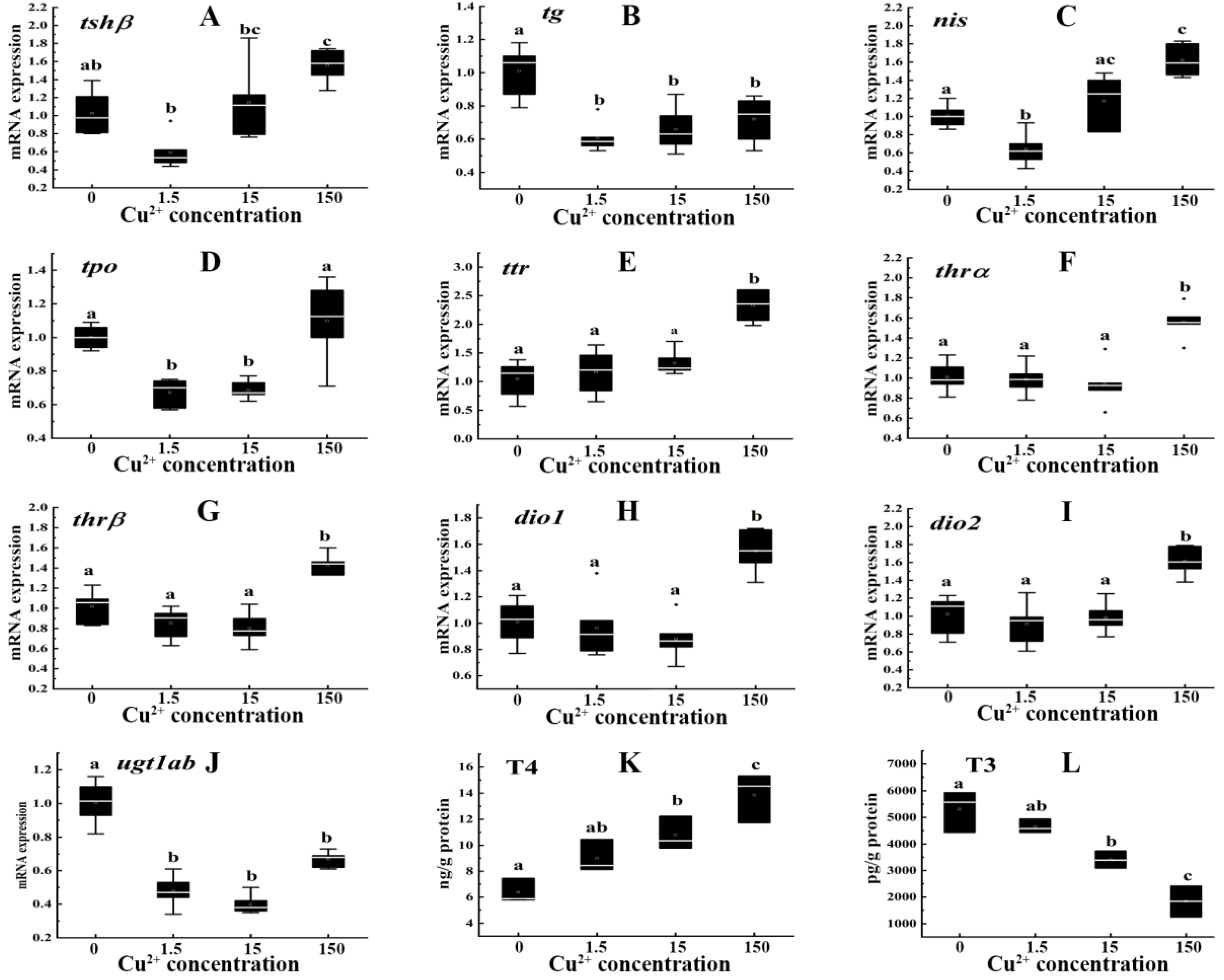
Figure 1. Transcription levels of tshβ (A), tg (B), nis (C), tpo (D), ttr (E), thrα (F), thrβ (G), dio1 (H), dio2 (I), ugt1ab (J) and contents of T4 (K), T3 (L) in zebrafish embryos/larvae that were treated with Cu2+ (0, 1.5, 15, and 150 μg/L) for 120 h. Data are shown as mean ± SD (n = 6). Different letters denote the significant differences between groups.
Toxics 10 00756 g001
Figure 2. Transcription levels of tshβ (A), tg (B), nis (C), tpo (D), ttr (E), thrα (F), thrβ (G), dio1 (H), dio2 (I), ugt1ab (J) and contents of T4 (K), T3 (L) in zebrafish embryos/larvae that were treated with Zn2+ (0, 20, 200 and 2000 μg/L) for 120 h. Data are shown as mean ± SD (n = 6). Different letters denote the significant differences between groups.
Toxics 10 00756 g002
Figure 3. PCA of parameters in zebrafish embryos/larvae that were treated with Cu2+ (0, 1.5, 15 and 150 μg/L) (A) and Zn2+ (0, 20, 200 and 2000 μg/L) (B) for 120 h.
Toxics 10 00756 g003
Figure 4. Heatmap of correlation coefficients between the THs (T4 and T3) contents and the gene expression levels in the zebrafish embryos/larvae that were treated with Cu2+ (0, 1.5, 15 and 150 μg/L) (A) and Zn2+ (0, 20, 200 and 2000 μg/L) (B) for 120 h. The scale indicates the level of positive (red) or negative (blue) correlation, and * indicates significance (* p < 0.05).
Toxics 10 00756 g004
Table 1. The changes of hatching, malformation, and survival rates in zebrafish embryos/larvae treatment with Cu2+ and Zn2+. Data are shown as the mean ± SD (n = 6). * p < 0.05, denotes a statistically significant difference between the treatment and control.
Cu2+ (μg/L)01.515150
Hatching (%)89.83 ± 1.2988.25 ± 1.3287.50 ± 2.1485.58 ± 1.87
Malformation (%)0.83 ± 0.141.92 ± 0.382.25 ± 0.665.12 ± 0.80 *
Survival (%)89.25 ± 1.4087.50 ± 1.4087.50 ± 2.484.83 ± 2.13
Zn2+ (μg/L)0202002000
Hatching (%)88.83 ± 1.5387.08 ± 1.2387.00 ± 2.2983.83 ± 1.61
Malformation (%)1.08 ± 0.381.75 ± 0.663.5 ± 0.754.67 ± 1.13 *
Survival (%)88.33 ± 1.5386.25 ± 0.8685.92 ± 2.4783.33 ± 1.84
Publisher’s Note: MDPI stays neutral with regard to jurisdictional claims in published maps and institutional affiliations.