Conventional Chromatographic Techniques and Biosensors for Mercury Speciation in Seafood: A Systematic Review
 , , , , ,

Abstract
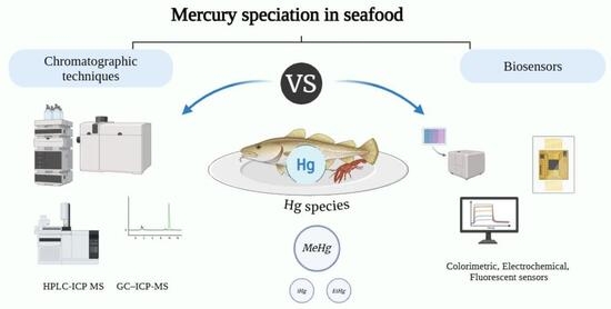
1. Introduction
1.1. Mercury Exposure and Toxicity: A Public Health Concern
1.2. Importance of Mercury Speciation in Seafood Safety
1.3. Analytical Approaches for Mercury Speciation in Seafood
2. Methods
2.1. Literature Search
2.2. Study Selection Criteria
- (i)
- mercury speciation analysis was performed in fish or seafood products;
- (ii)
- the analytical approach was based on chromatographic techniques (including HPLC and non-HPLC methods) or biosensor-based methods; and
- (iii)
- the study was a peer-reviewed original research article published in English.
2.3. Selection Process of the Articles
3. Results
3.1. Results of the Bibliographic Search
3.2. Sample Preparation for the Mercury Speciation in Seafood Samples Prior to Chromatographic Analysis
3.2.1. Microwave-Assisted Extraction (MAE)
3.2.2. Ultrasound-Assisted Extraction (UAE)
3.2.3. Solid Phase Extraction (SPE)
3.2.4. Solid Phase Microextraction (SPME)
3.3. Conventional Chromatographic Techniques
3.3.1. Liquid Chromatography (LC) and Related Separation Approaches
3.3.2. Gas Chromatography (GC)
3.3.3. Capillary Electrophoresis (CE)
3.4. Detection Techniques
3.5. Biosensors
3.5.1. Electrochemical-Based Biosensors
3.5.2. Colorimetric Biosensors
3.5.3. Fluorescence-Based Biosensors
3.6. Comparative Analytical Performance of Chromatographic Versus Sensor-Based Approaches
4. Conclusions
5. Future Perspectives
Supplementary Materials
Author Contributions
Funding
Institutional Review Board Statement
Informed Consent Statement
Data Availability Statement
Conflicts of Interest
Abbreviations
| APDC | Ammonia pyrrolidine dithiocarbamate | ID | Isotope dilution |
| NH4Ac | Ammonium acetate | LODs | Limits of detection |
| AcOH | Acetic acid | LC | Liquid chromatography |
| AFS | Atomic fluorescence spectrometry | Hg | Mercury |
| AAS | Atomic absorption spectrometry | MeHg | Methylmercury |
| CRM | Certified reference material | 2-ME | 2-Mercaptoethanol |
| CV-AFS | Cold vapor atomic fluorescence spectrometry | MeOH | Methanol |
| CVG | Cold vapor generation | MSPE | Magnetic solid phase extraction |
| CE | Capillary electrophoresis | MNPs | Magnetic nanoparticles |
| CE–ICP-MS | CE coupled to inductively coupled plasma mass spectrometry | MAE | Microwave-assisted extraction |
| CNTs | Carbon nanotubes | HNO3 | Nitric acid |
| DBD-OES | Dielectric barriers discharge optical emission spectrometry | NPs | Nanoparticles |
| EtHg | Ethylmercury | PhHg | Phenylmercury |
| GC | Gas chromatography | RP | Reversed phase |
| GC-DBD-OES | GC coupled to DBD-OES | SPE | Solid phase extraction |
| HPLC | High-performance liquid chromatography | THg | Total mercury |
| HPLC-ICP-MS | HPLC coupled to inductively coupled plasma mass spectrometry | TMAH | Tetramethylammonium hydroxide |
| HCl | Hydrochloric acid | NaBEt4 | Sodium tetraethylborate |
| ICP-MS | Inductively coupled plasma mass spectrometry | NaBPr4 | Sodium tetrapropylborate |
| IIP | Ionic imprinted polymer | NaBPh4 | Sodium tetraphenylborate |
| IIP-SPE | Ionic imprinted polymer–solid phase extraction | UAE | Ultrasound-assisted extraction |
| iHg | Inorganic mercury |
References
- Favilli, L.; Giacomino, A.; Malandrino, M.; Inaudi, P.; Diana, A.; Abollino, O. Strategies for Mercury Speciation with Single and Multi-Element Approaches by HPLC-ICP-MS. Front. Chem. 2022, 10, 1082956. [Google Scholar] [CrossRef] [Scilit]
- Ferreira, S.L.C.; Lemos, V.A.; Silva, L.O.B.; Queiroz, A.F.S.; Souza, A.S.; da Silva, E.G.P.; dos Santos, W.N.L.; das Virgens, C.F. Analytical Strategies of Sample Preparation for the Determination of Mercury in Food Matrices—A Review. Microchem. J. 2015, 121, 227–236. [Google Scholar] [CrossRef] [Scilit]
- Clémens, S.; Monperrus, M.; Donard, O.F.X.; Amouroux, D.; Guérin, T. Mercury Speciation in Seafood Using Isotope Dilution Analysis: A Review. Talanta 2012, 89, 12–20. [Google Scholar] [CrossRef] [Scilit]
- Zmozinski, A.V.; Carneado, S.; Ibáñez-Palomino, C.; Sahuquillo, À.; López-Sánchez, J.F.; da Silva, M.M. Method Development for the Simultaneous Determination of Methylmercury and Inorganic Mercury in Seafood. Food Control 2014, 46, 351–359. [Google Scholar] [CrossRef] [Scilit]
- EFSA Panel on Contaminants in the Food Chain (CONTAM). Scientific Opinion on the Risk for Public Health Related to the Presence of Mercury and Methylmercury in Food. EFSA J. 2012, 10, 2985.
- European Commission Commission Regulation (EU). 2023/915 of 25 April 2023 on Maximum Levels for Certain Contaminants in Food and Repealing Regulation (EC) No 1881/2006; Publications Office of the European Union: Luxembourg, 2023. [Google Scholar]
- Kuballa, T.; Moellers, M.; Schoeberl, K.; Lachenmeier, D.W. Survey of Methylmercury in Fish and Seafood from the Southwestern German Market. Eur. Food Res. Technol. 2011, 232, 737–742. [Google Scholar] [CrossRef] [Scilit]
- Médieu, A.; Point, D.; Allain, V.; Bodin, N.; Lemire, M.; Ayotte, P.; Dhurmeea, Z.; Waeles, M.; Laffont, L.; Le Gohalen, A.; et al. Species-Specific Mercury Speciation in Billfishes and Its Implications for Food Safety Monitoring and Dietary Advice. Environ. Int. 2025, 195, 109252. [Google Scholar] [CrossRef] [Scilit] [PubMed]
- Amde, M.; Yin, Y.; Zhang, D.; Liu, J. Methods and Recent Advances in Speciation Analysis of Mercury Chemical Species in Environmental Samples: A Review. Chem. Speciat. Bioavailab. 2016, 28, 51–65. [Google Scholar] [CrossRef] [Scilit]
- Saleh, T.A.; Fadillah, G.; Ciptawati, E.; Khaled, M. Analytical Methods for Mercury Speciation, Detection, and Measurement in Water, Oil, and Gas. TrAC Trends Anal. Chem. 2020, 132, 116016. [Google Scholar] [CrossRef] [Scilit]
- Viana, J.L.M.; Menegário, A.A.; Fostier, A.H. Preparation of Environmental Samples for Chemical Speciation of Metal/Metalloids: A Review of Extraction Techniques. Talanta 2021, 226, 122119. [Google Scholar] [CrossRef] [Scilit]
- Yang, H.; Jian, R.; Liao, J.; Cui, J.; Fang, P.; Zou, Z.; Huang, K. Recent Development of Non-Chromatographic Atomic Spectrometry for Speciation Analysis of Mercury. Appl. Spectrosc. Rev. 2022, 57, 441–460. [Google Scholar] [CrossRef] [Scilit]
- Page, M.J.; McKenzie, J.E.; Bossuyt, P.M.; Boutron, I.; Hoffmann, T.C.; Mulrow, C.D.; Shamseer, L.; Tetzlaff, J.M.; Akl, E.A.; Brennan, S.E.; et al. The PRISMA 2020 Statement: An Updated Guideline for Reporting Systematic Reviews. BMJ 2021, 372, n71. [Google Scholar] [CrossRef] [Scilit] [PubMed]
- Koplík, R.; Klimešová, I.; Mališová, K.; Mestek, O. Determination of Mercury Species in Foodstuffs Using LC-ICP-MS: The Applicability and Limitations of the Method. Czech J. Food Sci. 2014, 32, 249. [Google Scholar] [CrossRef] [Scilit]
- Sedláčková, L.; Kružíková, K.; Svobodová, Z. Mercury Speciation in Fish Muscles from Major Czech Rivers and Assessment of Health Risks. Food Chem. 2014, 150, 360–365. [Google Scholar] [CrossRef] [Scilit] [PubMed]
- Montero-Alvarez, A.; del Rosario Fernández de la Campa, M.; Sanz-Medel, A. Mercury Speciation in Cuban Commercial Edible Fish by HPLC-ICP-MS Using the Double Spike Isotope Dilution Analysis Strategy. Int. J. Environ. Anal. Chem. 2014, 94, 36–47. [Google Scholar] [CrossRef] [Scilit]
- Watanabe, T.; Kikuchi, H.; Matsuda, R.; Hayashi, T.; Akaki, K.; Teshima, R. Performance Evaluation of an Improved GC-MS Method to Quantify Methylmercury in Fish. Food Hyg. Saf. Sci. 2015, 56, 69–76. [Google Scholar] [CrossRef] [Scilit] [PubMed]
- Schmidt, L.; Bizzi, C.A.; Duarte, F.A.; Muller, E.I.; Krupp, E.; Feldmann, J.; Flores, E.M.M. Evaluation of Hg Species after Culinary Treatments of Fish. Food Control 2015, 47, 413–419. [Google Scholar] [CrossRef] [Scilit]
- Brombach, C.-C.; Gajdosechova, Z.; Chen, B.; Brownlow, A.; Corns, W.T.; Feldmann, J.; Krupp, E.M. Direct Online HPLC-CV-AFS Method for Traces of Methylmercury without derivatisation: A Matrix-Independent Method for Urine, Sediment and biological Tissue Samples. Anal. Bioanal. Chem. 2015, 407, 973–981. [Google Scholar] [CrossRef] [Scilit]
- Döker, S.; Boşgelmez, I.I. Rapid Extraction and Reverse Phase-Liquid Chromatographic Separation of Mercury(II) and Methylmercury in Fish Samples with Inductively Coupled Plasma Mass Spectrometric Detection Applying Oxygen Addition into Plasma. Food Chem. 2015, 184, 147–153. [Google Scholar] [CrossRef] [Scilit] [PubMed]
- Chen, B.; Wu, Y.; Guo, X.; He, M.; Hu, B. Speciation of Mercury in Various Samples from the Micro-Ecosystem of East Lake by Hollow Fiber-Liquid-Liquid-Liquid Microextraction-HPLC-ICP-MS. J. Anal. At. Spectrom. 2015, 30, 875–881. [Google Scholar] [CrossRef] [Scilit]
- Deng, D.; Zhang, S.; Chen, H.; Yang, L.; Yin, H.; Hou, X.; Zheng, C. Online Solid Sampling Platform Using Multi-Wall Carbon Nanotube Assisted Solid Phase Dispersion for Mercury Speciation in Fish by HPLC-ICP-MS. J. Anal. At. Spectrom. 2015, 30, 882–887. [Google Scholar] [CrossRef] [Scilit]
- Leon-Perez, D.E.; Munoz-Jimenez, A.M.; Jimenez-Cartagena, C. Determination of Mercury Species in Fish and Seafood by Gas-Mass Spectrometry: Validation Study. Food Anal. Methods 2015, 8, 2383–2391. [Google Scholar] [CrossRef] [Scilit]
- Valdersnes, S.; Fecher, P.; Maage, A.; Julshamn, K. Collaborative Study on Determination of Mono Methylmercury in Seafood. Food Chem. 2016, 194, 424–431. [Google Scholar] [CrossRef] [Scilit]
- Lin, Y.; Yang, Y.; Li, Y.; Yang, L.; Hou, X.; Feng, X.; Zheng, C. Ultrasensitive Speciation Analysis of Mercury in Rice by Headspace Solid Microextraction Using Porous Carbons and Gas-Dielectric Barrier Discharge Optical Emission. Environ. Sci. Technol. 2016, 50, 2468–2476. [Google Scholar] [CrossRef] [Scilit]
- Zhu, S.; Chen, B.; He, M.; Huang, T.; Hu, B. Speciation of Mercury in Water and Fish Samples by HPLC-ICP-MS after Magnetic Solid Phase Extraction. Talanta 2017, 171, 213–219. [Google Scholar] [CrossRef] [Scilit]
- Maage, A.; Nilsen, B.M.; Julshamn, K.; Froyland, L.; Valdersnes, S. Total Mercury, Methylmercury, Inorganic Arsenic and Other Elements in Meat from Minke Whale (Balaenoptera acutorostrata) from the North Atlantic Ocean. Bull. Environ. Contam. Toxicol. 2017, 99, 161–166. [Google Scholar] [CrossRef] [Scilit]
- Liu, Z.; Xing, Z.; Li, Z.; Zhu, Z.; Ke, Y.; Jin, L.; Hu, S. The Online Coupling of High Performance Liquid Chromatography with atomic Fluorescence Spectrometry Based on Dielectric Barrier Discharge Chemical Vapor Generation for the Speciation of Mercury. J. Anal. At. Spectrom. 2017, 32, 678–685. [Google Scholar] [CrossRef] [Scilit]
- Yao, C.-H.; Jiang, S.-J.; Sahayam, A.C.; Huang, Y.-L. Speciation of Mercury in Fish Oils Using Liquid Chromatography Coupled Plasma Mass Spectrometry. Microchem. J. 2017, 133, 556–560. [Google Scholar] [CrossRef] [Scilit]
- Grijalba, A.C.; Quintas, P.Y.; Fiorentini, E.F.; Wuilloud, R.G. Usefulness of Ionic Liquids as Mobile Phase Modifiers in HPLC-CV-AFS for Mercury Speciation Analysis in Food. J. Anal. At. Spectrom. 2018, 33, 822–834. [Google Scholar] [CrossRef] [Scilit]
- Narukawa, T.; Iwai, T.; Chiba, K.; Feldmann, J. A Method for Methylmercury and Inorganic Mercury in Biological Samples High Performance Liquid Chromatography Inductively Coupled Plasma Spectrometry. Anal. Sci. 2018, 34, 1329–1334. [Google Scholar] [CrossRef] [Scilit]
- Londonio, A.; Hasuoka, P.E.; Pacheco, P.; Gil, R.A.; Smichowski, P. Online Solid Phase Extraction-HPLC-ICP-MS System for Mercury and Methylmercury Preconcentration Using Functionalised Carbon Nanotubes for Their Determination in Dietary Supplements. J. Anal. At. Spectrom. 2018, 33, 1737–1744. [Google Scholar] [CrossRef] [Scilit]
- Cheng, H.; Chen, X.; Shen, L.; Wang, Y.; Xu, Z.; Liu, J. Ion-Pairing Reversed-Phase Chromatography Coupled to Inductively Coupled Mass Spectrometry as a Tool to Determine Mercurial Species in freshwater Fish. J. Chromatogr. A 2018, 1531, 104–111. [Google Scholar] [CrossRef] [Scilit]
- Chen, Y.; Chen, L.; Wang, X.; Xi, Z.; Wu, Y.; Fu, F. DNA Binding in Combination with Capillary Electrophoresis and Inductively Coupled Plasma Mass Spectrometry for the Rapid Speciation Analysis of Mercury. Sep. Sci. Plus 2018, 1, 52–58. [Google Scholar] [CrossRef] [Scilit]
- Cheng, H.; Zhang, W.; Wang, Y.; Liu, J. Graphene Oxide as a Stationary Phase for Speciation of Inorganic and Organic Species of Mercury, Arsenic and Selenium Using HPLC with ICP-MS. Microchim. Acta 2018, 185, 425. [Google Scholar] [CrossRef] [Scilit]
- Dezfouli, A.B.; Salar-Amoli, J.; Ali-Esfahani, T.; Hosseini, H.; Ghanati, K. Evaluating Total Mercury and Methyl Mercury Contents in Canned Tuna Fish of the Persian Gulf. Iran. J. Pharm. Res. 2018, 17, 585–592. [Google Scholar]
- Entwisle, J.; Malinovsky, D.; Dunn, P.J.H.; Goenaga-Infante, H. Hg Isotope Ratio Measurements of Methylmercury in Fish Tissues Using with off Line Cold Vapour Generation MC-ICPMS. J. Anal. At. Spectrom. 2018, 33, 1645–1654. [Google Scholar] [CrossRef] [Scilit]
- Liu, H.; Luo, J.; Ding, T.; Gu, S.; Yang, S.; Yang, M. Speciation Analysis of Trace Mercury in Sea Cucumber Species of Apostichopus Japonicus Using High-Performance Liquid Chromatography Conjunction with Inductively Coupled Plasma Mass Spectrometry. Biol. Trace Elem. Res. 2018, 186, 554–561. [Google Scholar] [CrossRef] [Scilit] [PubMed]
- Wang, S.; Song, X.; Hu, J.; Zhang, R.; Men, L.; Wei, M.; Xie, T.; Cao, J. Direct Speciation Analysis of Organic Mercury in Fish and Kelp by On-Line Complexation and Stacking Using Capillary Electrophoresis. Food Chem. 2019, 281, 41–48. [Google Scholar] [CrossRef] [Scilit]
- Cao, D.; Yin, Y.; He, B. Speciation of Organomercury Compounds by Capillary Electrophoresis with Pre-Column Derivatization and on-Line Stacking. Chin. Chem. Lett. 2019, 30, 650–652. [Google Scholar] [CrossRef] [Scilit]
- Linhart, O.; Kolorosova-Mrazova, A.; Kratzer, J.; Hranicek, J.; Cerveny, V. Mercury Speciation in Fish by High-Performance Liquid Chromatography(HPLC) and Post-Column Ultraviolet (UV)-Photochemical Vapor Generation(PVG): Comparison of Conventional Line-Source and High-Resolution Source (HR-CS) Atomic Absorption Spectrometry (AAS). Anal. Lett. 2019, 52, 613–632. [Google Scholar] [CrossRef] [Scilit]
- Li, L.; Bi, R.; Wang, Z.; Xu, C.; Li, B.; Luan, L.; Chen, X.; Xue, F.; Zhang, S.; Zhao, N. Speciation of Mercury Using High-Performance Liquid-Inductively Coupled Plasma Mass Spectrometry Following by Dithizone Functionalized Magnetite-Reduced Graphene Oxide. Spectrochim. Acta Part B 2019, 159, 105654. [Google Scholar] [CrossRef] [Scilit]
- He, Y.; He, M.; Nan, K.; Cao; Beibei, R.; Chen, B.; Hu, B. Magnetic Solid-Phase Extraction Using Sulfur-Containing Functional Polymer for High-Performance Liquid Chromatography-Inductively Plasma-Mass Spectrometric Speciation of Mercury in Environmental. J. Chromatogr. A 2019, 1595, 19–27. [Google Scholar] [CrossRef] [Scilit]
- Krata, A.A.; Wojciechowski, M.; Vassileva, E.; Bulska, E. Reference Measurements of Mercury Species in Seafood Using Isotope Dilution Inductively Coupled Plasma Mass Spectrometry. J. Food Compos. Anal. 2020, 86, 103381. [Google Scholar] [CrossRef] [Scilit]
- Rosera, T.J.; Janssen, S.E.; Tate, M.T.; Lepak, R.F.; Ogorek, J.M.; DeWild, J.F.; Babiarz, C.L.; Krabbenhoft, D.P.; Hurley, J.P. Isolation of Methylmercury Using Distillation and Anion-Exchange Chromatography for Isotopic Analyses in Natural Matrices. Anal. Bioanal. Chem. 2020, 412, 681–690. [Google Scholar] [CrossRef] [Scilit]
- Narukawa, T.; Iwai, T.; Chiba, K. Simultaneous Speciation Analysis of Inorganic Arsenic and Methylmercury in Edible Oil by High-Performance Liquid Chromatography-Inductively Coupled Plasma Mass Spectrometry. Talanta 2020, 210, 120646. [Google Scholar] [CrossRef] [Scilit] [PubMed]
- Garcia, M.; Vassileva, E.; Azemard, S.; Canals, A. Reference Measurements for Priority and Essential Trace Elements and Methyl Mercury with Isotope Dilution Inductively Coupled Plasma-Mass for Seafood Safety Assessment and CRM Production. Food Anal. Methods 2020, 13, 390–402. [Google Scholar] [CrossRef] [Scilit]
- Ghosn, M.; Chekri, R.; Mahfouz, C.; Khalaf, G.; Guérin, T.; Amara, R.; Jitaru, P. Toward a Routine Methodology for Speciation Analysis of Methylmercury in Fishery Products by HPLC Coupled to ICP-MS Following the Validation Based on the Accuracy Profile Approach. Int. J. Environ. Anal. Chem. 2022, 102, 3343–3356. [Google Scholar] [CrossRef] [Scilit]
- Jinadasa, K.K.; Herbello-Hermelo, P.; Pena-Vazquez, E.; Bermejo-Barrera, P.; Moreda-Pineiro, A. Mercury Speciation in Edible Seaweed by Liquid Chromatography—Inductively Coupled Plasma Mass Spectrometry after Ionic Imprinted-Solid Phase Extraction. Talanta 2021, 224, 121841. [Google Scholar] [CrossRef] [Scilit]
- Chen, Y.-C.; Jiang, S.-J. Simultaneous Speciation of Arsenic and Mercury in Fish By-Performance Liquid Chromatography Inductively Coupled Plasma Mass. J. Anal. At. Spectrom. 2021, 36, 938–945. [Google Scholar] [CrossRef] [Scilit]
- Xie, Q.; Wang, Y.; Li, S.; Zhang, C.; Tian, X.; Cheng, N.; Zhang, Y.; Wang, D. Total Mercury and Methylmercury in Human Hair and Food: Implications for the Exposure and Health Risk to Residents in the Three Gorges Reservoir, China. Environ. Pollut. 2021, 282, 117041. [Google Scholar] [CrossRef] [Scilit]
- He, Y.; He, M.; Chen, B.; Hu, B. Preparation of Functional Magnetic Porous Organic Polymer as Sorbent for mercury Speciation Followed by HPLC-ICP-MS Analysis. J. Anal. At. Spectrom. 2021, 36, 1568–1575. [Google Scholar] [CrossRef] [Scilit]
- Zhou, D.B.; Xiao, Y.B.; Han, F.; Lv, Y.N.; Ding, L.; Song, W.; Liu, Y.X.; Zheng, P.; Chen, D. Magnetic Solid-Phase Extraction Based on Sulfur-Functionalized Magnetic Metal-Organic Frameworks for the Determination of Methylmercury and Inorganic Mercury in Water and Fish Samples. J. Chromatogr. A 2021, 1654, 462465. [Google Scholar] [CrossRef] [Scilit]
- Wu, X.; Chen, L.; Li, X.; Cao, X.; Zheng, X.; Li, R.; Zhang, J.; Luo, X.; Mai, B. Trophic Transfer of Methylmercury and Brominated Flame Retardants in Adjacent Riparian and Aquatic Food Webs: 13C Indicates Biotransport of Contaminants through Food Webs. Environ. Pollut. 2022, 306, 119433. [Google Scholar] [CrossRef] [Scilit]
- Spanu, D.; Butti, L.; Boldrocchi, G.; Bettinetti, R.; Recchia, S.; Monticelli, D. Selective Organomercury Determination by ICP-MS Made Easy. Anal. Chim. Acta 2022, 1206, 339553. [Google Scholar] [CrossRef] [Scilit]
- Storelli, A.; Barone, G.; Garofalo, R.; Busco, A.; Storelli, M.M. Determination of Mercury, Methylmercury and Selenium Concentrations in Elasmobranch Meat: Fish Consumption Safety. Int. J. Environ. Res. Public Health 2022, 19, 788. [Google Scholar] [CrossRef] [Scilit]
- Li, R.; Pan, Y.; Sun, C.; Lin, C.; Chen, S.; Wu, Y.; Fu, F. A Broad-Applicability Method for Mercury Speciation in Various Seafoods Microwave-Assisted Extraction and Ion Chromatography-Inductively Plasma Mass Spectrometry. Anal. Methods 2023, 15, 1802–1811. [Google Scholar] [CrossRef] [Scilit]
- Suratno, S.; Wahono, S.K.; Siswanta, D.; Aprilita, N.H. Development of Methylmercury Analysis by Ultra-High Performance Liquid Coupled with ICP-MS and Its Application on Sharks’ Meat. Indones. J. Chem. 2023, 23, 823–830. [Google Scholar] [CrossRef] [Scilit]
- Ribeiro, M.; Galli, E.; Guérin, T.; Silva, J.A.L.; Castanheira, I.; Leufroy, A.; Jitaru, P. Simultaneous Speciation Analysis of Hg and Se in Fish by High-Performance Liquid Chromatography and Inductively Coupled Plasma-Mass Spectrometry Following Microwave-Assisted Enzymatic Hydrolysis. Anal. Bioanal. Chem. 2023, 415, 7175–7186. [Google Scholar] [CrossRef] [Scilit]
- Crowther, E.R.; Demers, J.D.; Blum, J.D.; Brooks, S.C.; Johnson, M.W. Coupling of Nitric Acid Digestion and Anion-Exchange Resin Separation for the Determination of Methylmercury Isotopic Composition within Organisms. Anal. Bioanal. Chem. 2023, 415, 759–774. [Google Scholar] [CrossRef] [Scilit] [PubMed]
- Anil, A.S.; Alam, S.; Thakur, L.K. Optimized Mercury Speciation Analysis Using LC-ICP-MS and Microwave Assisted Extraction for Precise Determination of Methylmercury in Fish, Rice and Soil. J. Food Compos. Anal. 2024, 129, 106092. [Google Scholar] [CrossRef] [Scilit]
- Ghaffour, D.; Leufroy, A.; Jitaru, P. Experimental Design Optimisation and Validation by Accuracy Profile of a Novel Method for Hg Speciation Analysis by HPLC-ICP-MS and Application to Total Diet Studies. Anal. Bioanal. Chem. 2024, 416, 4353–4367. [Google Scholar] [CrossRef] [Scilit] [PubMed]
- Kakimoto, S.; Yoshimitsu, M.; Kakutani, N. Simple and Rapid Analytical Method for the Determination of Methylmercury in Fish and Shellfish Using Solid-Phase Extraction and Gas Chromatography–Mass Spectrometry. J. Chromatogr. B Analyt. Technol. Biomed. Life Sci. 2025, 1250, 124383. [Google Scholar] [CrossRef] [Scilit] [PubMed]
- Zeng, P.; Guo, F.; Wang, Y.; Hu, H.; Liu, J.; Cheng, H. Development of a Tetra-Elemental Speciation Method for Simultaneously Quantifying Cd, Sn, Hg, and Pb Compounds in Shrimp and Fish by High-Performance Liquid Chromatography–Inductively Coupled Plasma Mass Spectrometry. Microchem. J. 2025, 208, 112609. [Google Scholar] [CrossRef] [Scilit]
- Reyes, L.H.; Mizanur Rahman, G.M.; Fahrenholz, T. Comparison of Methods with Respect to Efficiencies, Recoveries, and Quantitation of Mercury Species Interconversions in Food Demonstrated Using Tuna Fish. Anal. Bioanal. Chem. 2008, 390, 2123–2132. [Google Scholar] [CrossRef] [Scilit]
- Chen, S.S.; Chou, S.S.; Hwang, D.F. Determination of Methylmercury in Fish Using Focused Microwave Digestion Following by Cu2+ Addition, Sodium Tetrapropylborate Derivatization, n-Heptane Extraction, and Gas Chromatography-Mass Spectrometry. J. Chromatogr. A 2004, 1024, 209–215. [Google Scholar] [CrossRef] [Scilit]
- Jinadasa, B.K.K.K.; Moreda-Piñeiro, A.; Fowler, S.W. Ultrasound-Assisted Extraction in Analytical Applications for Fish and Aquatic Living Resources, a Review. Food Rev. Int. 2023, 39, 2684–2707. [Google Scholar] [CrossRef] [Scilit]
- López, I.; Cuello, S.; Cámara, C.; Madrid, Y. Approach for Rapid Extraction and Speciation of Mercury Using a Microtip Ultrasonic Probe Followed by LC-ICP-MS. Talanta 2010, 82, 594–599. [Google Scholar] [CrossRef] [Scilit]
- Zhang, W.B.; Xue, J.J.; Yang, X.A.; Wang, S.B. Determination of Inorganic and Total Mercury in Seafood Samples by a New Ultrasound-Assisted Extraction System and Cold Vapor Atomic Fluorescence Spectrometry. J. Anal. At. Spectrom. 2011, 26, 2023–2029. [Google Scholar] [CrossRef] [Scilit]
- Xie, L.; Jiang, R.; Zhu, F.; Liu, H.; Ouyang, G. Application of Functionalized Magnetic Nanoparticles in Sample Preparation. Anal. Bioanal. Chem. 2014, 406, 377–399. [Google Scholar] [CrossRef] [Scilit] [PubMed]
- Rodríguez-Reino, M.P.; Rodríguez-Fernández, R.; Peña-Vázquez, E.; Domínguez-González, R.; Bermejo-Barrera, P.; Moreda-Piñeiro, A. Mercury Speciation in Seawater by Liquid Chromatography-Inductively Coupled Plasma-Mass Spectrometry Following Solid Phase Extraction Pre-Concentration by Using an Ionic Imprinted Polymer Based on Methyl-Mercury-Phenobarbital Interaction. J. Chromatogr. A 2015, 1391, 9–17. [Google Scholar] [CrossRef] [Scilit]
- Mester, Z.; Sturgeon, R. Trace Element Speciation Using Solid Phase Microextraction. Spectrochim. Acta Part B At. Spectrosc. 2005, 60, 1243–1269. [Google Scholar] [CrossRef] [Scilit]
- Montoro-Leal, P.; Garcia-Mesa, J.C.; Morales-Benitez, I.; Vazquez-Palomo, L.; del Mar López Guerrero, M.; Alonso, E.V.I. Synthesis of a Novel Magnetic Nanomaterial for the Development of a multielemental Speciation Method of Lead, Mercury, and Vanadium via HPLC-ICP MS. Microchim. Acta 2023, 190, 296. [Google Scholar] [CrossRef] [Scilit]
- Chen, X.; Han, C.; Cheng, H.; Liu, J.; Xu, Z.; Yin, X. Determination of Mercurial Species in Fish by Inductively Coupled Plasma Mass Spectrometry with Anion Exchange Chromatographic Separation. Anal. Chim. Acta 2013, 796, 7–13. [Google Scholar] [CrossRef] [Scilit]
- Masbou, J.; Point, D.; Guillou, G.; Sonke Jeroen, E.; Lebreton, B.; Richard, P. Carbon Stable Isotope Analysis of Methylmercury Toxin in Biological by Gas Chromatography Isotope Ratio Mass Spectrometry. Anal. Chem. 2015, 87, 11732–11738. [Google Scholar] [CrossRef] [Scilit]
- Hight, S.C.; Cheng, J. Determination of Methylmercury and Estimation of Total Mercury in Seafood Using High Performance Liquid Chromatography (HPLC) and Inductively Coupled Plasma-Mass Spectrometry (ICP-MS): Method Development and Validation. Anal. Chim. Acta 2006, 567, 160–172. [Google Scholar] [CrossRef] [Scilit]
- Liu, Z.; Zhu, Z.; Zheng, H.; Hu, S. Plasma Jet Desorption Atomization-Atomic Fluorescence Spectrometry and Its Application to Mercury Speciation by Coupling with Thin Layer Chromatography. Anal. Chem. 2012, 84, 10170–10174. [Google Scholar] [CrossRef] [Scilit]
- Grinberg, P.; Campos, R.C.; Mester, Z.; Sturgeon, R.E. A Comparison of Alkyl Derivatization Methods for Speciation of Mercury Based on Solid Phase Microextraction Gas Chromatography with Furnace Atomization Plasma Emission Spectrometry Detection. J. Anal. At. Spectrom. 2003, 18, 902–909. [Google Scholar] [CrossRef] [Scilit]
- Fernández, R.G.; Bayón, M.M.; Alonso, J.I.G.; Sanz-Medel, A. Comparison of Different Derivatization Approaches for Mercury Speciation in Biological Tissues by Gas Chromatography/Inductively Coupled Plasma Mass Spectrometry. J. Mass Spectrom. 2000, 35, 639–646. [Google Scholar] [CrossRef] [Scilit]
- Berzas Nevado, J.J.; Rodríguez Martin-Doimeadios, R.C.; Krupp, E.M.; Guzmán Bernardo, F.J.; Rodríguez Fariñas, N.; Jiménez Moreno, M.; Wallace, D.; Patiño Ropero, M.J. Comparison of Gas Chromatographic Hyphenated Techniques for Mercury Speciation Analysis. J. Chromatogr. A 2011, 1218, 4545–4551. [Google Scholar] [CrossRef] [Scilit] [PubMed]
- Chen, C.; Peng, M.; Hou, X.; Zheng, C.; Long, Z. Improved Hollow Fiber Supported Liquid-Liquid-Liquid Membrane Microextraction for Speciation of Inorganic and Organic Mercury by Capillary Electrophoresis. Anal. Methods 2013, 5, 1185–1191. [Google Scholar] [CrossRef] [Scilit]
- Lancaster, S.T.; Brombach, C.-C.; Corns, W.T.; Feldmann, J.; Krupp, E.M. Determination of Methylmercury Using Liquid Chromatography—Photochemical Vapour Generation—Atomic Fluorescence Spectroscopy(LC-PVG-AFS): A Simple, Green Analytical Method. J. Anal. At. Spectrom. 2019, 34, 1166–1172. [Google Scholar] [CrossRef] [Scilit]
- Chen, J.; Chen, H.; Jin, X.; Chen, H. Determination of Ultra-Trace Amount Methyl-, Phenyl- and Inorganic Mercury in Environmental and Biological Samples by Liquid Chromatography with Inductively Coupled Plasma Mass Spectrometry after Cloud Point Extraction Preconcentration. Talanta 2009, 77, 1381–1387. [Google Scholar] [CrossRef] [Scilit]
- Ali, S.; Mansha, M.; Baig, N.; Khan, S.A. Recent Trends and Future Perspectives of Emergent Analytical Techniques for Mercury Sensing in Aquatic Environments. Chem. Rec. 2022, 22, e202100327. [Google Scholar] [CrossRef] [Scilit]
- Ashrafi, A.M.; Koudelkova, Z.; Sedlackova, E.; Richtera, L.; Adam, V. Review—Electrochemical Sensors and Biosensors for Determination of Mercury Ions. J. Electrochem. Soc. 2018, 165, B824–B834. [Google Scholar] [CrossRef] [Scilit]
- Huang, L.; Liu, G.; Fu, Y. Recent Developments in Biosensing Strategies for the Detection of Small Molecular Contaminants to Ensure Food Safety in Aquaculture and Fisheries. Trends Food Sci. Technol. 2023, 133, 15–27. [Google Scholar] [CrossRef] [Scilit]
- Mabes Raj, A.F.P.A.; Chouhan, R.S.; Košak, A.; Horvat, M.; Lobnik, A.; Rijavec, T.; Lapanje, A. Recent Progress and Advancement in Detecting Methylmercury Using a Battery of Biosensors and Biomolecular-Based Techniques: An Updated Overview. TrAC Trends Anal. Chem. 2025, 184, 118157. [Google Scholar] [CrossRef] [Scilit]
- Chen, Z.; Wang, X.; Cheng, X.; Yang, W.; Wu, Y.; Fu, F.F. Specifically and Visually Detect Methyl-Mercury and Ethyl-Mercury in Fish Sample Based on DNA-Templated Alloy Ag-Au Nanoparticles. Anal. Chem. 2018, 90, 5489–5495. [Google Scholar] [CrossRef] [Scilit]
- Saranchina, N.V.; Dudkina, A.A.; Gavrilenko, M.M.; Gavrilenko, N.A. A Simple Method for Colorimetric and Naked-Eye Detection of Mercury in Fish Products. Mater. Sci. Forum 2019, 970, 219–226. [Google Scholar] [CrossRef] [Scilit]
- Huang, Y.; Wang, S.; Wang, Z.; Wang, L.; Gao, H.; Xue, Y.; Lou, X. Task-Specific Ionic Liquid-Enabled Mercury Sensor for Sensitive Detection of Total Mercury in Food Digestion Solution. Sens. Actuators B Chem. 2019, 285, 62–67. [Google Scholar] [CrossRef] [Scilit]
- Erdemir, E.; Suna, G.; Gunduz, S.; Şahin, M.; Eğlence-Bakır, S.; Karakuş, E. Rapid, Ultrasensitive, Highly Selective Detection of Toxic Hg(II) Ions in Seabass, Swordfish and Water Samples. Food Chem. 2022, 371, 131309. [Google Scholar] [CrossRef] [Scilit] [PubMed]
- Shen, T.; Yue, Q.; Jiang, X.; Wang, L.; Xu, S.; Li, H.; Gu, X.; Zhang, S.; Liu, J. A Reusable and Sensitive Biosensor for Total Mercury in Canned Fish Based on Fluorescence Polarization. Talanta 2013, 117, 81–86. [Google Scholar] [CrossRef] [Scilit]
- Deng, L.; Li, Y.; Yan, X.; Xiao, J.; Ma, C.; Zheng, J.; Liu, S.; Yang, R. Ultrasensitive and Highly Selective Detection of Bioaccumulation of Methyl-Mercury in Fish Samples via Ag0/Hg0 Amalgamation. Anal. Chem. 2015, 87, 2452–2458. [Google Scholar] [CrossRef] [Scilit]
- Huang, L.; Li, P.; Lin, C.; Wu, Y.; Chen, Z.; Fu, F. DNA-Templated Fluorescent Silver Nanoclusters on-off Switch for Specific and Sensitive Determination of Organic Mercury in Seafood. Biosens. Bioelectron. 2021, 183, 113217. [Google Scholar] [CrossRef] [Scilit]
- Costas-Mora, I.; Romero, V.; Lavilla, I.; Bendicho, C. In Situ Building of a Nanoprobe Based on Fluorescent Carbon Dots for Methylmercury Detection. Anal. Chem. 2014, 86, 4536–4543. [Google Scholar] [CrossRef] [Scilit]
- El-Raheem, H.A.; Hassan, R.Y.A.; Khaled, R.; Farghali, A.; El-Sherbiny, I.M. New Sensing Platform of Poly(Ester-Urethane)Urea Doped with Gold Nanoparticles for Rapid Detection of Mercury Ions in Fish Tissue. RSC Adv. 2021, 11, 31845–31854. [Google Scholar] [CrossRef] [Scilit]
- Zhang, Y.; Wu, Y.; Su, L.; Zhu, C.; Wu, X. An Ultrasensitive Electrochemical Sensor Based on in Situ Synthesized Manganese Dioxide/Gold Nanoparticles Nanocomposites for Rapid Detection of Methylmercury in Foodstuffs. Anal. Methods 2022, 14, 2329–2336. [Google Scholar] [CrossRef] [Scilit] [PubMed]
- Ran, Q.; Feng, H.; Chang, G.; Luo, M.; Xu, S. Thymine-Mediated Electrochemical Aptasensor for Sensitive and Simultaneous Detection of Hg2+ and CH3Hg+ in Fish Samples. Electrochim. Acta 2023, 461, 142406. [Google Scholar] [CrossRef] [Scilit]
- Rotariu, L.; Lagarde, F.; Jaffrezic-Renault, N.; Bala, C. Electrochemical Biosensors for Fast Detection of Food Contaminants—Trends and Perspective. TrAC Trends Anal. Chem. 2016, 79, 80–87. [Google Scholar] [CrossRef] [Scilit]
- Radulescu, M.C.; Danet, A.F. Mercury Determination in Fish Samples by Chronopotentiometric Stripping Analysis Using Gold Electrodes Prepared from Recordable CDs. Sensors 2008, 8, 7157–7171. [Google Scholar] [CrossRef] [Scilit] [PubMed]
- Inaudi, P.; Mondino, E.; Abollino, O.; Malandrino, M.; Argenziano, M.; Favilli, L.; Boschini, R.; Giacomino, A. On-Site Determination of Methylmercury by Coupling Solid-Phase Extraction and Voltammetry. Molecules 2022, 27, 3178. [Google Scholar] [CrossRef] [Scilit]
- Amine, A.; Mohammadi, H. Procedure 20 Determination of Methyl Mercury in Fish Tissue Using Electrochemical Glucose Oxidase Biosensors Based on Invertase Inhibition. Compr. Anal. Chem. 2007, 49, e139–e149. [Google Scholar]
- Bunney, J.; Williamson, S.; Atkin, D.; Jeanneret, M.; Cozzolino, D.; Chapman, J.; Power, A.; Chandra, S. The Use of Electrochemical Biosensors in Food Analysis. Curr. Res. Nutr. Food Sci. 2017, 5, 183–195. [Google Scholar] [CrossRef] [Scilit]



| n. | Type of Seafood | Analytes | Sample Prepa Ration | Sample Prep Time | Analytical Method | Conc in Sample | Chromatographic Column | Mobile Phase | LOD (µg/kg, Method)/(µg/L, Instrumental) | Recovery | Linearity Range | Used CRM | Analysis Time | References |
|---|---|---|---|---|---|---|---|---|---|---|---|---|---|---|
| 1 | CRM | iHg MeHg | Repeated extraction with HCL and 2-ME, centrifugation, buffering with NH4Ac then filtration | 150 min | HPLC-ICP-MS | iHg: 30–290 µg/kg MeHg: 1–4280 µg/kg | Column:Purospher® STAR RP-8e, 75 × 4 mm, 3 μm Guard column: Purospher® STAR RP-8e, 4 × 4 mm, 5 μm | 0.02 M NH4Ac, 0.2% 2-ME, 1% MeOH | – | MeHg: 98–102% | 0.01–0.05 μg/L | DORM-2 CRM-422 NIST SRM 2976 SRM-1570a CRM-185 | 12 min. | [14] |
| 2 | Fish (chub muscle) | MeHg | Acidic digestion and extraction with toluene | – | GC-ECD | MeHg: 65–231 µg/kg | Capillary column DB 608 (30 m × 0.53 mm × 0.83 µm) | Helium | 21 µg/kg | MeHg: 89 ± 2.5% | – | BCR-463 BCR-464 | – | [15] |
| 3 | 24 seafood samples | iHg MeHg | MAE with HCl | <1 h | LC-UV-CV-AFS | MeHg: 3–2230 µg/kg iHg: 6–85 µg/kg | Analytical RP-C18 column (ODS Hypersyl 250 mm A 4.6 mm id, 5 mm, Thermo Hypersil-Keystone) | 20% APDC and NH4Ac solution and 80% MeOH | iHg: 0.4 µg/kg MeHg: 0.3 µg/kg | MeHg: 80–102% | – | DOLT-4 TORT-2 BCR-463 | 8 min. | [4] |
| 4 | 12 species of commercial fish in Cuba | iHg MeHg | UAE: 0.1% 2-ME, 0.05% L-cysteine and 0.1% HCl | 30 min | ID-LC-ICP-MS | MeHg: 40–1920 µg/kg iHg: <62 mg/kg | Tracer Spherisorb C8 5 μm (100 mm × 4.6 mm) | 0.05% 2-ME, 0.075% L-cysteine and 0.06 M NH4Ac buffer | MeHg: 7.7 µg/kg iHg: 5.2 µg/kg | MeHg: 93–99% | – | DOLT-2 DORM-2 | <10 min | [16] |
| 5 | Cod, tuna, mackerel, and bonito | MeHg | Extraction with acetone, toluene; treatment of residue with KBr, copper sulfate, sulfuric acid. Treatment of toluene layers with L-Cys, HCl; derivatization of 4 mL aliquot with phosphate buffer, NaBPh4 | 50 min | GC-MS | MeHg: 150 µg/kg | InertCap 5MS/NP (0.25 mm i.d. × 30 m long, 0.25 μm) column | Helium | – | 80–110% | – | CRM-7402a BCR-463 BCR-464 CRM-7403a | 9 min | [17] |
| 6 | 6 fish species (bearded brotula, bluewing searobin, flatfish, pirarucu, salmon, and yellowfin tuna) | iHg MeHg | Extraction with 6 mL of L-Cys, centrifugation, and filtration | 1–18 h | LC-CVG-ICP-MS | MeHg: <500 µg/kg | Si-C18 RP-column (Discovery, 250 × 4 mm) and guard column Si-C18 (Allchrom, 4 × 3 mm), both with particle size of 5 μm | 0.1% L-cysteine solution at pH 4 | iHg: 1.7 µg/kg MeHg: 2.3 µg/kg | 97–102% | 0.10–5 μg/L | DORM-2 DOLT-3 | 6 min | [18] |
| 7 | Long-finned pilot whale samples (liver, kidney and muscle) | MeHg | MAE in 2 mL TMAH (60 °C). Ultracentrifugation, dilution of 1 mL clear digest in water, acidification with HCl, topped up to 50 mL | 40 min | HPLC-CV-AFS and species-specific isotope dilution GC-ICP-MS | MeHg: >1000 µg/kg by GC | Eclipse XDB C8 (4.6 × 150 mm, 5 μm) for HPLC; 100% PDMS Silcosteel® (30 m × 0.59 mm × 1.0 μm) for GC | 75% MeOH, 1.5 mM APDC (HPLC), helium (GC) | 0.0015 μg/L (HPLC) | 88–104% | 0–0.5 μg/L (HPLC) | DOLT-2 DOLT-4 TORT-2 DORM-2 | 15 min | [19] |
| 8 | Fish (Black Sea) | iHg MeHg | Homogenized with 0.15% 2-ME, 0.01 M HCl, microwaved (60 °C), centrifugation, filtration | 2 min | HPLC-ICP–MS | <500 µg/kg for muscle meat of fish and fishery products | Zorbax C18 (100 × 4.6 mm, 3.5 μm) | 55.0% MeOH, 0.1% 2-ME, 60 mM NH4Ac, pH 4.0 | iHg: 0.2 µg/kg MeHg: 0.1 µg/kg | 97.6–98.5% | 0.0–20.0 μg/L | TORT-2 DORM-2 | 4 min | [20] |
| 9 | Silver carp muscles (pelagic fish) | iHg MeHg EtHg PhHg | Sonication with 5 M HCl, centrifugation. Second extraction, combination of supernatants, adjusted to pH 7 with NaOH, diluted with water | 80 min | HPLC-ICP-MS | MeHg, iHg: from 9–88.2 µg/kg, EtHg, PhHg: not detected | C18 column (Lichrospher- ODS, 200 × 4.6 mm, 5 μm) | MeOH-buffer solution (2.5 mM L-Cys, 12.5 mM (NH4)2HPO4, 0.05% triethylamine, pH 7.0) | iHg:0.0041 μg/L MeHg: 0.0029 μg/L EtHg: 0.0047 μg/L PhHg: 0.0056 μg/L | MeHg: 92.67% | iHg, EtHg, PhHg: 0.02–5 μg/L MeHg: 0.01–5 μg/L | DORM-2 | 13.3 min | [21] |
| 10 | Grouper, pufferfish | iHg MeHg | MWCNT-assisted MSPD extraction: Blended 4 mg MWCNTs, 10 mg sample. Transferred mixture with 0.20 g C18 co-sorbent to polypropylene column | 35 min | MSPD-HPLC-ICP-MS | iHg: 0.047 µg/kg (grouper), 0.016 µg/kg (pufferfish). MeHg: 0.277 µg/kg (grouper), 0.193 µg/kg (pufferfish) | Agilent zorbax SB-C18 (4.6 mm i.d. × 250 mm, 5 μm) | 8% MeOH, 0.12% L-Cys, 10 mM NH4Ac, pH 7.5 | iHg: 9.9 μg/L MeHg: 8.4 μg/L | iHg: 94% MeHg: 106% | – | DORM-2 DORM-3 | [22] | |
| 11 | Canned tuna in water and oil, 6 types of commercial seafood, 2 frozen fish (Merluccius merluccius and Salmo salar) | MeHg EtHg | Sonication with methanolic KOH. Adjusted pH with AcOH-NaOAc buffer. Added 0.8 mL NaBPh4 (2.0% m/v) and 2 mL hexane | 90 min | GC-MS | MeHg: <LOD in most samples, while those quantified were <1000 µg/kg for predator fish and <500 µg/kg for the others. EtHg: not detected | Thermo non-polar TG-5MS (30 m × 0.25 mm I.D., 0.25- μm) capillary column (5% diphenyl, 95% dimethylpolysiloxane) | Helium | MeHg: 1.61 µg/kg EtHg: 1.95 µg/kg | 70–120% | 20–900 μg/L | NIST SRM 2976 CRM-7402a | 9 min | [23] |
| 12 | 7 seafood products (muscle tissue, squid muscle, crab claw meat, whale meat, cod muscle, halibut muscle and dogfish liver) | MeHg | Digestion: TMAH overnight for complete dissolution. Buffer addition: 1 mL pH 5 AcOH-NaOAc buffer, 600 μL HNO3. Derivatization: 1 mL hexane, 500 μL NaBEt4, centrifugation | – | GC–ICP-IDMS | MeHg: 35–3580 µg/kg (as Greenland halibut muscle had the highest concentration) | 30 m × 0.32 mm id column with HP-5–5% phenyl methyl siloxane; 0.25 μm | Helium | – | 99.9% | 50–500 μg/kg | NIST-SRM1566 BCR-464 DOLT-4 | – | [24] |
| 13 | CRM | iHg MeHg EtHg | Derivatization: with NaBPh4, preconcentrated via headspace SPME with porous carbons. CRMs treated with 25% KOH-MeOH, heated (65 °C), diluted to 25 mL with MeOH | 4 h | GC-DBD-OES | nd | HP-5 capillary column (30 m length × 0.32 mm i.d. × 0.25 μm) | Argon | iHg: 0.5 µg/kg MeHg: 0.75 µg/kg EtHg: 1 µg/kg | 90–105% | iHg: 0.5–30 μg/L MeHg: 0.5–50 μg/L EtHg: 1–100 μg/L | DORM-4 TORT-3 | 10 min | [25] |
| 14 | Bream and crucian fish | iHg MeHg PhHg | MSPE using Fe3O4@SiO2@GMA-MPTS as sorbents | 20 min | MSPE-HPLC-ICP-MS | MeHg: 35.6 µg/kg and 19.1 µg/kg in bream and crucian | C18 column (Hypersil ODS2, 250 × 4.6 mm, 5 μm) | 50:50 MeOH-buffer solution (0.1% 2-ME, 50 mM NH4Ac, adjusted to pH 5 with 0.1 M HAc) | iHg: 0.0007 μg/L MeHg: 0.0007 μg/L PhHg: 0.0005 μg/L | iHg: 94–106% MeHg: 95–105% EtHg: 86–94% | 0.005–5 μg/L | DORM-2 | 8 min | [26] |
| 15 | Meat samples of 84 minke whales | MeHg | Sample digested with TMAH, pH adjusted using NaAc/HNO3, derivatized with NaBEt4. EtHg extracted into hexane | – | GC–ICP–MS | MeHg: 140– 230 µg/kg | ICSep ION-120 ion exchange column | – | MeHg: 30 µg/kg | 96–102% | – | TORT-2 | – | [27] |
| 16 | Tuna | iHg MeHg EtHg | Mixed, shaken, CH2Cl2 added, titrated with HCl. Phases separated, CH2Cl2 transferred to a glass tube. Sodium thiosulfate added, shaken, and centrifuged | – | HPLC-DBD-plasma-CVG-AFS | – | Zorbax SB-C18 (4.6 mm × 75 mm, 3.5 μm) | 0.01% 2-ME, 10% MeOH and 0.06 M NH4Ac at pH 6.8 | iHg: 1.6 μg/L MeHg: 0.42 μg/L PhHg: 0.75 μg/L | iHg: 98.8% MeHg: 92.8% EtHg: 91.9% | 5–200 μg/L | GBW10029 | <14 min | [28] |
| 17 | Three fish oil and fish liver samples | iHg MeHg EtHg | MAE with 2-ME, MeOH, lipase, Triton X-100, HNO3 at 60 °C, cooled, and centrifuged | 5 min | HPLC ICP-MS | RP-C8 column PerkinElmer C8 (3.0 mm × 33 mm; 3- μm) | 0.6% 2-ME, 3% MeOH | 0.013–0.026 μg/L | 93–107% | 0.2–10 μg/L | DOLT-3 | 6 min | [29] | |
| 18 | Fresh fish, canned fish | iHg MeHg EtHg | Sonication with HCl, centrifugation, pH adjusted to 2.0 with a NaOH and citric acid/citrate | 1 night and 4 h | HPLC-CV-AFS | MeHg: 0.17– 0.26 mg/g iHg: 0.14 mg/g EtHg: 0.13– 0.19 mg/g in canned seafood MeHg: 0.15–0.25 mg/g iHg: 0.13 in fresh seafood | Thermo Scientific–Hypersil GOLD aQ–C18 column (150 mm × 4.6 mm i.d.) | A: 0.4% 1-octyl-3-methylimidazolium chloride [C8mim] Cl,100 mM NaCl, and 20 mM buffer citric acid/citrate at pH 2.0 B: MeOH | iHg: 0.1 μg/L MeHg: 0.05 μg/L EtHg: 0.06 μg/L | 94.6–99.2% | iHg: 0.4–1000 μg/L MeHg EtHg: 0.2–1000 μg/L | – | 12 min | [30] |
| 19 | CRM | iHg MeHg EtHg | Heat-assisted alkaline extraction with TMAH at 80 °C | 2 h | HPLC-ICP-MS | MeHg: 58–5100 μg/kg | (i) Octadecylsilyl column (VP-ODS5 μm, i.d. 4.6 × 250 mm), (ii) Adamantyl column (ADME, 3 μm, i.d. 4.6 × 150 mm) | 0.5 g/L L-Cys/1% MeOH (pH 2.3 with HCl) | MeHg: 0.08 and 0.13 µg/kg for ADME and VP-ODS5 columns | MeHg: 101% iHg: 103% | Up to 50 µg/kg | CRM-7402a CRM-7403a DORM-2 NIES CRM No. 13 | 6 and 4 min for ADME and VP-ODS5 columns | [31] |
| 20 | CRM | iHg MeHg | UAE: 5 M HCl | 15 min | SPE-HPLC-ICP-MS | iHg: 18–161 μg/kg MeHg: 141– 3130 μg/kg | Phenomenex SphereClone ODS C18 (5 mm, 250 mm 4.6 mm i.d.) | 0.5% formic acid; 0.2% 2-ME; 20% MeOH | iHg: 0.015 μg/L MeHg: 0.017 μg/L | >93% | 0.01–10 μg/L | BCR 643 TORT-3 | 10 min | [32] |
| 21 | Freshwater fish | iHg MeHg EtHg PhHg | UAE: 40 °C, 5 M HCl, 2 mm L-cysteine | 15 min | IP-RPC-ICP-MS | MeHg: <0.6 μg/g iHg: 3.7–2915 μg/g EtHg: <1.1 μg/g PhHg: <1.7 μg/g | Zorbax C18 25 mm × 4.6 mm × 5 μm | 2.0 mM SDBS, 2.0 mM Cys, 1.0 mM Phe (pH 3.0) and 4.0 mM TBAH, 2.0 mM MPS, 2.0 mM Phe (pH 6.0) | iHg: 0.02 μg/L MeHg: 0.01 μg/L EtHg: 0.03 μg/L PhHg: 0.04 μg/L | iHg: 94–104% MeHg: 93–106% EtHg: 91–104% PhHg: 95–101% | 0.1–100 µg/kg | GBW10029 BCR-463 | 3 min | [33] |
| 22 | Dried fish muscle | iHg MeHg | Sample was 50 μm DNA (HT7 sequence), agitated, then diluted to 0.5 mL with buffer | 15 min | CE–ICP-MS | iHg: 0.018–0.161 μg/g MeHg: 0.473–0.9723 μg/g | 0.72 mM Tris buffer, 0.72 mM boric acid, 16 mM EDTA, and 0.3 mM CTAB (pH = 8.0) | iHg: 0.12 μg/L MeHg: 0.10 μg/L | 95–101% | 5.0–200 μg/L | 11 min | [34] | ||
| 23 | Fish tissue | iHg MeHg EtHg PhHg | UAE with L-cysteine in HCl, 40 °C, centrifuged, neutralized | 15 min | HPLC-ICP-MS | MeHg: 0.81–0.04 mg/kg iHg: 0.018–0.004 mg/kg | GSiO2 column (graphene oxide on silica) | NH4Ac, 2-thiosalicylic acid, Phe, and positively charged BHDMA | iHg: 0.016 μg/L MeHg: 0.027 μg/L EtHg: 0.032 μg/L PhHg: 0.068 μg/L | 92–96%. | 0.5–500 μg/L | GBW10029 | 12 min | [35] |
| 24 | 40 canned tuna samples from Tehran, Iran (white and light style) | THg MeHg | – | GC-MS | MeHg: 207– 352 μg/kg iHg: 111–172 μg/kg in light tuna MeHg: 88–135 μg/kg iHg: 136–303 μg/kg in white tuna | CP-Sil5CB (100% polydimethylsiloxan) fused-silica capillary column (25 m × 0.32 mm i.d. and 1.2 μm) | Helium gas | iHg: 3 µg/kg MeHg: 5 µg/kg | – | – | [36] | |||
| 25 | Fish tissues | iHg MeHg | MAE with HNO3 and HCl | HPLC and CVG MC-ICPMS | MeHg: 229.05 μg/kg in white tuna, 113.37 μg/kg in light-style tuna | C18 column (PROTECOL C18G123 100 × 10 mm) | 0.05% m/m L-Cys, 0.006% v/m AcOH adjusted to pH 4 with ammonia | – | – | – | NIST SRM 1947 BCR-463 | 15 min | [37] | |
| 26 | Sea cucumber (Apostichopus japonicus) | iHg MeHg EtHg | UAE with HCl, L-cystein and 2-ME | 30 min | HPLC-ICP-MS | iHg: 0.04–1.9 mg/kg MeHg: 0.04–2.5 mg/kg | Agilent Zorbax SB-C18 analytical and guard columns | 8% MeOH and 92% H2O | iHg: 0.12 μg/L MeHg: 0.08 μg/L EtHg: 0.20 μg/L | 91.3–104.1% | GBW10024 | 10 min | [38] | |
| 27 | Fish muscle and kelp samples | Sonication in 5 HCL | CE-DAD | 0.038–0.054 mg/mL | Bare fused-silica capillary | Sodium tetraborate | 11–16 μg/L | 84.63–111.39% for fish and 75.68–114.76% for kelp | 50–2000 μg/L | [39] | ||||
| 28 | Fish tissues (Chinese rare minnow) | MeHg EtHg PhHg | Alkaline extraction, dissolved in Na2S2O3 | CE-UV | 30 mM borate buffer at pH 9.2 | MeHg: 76.4 μg/L EtHg: 83.0 μg/L PhHg: 76.9 μg/L | 400–2000 μg/L | DORM-2 | 25 min | [40] | ||||
| 29 | Two fish samples | iHg MeHg EtHg PhHg | Alkaline extraction with TMAH and HCl at 75 °C | 30 min | HPLC-UVPVG and AAS detector | iHg: 0.05–1.16 mg/kg MeHg: 0.4–3.63 mg/kg | Gemini column (C18, 110 A, 3 lm, 150 mm length, 2.4 mm i.d.) | Organic solvent (MeOH, acetonitrile, and ethanol), buffer system (AcOH and NH4 or NaOAc), and photochemical reagent (2-ME) | iHg: 0.47 μg/L MeHg: 0.84 μg/L EtHg: 0.80 μg/L PhHg: 2.0 μg/L | – | Up to 100 μg/L | DOLT-4 BCR-464 | [41] | |
| 30 | Environmental water and fish samples | iHg MeHg | MSPE | – | MSPE-HPLC-ICP-MS | iHg: 0.12–0.18 mg/kg MeHg: 0.4–3.63 mg/kg | C18 column (Hypersil ODS2, 250 × 4.6 mm, 5 m particle size) | 1.0 g/L L-Cys, 60 mM NH4Ac and 2% (vv) MeOH | iHg: 0.00048 μg/L MeHg: 0.00017 μg/L | iHg: 96–103% MeHg: 95–97% | iHg: 0.000016–0.005 µg/L MeHg: 0.0006–0.5 µg/L | DORM-4 | – | [42] |
| 31 | Water, soil, rice and fish | iHg MeHg PhHg | MSPE using Fe3O4@SiO2@GMA-S-SH as sorbents | 7 min | MSPE-HPLC-ICP-MS | iHg: 0.62 mg/kg MeHg: 4.11 mg/kg | Shim-pack CLC-ODS C18 column (15.0 mm × 6.0 mm × 5.0 mm) | 8.0 mM L-Cys, 12.5 mM (NH4)2HPO4, 0.05% triethylamine, pH 7.0, MeOH (8:92) | iHg: 0.0004 μg/L MeHg: 0.00049 μg/L PhHg: 0.0014 μg/L | 84.3–116% | 0.005–5 µg/L | DORM-2 | 17 min | [43] |
| 32 | Clam, oyster and fish homogenate | iHg MeHg | Solid–liquid extraction with magnetic stirring: samples spiked with isotopically enriched MeHg then extracted with 15% HCl at 50 °C | 40 min | HPLC and species-specific isotope dilution ICP-MS | – | Symmetry Shield RP C18, 150 × 3.9 mm, 5 μm and Guard column 3.9 mm × 5 mm, 5 μm | 2-ME, 0.4% L-Cys, 0.06 M NH4Ac and 2% MeOH | iHg: 0.15 µg/kg MeHg: 0.42 µg/kg | – | – | IAEA-461 IAEA-470 IAEA-476 | 5 min | [44] |
| 33 | CRM | iHg MeHg | Anion exchange chromatography using AG 1-X4 that further isolates from the sample matrix, after a distillation treatment | – | MC-ICP-MS | AG 1-X4 anion exchange column | 0.05% (w/v−1) L-cysteine and 1% citric acid trisodium salt dihydrate | MeHg: 5 μg/L | 97 ± 4% | 0.5–1 μg/L | DOLT-2 TORT-2 | [45] | ||
| 34 | CRM | iAs iHg MeHg | UAE with TMAH at 80 °C | 60 min | HPLC-ICP-MS | MeHg: 0.586 mg/kg | RP-C18 ODS column | 5 mM sodium 1-butanesulfonate, 2 mM NH4H2PO4, 4 mM TMAH, 5 mM L-Cys, and 0.1% MeOH (pH 2.3) | MeHg: 0.09 µg/kg | 94–102% | – | NMIJ CRM 740 | 12 min | [46] |
| 35 | Fish from the eastern Irish Sea | iHg MeHg Cu Pb Zn | MAE with HCl at 800 W and ion exchange chromatography | 30 min | ID ICP-MS | MeHg: 0.53 mg/kg | Anion exchange column | – | – | – | IAEA-476 | [47] | ||
| 36 | Canned tuna fish tissues | iHg MeHg | MAE with L-cysteine HCl·H2O at 60 °C, dilution with MeOH, centrifugation and filtration | 15 min | HPLC-ICP-MS | MeHg: 0.500–0.800 g | RP-C18, Agilent peptide mapping (2.1 × 150 mm, 2.7 μm) and precolumn C18 poroshell (2.1 × 50 mm; 2.7 μm) | 5% MeOH and 95% L-cysteine. HCl. H2O | 80–120% | 1–10 μg/L | DOLT-5 BCR-464 TORT-3 | <3 min | [48] | |
| 37 | Tuna fish and seaweeds | iHg MeHg | UAE with HCl, L-cysteine, 2-ME followed by IIP-SPE | 60 min | HPLC-ICP-MS | iHg: 0.001 mg/kg MeHg: 0.05 mg/kg | Kinetex C18 100 Å (100 mm × 2.10 mm, 5 μm) connected with a C18 guard column | 0.4% 2-ME, 10% MeOH, pH 2.0 | iHg: 0.02 μg/kg MeHg: 0.007 μg/kg | iHg: 86–108% MeHg: 89–112% | iHg: 0–50 μg/L MeHg: 0–20 μg/L | BCR-463 | 8 min | [49] |
| 38 | Freshwater tilapia, seawater tilapia, swordfish, freshwater bass and flatfish | iHg MeHg As | MAE with HCl and protease XIV kept in a pool of water which was heated to 70 °C | 70 min | HPLC-DRC-ICP-MS | iHg: nd–134 μg/kg MeHg: 11.7–133 μg/kg | Zorbax SB-Aq C18 column (5 μm, 4.6 mm i.d. × 250 mm) | A: 5 mM 1-octanesulfonate, 5 mM acetate buffer and 1% isopropyl alcohol (IPA) at pH 4.0 B: 2 mM L-cysteine in 1% IPA (pH 4) | MeHg: 0.013 μg/L iHg: 0.015 μg/L | 97–103% | 0.2–10 μg/L | DORM-3 | 4.5 min | [50] |
| 39 | Carassius auratus, Aristichthys nobilis and silver carp fish | MeHg | Digestion with KOH in MeOH at 75 °C, diluted by aqueous ethylation | 3 h | GC-CVAFS | MeHg: 12.9–27.5 μg/kg | – | – | 0.02 µg/kg | 90–110% | – | TORT-2 | [51] | |
| 40 | Crucian and grass carp fish | iHg MeHg EtHg PhHg | UAE with HCl, centrifugation, filtration. Then MSPE (Fe3O4@BD-TpMA-S-SH MOP) was performed | 60 min | HPLC-ICP-MS | MeHg: 3.89–7.95 μg/kg iHg: 51.6–71.9 μg/kg EtHg: nd PhHg: nd | C18 column (Hypersil ODS2, 250 × 4.6 mm, 5 μm) | 8.0 mM L-cysteine, 12.5 mM (NH4)2HPO4, 0.05% triethylamine, pH 7.0, MeOH (8:92) | iHg: 0.00043 μg/L MeHg: 0.00055 μg/L EtHg: 0.00069 μg/L PhHg: 0.0011 μg/L | 82.6–110% | iHg, MeHg, EtHg: 0.002–5 µg/L PhHg: 0.005–5 µg/L | DORM-2 | 1000 s (=16.67 min) | [52] |
| 41 | Sea perch and water | iHg MeHg | Sonication with HCl, centrifugation, filtration. Then MSPE: (Fe3O4@UiO66-SH)9, elute with HNO3 and thiourea | 17 min | HPLC-ICP-MS | iHg: 8.41 MeHg: <LOD | A Pgrandsil-FE-C18 column (150 × 4.6 mm id, 5 μm) | 0.1% L-cysteine, 0.5% MeOH and 10 mM NH4Ac mixture solutions, at pH 7 | 0.00007–0.00013 μg/L | 91.4–95.1% | 0.02–1 µg/L | GBW10029 | 6 min | [53] |
| 42 | Shrimp, tilapia, catfish, and water snake | MeHg | Digested with KOH/MeOH at 80 °C, centrifugation, filtration, 300 μL buffer added, and 40 μL derivatizing reagent (NaBEt4) | 5 h–10 min | GC-CVAFS | MeHg: 2.51–254 μg/kg dw | DB-5MS capillary column (15 m × 0.25 mm × 0.10 μm) | Ar and N2 | – | – | – | – | – | [54] |
| 43 | Three zooplankton samples, four scalloped hammerhead shark muscle samples and two milk shark muscle samples | MeHg | UAE with HCl, centrifugation | 15 min | FC-ICP-MS | MeHg: 0.12–5 mg/kg | Short, homemade anion exchange column | 1.6 µg/kg | 0.024–10 µg/kg | BCR-463 BCR-464 BCR414 and DOLT-5 | 100 s | [55] | ||
| 44 | 15 elasmobranch species | MeHg | Sonication with EtHg chloride in MeOH and HCl, NaCl added, centrifugation, toluene extraction, L-cysteine back-extraction, derivatization with CuSO4 and NaBPh4 | Trace Ultra GC connected with Polaris Q MS | MeHg: 0.41–0.63 µg/g | A SPB-608 capillary column (30 m × 0.53 mm id., 0.5 µm) | Helium (99.99%) | 0.03 µg/kg | 96–101% | TORT-3 | [56] | |||
| 45 | Seaweed, fish and shellfish | iHg MeHg EtHg | MAE with HNO3–L-cysteine at 120 °C, cooled, centrifuged | 6.5 h | IC-HPLC-ICP-MS | iHg: 0.07–0.13 µg/g MeHg: 0.09–0.52 µg/g EtHg: <0.10 in fish iHg: 0.05–0.08 µg/g MeHg: 0.05–0.15 µg/g EtHg: <LOD in shellfish | Two consecutive strong cation guard columns (Zorbax 300SCX, 4.6 × 12.5 mm) | 1.0 mM Cys in 5 mM HNO3 | iHg, MeHg, EtHg: 0.02–0.05 μg/L | 92–105% | 0.5–10 μg/L | Fish (P40219) | 20 min | [57] |
| 46 | Shark meat | MeHg | Sonication with L-cysteine in HNO3 | 30 min | HPLC-ICP-MS | MeHg: 0.22–1.22 mg/kg | C18 Hypersil Gold column | 0.5% L-cysteine in 2% HNO3 and 100% MeOH | MeHg: 8.6 × 10⁻⁷ μg/L | 96.90% | 1–100 μg/L | DORM4 | 300 s | [58] |
| 47 | Rainbow trout, tuna, swordfish, and dogfish | iHg MeHg Se | MAE with protease XIV in buffer, pH adjusted with HNO3 and 2-ME, then diluted with water | 60 min | HPLC-ICP-MS | MeHg: 0.026–1.77 mg/kg | Dionex IonPac™ CS5A A cation exchange column p4 × 250 mm, 9 μm | Mixture of MeOH 5%, 45 mM HNO3, 0.015%2-ME, and 1.5 mM sodium 3-mercapto-1-propanesulfonate | MeHg: 0.011 μg/L | – | – | ERM-CE101 ERM-BB422 CRM-7402a DOLT-5 | 15 min | [59] |
| 48 | Asian clams (soft tissue), megaloptera larvae (whole body), mayfly larvae (whole body), crayfish (muscle tissue), shiner (skinless fillet) | MeHg | Digested with HNO3, 60 °C, centrifuged, diluted | 12 h | GC-CVAFS | Column containing anion exchange resin | MeHg: 0.00007 μg/L | 93.4 ± 2.9% | 0.0005–0.1 µg/L | DORM-3 TORT-2 DOLT-2 DOLT-5 | [60] | |||
| 49 | Rice, fish, soil | iHg MeHg | MAE with HCl (2, 4, 5, 7.5 M) and KOH (10, 20, 25, 30%), various temp (50 to 125◦C) | 50–60 min | HPLC-ICP-MS | MeHg: 0.68–2.26 µg/kg in fish | Zorbax Extended C18 column | 0.06 M NH4Ac, 0.1% 2 -ME and 5% MeOH at pH 6.8 | MeHg: 5 μg/kg | 90–110% | 0–20 μg/L | – | 20 min | [61] |
| 50 | Fishery products | iHg MeHg | MAE with 2-ME and MeOH, diluted with water, filtered | HPLC-ICP-MS | iHg: 1.6–43.4 MeHg: 3.1–190.8 µg/kg | Advance Bio Peptide Map 2.7 μm, 2.1 × 150 mm | 2-ME at 0.25%, MeOH at 1% | iHg: 0.005 μg/kg MeHg: 0.001 μg/kg | iHg: 80–120% MeHg: 90–120% | iHg: 0–1 μg/L MeHg: 0–5 μg/L | TORT-3 SRM 1566-b SQID-1 CRM-7402a | <8 min | [62] | |
| 51 | Fish and shellfish (sea bream, shrimp, crab, scallop, salmon, sardine) | MeHg | QuEChERS-based extraction + HLB SPE + phenyl derivatization | GC–MS | 86.1–98.3% | NMIJ CRM 7402-a, 7403-a, IAEA-436A | [63] | |||||||
| 52 | Fish, shrimp | MeHg, iHg, Cd, Sn, Pb species | Acid extraction with HCl followed by filtration | ~30 min | HPLC–ICP-MS | Amphion II column | Aqueous solution containing sodium dodecylbenzenesulfonate and cysteine | 0.08–0.10 µg kg−1 (MeHg) | 86–105% | 0.1–50 µg/L | ~10 min | [64] |
| Method | Advantages | Disadvantages |
| MAE |
|
|
| UAE |
|
|
| SPE |
|
|
| SPME |
|
|
| Principle | Advantages | Disadvantages | |
|---|---|---|---|
| LC | Analytes separate depending on polarity, solubility, ionic charge and size, or molecular mass (based on their interaction with mobile and stationary phases) | High resolving power Compatible with various detectors No need for laborious, time-consuming and chemical-wasting derivatization (required for GC) Can detect non-volatile compounds | Moderate to high cost depending on the system and detectors |
| GC | Separation based on volatility and interaction with a gas mobile phase and solid stationary phase | Excellent resolving power Transfer analytes from the column to the detector without sample nebulization, enhancing LODs compared to LC Separation of volatile and semi-volatile compounds | Complex sample preparation (derivatization is required) |
| CE | Separate compounds based on their size, charge and interaction with electric field | High automatization A small volume of injected samples is needed Simple sample preparation | Reduced sensitivity due to the short optical path length and small diameter of the capillary Weak UV absorption Requirement for derivatization Require control of buffer and pH |
| Sample | Target Analyte | Sample Preparation | Sample Preparation Time | Recognition Element | Signal Transduction | Assay Time | LOD (µg/kg, Method)/(µg/L, Instrumental) | Ref. | |
|---|---|---|---|---|---|---|---|---|---|
| 1 | Fish muscles | MeHg EtHg | Acid extraction with Tris–HNO3 buffer | – | DNA-templated Ag–Au alloy nanoparticles | Colorimetric (UV–Vis/naked eye) | ~60–70 min | 1000 μg/kg | [88] |
| 2 | Fish products | iHg | SPE into transparent polymer matrix (TPP) with immobilized Cu (DDTC)2 | 10 min | Hg2+-responsive chromogenic reagent | Colorimetric (spectrophotometry/naked eye) | ~10 min | 8 μg/L | [89] |
| 3 | Pig liver and fish | iHg | Acid digestion followed by TSIL extraction | 80 min | Task-specific ionic liquid (TSIL) optode | Colorimetric (optical absorbance) | ~20 min | 0.1 μg/L | [90] |
| 4 | Seabass, swordfish | iHg | MAE with HNO3 200 °C | 30 min | TPA–TSC fluorescent probe | Fluorescence turn-off | <30 s | <0.04 μg/L | [91] |
| 5 | Canned fish | THg | MAE with HNO3 and H2O2 | – | Thymine-rich ssDNA on streptavidin-modified MNPs | Fluorescence polarization | – | 0.09 μg/L | [92] |
| 6 | Fish | MeHg | Alkaline MAE | – | MeHg-specific DNA probe with Ag0/Hg0 amalgamation | Fluorescence | – | 0.089 μg/L | [93] |
| 7 | Fish tissue | MeHg EtHg | MAE with HCl | – | DNA extraction by TSIL coupled with sensors for matrix interference | Fluorescence on–off | 85 min | 1.08 μg/L | [94] |
| 8 | Water and fish tissue | MeHg | UAE with HCl | 4 min | Carbon-dot nanoprobe | Fluorescence (microfluorospectrometry) | 1.18 μg/L | [95] | |
| 9 | Fish and shrimps | iHg | MAE with HNO3 | 4 h | Poly(ester-urethane) urea–AuNP composite | Electrochemical | <300 s | 0.235 μg/L | [96] |
| 10 | Seafood products | MeHg | UAE with HCl, pH adjustment | – | MnO2/AuNP nanocomposite-modified electrode | Electrochemical | – | 0.051 μg/L | [97] |
| 11 | Fish muscle | iHg MeHg | UAE with HCl and derivatization | 2.5 h | Thymine-rich DNA aptamer | Electrochemical | ~60 min | iHg: 0.012 µg/L; MeHg: 0.029 µg/L | [98] |
Disclaimer/Publisher’s Note: The statements, opinions and data contained in all publications are solely those of the individual author(s) and contributor(s) and not of MDPI and/or the editor(s). MDPI and/or the editor(s) disclaim responsibility for any injury to people or property resulting from any ideas, methods, instructions or products referred to in the content. |
© 2026 by the authors. Licensee MDPI, Basel, Switzerland. This article is an open access article distributed under the terms and conditions of the Creative Commons Attribution (CC BY) license.
Share and Cite
Abouelenein, D.; Henares, M.; Fuentes, A.; Fernández-Segovia, I.; M. Barat, J.; Loeschner, K.; Duedahl-Olesen, L.; Gómez-Gómez, M.; Griol, A.; J. Sloth, J. Conventional Chromatographic Techniques and Biosensors for Mercury Speciation in Seafood: A Systematic Review. Foods 2026, 15, 971. https://doi.org/10.3390/foods15060971
Abouelenein D, Henares M, Fuentes A, Fernández-Segovia I, M. Barat J, Loeschner K, Duedahl-Olesen L, Gómez-Gómez M, Griol A, J. Sloth J. Conventional Chromatographic Techniques and Biosensors for Mercury Speciation in Seafood: A Systematic Review. Foods. 2026; 15(6):971. https://doi.org/10.3390/foods15060971
Chicago/Turabian StyleAbouelenein, Doaa, Miguel Henares, Ana Fuentes, Isabel Fernández-Segovia, José M. Barat, Katrin Loeschner, Lene Duedahl-Olesen, Maribel Gómez-Gómez, Amadeu Griol, and Jens J. Sloth. 2026. "Conventional Chromatographic Techniques and Biosensors for Mercury Speciation in Seafood: A Systematic Review" Foods 15, no. 6: 971. https://doi.org/10.3390/foods15060971
APA StyleAbouelenein, D., Henares, M., Fuentes, A., Fernández-Segovia, I., M. Barat, J., Loeschner, K., Duedahl-Olesen, L., Gómez-Gómez, M., Griol, A., & J. Sloth, J. (2026). Conventional Chromatographic Techniques and Biosensors for Mercury Speciation in Seafood: A Systematic Review. Foods, 15(6), 971. https://doi.org/10.3390/foods15060971