Review

Phenolic Antioxidants in Food: A Comparative Review of Chromatographic, Spectroscopic, and Electrochemical Detection Methods


School of Food Science and Engineering, Jiangsu University, No. 301 Xuefu Road, Zhenjiang 212013, China
*
Author to whom correspondence should be addressed.
Foods 2026, 15(18), 3270; https://doi.org/10.3390/foods15183270
Submission received: 6 August 2026 / Revised: 7 September 2026 / Accepted: 14 September 2026 / Published: 16 September 2026
(This article belongs to the Special Issue Intelligent Sensor Systems and Digital Technologies for Food Analysis)

Abstract

The detection of phenolic antioxidants in food is essential for ensuring food safety. Chromatographic analysis, spectroscopic analysis, and electrochemical detection are three mainstream techniques widely applied for qualitative and quantitative analysis of antioxidants in complex matrices. However, existing reviews mostly focus on single techniques, lack systematic cross-platform comparisons. This paper reviews the classification of phenolic antioxidants, summarizes recent advances in each detection technique, and provides a horizontal comparison in terms of sensitivity, selectivity, analytical efficiency, cost, and applicable scenarios. Chromatography remains the gold standard for confirmatory analysis, spectroscopy is suitable for rapid screening, and electrochemistry shows unique potential for on-site real-time monitoring. Each technique has distinct advantages and limitations, and their integration represents a promising direction for comprehensive food safety monitoring. Future development trends, including intelligence, portability, and multi-technique integration, are also discussed to guide practical method selection for food-safety detection.

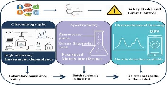
Graphical Abstract

1. Introduction

Oxidative deterioration is a major challenge affecting food quality and shelf life, triggering off-flavour formation, nutrient loss, and generation of potentially harmful secondary products [1,2,3,4]. To prevent such deterioration, the addition of antioxidants is the most widely adopted industrial strategy [5,6,7]. Food antioxidants are generally classified into natural and synthetic categories classified by source [8,9,10]. Natural phenolic antioxidants are mainly extracted from plants, including tea polyphenols, gallic acid, proanthocyanidins, etc. Nevertheless, natural antioxidants commonly suffer drawbacks such as poor stability, limited oil solubility, high cost, and large variability in antioxidant efficacy originating from raw-material sources and extraction processes [11]. Therefore, synthetic phenolic antioxidants, due to their stable chemical properties, significant antioxidant effects, and low production costs, continue to hold a dominant position in the food and even cosmetic industries.
Despite their widespread use, the safety of synthetic phenolic antioxidants has drawn considerable regulatory and scientific scrutiny. Toxicological studies in laboratory animals have demonstrated that high-dose exposure to BHA (Butylated hydroxyanisole) is associated with hepatocellular lesions and hyperplasia, leading the International Agency for Research on Cancer (IARC) to classify BHA as a Group 2B agent (possibly carcinogenic to humans) based on sufficient evidence in experimental animals [12]. Endocrine-disrupting potential of BHA has also been suggested in in vitro studies [13]. For TBHQ (t-Butylhydroquinone), subchronic and chronic exposure studies have indicated neurotoxic potential and DNA-damaging effects at elevated doses in rodent models [13,14,15,16]. PG (Propyl gallate) has been associated with cytotoxic and organ toxic effects at high concentrations in animal studies and in vitro models [17,18]. Although the direct translation of these animal findings to human health risks at normal dietary exposure levels remains a subject of ongoing research, the precautionary principle in food-safety regulation has prompted global regulatory bodies to establish strict maximum permitted levels and acceptable daily intakes (ADIs) for these compounds [19].
Given the potential risks associated with certain synthetic antioxidants as identified in toxicological studies, food safety regulatory agencies in major economies worldwide have established clear regulations regarding their scope of use and maximum permitted levels. For instance, the “National Food Safety Standard—Use Standards of Food Additives” (GB 2760-2024) of China specifies that the maximum allowable usage amounts of BHA, BHT (butylated hydroxytoluene) and TBHQ in oils are 0.2 g/kg, while the maximum allowable concentration of PG is 0.1 g/kg [20]. The European Food Safety Authority (EFSA) has set acceptable daily intakes (ADIs) of 1.0 mg/kg bw/day for BHA, 0.25 mg/kg bw/day for BHT, 0.7 mg/kg bw/day for TBHQ, and 0.5 mg/kg bw/day for propyl gallate (PG) [21,22,23,24]. In the United States, the FDA has classified BHA and BHT as GRAS (Generally Recognized as Safe) substances, with their use subject to good manufacturing practices (GMP) [25,26]. These regulatory frameworks require reliable analytical methods for enforcement and compliance monitoring.
Although the detection of food antioxidants has received widespread attention and there are numerous review articles on the subject, a thorough review reveals that most existing reviews focus on a single aspect—such as the optimization of chromatographic methods, the modification of sensor materials, the evaluation of natural antioxidant activity, or the determination of total antioxidant capacity—and lack a systematic cross-comparison of different detection techniques. In terms of sensitivity, selectivity, analytical throughput, sample pretreatment complexity, and equipment cost, no comprehensive assessment currently exists. A unified standard for evaluating the advantages and disadvantages of these three technologies has not yet been established. Moreover, the applicability and anti-interference ability of detection methods to different food matrices also lack systematic analysis. To address this gap, this review provides a comprehensive and critical comparison of the three major detection platforms, with the aim of offering practical guidance for method selection in food safety monitoring and identifying future research directions. Therefore, this paper first introduces the classification system for phenolic antioxidants in food and the requirements for their detection. It then systematically summarizes the current status and research progress of the three mainstream detection techniques—chromatographic analysis, spectroscopic analysis, and electrochemical detection—and conducts a comparative analysis from multiple perspectives, including detection performance, scope of application, complexity of sample preparation, and application scenarios. Based on this, the advantages and limitations of the three types of technologies are analyzed. Finally, the paper looks ahead to the development trends in smart sensing, portable detection, and multi-technology integration, with the aim of providing guidance for the appropriate selection of detection methods and technological iteration in food safety monitoring.

2. The Development of Antioxidant Classification and Multi-Dimensional Comparison

2.1. Analytical Objectives of Antioxidant Detection

Before discussing specific detection techniques, it is essential to clarify that “antioxidant detection” in food analysis encompasses three conceptually distinct analytical objectives, which differ fundamentally in their methodological requirements and interpretative value.

2.1.1. Quantitative Analysis of Individual Antioxidant Compounds

The determination of specific target antioxidants (e.g., BHA, TBHQ, PG) in food matrices. This objective requires chromatographic separation or highly selective sensors (e.g., molecularly imprinted electrochemical sensors) to distinguish the target from co-existing interferents. Quantification is achieved through external or internal standard calibration.

2.1.2. Total Phenolic Content (TPC) Determination

The measurement of the total amount of phenolic substances in a sample, typically using the Folin–Ciocalteu colorimetric method. This approach provides a composite index reflecting the overall phenolic pool but offers no information on individual compound identities or concentrations.

2.1.3. Total Antioxidant Capacity (TAC)/Activity Assessment

The evaluation of the integrated free-radical scavenging or reducing capacity of a sample using in vitro assays such as ABTS (2,2’-Azinobis (3-ethylbenzothiazoline-6-sulfonic acid)), DPPH (1,1-Diphenyl-2-picrylhydrazyl), or FRAP (Ferric Reducing Antioxidant Power). This parameter reflects the overall antioxidant performance of the sample matrix, which is particularly relevant for functional foods and natural product extracts, but does not correlate directly with the concentration of any single antioxidant compound.
These three analytical objectives are not interchangeable and require distinctly different methodological approaches. The following sections will systematically discuss the three major detection platforms—chromatography, spectroscopy, and electrochemistry—with explicit reference to which analytical objective(s) each technique can address.

2.2. Classification of Phenolic Antioxidants

From a chemical perspective, food antioxidants do not constitute a homogeneous group with a single structure. Instead, they are composed of numerous compounds with different sources, solubilities, and mechanisms of action. They may originate from secondary metabolism of plants, or be artificially synthesized; some are lipophilic and suitable for lipid systems, while others are hydrophilic and active in aqueous media; certain antioxidants directly terminate free-radical chain reactions via phenolic hydroxyl groups, while others exert auxiliary effects through indirect pathways such as chelating metal ions or quenching singlet oxygen [27,28,29,30,31]. Different antioxidants vary significantly in terms of efficacy, strength, safety and cost. Therefore, before discussing the detection methods, a multi-dimensional classification framework should be established to compare the advantages and disadvantages of various antioxidants (Table 1). Although the subsequent detection methodology discussion focuses primarily on phenolic antioxidants, a comprehensive classification including non-phenolic types is presented here to provide a complete context for understanding the structural and physicochemical properties that influence analytical method selection.
Antioxidants can be categorized along multiple independent dimensions, including source, solubility, and chemical structure. These dimensions are not mutually exclusive; a single antioxidant may fall into different categories across dimensions (e.g., BHA is synthetic, lipophilic, and phenolic). When categorized by source, antioxidants can be classified into natural and synthetic antioxidants. Natural antioxidants refer to antioxidant-active substances obtained from plant, microbial, or animal sources through processes such as physical extraction, enzymatic hydrolysis, or solvent extraction, and which have not undergone artificial chemical modification [32,33,34]. They are generally regarded as safer and healthier options and are widely present in daily diets. Common types include vitamins (such as vitamin C, vitamin E), carotenoids (such as lycopene, beta-carotene, astaxanthin), polyphenols (such as tea polyphenols, resveratrol in grapes, anthocyanins in berries, and isoflavones in soybeans), and rosemary extract, etc. [35,36]. In addition to directly eliminating free radicals, many natural antioxidants can also exert comprehensive health benefits by regulating the body’s own endogenous antioxidant system and anti-inflammatory pathways. However, their limitations lie in the high extraction costs and the poor stability and varying bioavailability of some components. Synthetic antioxidants are antioxidant-active substances prepared through chemical synthesis and with specifically designed molecular structures [37,38,39]. Common varieties include BHA, BHT and TBHQ, which have stable chemical properties and strong antioxidant effects, and can significantly extend the shelf life of food. However, although their use in compliance is considered safe, consumers have always had concerns about the long-term safety of consuming them. Some compounds have potential toxic risks at high doses, which has driven the food industry to continuously seek natural alternatives.
Based on solubility, antioxidants can be classified into oil-soluble and water-soluble types. Oil-soluble antioxidants are mainly soluble in oils and organic solvents, and can exert antioxidant effects in lipid environments such as cell membranes and lipoproteins [40]. Common examples include vitamin E (tocopherol), butylated hydroxyanisole (BHA), and BHT, etc. They are adept at protecting foods rich in oils from oxidative rancidity. In the human body, they mainly function to protect the lipid layer of cell membranes from free radical attacks [41]. Their key characteristic is fat-solubility, enabling accumulation within the human body; therefore, dosage intake should be carefully controlled. Water-soluble antioxidants mainly dissolve in water and function in aqueous environments such as plasma and cytoplasm. Common representatives include vitamin C, tea polyphenols, and anthocyanins, etc. [42,43]. They are mainly applied in water-based food systems. In the human body, they are responsible for eliminating free radicals in the blood and cytoplasm, and can also work in synergy with oil-soluble antioxidants [44,45]. For instance, vitamin C can regenerate the oxidized vitamin E, restoring its activity. They exhibit broad-spectrum activity yet are poorly retained in the human body and require continuous dietary supplementation.
Based on chemical structure, antioxidants can be classified into phenolic and non-phenolic types. Phenolic antioxidants are structurally defined as compounds bearing one or more phenolic hydroxyl groups (i.e., hydroxyl groups directly attached to an aromatic ring). Their primary antioxidant mechanism involves the donation of a hydrogen atom from the phenolic OH group to free radicals via hydrogen atom transfer (HAT), thereby terminating radical chain reactions while forming relatively stable phenoxyl radicals through resonance stabilization. Additionally, phenolic compounds can act through single electron transfer (SET) mechanisms, and their antioxidant activity is influenced by factors such as redox potential, pH, solvent, and concentration. Some phenolic varieties also exhibit metal-chelating capacity, indirectly inhibiting oxidation reactions [46,47,48,49]. Typical representatives include natural sources of tea polyphenols, tocopherol (vitamin E), rosmarinic acid, resveratrol, as well as synthetic substances such as BHA, BHT, and TBHQ [50,51,52]. Phenolic compounds usually possess excellent redox cycling capabilities. Some varieties can also chelate metal ions, indirectly inhibiting the occurrence of oxidation reactions. Non-phenolic antioxidants refer to substances whose structures do not contain phenolic hydroxyl groups and exert their antioxidant effects through other chemical mechanisms. Their mechanisms of action include either relying on the enol structure (such as vitamin C) to provide reducing hydrogen atoms, or by capturing reactive oxygen species or peroxide intermediate products to interrupt the oxidation process [53,54]. Typical examples include vitamin C (ascorbic acid), coenzyme Q10, carotenoids (such as lycopene and astaxanthin), and alpha-lipoic acid [55,56,57]. It is precisely because of the significant differences in the sources, solubility and chemical structure of antioxidants that the detection of antioxidants becomes even more important.
Table 1. A Comparison of Different Categories of Antioxidants.
CategoryRepresentativeSourceSolubility (logP)Structural
Features
Primary Antioxidant MechanismSafety/Regulatory StatusAnalytical PropertiesMain Analytical Challenges
Synthetic monophenolsBHASynthetic~3.52(3)-tert-butyl-4-hydroxyanisole (mixture of isomers)HAT donor; moderate chain-breaking activity [58]IARC Group 2B; EFSA ADI: 1.0 mg/kg bw/dayUV λmax 288 nm; electroactive; volatile [59]Moderate UV response; co-elution with lipid matrix
Synthetic polyphenolsTBHQSynthetic~2.92-tert-butyl-1,4-dihydroxybenzeneStrong HAT and SET donor; most effective among synthetic types [60]EFSA ADI: 0.7 mg/kg bw/day; banned in JapanUV λmax 290 nm; highly electroactive; thermally labile [61]Prone to oxidation; thermal decomposition
Synthetic polyphenolsPGSynthetic~2.0Propyl 3,4,5-trihydroxybenzoateStrong HAT donor; metal chelation [62]EFSA ADI: 0.5 mg/kg bw/dayUV λmax 272 nm; highly electroactive; polar [63]Poor retention on conventional C18 columns
Natural tocopherolsα-, γ-, δ- tocopherolNatural>7Chromanol ring with phytyl side chain; phenolic OHHAT + singlet oxygen quenching; activity: δ > γ > α for HAT [64]GRAS (FDA 21 CFR 182.3890)UV λmax 292 nm; weak electroactive; fluorescence (ex 295/em 330 nm) [65]Fluorescence requires optimization; poor thermal stability for GC
Natural tea polyphenolsEGCGNatural~1.5Gallate ester with 8 phenolic OH groupsVery strong HAT/SET donor; metal chelation; pro-oxidant at high doses [66]Generally recognized as safe; rare hepatotoxicity at high supplement dosesUV λmax 280, 325 nm; highly electroactive; non-volatile [67]Poor stability at neutral pH/high temperature; matrix interference
Non-phenolic ascorbic acidAscorbic acidNatural/synthetic~−1.9Enediol structure (no phenolic ring)SET donor; regenerates tocopherol; pro-oxidant with metal ions [68]GRAS (FDA 21 CFR 182.3013)UV λmax 265 nm; electroactive; unstable in solution [69]Unstable to heat, light, and oxygen
Non-phenolic carotenoidsβ-caroteneNatural~15Polyene chain (no phenolic OH)Singlet oxygen quenching; poor HAT donor [70]Approved as food colorant/nutrient supplement;UV–Vis λmax 450, 475 nm; non-electroactive; non-volatile [71]Requires normal-phase LC; easily oxidized

3. Classification and Research Progress of Detection Technology

The detection methods for antioxidants in food have continuously evolved with the advancement of analytical chemistry techniques, forming a multi-level technical system ranging from classical chemical analysis to modern instrumental analysis and to on-site rapid detection. According to the different detection principles, the existing methods can be classified into three major categories: chromatographic analysis, spectroscopic analysis, and electrochemical detection. Among these, chromatography has become the gold standard for confirmatory analysis in laboratories due to its exceptional separation capabilities and quantitative accuracy; spectroscopic techniques are well-suited for high-throughput screening because of their ease of operation and rapid response; and electrochemical detection has garnered significant attention in real-time on-site monitoring due to its high sensitivity and great potential for miniaturization. Each of these three types of technology has its own strengths, and they complement one another in various application scenarios. The following section first outlines the overall development trajectory of detection technologies, followed by a systematic discussion of research progress for each category.

3.1. Development Process of Detection Technology

As the food matrix components become increasingly complex, food safety regulatory standards become stricter, and the actual demand for on-site rapid quality control in the food industry keeps rising, the food antioxidant detection technology has undergone multiple technological updates. The antioxidant detection technology can be divided into three stages.

3.2. Simple Instrument Analysis Stage in the Laboratory

During this period, traditional analytical methods such as chemical titration and spectrophotometry were mainly relied upon. For instance, iodometric methods based on the reductivity of antioxidants, determination of iron reductivity, and colorimetric quantitative analysis using color reactions (such as the color reaction between BHA and 2,6-dichloroquinone-4-chloroimide) were employed. These methods required simple instruments and were suitable for routine laboratory analysis, but had limited sensitivity and were susceptible to interference from complex food matrices, making them unable to meet the requirements for precise quantification and simultaneous detection of multiple components. This stage mainly focused on qualitative analysis and rough quantification.

3.3. Precision Instrument Analysis Stage

With the widespread use of sophisticated analytical instruments and the gradual improvement of chromatographic theory, the detection methods for antioxidants have evolved from simple qualitative testing to precise quantitative testing, and the detection accuracy has also increased significantly. Representative techniques include high-performance liquid chromatography (HPLC), gas chromatography (GC), liquid chromatography tandem mass spectrometry (LC-MS/MS), and gas chromatography-tandem mass spectrometry (GC-MS/MS) [72,73,74,75,76]. These techniques rely on chromatographic separation to achieve component splitting, combined with mass spectrometry and ultraviolet detectors to complete precise quantification. They feature excellent detection repeatability, low quantification error, and significantly improved sensitivity compared to traditional chemical methods. For example, Xu et al. [77] developed a new method combining dispersed liquid–liquid microextraction with high-performance liquid chromatography for the simultaneous determination of six synthetic phenolic antioxidant compounds in edible oil. By optimizing parameters such as extraction solvent, dispersion solvent, and ultrasonic time, this method requires only 25 mg of sample and 1 mL of organic solvent, and can complete the sample preparation in approximately 5 min. Method validation demonstrated that each target compound had good linearity within the range of 0.1–500 mg/L, with spiked recovery rates ranging from 86.3% to 102.5%, and RSD (relative standard deviation) values below 3.5%. The results of actual sample determination showed that TBHQ was the most frequently detected component, and none of the analyzed samples exceeded the regulatory limit. This method is simple, rapid, and requires only minimal organic solvent consumption, providing an effective tool for the quality control of antioxidants in edible oil. The introduction of chromatographic instrument analysis technology has effectively made up for the shortcomings of early detection methods in terms of accuracy and specificity, laying a methodological foundation for the precise detection of phenolic antioxidant residues in food.

3.4. Rapid Detection Stage

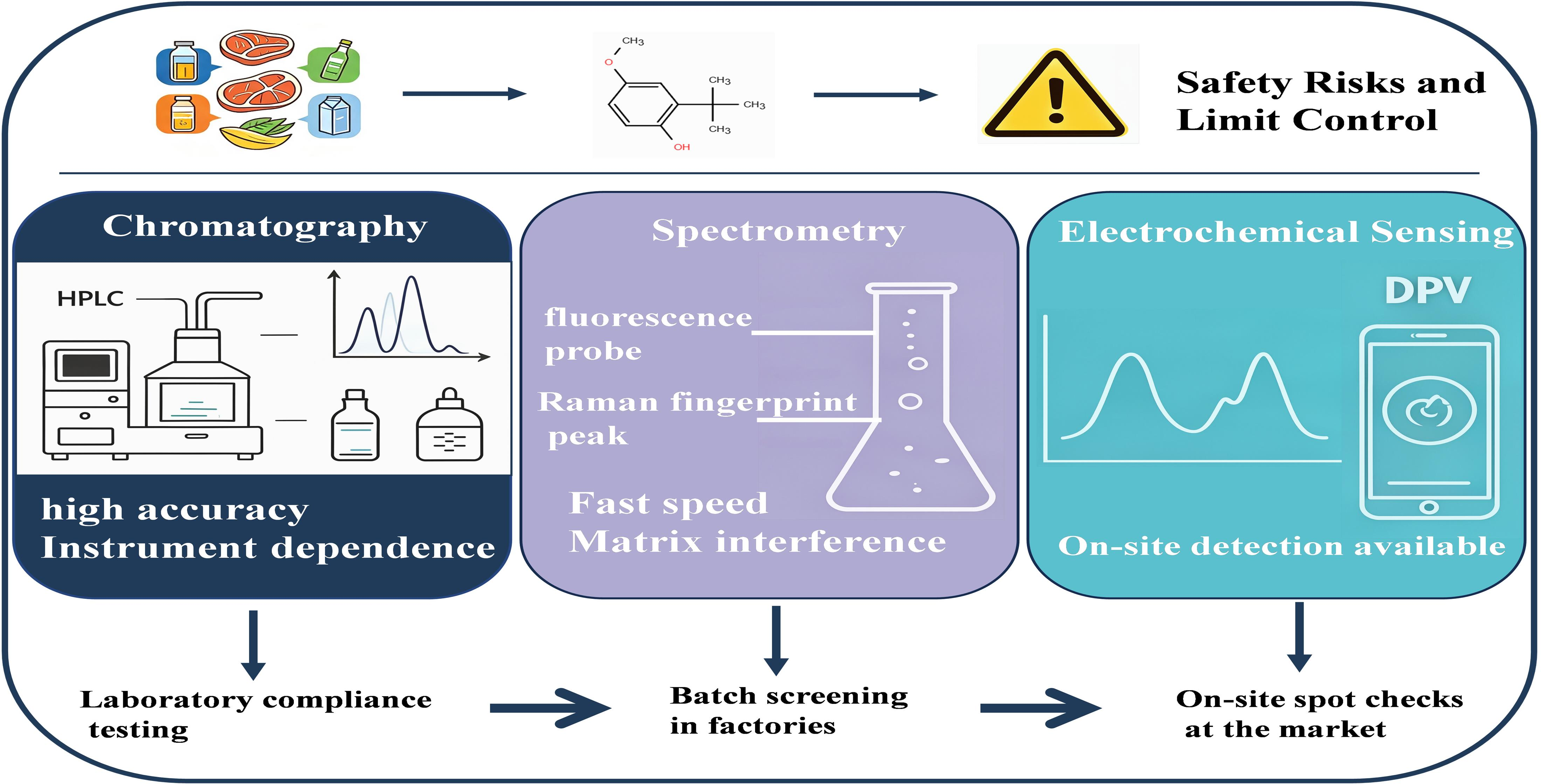
With the increasing globalization of the food supply chain, the chain from the farm to the table has become longer, and the risk points have increased. The traditional centralized laboratory testing mode is increasingly unable to meet the timeliness requirements. At the same time, the production process of food also needs to strictly control the content of harmful substances. Therefore, on-site rapid detection of antioxidants has emerged. The emerging technologies in this stage include spectroscopy analysis, electrochemical sensing, and biological sensing, etc. These technologies have removed the limitations of large-scale precision instruments and simplified the sample pre-treatment process. The detection time has been reduced from several hours to a few minutes [78,79,80,81,82]. For instance, Soylu et al. [83] developed an electrochemical sensor based on a molecularly imprinted polymer (MIP)-modified NiFe2O4/multi-walled carbon nanotube (MWCNT) nanocomposite for the detection of tert-butylhydroquinone (TBHQ) in edible oil. By modifying the surface of a glassy carbon electrode with MIP/MWCNT/NiFe2O4, the conductivity, specific surface area, and active sites were significantly enhanced. Under optimal conditions, the linear range of differential pulse voltammetry for detecting TBHQ was 0.5–25 μM and 25–600 μM, with a detection limit as low as 0.05 μM. This sensor exhibited excellent stability, selectivity, and repeatability, and the spiked recovery rate in actual oil samples was 96.90–104.20%, with RSD lower than 2.94%, providing an effective new method for the rapid detection of TBHQ in edible oil (Figure 1). The new rapid detection technology has enabled the detection to gradually shift from offline precise detection in the laboratory to on-site, real-time and intelligent rapid screening. However, it should be noted that these advantages are context-dependent; spectroscopic methods still face selectivity limitations, while electrochemical sensors require careful evaluation of long-term stability and reproducibility under real-world conditions.

3.5. Overview of Sample Pretreatment Techniques

Regardless of the detection technique used, sample preparation is a critical step that affects the accuracy and reproducibility of analytical results. Food matrices have complex compositions; co-existing substances such as fats, proteins, pigments, and carbohydrates not only interfere with the separation of target compounds and their signal responses but may also contaminate chromatographic columns or passivate electrode surfaces. Therefore, sample preparation steps are necessary to extract, purify, and enrich the target antioxidants.
The traditional pre-treatment methods are mainly represented by liquid–liquid extraction and solid-phase extraction. Liquid–liquid extraction extracts the target substances based on the distribution difference between the organic phase and the aqueous phase. It is simple to operate and has low cost, but it consumes a large amount of organic solvents, frequently causes emulsification, and is difficult to handle high lipid matrices [84]. Solid-phase extraction selectively retains the target substances through the adsorbent and then elutes them. Its purification effect is superior to that of liquid–liquid extraction. However, the operation process is cumbersome and time-consuming, and the solid-phase extraction column is a one-time consumable material, resulting in high detection costs [85,86].
In recent years, to meet the demands for rapid detection and high-throughput analysis, miniaturized and green pre-treatment technologies have developed rapidly. Dispersed liquid–liquid microextraction rapidly injects trace extraction solvents and dispersants into the sample solution, forming a cloud-like microemulsion, which greatly increases the contact area between the extraction solvent and the target substance, and can complete the extraction within several minutes. For example, Mogaddam et al. [87] developed a dispersion-liquid–liquid microextraction (DLLME) pretreatment method based on heated liquid–liquid extraction combined with a switchable low-eutectic solvent (DES) for the extraction of three phenolic antioxidants from edible oils. In this method, a trace amount of DES extractant is dissolved in an alkaline solution to form a homogeneous system; upon injection of an acidic solution, an acid-base reaction occurs, causing the target compounds to migrate into the fine DES droplets, thereby completing the extraction. The entire extraction process requires no organic solvents and is simple and cost-effective. Method validation results showed that the limits of detection for the three antioxidants ranged from 0.13 to 0.42 ng/mL, with enrichment factors of 370–445, extraction recoveries of 74–89%, and relative standard deviations ≤ 7.4%. This method was successfully applied to the quantitative analysis of antioxidants in actual edible oil samples.
Overall, the development trend of sample pretreatment techniques is towards miniaturization, automation, low solvent consumption, and online integration. Chromatography has the highest dependence on pretreatment, followed by spectroscopy. Electrochemistry, on the other hand, has relatively less stringent requirements for pretreatment as it can directly detect in complex matrices. Therefore, electrochemistry has significant advantages in field applications, although challenges such as electrode fouling and matrix effects require careful consideration.

3.6. Development and Progress of Chromatographic Analysis Technology

Chromatographic analysis is the most mature and widely applied technical method for detecting food antioxidants. The mainstream chromatography techniques are divided into two branches: gas phase and liquid phase. In the field of antioxidant detection, each category has its specific applicable objects and technical characteristics.
The evolution of chromatographic techniques for antioxidant analysis has progressed through several stages: from conventional liquid chromatography (LC) and gas chromatography (GC) for single-compound analysis, to high-performance liquid chromatography (HPLC) with UV or fluorescence detection for multi-component separation, and more recently to ultra-high-performance liquid chromatography (UHPLC) and comprehensive two-dimensional chromatography (LC × LC, GC × GC) coupled with high-resolution mass spectrometry (HRMS) for untargeted profiling and trace-level quantification. The development of reversed-phase HPLC (RP-HPLC) with C18 stationary phases, combined with diode array detection (DAD), enabled simultaneous separation and spectral identification of multiple phenolic antioxidants. The hyphenation of chromatography with mass spectrometry—from GC-MS to LC-MS/MS and HRMS—further enhanced sensitivity and structural elucidation capability, making these techniques indispensable for confirmatory analysis in food safety laboratories.
Liquid chromatography is currently the most widely used technique in antioxidant analysis. It can be further classified based on the separation mechanism into normal-phase high-performance liquid chromatography and reversed-phase liquid chromatography [88]. Polar chromatography is mainly used for separating polar compounds such as phenolic acids and flavonoids that are derived from natural sources [89,90,91]. Reversed-phase high-performance liquid chromatography is the most commonly used method. By using a C18 column in combination with a mobile phase system of acetonitrile-water or methanol-water, it can simultaneously separate BHA, BHT, TBHQ, PG, and a variety of natural phenolic antioxidants within one analytical cycle [92,93]. For example, Casagrande et al. [94] developed a reverse-phase liquid chromatography method for the direct analysis of four synthetic phenolic antioxidants (PG, TBHQ, BHA, and BHT) in biodiesel. Although biodiesel is not a food matrix, this study provides valuable methodological insights into the separation of phenolic antioxidants from lipid-rich matrices using a phenyl column via π-π interactions, which are transferable to edible oil analysis. The study found that BHT co-eluted with fatty acid methyl esters on a C18 column, whereas an alkylphenyl column achieved good separation of the antioxidants from the matrix through π-π interactions. This method exhibits good linearity in the range of 10–80 ppm (R2 > 0.9986), with detection limits of 0.010–0.030 ppm and spiked recovery rates of 70–103%.
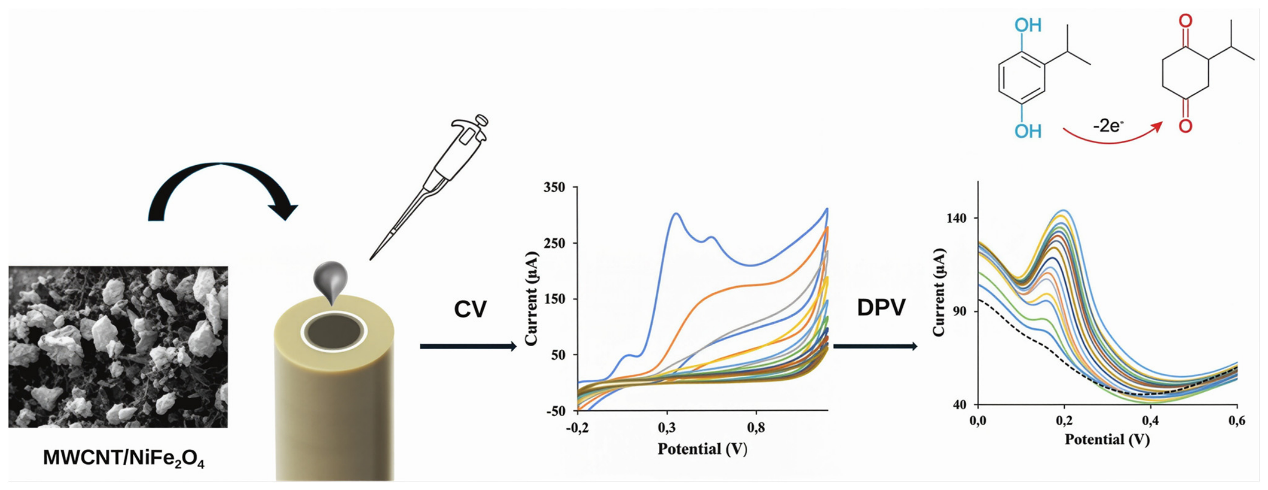
Gas chromatography utilizes the difference in distribution between the sample components in the gaseous state and the stationary phase of the chromatographic column to achieve separation. It is suitable for those antioxidants with strong volatility and good thermal stability [95,96,97,98]. BHA and BHT are the most commonly analyzed compounds in gas chromatography. These two synthetic phenolic antioxidants have low molecular weights and moderate boiling points; they vaporize readily at an injection port temperature of around 200 °C without undergoing thermal degradation, allowing them to be analyzed directly by gas chromatography without the need for complex derivatization pretreatment. For food matrix applications, Znidersic et al. [99] developed an SPME-GC-MS/MS method for the simultaneous determination of nine compounds (including phenolic antioxidants, parabens, plasticizers, and flame retardants) expected to migrate from food contact materials into beverages and vinegar, with no derivatization required. The method achieved detection limits of 0.005–0.2 μg/L, mean recoveries of 98–109%, and RSD values of 0.8–5.4%. Similarly, Chen et al. [100] established a headspace solid-phase microextraction (HS-SPME) coupled with GC-MS method for the analysis of synthetic phenolic antioxidants in food-grade lubricant samples, demonstrating recoveries of 80.7–109.6%. These studies confirm the applicability of GC-MS for volatile antioxidant analysis in food-related matrices. Additionally, Ghosh et al. [101] demonstrated the utility of a headspace needle trap combined with portable GC-MS for rapid BHT quantification in cosmetics, highlighting the potential of portable GC-MS for on-site screening applications (Figure 2). For phenolic compounds with high boiling points or those prone to decomposition upon heating—such as TBHQ and gallate esters—the application of gas chromatography is limited, often requiring derivatization to reduce polarity and improve thermal stability, which adds to the number of steps and increases analysis time. For example, Xu et al. [102] successfully determined TBHQ and its metabolites in fats and oils simultaneously using gas chromatography–mass spectrometry (GC-MS). GC generally delivers higher separation efficiency and faster analysis than HPLC, yet sample-preparation workflows are more complex and demand higher operator proficiency. In addition, thermally unstable compounds may decompose during vaporization, limiting the application of GC in the detection of certain phenolic antioxidants.
Beyond conventional HPLC and GC, ultra-high performance liquid chromatography (UHPLC) with sub-2 μm particles has further improved separation efficiency and reduced analysis time. Liquid chromatography-tandem mass spectrometry (LC-MS/MS) has become increasingly important for antioxidant analysis, offering superior sensitivity, structural confirmation capability, and the ability to simultaneously quantify multiple targets in complex food matrices [103,104,105]. This is particularly advantageous for thermally labile and polar phenolic antioxidants that are not amenable to GC analysis.
In summary, the choice between GC and LC depends primarily on the physicochemical properties of the target antioxidants: GC is suitable for volatile and thermally stable compounds such as BHA and BHT, often with minimal derivatization; LC is preferred for polar, non-volatile, or thermally labile compounds such as TBHQ, PG, and natural polyphenols, and is more compatible with high-sensitivity mass spectrometric detection. Derivatization is generally required for GC analysis of phenolic antioxidants containing multiple hydroxyl groups to reduce polarity and improve thermal stability.

3.7. Development and Progress of Spectroscopic Analysis Technology

The mainstream spectroscopic techniques for food antioxidant detection include ultraviolet-visible absorption spectroscopy, molecular fluorescence spectroscopy, Fourier transform infrared spectroscopy, and surface-enhanced Raman spectroscopy (SERS) [36,106,107]. The application of spectroscopic techniques for antioxidant analysis has progressed from conventional UV-Vis absorption and fluorescence methods to more advanced techniques such as SERS, enabled by the development of nanostructured substrates and chemometric algorithms. They achieve the detection by relying on the optical characteristic signals of the phenolic substances themselves. Unlike chromatography, which requires a complex separation process, the sample pretreatment is relatively simple and analysis proceeds rapidly.
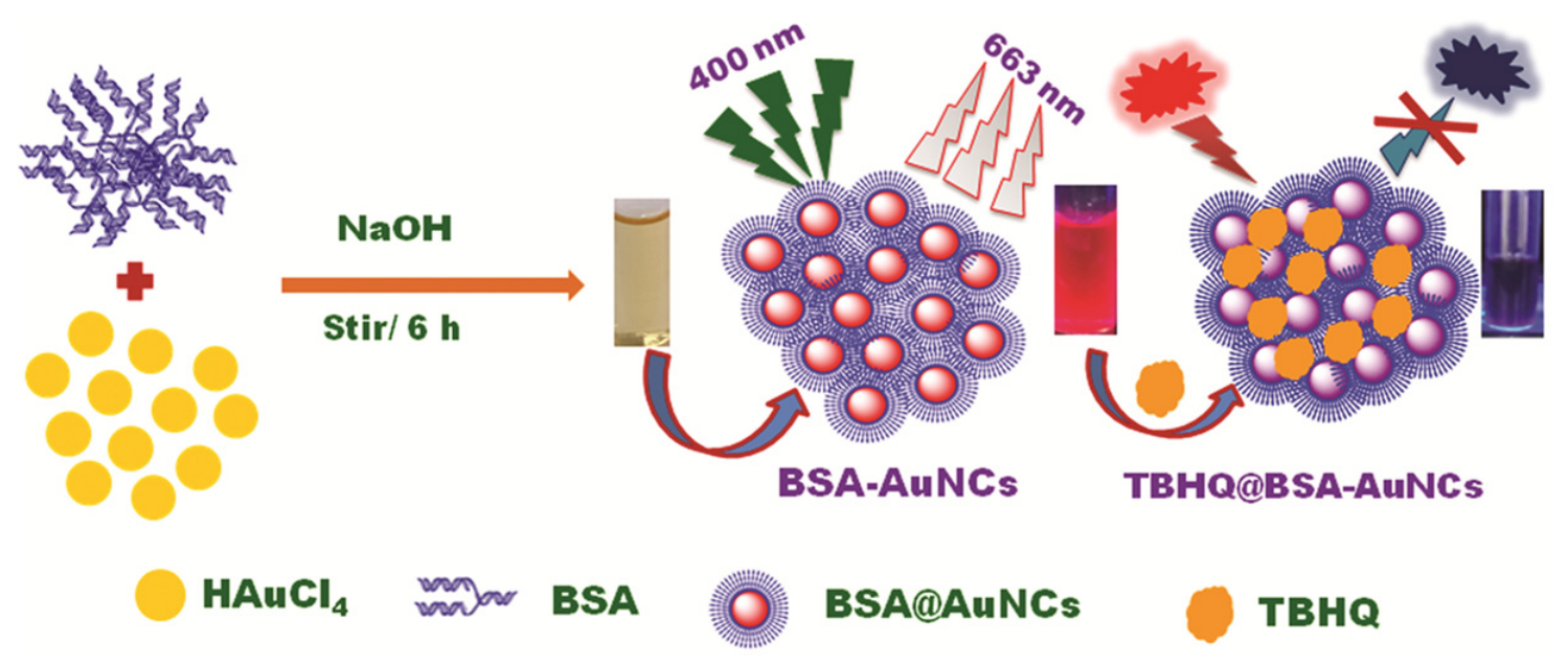
Compared to surface-enhanced Raman spectroscopy, UV-visible absorption spectroscopy, Fourier transform infrared spectroscopy, and molecular fluorescence spectroscopy exhibit distinct application boundaries for antioxidant analysis. UV-visible absorption spectroscopy is typically combined with various colorimetric reaction systems to evaluate the overall antioxidant strength or total phenolic content of a sample, among other parameters, yielding a comprehensive numerical value. Molecular fluorescence spectroscopy mostly relies on the quenching or enhancement effects of antioxidants on fluorescent probes to indirectly reflect their activity levels, providing information on total quantity or total activity; it lacks the ability to identify individual substances in multicomponent mixtures. However, if highly selective probes are designed, sensitive detection of a specific single target can be achieved. For instance, Shankar et al. [108] developed a dual-mode (fluorescence/colorimetric) sensing method for detecting the food antioxidant TBHQ based on gold nanoclusters coated with bovine serum albumin (BSA-AuNCs). TBHQ quenches the red fluorescence of the gold nanoclusters through electrostatic interaction. This method has high selectivity for TBHQ, and common phenolic analogues do not interfere. The detection limit is as low as 72 nM, and it has been successfully applied to the detection of TBHQ in edible oil samples (Figure 3). The Fourier transform infrared spectroscopy relies on the superimposition of O–H stretching vibrations in phenolic compounds, C=C vibrations in aromatic rings, and absorption peaks of oxygen-containing functional groups to form an infrared fingerprint spectrum. By comparing it with the standard sample spectrum or using chemometric methods such as principal component analysis, the type identification of the sample can be completed. FTIR coupled with chemometric algorithms enables reliable quantitative analysis; however, its quantitative performance is readily compromised by severe food-matrix interference, restricting its practical application for trace-level target analysis.
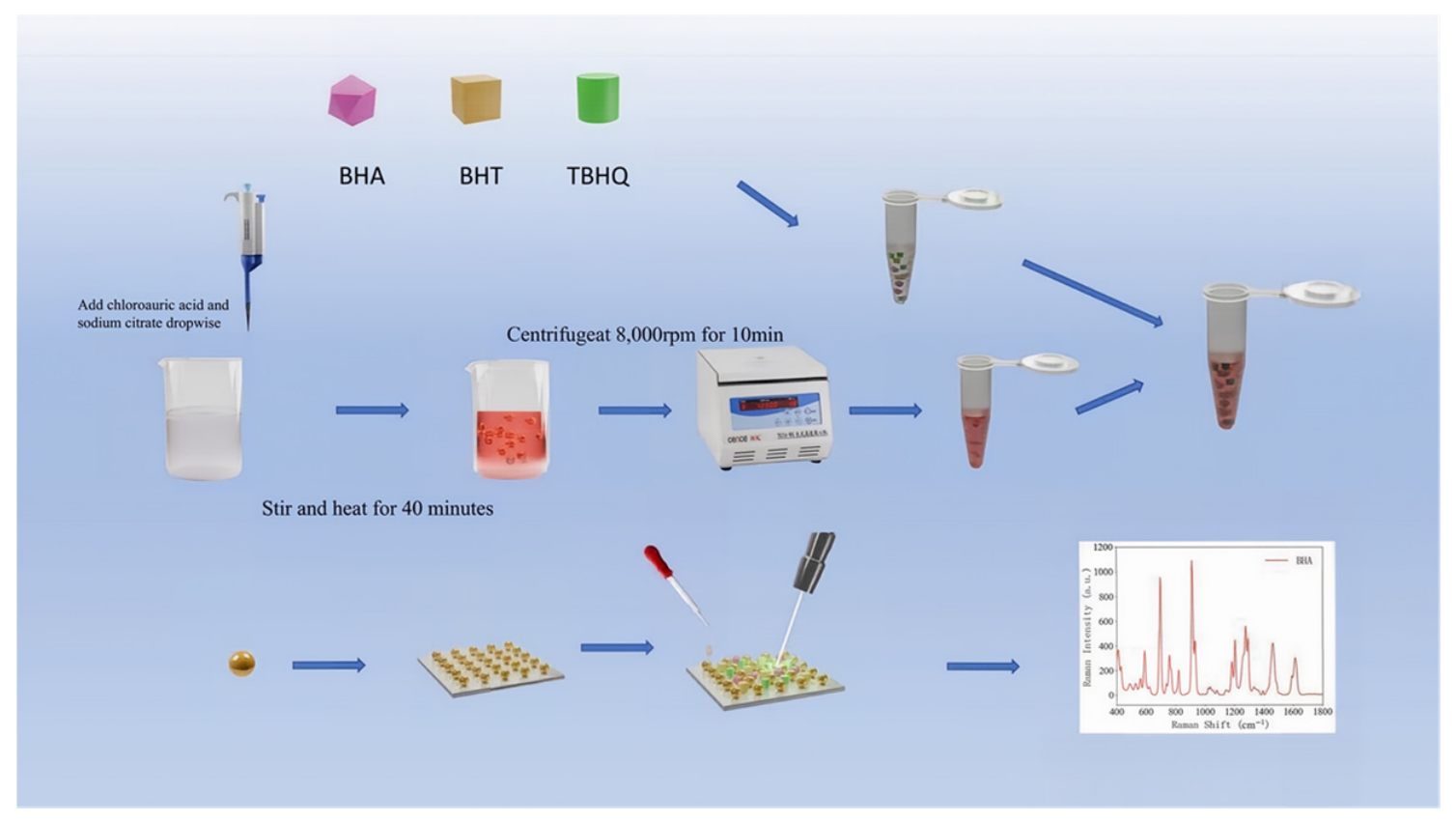
Among these spectroscopic techniques, SERS is particularly attractive for antioxidant detection because its Raman scattering peaks correspond to molecular vibrational modes, providing unique fingerprint spectra for individual phenolic antioxidants. Unlike UV-Vis absorption, conventional fluorescence, and FTIR spectroscopy—which are more commonly employed for total phenolic content (TPC) or total antioxidant capacity (TAC) assessment—SERS enables the identification and quantification of specific antioxidant compounds in complex matrices [109,110,111,112,113]. Unlike the other three types of spectroscopic techniques, the Raman scattering peaks correspond to the vibration modes of chemical bonds within molecules. Each phenolic antioxidant has its own unique Raman fingerprint spectrum. Surface-enhanced Raman spectroscopy technology has made certain progress in antioxidant detection. It can achieve a relatively low detection limit for synthetic antioxidants such as BHA and TBHQ, and the detection time has been reduced from several tens of minutes in chromatography to a few minutes or even shorter. The sample pretreatment is also much simpler than chromatography [114,115,116,117]. Pan et al. [118] developed a quantitative detection method for tert-butylhydroquinone (TBHQ) in plant oils by combining surface-enhanced Raman spectroscopy with chemometrics. The minimum detection concentration of this method in the standard solution was 5 mg/L, and the detection limit in plant oils was 10 mg/kg. A calibration model was established using partial least squares and support vector machine regression, and the predicted values of TBHQ content in actual oil samples were highly correlated with the true values. This provided a new idea for the rapid detection of antioxidants in complex food matrices. Wang et al. [119] used gold nanoparticles (AuNPs) as the active substrate for surface-enhanced Raman spectroscopy (SERS) and combined machine learning algorithms to establish a rapid detection method for three phenolic antioxidants (BHA, BHT, and TBHQ) in edible oils. The enhancement factor of the SERS substrate for rhodamine 6G was 4.4 × 10⁵. The three antioxidants showed good concentration responses in the range of 20–1000 mg/kg, and the detection limit was lower than 10 mg/kg. On this basis, a classification model was constructed using CNN (convolutional neural network) and other machine learning algorithms, and the recognition accuracy for six addition scenarios (single and binary mixtures) reached 99.54%; further, PLS-DA (partial least squares discriminant analysis) was used for over-limit discrimination analysis, with an accuracy rate exceeding 94%. This study provided a new idea for on-site and rapid SERS screening of antioxidants in edible oils (Figure 4).

3.8. Development Progress of Electrochemical Detection Technology

Electrochemical analysis is a quantitative detection technique based on the current, potential or impedance signals generated when the analyte undergoes oxidation-reduction reactions at the electrode surface. The content of the analyte is determined by measuring the intensity of the electrical signal [120,121,122,123]. Compared with chromatography and spectroscopy, electrochemical detection has advantages in terms of instrument cost, response speed, operational convenience and miniaturization potential. Due to the continuous development of modified electrode materials, detection performance has been significantly improved. The evolution of electrochemical detection can be divided into four stages based on electrode modification strategies.
Bare Electrode Stage. Early studies directly used glassy carbon, gold, or carbon paste electrodes for phenolic antioxidant detection. These bare electrodes generally deliver only micromolar-level limits of detection. Moreover, co-existing electroactive interferents (e.g., ascorbic acid, uric acid, dopamine) with similar oxidation potentials produce overlapping signals, resulting in poor selectivity [124,125]. Electrode passivation induced by oxidation-product accumulation further degrades reproducibility [80]. Surface modification using nanomaterials provides an effective route to overcome these intrinsic drawbacks and improve overall sensing performance.
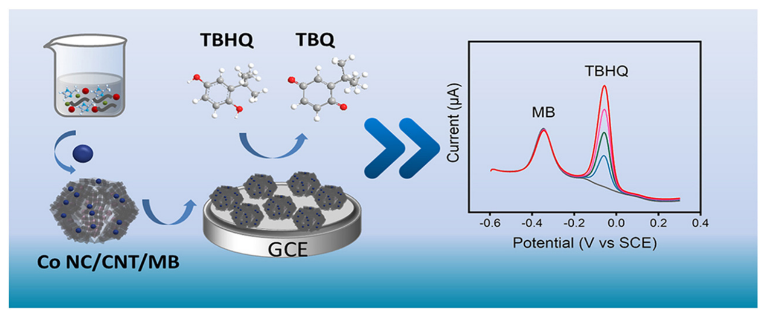
Carbonaceous and Metallic Nanomaterial Modification. The introduction of carbon-based nanomaterials (carbon nanotubes, graphene, carbon black) and noble metal nanoparticles (AuNPs, AgNPs, PtNPs) significantly enhanced electrode conductivity, surface area, and catalytic activity, substantially improving detection sensitivity [126,127,128,129,130,131]. Representative examples include a ratiometric sensor based on Co/N-doped carbon nanotube composites for TBHQ detection (LOD: 0.054 μM) [132], and a CB@Au/poly (p-ABSA) nanocomposite sensor achieving a 10 nM LOD for TBHQ in edible oil [14] (Figure 5).
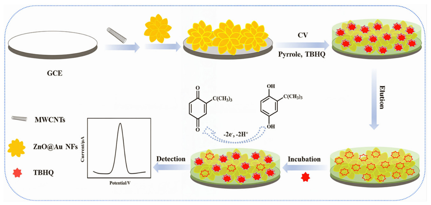
Functional Porous Material Modification. The introduction of MXenes, metal-organic frameworks (MOFs), covalent organic frameworks (COFs), and metal oxide/sulfide nanomaterials further enhanced sensing performance [133,134,135,136]. These materials offer high porosity, large surface areas, ordered pore structures, and abundant active sites, which facilitate analyte enrichment and improve sensitivity. For example, Srinivasan et al. [137] constructed a Mn-MOF@f-CNF/SPE sensor for TBHQ detection, achieving a linear range of 0.01–800 μM with a 1.36 nM LOD, and successfully applied it to potato chips and instant noodles. Han et al. [138] developed a ZnO@Au flower-like nanocomposite with a molecularly imprinted polymer for TBHQ detection, exhibiting a linear range of 0.5–200 μmol/L and a 58 nmol/L LOD (Figure 6).
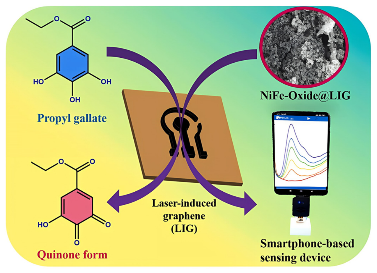
Integrated Intelligent Sensing. By integrating microfluidic channels, flexible conductive substrates, and smartphone signal acquisition modules, integrated sensing platforms have been developed for on-site and real-time monitoring. Chinnapaiyan et al. [139] fabricated a flexible laser-induced graphene (LIG) electrode modified with porous NiFe oxide for smartphone-based electrochemical detection of PG in edible oils, achieving a 0.01 μM LOD (Figure 7).
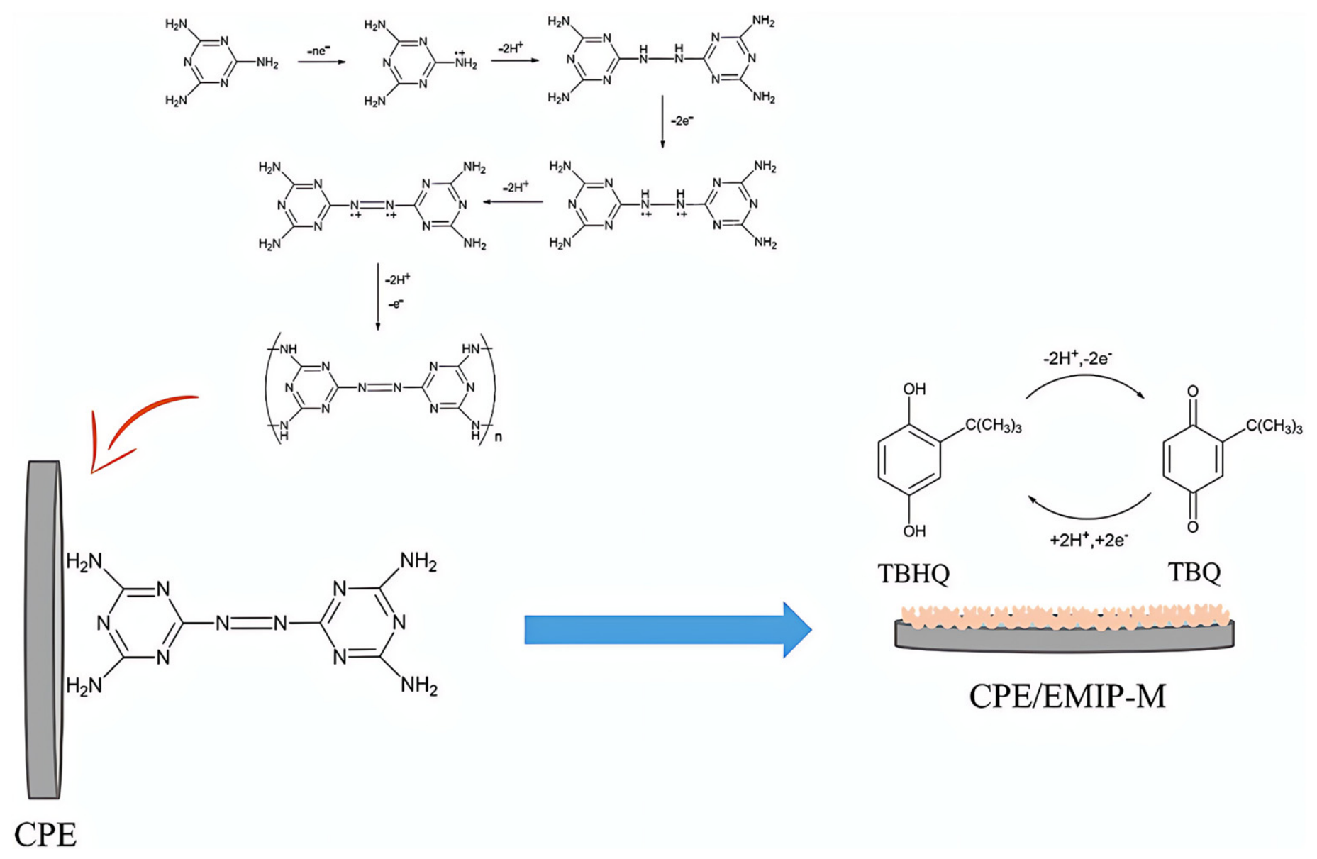
Despite these advances, selectivity remains a key challenge for electrochemical sensing in food applications. Common electroactive endogenous compounds (ascorbic acid, dopamine, uric acid) can produce overlapping signals, while polymerization and adsorption of phenolic oxidation products may cause electrode fouling. To address these limitations, molecularly imprinted polymers (MIPs) and metal–organic frameworks (MOFs) have been introduced to provide specific recognition cavities [140,141,142]. Noviandri et al. [143] used melamine as the functional monomer and TBHQ as the template molecule to prepare a molecularly imprinted polymer (MIP)-modified electrode through an electrochemical polymerization method on the surface of a carbon paste electrode. This electrode was used for the square wave voltammetric detection of TBHQ. The electrochemical molecularly imprinted polymer (EMIP-M) provided a specific recognition cavity, endowing the sensor with good selectivity. Under optimized conditions, the linear detection range of this sensor was 1–1000 μM, and the detection limit was 0.29 μM. The detection results in actual food samples showed no significant difference from those obtained by the HPLC method (Figure 8).
By analyzing the development progress of various technologies, it can be observed that the chromatography, spectroscopy, and electrochemistry detection techniques have been continuously evolving and upgrading along the directions of precise separation, rapid screening, and in situ online detection. Currently, none of these technologies can completely replace the other two types [144,145,146]. This also indicates that in actual testing work, multiple factors such as the characteristics of the sample to be tested, the detection sensitivity standards, and the on-site operational conditions need to be considered. Appropriate detection methods should be selected flexibly. In practical applications, the three techniques do not operate independently, but have formed a good complementary system. Among them, Chromatography delivers outstanding separation performance and can provide precise benchmark verification basis for spectral and electrochemical detection results; spectral technology has a fast response speed and is more suitable for the preliminary screening of large quantities of samples; electrochemical technology has a simple equipment structure and is easy to be miniaturized, effectively compensating for the technical shortcomings of real-time rapid detection in the field. Through multi-dimensional comparative analysis of the three types of detection technologies (Table 2), this article can provide reliable references for practical method selection.

4. Summary and Prospect

This article systematically reviews the classification system of phenolic antioxidants in food and the latest research progress in their detection techniques, with a focus on evaluating the advantages and limitations of the three major mainstream technical paths: chromatographic analysis, spectroscopic analysis, and electrochemical detection. Due to significant differences in sources, solubility, and chemical structure, the detection methods for food antioxidants have evolved from laboratory chemical titration, precise instrumental analysis to on-site rapid screening. Currently, chromatographic technology, with its outstanding separation ability and excellent quantitative accuracy, remains the gold standard for laboratory confirmatory analysis; spectroscopic technology (especially SERS) is suitable for initial screening of large quantities of samples due to its simple operation and rapid response; while electrochemical sensing technology demonstrates unique potential in on-site real-time detection and wearable monitoring scenarios due to its low equipment cost, high sensitivity, and ease of miniaturization. Although all these methods have made significant progress, no single technology can simultaneously meet the combined requirements of high sensitivity, high selectivity, rapid response, and simultaneous detection of multiple components. Chromatography is limited by expensive equipment, complex pre-treatment, and large consumption of organic solvents; spectroscopy is susceptible to interference from food matrices and has insufficient selectivity; electrochemical sensing faces bottlenecks such as electrode contamination and interference from electroactive substances. Therefore, in practical applications, it is necessary to flexibly select or combine multiple technologies based on the detection purpose, sample characteristics, and on-site conditions to form a complementary detection strategy.
The development of future food antioxidant detection technologies should focus on the deep integration and systematic integration of multiple technical paths to achieve full coverage from laboratory precise analysis to on-site real-time monitoring. By fully leveraging the complementary advantages of chromatography in separation and confirmation, spectroscopic methods in rapid screening, and electrochemical sensing in portable detection, it is expected to construct a more flexible and adaptable detection system, satisfying differentiated requirements for diverse food matrices and application scenarios. At the same time, continuous innovation in new nanomaterials, porous framework materials, and molecular imprinting polymers will provide strong support for improving sensor selectivity and enrichment efficiency, promoting further improvement in detection performance. Future research should also focus on establishing a comprehensive antioxidant detection method database, systematically tracking the applicability and impact of different detection strategies on various food matrices and target substances, and developing efficient mixed-association strategies to improve the detection sensitivity and selectivity in complex systems. Beyond technological innovation, translating these methods into practice also demands rigorous validation in real food matrices, standardized protocols for inter-laboratory reproducibility, and regulatory acceptance supported by thorough performance data. Closing the gap between academic development and industrial adoption remains a key challenge. These advancements will drive the development of fields such as food safety regulation, on-site rapid screening, online real-time quality control, and green analytical chemistry, ensuring that detection technologies continue to play a foundational role in ensuring food quality and consumer health.

Author Contributions

Y.Z.: Conceptualization, Methodology, Validation, Formal analysis, Investigation, Writing—Original Draft, Writing-Review & Editing. Q.L.: Conceptualization, Methodology, Investigation, Resources, Writing-Review & Editing. J.T.: Conceptualization, Methodology, Writing-Review & Editing. Z.Z.: Investigation, Resources, Writing-Review & Editing. B.Z.: Conceptualization, Supervision, Methodology, Investigation, Resources, Writing-Review & Editing. All authors have read and agreed to the published version of the manuscript.

Funding

This work was supported by the National Natural Science Foundation of China (No. 22178153), National Key Research and Development Program of China (No. 2021YFC2103800), Priority Academic Program Development of Jiangsu Higher Education Institutions and the 24th Batch of Undergraduate Research Project at Jiangsu University (Y24A044).

Institutional Review Board Statement

Not applicable.

Informed Consent Statement

Not applicable.

Data Availability Statement

No new data were created or analyzed in this study. Data sharing is not applicable to this article.

Acknowledgments

All the authors thank to the National Natural Science Foundation of China (No. 22178153), National Key Research and Development Program of China (No. 2021YFC2103800), Priority Academic Program Development of Jiangsu Higher Education Institutions and the 24th Batch of Undergraduate Research Project at Jiangsu University (Y24A044).

Conflicts of Interest

The authors declare no conflicts of interest.

References

  1. Lv, Q.L.; Xu, Q.Q.; Wang, Q.Q.; Xie, Y.X.; Liu, Z.G.; Guo, Z. Anchoring NiO/Ni nanoparticles onto carbon spheres for efficient electrochemical simultaneous detection of antioxidants TBHQ and BHA. Talanta 2026, 302, 129419. [Google Scholar] [CrossRef] [Scilit]
  2. Sakthivel, N.K.; Veerasamy, R.P.; Akremi, A.; Alshahrani, T.; Govindasamy, M. Enhanced electrochemical detection of propyl gallate in foods using molybdenum diselenide decorated on graphitic carbon nitride modified electrode. Food Chem. 2025, 493, 145836. [Google Scholar] [CrossRef] [Scilit]
  3. Deng, J.H.; Chen, Z.Y.; Jiang, H.; Chen, Q.S. High-precision detection of dibutyl hydroxytoluene in edible oil via convolutional autoencoder compressed Fourier-transform near-infrared spectroscopy. Food Control 2025, 167, 110808. [Google Scholar] [CrossRef] [Scilit]
  4. Zhang, Y.Y.; Li, M.; Cui, Y.; Hong, X.; Du, D.L. Using of Tyramine Signal Amplification to Improve the Sensitivity of ELISA for Aflatoxin B1 in Edible Oil Samples. Food Anal. Methods 2018, 11, 2553–2560. [Google Scholar] [CrossRef] [Scilit]
  5. Aladhadh, M. Antioxidants: Mechanisms, Benefits, and the Importance of Extremophilic Microorganisms. Microorganisms 2026, 14, 838. [Google Scholar] [CrossRef] [Scilit]
  6. Tammina, S.K.; Krishnamoorthy, R.; Manzoor, A.; Priyadarshi, R.; Sridhar, K.; Banat, F. A Comprehensive Review on the Significance of Antioxidants and Their Influence on Biopolymer Packaging Films: Recent Advances and Challenges. Food Front. 2026, 7, e70227. [Google Scholar] [CrossRef] [Scilit]
  7. Zaitsev, S.Y. Major Antioxidants and Methods for Studying Their Total Activity in Milk: A Review. Methods Protoc. 2025, 8, 139. [Google Scholar] [CrossRef] [Scilit]
  8. Hadidi, M.; Orellana-Palacios, J.C.; Aghababaei, F.; Gonzalez-Serrano, D.J.; Moreno, A.; Lorenzo, J.M. Plant by-product antioxidants: Control of protein-lipid oxidation in meat and meat products. Lwt-Food Sci. Technol. 2022, 169, 114003. [Google Scholar] [CrossRef] [Scilit]
  9. Mishra, S.K.; Belur, P.D.; Iyyaswami, R. Comparison of efficacy of various natural and synthetic antioxidants in stabilizing the fish oil. J. Food Process. Preserv. 2022, 46, e16970. [Google Scholar] [CrossRef] [Scilit]
  10. Polvarini, G.D.; Sant’ Ana, C.T.; Toshimitu, P.E.M.; Silva, E.C.; Silva, P.I.; Saraiva, S.H.; Villanova, J.C.O.; Costa, A.G.V. Green Coffee Extract (Coffea canephora) as an Antioxidant in Mayonnaise in Replacement of Synthetic Agents. Eur. J. Lipid Sci. Technol. 2025, 127, e70007. [Google Scholar] [CrossRef] [Scilit]
  11. Iqbal, N.; Kibtia, M.; Khoder, R.M.; Javed, M.; Khalifa, I.; Kanwal, R.; Zheng, R.Y.; Xiong, S.B.; Liu, Y.M. Valorization of peanut shell polyphenols as natural antioxidants for preserving silver carp mince during refrigerated storage. Food Chem. 2025, 493, 145762. [Google Scholar] [CrossRef] [Scilit]
  12. Felter, S.P.; Zhang, X.L.; Thompson, C. Butylated hydroxyanisole: Carcinogenic food additive to be avoided or harmless antioxidant important to protect food supply? Regul. Toxicol. Pharmacol. 2021, 121, 104887. [Google Scholar] [CrossRef] [Scilit]
  13. Zhang, X.J.; Diao, M.N.; Zhang, Y.F. A review of the occurrence, metabolites and health risks of butylated hydroxyanisole (BHA). J. Sci. Food Agric. 2023, 103, 6150–6166. [Google Scholar] [CrossRef] [Scilit]
  14. Han, E.; Gao, T.; Bao, M.; Cai, J.R. Tert-butylhydroquinone determination in food using electrochemical sensor based on polyaminobenzenesulfonic acid/gold nanoparticles-functionalized carbon black. J. Food Compos. Anal. 2025, 148, 108685. [Google Scholar] [CrossRef] [Scilit]
  15. Ren, J.; Li, Z.; Li, X.F.; Yang, L.; Bu, Z.L.; Wu, Y.H.; Li, Y.T.; Zhang, S.S.; Meng, X.L. Exploring the Mechanisms of the Antioxidants BHA, BHT, and TBHQ in Hepatotoxicity, Nephrotoxicity, and Neurotoxicity from the Perspective of Network Toxicology. Foods 2025, 14, 1095. [Google Scholar] [CrossRef] [Scilit]
  16. Yang, X.H.; Li, C.; Yu, G.C.; Sun, L.; Guo, S.M.; Sai, L.L.; Bo, C.X.; Xing, C.H.; Shao, H.; Peng, C.; et al. paper Ligand-independent activation of AhR by hydroquinone mediates benzene-induced hematopoietic toxicity. Chem.-Biol. Interact. 2022, 355, 109845. [Google Scholar] [CrossRef] [Scilit]
  17. Mou, S.; Hummer, B.T.; Yuan, J.Q.; Huang, Y.; Liang, M.N.; Faggioni, R.; Roskos, L.K.; Rosenbaum, A.I. Investigations of Enteric-Coated Tablet Propyl Gallate-Induced Nephrotoxicity in Beagles as well as Human and Dog Renal Proximal Tubule Epithelial Cells. Acs Pharmacol. Transl. Sci. 2025, 8, 1282–1291. [Google Scholar] [CrossRef] [Scilit]
  18. Park, W.H. Propyl gallate induces human pulmonary fibroblast cell death through the regulation of Bax and caspase-3. Ann. Med. 2024, 56, 2319853. [Google Scholar] [CrossRef] [Scilit]
  19. Araújo, M.V.L.; Oi, L.A.; de Arruda, L.M.; Gomes, L.B.; Bragotto, A.P.A. Use of antioxidants as food additives and exposure assessment in Brazil. Food Control 2026, 181, 111719. [Google Scholar] [CrossRef] [Scilit]
  20. GB 2760-2024; National Food Safety Standard—Use Standards of Food Additives. China Standards Press: Beijing, China, 2024.
  21. Aguilar, F.; Crebelli, R.; Dusemund, B.; Galtier, P.; Gilbert, J.; Gott, D.M.; Gundert-Remy, U.; König, J.; Lambré, C.; Leblanc, J.C.; et al. Scientific Opinion on the re-evaluation of butylated hydroxytoluene BHT (E 321) as a food additive EFSA Panel on Food Additives and Nutrient Sources added to Food (ANS). Efsa J. 2012, 10, 2588. [Google Scholar]
  22. Aguilar, F.; Crebelli, R.; Dusemund, B.; Galtier, P.; Gilbert, J.; Gott, D.M.; Gundert-Remy, U.; König, J.; Lambré, C.; Leblanc, J.C.; et al. Statement on the safety assessment of the exposure to butylated hydroxyanisole E 320 (BHA) by applying a new exposure assessment methodology. Efsa J. 2012, 10, 2770. [Google Scholar] [CrossRef] [Scilit]
  23. Aguilar, F.; Crebelli, R.; Dusemund, B.; Galtier, P.; Gott, D.; Gundert-Remy, U.; König, J.; Lambré, C.; Leblanc, J.C.; Mortensen, A.; et al. Scientific Opinion on the re-evaluation of propyl gallate (E 310) as a food additive. Efsa J. 2014, 12, 3642. [Google Scholar] [CrossRef] [Scilit]
  24. Aguilar, F.; Crebelli, R.; Di Domenico, A.; Dusemund, B.; Frutos, M.J.; Galtier, P.; Gott, D.; Gundert-Remy, U.; Lambré, C.; Leblanc, J.C.; et al. Statement on the refined exposure assessment of tertiary-butyl hydroquinone (E 319). Efsa J. 2016, 14, 4363. [Google Scholar] [CrossRef] [Scilit][Green Version]
  25. U.S. Food and Drug Administration. Code of Federal Regulations, Title 21, Part 182—Substances Generally Recognized as Safe, Section 182.3169—Butylated Hydroxyanisole; U.S. Food and Drug Administration: Silver Spring, MD, USA, 2026.
  26. U.S. Food and Drug Administration. Code of Federal Regulations, Title 21, Part 182—Substances Generally Recognized as Safe, Section 182.3173—Butylated Hydroxytoluene; U.S. Food and Drug Administration: Silver Spring, MD, USA, 2026.
  27. Eze, C.N.; Aduba, C.C.; Ezema, B.O.; Ayoka, T.O.; Nnadi, C.O.; Onyeaka, H. The role of antioxidants in food safety and preservation: Mechanisms, applications, and challenges. Cogent Food Agric. 2025, 11, 2591436. [Google Scholar] [CrossRef] [Scilit]
  28. Lee, Y.; Hwang, C.Y.; Cho, E.S.; Seo, M.J. Water-soluble carotenoid: Focused on natural carotenoid crocin. Food Sci. Biotechnol. 2025, 34, 1119–1138. [Google Scholar] [CrossRef] [Scilit]
  29. Zhu, X.; Chen, X.; Wang, M.; Hu, H.H. Astaxanthin: A Compound in the Prevention of Chronic Diseases and as a Potential Adjuvant Treatment Agent. Antioxidants 2025, 14, 715. [Google Scholar] [CrossRef] [Scilit]
  30. Özdemir, K.; Demir, Y. Phenolic Compounds in Exercise Physiology: Dual Role in Oxidative Stress and Recovery Adaptation. Food Sci. Nutr. 2025, 13, e70714. [Google Scholar] [CrossRef] [Scilit]
  31. Zúñiga-Núñez, D.; Barrias, P.; Cárdenas-Jirón, G.; Ureta-Zañartu, M.S.; Lopez-Alarcón, C.; Vieyra, F.E.M.; Borsarelli, C.D.; Alarcon, E.I.; Aspée, A. Atypical antioxidant activity of non-phenolic amino-coumarins. RSC Adv. 2018, 8, 1927–1933. [Google Scholar] [CrossRef] [Scilit]
  32. Hoang, H.T.; Moon, J.Y.; Lee, Y.C. Natural Antioxidants from Plant Extracts in Skincare Cosmetics: Recent Applications, Challenges and Perspectives. Cosmetics 2021, 8, 106. [Google Scholar] [CrossRef] [Scilit]
  33. Hrebien-Filisinska, A. Application of natural antioxidants in the oxidative stabilization of fish oils: A mini-review. J. Food Process. Preserv. 2021, 45, e15342. [Google Scholar] [CrossRef] [Scilit]
  34. Yin, F.W.; Sun, X.L.; Zheng, W.L.; Luo, X.; Zhang, Y.Y.; Yin, L.F.; Jia, Q.; Fu, Y.Q. Screening of highly effective mixed natural antioxidants to improve the oxidative stability of microalgal DHA-rich oil. RSC Adv. 2021, 11, 4991–4999. [Google Scholar] [CrossRef] [Scilit]
  35. Lourenço, S.C.; Moldao-Martins, M.; Alves, V.D. Antioxidants of Natural Plant Origins: From Sources to Food Industry Applications. Molecules 2019, 24, 4132. [Google Scholar] [CrossRef] [Scilit]
  36. Zhang, D.; Wang, Y.; Sun, X.X.; Liu, Y.X.; Zhou, Y.J.; Shin, H.C.; Wang, Y.; Shen, L.Q.; Wang, C.T.; Wang, S.Y.; et al. Voltammetric, spectroscopic, and cellular characterization of redox functionality of eckol and phlorofucofuroeckol-A: A comparative study. J. Food Biochem. 2019, 43, e12845. [Google Scholar] [CrossRef] [Scilit]
  37. Wang, H.L.; Liu, X.T.; Tu, M.L.; Xu, X.; Yang, S.; Chen, D. Current sample preparation methods and analytical techniques for the determination of synthetic antioxidants in edible oils. J. Sep. Sci. 2022, 45, 3874–3886. [Google Scholar] [CrossRef] [Scilit]
  38. Thomas, D.; Rasheed, Z.; Jagan, J.S.; Kumar, K.G. Study of kinetic parameters and development of a voltammetric sensor for the determination of butylated hydroxyanisole (BHA) in oil samples. J. Food Sci. Technol.-Mysore 2015, 52, 6719–6726. [Google Scholar] [CrossRef] [Scilit]
  39. Ziyatdinova, G.; Os’kina, K.; Ziganshina, E.; Budnikov, H. Simultaneous determination of TBHQ and BHA on a MWNT-Brij® 35 modified electrode in micellar media. Anal. Methods 2015, 7, 8344–8351. [Google Scholar] [CrossRef] [Scilit]
  40. Kharat, M.; Skrzynski, M.; Decker, E.A.; McClements, D.J. Enhancement of chemical stability of curcumin-enriched oil-in-water emulsions: Impact of antioxidant type and concentration. Food Chem. 2020, 320, 126653. [Google Scholar] [CrossRef] [Scilit]
  41. Shen, C.Y.; Ankolekar, C.; Ban, L. Comparison of Oil Soluble Green Tea Extract with Common Antioxidantive Ingredients in Bulk Oil under Different Storage Conditions. J. Am. Oil Chem. Soc. 2021, 98, 65–72. [Google Scholar] [CrossRef] [Scilit]
  42. Chen, Y.; Zhang, W.J.; Zhao, T.; Li, F.; Zhang, M.; Li, J.; Zou, Y.; Wang, W.; Cobbina, S.J.; Wu, X.Y.; et al. Adsorption properties of macroporous adsorbent resins for separation of anthocyanins from mulberry. Food Chem. 2016, 194, 712–722. [Google Scholar] [CrossRef] [Scilit]
  43. Zhai, X.D.; Shi, J.Y.; Zou, X.B.; Wang, S.; Jiang, C.P.; Zhang, J.J.; Huang, X.W.; Zhang, W.; Holmes, M. Novel colorimetric films based on starch/polyvinyl alcohol incorporated with roselle anthocyanins for fish freshness monitoring. Food Hydrocoll. 2017, 69, 308–317. [Google Scholar] [CrossRef] [Scilit]
  44. Ermurat, Y. Development of Nutraceutical and Nutritional Design Method for Liposomal Curcumin and Vitamin C Using Microstructural and Elemental Analysis. Kem. Ind.-J. Chem. Chem. Eng. 2025, 74, 1–6. [Google Scholar] [CrossRef] [Scilit]
  45. Verma, K.; Nema, P.K.; Badgujar, P.C.; Venkatesh, K.; Rajendran, A.; Loushigam, G.L. Vitamin C in fruits and vegetables: Stability, processing effects, analytical detection, nutrient interactions, bioavailability, and health implications. Food Chem. 2026, 505, 148058. [Google Scholar] [CrossRef] [Scilit]
  46. Feng, J.H.; Wang, H.Y.; Liu, Y.Y.; Liu, W.N.Y.; Lin, X.Y. Zirconium-based metal-organic framework and functionalized carbon black composite for simultaneous electrochemical detection of tert-butylhydroquinone and butylhydroxyanisole. Appl. Organomet. Chem. 2024, 38, e7600. [Google Scholar] [CrossRef] [Scilit]
  47. Penha, R.D.P.; Maciel, A.P.; Bezerra, C.W.B.; Damos, F.S.; Luz, R.C.S. Exploiting a carbon black paste electrode modified with palladium nanoparticles entrapped in aluminum hydroxide matrix for selective detection of 2(3)-t-Butyl-4-hydroxyanisole antioxidant in biodiesel samples. J. Electroanal. Chem. 2024, 973, 118683. [Google Scholar] [CrossRef] [Scilit]
  48. Angelis, P.N.; Mendonça, J.D.; da Rocha, L.R.; Capelari, T.B.; Prete, M.C.; Segatelli, M.G.; Borsato, D.; Tarley, C.R.T. Feasibility of a Nano-Carbon Black Paste Electrode for Simultaneous Voltammetric Determination of Antioxidants in Food Samples and Biodiesel in the Presence of Surfactant. Electroanalysis 2020, 32, 1198–1207. [Google Scholar] [CrossRef] [Scilit]
  49. Ahmad, N.; Naeem, M.; Ali, H.; Alabbosh, K.F.; Hussain, H.; Khan, I.; Siddiqui, S.A.; Khan, A.A.; Iqbal, B. From challenges to solutions: The impact of melatonin on abiotic stress synergies in horticultural plants via redox regulation and epigenetic signaling. Sci. Hortic. 2023, 321, 112369. [Google Scholar] [CrossRef] [Scilit]
  50. Xia, J.J.; Bin, Z.; Zhou, R.Y.; Onyinye, A. Lipase nanogel catalyzed synthesis of vitamin E succinate in non-aqueous phase. J. Sci. Food Agric. 2021, 101, 3186–3192. [Google Scholar] [CrossRef] [Scilit]
  51. Jaramillo-Vivanco, T.; Balslev, H.; Montúfar, R.; Cámara, R.M.; Giampieri, F.; Battino, M.; Cámara, M.; Alvarez-Suarez, J.M. Three Amazonian palms as underestimated and little-known sources of nutrients, bioactive compounds and edible insects. Food Chem. 2022, 372, 131273. [Google Scholar] [CrossRef] [Scilit]
  52. Dong, C.W.; Zhu, H.K.; Wang, J.J.; Yuan, H.B.; Zhao, J.W.; Chen, Q.S. Prediction of black tea fermentation quality indices using NIRS and nonlinear tools. Food Sci. Biotechnol. 2017, 26, 853–860. [Google Scholar] [CrossRef] [Scilit]
  53. Nagar, D.N.; Das, D.; Aduri, R.; Braganca, J.M. Characterization and Functional Evaluation of Carotenoids From Haloarcula rubripromontorii BS2. Microbiologyopen 2026, 15, e70228. [Google Scholar] [CrossRef] [Scilit]
  54. Jijón, S.; Czett, D.; Böddi, K.; Gulyás-Fekete, G.; Nagy, V.; Takátsy, A.; Balogh, G.T.; Deli, J.; Agócs, A. Synthesis, antioxidant capacity and aggregation of carotenoid-curcumin conjugates and hybrids. PLoS ONE 2026, 21, e0347640. [Google Scholar] [CrossRef] [Scilit]
  55. Cao, J.; Wang, J.P.; Wang, S.C.; Xu, X.M. A Mini-Review of Its Pharmacological and Nutritional Properties. J. Med. Food 2016, 19, 111–119. [Google Scholar] [CrossRef] [Scilit]
  56. Oladejo, A.O.; Ma, H.L.; Qu, W.J.; Zhou, C.S.; Wu, B.G. Effects of Ultrasound on Mass Transfer Kinetics, Structure, Carotenoid and Vitamin C Content of Osmodehydrated Sweet Potato (Ipomea batatas). Food Bioprocess Technol. 2017, 10, 1162–1172. [Google Scholar] [CrossRef] [Scilit]
  57. Feng, Y.B.; Wu, B.G.; Yu, X.J.; Yagoub, A.A.; Sarpong, F.; Zhou, C.S. Effect of catalytic infrared dry-blanching on the processing and quality characteristics of garlic slices. Food Chem. 2018, 266, 309–316. [Google Scholar] [CrossRef] [Scilit]
  58. Zhang, X.R.; Yang, H.Y.; Peng, Z.; Gao, Y.; Su, B.; Li, K.; Zhang, C.L. Rapid quantitative evaluation of antioxidant activity using terahertz spectroscopy and microfluidic Technology: A study of Butylated hydroxyanisole. Microchem. J. 2025, 213, 113802. [Google Scholar] [CrossRef] [Scilit]
  59. Bazina, N.; Ahmed, T.G.; Almdaaf, M.; He, J.; Sarker, M.; Islam, M. BBCEAS-HLPC measurements for synthetic antioxidants (TBHQ, BHA, and BHT) in deep-UV region below 300 nm. Food Chem. 2025, 465, 142150. [Google Scholar] [CrossRef] [Scilit]
  60. Thbayh, D.K.; Fiser, B. Computational study of synthetic and natural polymer additives—Antioxidant potential of BHA, TBHQ, BHT, and curcumin. Polym. Degrad. Stab. 2022, 201, 109979. [Google Scholar] [CrossRef] [Scilit]
  61. Wu, Q.Y.; Ferreira, M.V.D.; Kamruzzaman, M. Fully portable smartphone-integrated device coupled with nanozyme-based assay for sensitive detection of TBHQ in edible oils. Food Chem. 2026, 518, 149610. [Google Scholar] [CrossRef] [Scilit]
  62. Zhao, X.L.; Zhang, H.H.; Wang, S.; Zhang, X.H.; Zhong, W.; Wang, X.G.; Gao, P. Optimizing oxidative stability of Camellia oleifera seed oil: Multimodal analysis of antioxidant efficacy, tocopherol loss, benzo a pyrene suppression, and kinetic mechanisms. Food Chem. 2026, 502, 147731. [Google Scholar] [CrossRef] [Scilit]
  63. Zhang, L.L.; Guan, Q.H.; Zhang, H.; Tang, L.H.; Xu, M. A spectroscopic and quantum chemical calculation method for the characterisation of metal ions complexed with propyl gallate and procyanidins. Sci. Rep. 2023, 13, 5030. [Google Scholar] [CrossRef] [Scilit]
  64. Tasaka, T.; Matsumoto, T.; Nagashima, U.; Nagaoka, S.I. Potential energy curve for singlet-oxygen quenching reaction by vitamin E. J. Photochem. Photobiol. A-Chem. 2023, 442, 114749. [Google Scholar] [CrossRef] [Scilit]
  65. Kopec, R.E.; Schweiggert, R.M.; Riedl, K.M.; Carle, R.; Schwartz, S.J. Comparison of high-performance liquid chromatography/tandem mass spectrometry and high-performance liquid chromatography/photo-diode array detection for the quantitation of carotenoids, retinyl esters, -tocopherol and phylloquinone in chylomicron-rich fractions of human plasma. Rapid Commun. Mass Spectrom. 2013, 27, 1393–1402. [Google Scholar] [CrossRef] [Scilit]
  66. Zhao, D.K.; Wang, X.; Wang, L.J.; Ma, Q.; Li, J.W.; Xu, B.C.; Dou, X.J.; Liu, L.L. Reevaluation of Physical and Chemical Singlet Oxygen Quenching Efficiency of Antioxidants via a Specific Methanol-Soluble Trap. Antioxidants 2026, 15, 625. [Google Scholar] [CrossRef] [Scilit]
  67. Kulandaivel, S.; Wang, Y.M.; Chen, S.F.; Lin, C.H.; Yeh, Y.C. A Cu-based metal-organic framework synthesized via a green method exhibits unique catecholase-like activity for epigallocatechin gallate detection in teas. Anal. Methods 2024, 16, 8307–8315. [Google Scholar] [CrossRef] [Scilit]
  68. Thbayh, D.K.; Reizer, E.; Kahaly, M.U.; Viskolcz, B.; Fiser, B. Antioxidant Potential of Santowhite as Synthetic and Ascorbic Acid as Natural Polymer Additives. Polymers 2022, 14, 3518. [Google Scholar] [CrossRef] [Scilit]
  69. Hagos, M.; Redi-Abshiro, M.; Chandravanshi, B.S.; Yaya, E.E. NEW ANALYTICAL METHODS FOR THE DETERMINATION OF ASCORBIC ACID CONTENT IN AQUEOUS EXTRACTS OF FLESH, PEEL AND SEEDS OF PUMPKIN (CUCURBITA MAXIMA). Bull. Chem. Soc. Ethiop. 2022, 36, 277–290. [Google Scholar] [CrossRef] [Scilit]
  70. Russo, G.L.; Moccia, S.; Russo, M.; Spagnuolo, C. Redox regulation by carotenoids: Evidence and conflicts for their application in cancer. Biochem. Pharmacol. 2021, 194, 114838. [Google Scholar] [CrossRef] [Scilit]
  71. Mussagy, C.U.; Alvarado-Holtheuer, C.N.; Passaes, A.; Farias, R.L. Solvatochromism and color modulation of carotenoids in bio-based solvents. Dye. Pigment. 2026, 250, 113671. [Google Scholar] [CrossRef] [Scilit]
  72. Chen, H.; Zuo, Y.G.; Deng, Y.W. Separation and determination of flavonoids and other phenolic compounds in cranberry juice by high-performance liquid chromatography. J. Chromatogr. A 2001, 913, 387–395. [Google Scholar] [CrossRef] [Scilit]
  73. Tsuji, S.; Nakano, M.; Terada, H.; Tamura, Y.; Tonogai, Y. Determination and confirmation of five phenolic antioxidants in foods by LC/MS and GC/MS. J. Food Hyg. Soc. Jpn. 2005, 46, 63–71. [Google Scholar] [CrossRef] [Scilit]
  74. Xie, T.H.; Lichtfouse, E.; Mochiwa, Z.O.; Wang, J.; Han, B.X.; Gao, L. Detection of aflatoxin B1 using DNA sensors: A review. Environ. Chem. Lett. 2025, 23, 1425–1443. [Google Scholar] [CrossRef] [Scilit]
  75. Ma, S.; Wang, M.; You, T.Y.; Wang, K. Using Magnetic Multiwalled Carbon Nanotubes as Modified QuEChERS Adsorbent for Simultaneous Determination of Multiple Mycotoxins in Grains by UPLC-MS/MS. J. Agric. Food Chem. 2019, 67, 8035–8044. [Google Scholar] [CrossRef] [Scilit]
  76. Fayek, N.M.; Zayed, A.; Zahran, H.A.; Ramadan, N.S.; Capanoglu, E.; Li, Z.H.; Fang, Y.J.; Khalifa, I.; Farag, M.A. Effect of cultivar type, ontogeny and extraction methods as determinant factors of olive leaf metabolome: A case study in 8 Egyptian cultivars as analyzed using LC/MS-based metabolomics. Ind. Crops Prod. 2024, 222, 120085. [Google Scholar] [CrossRef] [Scilit]
  77. Xu, S.J.; Liu, L.L.; Wang, Y.Q.; Zhou, D.Y.; Kuang, M.; Fang, D.; Yang, W.H.; Wei, S.J.; Xiao, A.P.; Ma, L. Simultaneous determination of six synthetic phenolic antioxidants in edible oils using dispersive liquid-liquid microextraction followed by high-performance liquid chromatography with diode array detection. J. Sep. Sci. 2016, 39, 3205–3211. [Google Scholar] [CrossRef] [Scilit]
  78. Bredács, M.; Kanatschnig, E.; Frank, A.; Oreski, G.; Pinter, G.; Gergely, S. Identifying active and degraded phenolic antioxidants in aged PE with IR-microscopy. Polym. Degrad. Stab. 2023, 212, 110345. [Google Scholar] [CrossRef] [Scilit]
  79. Petrucci, R.; Bortolami, M.; Di Matteo, P.; Curulli, A. Gold Nanomaterials-Based Electrochemical Sensors and Biosensors for Phenolic Antioxidants Detection: Recent Advances. Nanomaterials 2022, 12, 959. [Google Scholar] [CrossRef] [Scilit]
  80. Albayati, S.H.M.; Soylu, P. A simple molecularly imprinted electrochemical sensor for determination of propyl gallate in food samples. Anal. Biochem. 2024, 688, 115477. [Google Scholar] [CrossRef] [Scilit]
  81. Zhao, Y.; Ma, Y.Z.; Zhou, R.Y.; He, Y.; Wu, Y.T.; Yi, Y.H.; Zhu, G.B. Highly sensitive electrochemical detection of paraoxon ethyl in water and fruit samples based on defect-engineered graphene nanoribbons modified electrode. J. Food Meas. Charact. 2022, 16, 2596–2603. [Google Scholar] [CrossRef] [Scilit]
  82. Xu, Y.W.; Zhang, W.; Shi, J.Y.; Zou, X.B.; Li, Z.H.; Zhu, Y.D. Microfabricated interdigitated Au electrode for voltammetric determination of lead and cadmium in Chinese mitten crab (Eriocheir sinensis). Food Chem. 2016, 201, 190–196. [Google Scholar] [CrossRef] [Scilit]
  83. Soylu, P. NiFe2O4 modified multiwalled carbon nanotube based molecularly imprinted electrochemical sensor for tert-butylhydroquinone detection. Microchem. J. 2026, 224, 117820. [Google Scholar] [CrossRef] [Scilit]
  84. Liu, W.; Zhang, K.D.; Chen, J.N.; Yu, J.J. Ascorbic acid and choline chloride: A new natural deep eutectic solvent for extracting tert-butylhydroquinone antioxidant. J. Mol. Liq. 2018, 260, 173–179. [Google Scholar] [CrossRef] [Scilit]
  85. He, J.; Chen, S.; Jiang, Y.L.; Shen, Y.Z.; Zhu, J.; Wei, H.L.; Zhang, H.X.; Lu, K. Preparation and selective recognition of a novel solid-phase microextraction fiber combined with molecularly imprinted polymers for the extraction of parabens in soy sample. J. Sep. Sci. 2012, 35, 308–314. [Google Scholar] [CrossRef] [Scilit]
  86. Nigsch, L.S.; Kappacher, C.; Hense, D.; Huck, C.W.; Strube, O.I.; Rainer, M. Synthesis and optimization of a 2-isopropenyl-2-oxazoline based polymer for enhanced phenolic acid extraction. Anal. Methods 2025, 17, 8634–8645. [Google Scholar] [CrossRef] [Scilit]
  87. Mogaddam, M.R.A.; Farajzadeh, M.A.; Tuzen, M.; Jouyban, A.; Khandaghi, J. Organic solvent-free elevated temperature liquid-liquid extraction combined with a new switchable deep eutectic solvent-based dispersive liquid-liquid microextraction of three phenolic antioxidants from oil samples. Microchem. J. 2021, 168, 106433. [Google Scholar] [CrossRef] [Scilit]
  88. Guo, J.; Yu, X.J.; Zhou, C.S.; Wang, B.; Zhang, L.; Otu, P.; Chen, L.; Niu, Y.W.; Yao, D.Y.; Hua, C.H.; et al. Preparation of umami peptides from chicken breast by ultrasound-assisted gradient dilution feeding substrate and study of their formation mechanism. Food Biosci. 2024, 62, 105176. [Google Scholar] [CrossRef] [Scilit]
  89. Li, J.; Bi, Y.L.; Sun, S.D.; Peng, D. Simultaneous analysis of tert-butylhydroquinone, tert-butylquinone, butylated hydroxytoluene, 2-tert-butyl-4-hydroxyanisole, 3-tert-butyl-4-hydroxyanisole, α-tocopherol, γ-tocopherol, and δ-tocopherol in edible oils by normal-phase high performance liquid chromatography. Food Chem. 2017, 234, 205–211. [Google Scholar]
  90. Nielsen, M.M.; Hansen, Å. Rapid high-performance liquid chromatography determination of tocopherols and tocotrienols in cereals. Cereal Chem. 2008, 85, 248–251. [Google Scholar] [CrossRef] [Scilit]
  91. Verardo, V.; Cevoli, C.; Pasini, F.; Gómez-Caravaca, A.M.; Marconi, E.; Fabbri, A.; Caboni, M.F. Analysis of Oligomer Proanthocyanidins in Different Barley Genotypes Using High-Performance Liquid Chromatography-Fluorescence Detection-Mass Spectrometry and Near-Infrared Methodologies. J. Agric. Food Chem. 2015, 63, 4130–4137. [Google Scholar] [CrossRef] [Scilit]
  92. Chopra, S.; Padivitage, N.; Zhao, D.L.; Rustum, A. Simultaneous Determination of Esafoxolaner, Eprinomectin, Praziquantel, BHT and Their Related Substances in a Topical Finished Product by a Single Reversed-Phase High-Performance Liquid Chromatography Method. J. Chromatogr. Sci. 2023, 61, 863–874. [Google Scholar] [CrossRef] [Scilit]
  93. Contieri, L.S.; Mesquita, L.M.D.; Sanches, V.L.; Viganó, J.; Kamikawachi, R.C.; Vilegas, W.; Rostagno, M.A. Ultra-high-performance liquid chromatography using a fused-core particle column for fast analysis of propolis phenolic compounds. J. Sep. Sci. 2023, 46, e2200440. [Google Scholar] [CrossRef] [Scilit]
  94. Casagrande, M.; Kulsing, C.; Althakafy, J.T.; Piatnicki, C.M.S.; Marriott, P.J. Direct Analysis of Synthetic Phenolic Antioxidants, and Fatty Acid Methyl Ester Stability in Biodiesel by Liquid Chromatography and High-Resolution Mass Spectrometry. Chromatographia 2019, 82, 271–278. [Google Scholar] [CrossRef] [Scilit]
  95. Kisher, H.; Gould, O.; Honeychurch, K.C. Applications of Gas Chromatography and Gas Chromatography-Mass Spectrometry for the Determination of Illegal Drugs Used in Drink Spiking. Chemosensors 2025, 13, 205. [Google Scholar] [CrossRef] [Scilit]
  96. Kharbach, M.; Mansouri, M.A.; Taabouz, M.; Zhang, T.; Yu, H.W. Current advances in LC-MS/MS and GC-MS/MS coupled with chemometrics for integrated food analysis and quality control. Food Chem. 2026, 506, 147854. [Google Scholar] [CrossRef] [Scilit]
  97. He, P.H.; Hassan, M.M.; Tang, F.; Jiang, H.; Chen, M.J.; Liu, R.; Lin, H.; Chen, Q.S. Total Fungi Counts and Metabolic Dynamics of Volatile Organic Compounds in Paddy Contaminated by Aspergillus niger During Storage Employing Gas Chromatography-Ion Mobility Spectrometry. Food Anal. Methods 2022, 15, 1638–1651. [Google Scholar] [CrossRef] [Scilit]
  98. Tahir, H.E.; Zou, X.B.; Huang, X.W.; Shi, J.Y.; Mariod, A.A. Discrimination of honeys using colorimetric sensor arrays, sensory analysis and gas chromatography techniques. Food Chem. 2016, 206, 37–43. [Google Scholar] [CrossRef] [Scilit]
  99. Znidersic, L.; Mlakar, A.; Prosen, H. Development of a SPME-GC-MS/MS method for the determination of some contaminants from food contact material in beverages. Food Chem. Toxicol. 2019, 134, 110829. [Google Scholar] [CrossRef] [Scilit]
  100. Chen, Y.W.; Zhang, Y.J.; Xu, L. A Rapid Method for Analyzing Synthetic Phenolic Antioxidants in Food Grade Lubricant Samples Based on Headspace Solid-Phase Microextracion Coupled with Gas Chromatography-Mass Spectrometer. Food Anal. Methods 2021, 14, 2524–2533. [Google Scholar] [CrossRef] [Scilit]
  101. Ghosh, C.; Singh, V.; Grandy, J.; Pawliszyn, J. Development and validation of a headspace needle-trap method for rapid quantitative estimation of butylated hydroxytoluene from cosmetics by hand-portable GC-MS. RSC Adv. 2020, 10, 6671–6677. [Google Scholar] [CrossRef] [Scilit]
  102. Xu, M.Q.; Li, J.; Bi, Y.L.; Song, J.; Fu, C.Y. A method for evaluating the initial addition of tert-butylhydroquinone to oils during storage condition based on gas chromatography-mass spectrometry. J. Food Compos. Anal. 2024, 136, 106835. [Google Scholar] [CrossRef] [Scilit]
  103. Du, B.B.; Shen, M.J.; Pan, Z.B.; Zhu, C.Y.; Luo, D.; Zeng, L.X. Trace analysis of multiple synthetic phenolic antioxidants in foods by liquid chromatography-tandem mass spectrometry with complementary use of electrospray ionization and atmospheric pressure chemical ionization. Food Chem. 2022, 375, 131663. [Google Scholar] [CrossRef] [Scilit]
  104. Li, Q.L.; Chen, F.X.; Luo, Z.T.; Wang, M.R.; Han, X.; Zhu, J.F.; Li, J.E.; Liu, J.; Li, K.S.; Gong, P. Determination of nine bioactive phenolic components usually found in apple juice by simultaneous UPLC--MS/MS. Food Sci. Nutr. 2023, 11, 4093–4099. [Google Scholar] [CrossRef] [Scilit]
  105. Galal, S.A.B.; Mousa, M.M.; Elzanfaly, E.S.; Hussien, E.M.; Amer, E.A.H.; Zaazaa, H.E. Quantitative analysis of residual butylated hydroxytoluene and butylated hydroxyanisole in Salmo salar, milk, and butter by liquid chromatography—Tandem mass spectrometry. Food Chem. 2024, 453, 139653. [Google Scholar] [CrossRef] [Scilit]
  106. Yin, L.M.; Jayan, H.; Cai, J.R.; El-Seedi, H.R.; Guo, Z.M.; Zou, X.B. Development of a Sensitive SERS Method for Label-Free Detection of Hexavalent Chromium in Tea Using Carbimazole Redox Reaction. Foods 2023, 12, 2673. [Google Scholar] [CrossRef] [Scilit]
  107. Yin, L.M.; You, T.Y.; El-Seedi, H.R.; El-Garawani, I.M.; Guo, Z.M.; Zou, X.B.; Cai, J.R. Rapid and sensitive detection of zearalenone in corn using SERS-based lateral flow immunosensor. Food Chem. 2022, 396, 133707. [Google Scholar] [CrossRef] [Scilit]
  108. Shankar, S.; Gowthaman, N.S.K.; Arul, P.; Chen, F.; Lim, H.N.; Qin, F.X. Ultra-sensitive and selective determination of a phenolic food additive using protein capped gold nanoclusters: A dual in-line fluorometric and colorimetric sensing probe. New J. Chem. 2021, 45, 1278–1285. [Google Scholar] [CrossRef] [Scilit]
  109. Guo, Z.M.; Wu, X.C.; Jayan, H.; Yin, L.M.; Xue, S.S.; El-Seedi, H.R.; Zou, X.B. Recent developments and applications of surface enhanced Raman scattering spectroscopy in safety detection of fruits and vegetables. Food Chem. 2024, 434, 137469. [Google Scholar] [CrossRef] [Scilit]
  110. Liu, Z.Y.; Yang, R.Q.; Chen, H.L.; Zhang, X.A. Recent Advances in Food Safety: Nanostructure-Sensitized Surface-Enhanced Raman Sensing. Foods 2025, 14, 1115. [Google Scholar] [CrossRef] [Scilit]
  111. Xue, R.S.; Dai, J.Y.; Wang, X.J.; Chen, M.Y. Research Progress of Surface-Enhanced Raman Scattering (SERS) Technology in Food, Biomedical, and Environmental Monitoring. Photonics 2025, 12, 809. [Google Scholar] [CrossRef] [Scilit]
  112. Li, H.H.; Geng, W.H.; Hassan, M.M.; Zuo, M.; Wei, W.Y.; Wu, X.Y.; Ouyang, Q.; Chen, Q.S. Rapid detection of chloramphenicol in food using SERS flexible sensor coupled artificial intelligent tools. Food Control 2021, 128, 108186. [Google Scholar] [CrossRef] [Scilit]
  113. Li, D.Y.; Huang, R.B.; Cui, C.H.; Towey, D.; Zhou, L.; Tian, J.Y.; Zou, B. Ribonucleic-Acid protein interaction prediction based on deep learning: A comprehensive survey. Appl. Soft Comput. 2025, 184, 111522. [Google Scholar] [CrossRef] [Scilit]
  114. Song, X.Y.; Li, J.F.; Kong, Q.H.; Bai, H.; Xi, G.C. Molybdenum Nitride Porous Prisms with a Strong Plasmon Resonance Effect in the Visible Region for Surface-Enhanced Raman Spectroscopy. J. Phys. Chem. Lett. 2022, 13, 6777–6782. [Google Scholar] [CrossRef] [Scilit]
  115. Zhu, J.J.; Jiang, X.; Rong, Y.W.; Wei, W.Y.; Wu, S.D.; Jiao, T.H.; Chen, Q.S. Label-free detection of trace level zearalenone in corn oil by surface-enhanced Raman spectroscopy (SERS) coupled with deep learning models. Food Chem. 2023, 414, 135705. [Google Scholar] [CrossRef] [Scilit]
  116. Duan, N.; Chang, B.Y.; Zhang, H.; Wang, Z.P.; Wu, S.J. Salmonella typhimurium detection using a surface-enhanced Raman scattering-based aptasensor. Int. J. Food Microbiol. 2016, 218, 38–43. [Google Scholar] [CrossRef] [Scilit]
  117. Sun, Y.; Zhai, X.D.; Xu, Y.W.; Liu, C.; Zou, X.B.; Li, Z.H.; Shi, J.Y.; Huang, X.W. Facile fabrication of three-dimensional gold nanodendrites decorated by silver nanoparticles as hybrid SERS-active substrate for the detection of food contaminants. Food Control 2021, 122, 107772. [Google Scholar] [CrossRef] [Scilit]
  118. Pan, Y.; Lai, K.Q.; Fan, Y.X.; Li, C.Y.; Pei, L.; Rasco, B.A.; Huang, Y.Q. Determination of Tert-Butylhydroquinone in Vegetable Oils Using Surface-Enhanced Raman Spectroscopy. J. Food Sci. 2014, 79, T1225–T1230. [Google Scholar] [CrossRef] [Scilit]
  119. Wang, S.X.; Yang, D.L.; Wu, X.J.; Du, Z.R.; Zhang, X.; Zhao, X.; Wang, J.T.; Zhao, X.; Zhang, Y.A. Detection of antioxidants in edible oils using surface-enhanced Raman spectroscopy combined with machine learning. J. Photochem. Photobiol. A-Chem. 2026, 472, 116760. [Google Scholar] [CrossRef] [Scilit]
  120. Zhao, X.T.; Lu, J.Y.; Niu, W.Z.; Wang, Q.D.; Qi, Y.X.; Shi, L.; Qiao, Y.C.; Shang, J.Z.; Lu, L.M.; Li, S.; et al. Flower-like copper nanocomposite-based electrochemical sensor for highly selective detection of histidine. Microchem. J. 2026, 224, 117688. [Google Scholar] [CrossRef] [Scilit]
  121. Huang, Y.; Wang, T.; Zhao, X.; Wang, X.; Mao, Y.; Lv, X.; Wang, X.; Wang, S.; Zhang, L. Achieving Sheet-on-Sheet Structure of Pt and Ni(OH)2 for Electrochemical Detection of Ammonia-Nitrogen. Nano 2026, 19, 2650078. [Google Scholar] [CrossRef] [Scilit]
  122. Wang, R.J.; Huang, H.D.; Zhang, L.S.; Wang, R.; Luo, Y.G.; Jing, Y.Q.; Su, C.X. Electrochemical deposition of PANI/Co-MOF for highly sensitive electrochemical detection of copper(II) ions. Colloid Polym. Sci. 2026, 304, 1677–1686. [Google Scholar] [CrossRef] [Scilit]
  123. Zhou, W.S.; Li, C.H.; Sun, C.; Yang, X.D. Simultaneously determination of trace Cd2+ and Pb2+ based on L-cysteine/graphene modified glassy carbon electrode. Food Chem. 2016, 192, 351–357. [Google Scholar] [CrossRef] [Scilit]
  124. Ziyatdinova, G.K.; Zhupanova, A.S.; Budnikov, H.C. Electrochemical Sensors for the Simultaneous Detection of Phenolic Antioxidants. J. Anal. Chem. 2022, 77, 155–172. [Google Scholar] [CrossRef] [Scilit]
  125. Ziyatdinova, G.; Guss, E.; Yakupova, E. Electrochemical Sensors Based on the Electropolymerized Natural Phenolic Antioxidants and Their Analytical Application. Sensors 2021, 21, 8385. [Google Scholar] [CrossRef] [Scilit]
  126. Verma, S.; Thakur, D.; Pandey, C.M.; Kumar, D. Recent Prospects of Carbonaceous Nanomaterials-Based Laccase Biosensor for Electrochemical Detection of Phenolic Compounds. Biosensors 2023, 13, 305. [Google Scholar] [CrossRef] [Scilit]
  127. Zhang, J.; Chen, J.R.; Li, J.J.; Xie, Y.X. Detection of Tert-Butylhydroquinone in Edible Oils Using an Electrochemical Sensor Based on a Nickel-Aluminum Layered Double Hydroxide@Carbon Spheres-Derived Carbon Composite. Foods 2024, 13, 3431. [Google Scholar] [CrossRef] [Scilit]
  128. Rong, J.H.; Zhou, Z.J.; Wang, Y.; Han, J.; Li, C.M.; Zhang, W.L.; Ni, L. Immobilization of Horseradish Peroxidase on Multi-Armed Magnetic Graphene Oxide Composite: Improvement of Loading Amount and Catalytic Activity. Food Technol. Biotechnol. 2019, 57, 260–271. [Google Scholar]
  129. Zeng, K.; Wei, W.; Jiang, L.; Zhu, F.; Du, D.L. Use of Carbon Nanotubes as a Solid Support To Establish Quantitative (Centrifugation) and Qualitative (Filtration) Immunoassays To Detect Gentamicin Contamination in Commercial Milk. J. Agric. Food Chem. 2016, 64, 7874–7881. [Google Scholar] [CrossRef] [Scilit]
  130. Zeng, K.; Chen, B.; Li, Y.X.; Meng, H.; Wu, Q.Y.; Yang, J.; Liang, H.F. Gold nanoparticle-carbon nanotube nanohybrids with peroxidase-like activity for the highly-sensitive immunoassay of kanamycin in milk. Int. J. Food Sci. Technol. 2022, 57, 6028–6037. [Google Scholar] [CrossRef] [Scilit]
  131. Wu, S.J.; Duan, N.; Qiu, Y.T.; Li, J.H.; Wang, Z.P. Colorimetric aptasensor for the detection of Salmonella enterica serovar typhimurium using ZnFe2O4-reduced graphene oxide nanostructures as an effective peroxidase mimetics. Int. J. Food Microbiol. 2017, 261, 42–48. [Google Scholar] [CrossRef] [Scilit]
  132. Zhang, Y.X.; Liu, X.; Tian, Y.J.; Geng, Y.F.; Wang, J.L.; Ma, M. A ratiometric electrochemical sensing strategy based on the self-assembly of Co NC/CNT and methylene blue for effective detection of the food additive tert-butylhydroquinone. Talanta 2024, 266, 125024. [Google Scholar] [CrossRef] [Scilit]
  133. Liu, G.X.; Liu, Z.Y.; Sun, Y.M.; Sun, M.N.; Duan, J.S.; Tian, Y.; Du, D.L.; Li, M. Cascade Amplifying Electrochemical Bioanalysis for Zearalenone Detection in Agricultural Products: Utilizing a Glucose-Fenton-HQ System on Bimetallic-ZIF@CNP Nanocomposites. Foods 2024, 13, 3192. [Google Scholar] [CrossRef] [Scilit]
  134. Wang, X.; Xu, Y.W.; Li, Y.H.; Li, Y.X.; Li, Z.H.; Zhang, W.; Zou, X.B.; Shi, J.Y.; Huang, X.W.; Liu, C.; et al. Rapid detection of cadmium ions in meat by a multi-walled carbon nanotubes enhanced metal-organic framework modified electrochemical sensor. Food Chem. 2021, 357, 129762. [Google Scholar] [CrossRef] [Scilit]
  135. Shi, Y.Q.; Li, W.T.; Hu, X.T.; Zhang, X.A.; Huang, X.W.; Li, Z.H.; Zhai, X.D.; Shen, T.T.; Shi, J.Y.; He, Y.; et al. A novel sustainable biomass-based fluorescent probe for sensitive detection of salicylic acid in rice. Food Chem. 2024, 434, 137260. [Google Scholar] [CrossRef] [Scilit]
  136. Wang, F.; Zhu, Y.; Qian, L.; Yin, Y.H.; Yuan, Z.Y.; Dai, Y.T.; Zhang, T.; Yang, D.Y.; Qiu, F.X. Lamellar Ti 3 C 2 MXene composite decorated with platinum-doped MoS 2 nanosheets as electrochemical sensing functional platform for highly sensitive analysis of organophosphorus pesticides. Food Chem. 2024, 459, 140379. [Google Scholar] [CrossRef] [Scilit]
  137. Srinivasan, P.; Rajaji, U.; Sethuraman, M.G.; Karuppiah, C.; Yusuf, K.; Liu, T.Y. Rapid detection of tertiary butylated hydroquinone in food samples using Mn-MOF@f-CNF composite-modified screen-printed electrode. Microchim. Acta 2024, 191, 576. [Google Scholar] [CrossRef] [Scilit]
  138. Han, E.; Gao, T.; Bao, M.; Cai, J.R. Flower-like molecularly imprinted nanocomposite based on ZnO@Au for sensitive and selective electrochemical sensing of tert-butylhydroquinone. Talanta 2025, 292, 127989. [Google Scholar] [CrossRef] [Scilit]
  139. Chinnapaiyan, S.; Barveen, N.R.; Weng, S.C.; Kuo, G.L.; Cheng, Y.W.; Wahyuono, R.A.; Huang, C.H. Smartphone-based electrochemical sensing of propyl gallate in food samples by employing NiFe-Oxide decorated flexible laser-induced graphene electrode. Sens. Actuators B-Chem. 2025, 423, 136763. [Google Scholar] [CrossRef] [Scilit]
  140. Su, X.Y.; Zheng, K.Y.; Tian, X.Y.; Zhou, X.; Zou, X.B.; Xu, X.C.; Sun, Z.B.; Zhang, W. An advanced ratiometric molecularly imprinted sensor based on metal ion reoxidation for indirect and ultrasensitive glyphosate detection in fruit. Food Chem. 2023, 429, 136927. [Google Scholar] [CrossRef] [Scilit]
  141. Han, E.; Pan, Y.Y.; Li, L.; Cai, J.R. Bisphenol A detection based on nano gold-doped molecular imprinting electrochemical sensor with enhanced sensitivity. Food Chem. 2023, 426, 136608. [Google Scholar] [CrossRef] [Scilit]
  142. Li, J.K.; Li, C.; Guo, W.; Guo, Y.Q.; Zou, X.B.; Sun, Z.B. Recyclable magnetic HNTs@MIPs-Based SERS sensors for selective, sensitive, and reliable detection of capsaicin for gutter oil discrimination. Food Biosci. 2025, 66, 106179. [Google Scholar] [CrossRef] [Scilit]
  143. Noviandri, I.; Setyorini, D.A.; Pasaribu, R.; Nurhayati; Rosyiidah, N.A.; Setiyanto, H.; Rahayu, R.S.; Rusli, H.; Yudhistira, M. Selective carbon-based electrode modified with electrochemically molecularly imprinted poly-melamine (EMIP-M) for square wave voltammetry detection of TBHQ. Next Mater. 2026, 11, 101990. [Google Scholar] [CrossRef] [Scilit]
  144. Dong, X.X.; Huang, A.; He, L.L.; Cai, C.Y.; You, T.Y. Recent advances in foodborne pathogen detection using photoelectrochemical biosensors: From photoactive material to sensing strategy. Front. Sustain. Food Syst. 2024, 8, 1432555. [Google Scholar] [CrossRef] [Scilit]
  145. Sun, J.; Zhou, X.; Mao, H.P.; Wu, X.H.; Zhang, X.D.; Gao, H.Y. Identification of pesticide residue level in lettuce based on hyperspectra and chlorophyll fluorescence spectra. Int. J. Agric. Biol. Eng. 2016, 9, 231–239. [Google Scholar]
  146. Li, D.Y.; Huang, R.B.; Zhou, L.; Tian, J.Y.; Guo, S.K.; Zou, B. Predicting biomolecular interactions via a dual-stream graph neural network with motif constraint and diffusion-based regularization. Comput. Biol. Chem. 2026, 124, 109056. [Google Scholar] [CrossRef] [Scilit]
Figure 1. Modification procedure of GCE surface. Different colors represent distinct components and modification steps in the electrode fabrication process. Reproduced with permission from [83].
Foods 15 03270 g001
Figure 2. Schematic illustration of the experimental setup for BHT extraction from headspace using a Tenax/CAR (Carboxen) NTD (needle trap device) coupled with a heating block and gas aspiration. Reproduced with permission from [101].
Foods 15 03270 g002
Figure 3. Schematic diagram of the BSA-AuNCs-based fluorescence sensor for TBHQ detection. Reproduced with permission from [108].
Foods 15 03270 g003
Figure 4. Schematic illustration of the SERS-based detection of BHA, BHT, and TBHQ using gold nanoparticles. Reproduced with permission from [119].
Foods 15 03270 g004
Figure 5. Graphical abstract of a ratiometric electrochemical sensor for TBHQ detection constructed using Co NC/CNT composites and methylene blue (MB) as an internal standard. Reproduced with permission from [132].
Foods 15 03270 g005
Figure 6. Schematic illustration of the construction principle and redox mechanism of a molecularly imprinted electrochemical sensor based on MWCNTs and flower-like ZnO@Au NFs for TBHQ detection. Reproduced with permission from [138].
Foods 15 03270 g006
Figure 7. Schematic diagram of a smartphone-based electrochemical sensing method for propyl gallate in food samples using NiFe-oxide-decorated flexible laser-induced graphene electrodes. Reproduced with permission from [139].
Foods 15 03270 g007
Figure 8. Schematic illustration of the CPE/EMIP-M sensor constructed via in situ electropolymerization of melamine and molecular imprinting technology for the electrochemical detection of TBHQ. Reproduced with permission from [143].
Foods 15 03270 g008
Table 2. Comparison of antioxidant detection methods.
Comparison AspectsChromatographic TechniqueSpectroscopic TechnologyElectrochemical Detection
Analytic targetQualitative and quantitative analysis of individual target componentsTotal antioxidant capacity, or the quantitative measurement of specific known componentsTotal antioxidant capacity, as well as the quantification of specific active components
Analysis indexRetention time (qualitative), peak area/peak height (quantitative)Absorbance, fluorescence intensity, chemiluminescence intensityOxidation peak potential (qualitative), peak current (quantitative)
Key AdvantagesExcellent separation capability, with accurate qualitative and quantitative resultsThe operation is simple, fast, and the cost of the reagents is low.No complex preprocessing is required. the response is fast and the sensitivity is high.
Main limitationsThe instrument is expensive, requires professional training for operation, has a long analysis time, and needs a large amount of organic solvents.Low selectivity, prone to interference, and poor comparabilityThe electrode fouling and does not respond to non-electroactive antioxidants.
Sample treatmentComplex, both extraction and solid-phase purification are indispensable.Simple. some can be directly diluted before injection.Relatively simple. the Single-use electrodes do not require complex purification.
SelectivityExtremely highMedium (with only SERS/fluorescence probes showing strong specificity)Medium (the MIP modification can significantly enhance it)
Duration of single-sample testing10–60 min1–5 min0.5–3 min
Disclaimer/Publisher’s Note: The statements, opinions and data contained in all publications are solely those of the individual author(s) and contributor(s) and not of MDPI and/or the editor(s). MDPI and/or the editor(s) disclaim responsibility for any injury to people or property resulting from any ideas, methods, instructions or products referred to in the content.