Article

Bioaccessibility and Gut Microbiota Modulation of Phenolics in Prunus mume vs. Fructus mume

by
Qingzhuang Xie
1,
Zhaolun Tan
1,
Bangyan You
1,
Jinxin Luo
1,
Wei Huang
1,
Ruili Yang
1,* and
Wu Li
2,*
1
Guangdong Provincial Key Laboratory of Food Quality and Safety, College of Food Science, South China Agricultural University, Guangzhou 510642, China
2
School of Pharmacy and Food Engineering, Wuyi University, Jiangmen 529020, China
*
Authors to whom correspondence should be addressed.
Foods 2025, 14(23), 4067; https://doi.org/10.3390/foods14234067
Submission received: 22 October 2025 / Revised: 21 November 2025 / Accepted: 24 November 2025 / Published: 27 November 2025
(This article belongs to the Section Food Nutrition)

Abstract

Fructus mume (FM), the processed product of Prunus mume (PM), is a traditional Chinese medicine. The release characteristics and bioactivities of phenolics from PM and FM were compared in the present study. In oral and gastric digestion, both the total polyphenols content released and the antioxidant activities of PM were much higher than those of FM, whereas the opposite trend was observed in intestinal digestion and colonic fermentation. Specifically, during colonic fermentation, the total polyphenols content released of FM was 1.43-fold higher than that of PM, with corresponding antioxidant activities (DPPH and ABTS) of FM being 1.41- and 2.91-fold higher, respectively. Twenty-four individual phenolics were found after gastrointestinal digestions with neochlorogenic acid and chlorogenic acid as the predominant ones. During colonic fermentation, a comparatively higher content of cryptochlorogenic acid and benzoic acid was detected in FM, while a higher content of 3-(3,4-dihydroxyphenyl) propionic acid was detected in PM. Notably, FM has a better effect on regulating the gut microbiota composition than PM, as evidenced by a greater enrichment of beneficial bacteria such as Bifidobacterium and Megamonas, along with a stronger suppression of the pathogenic Escherichia–Shigella. These results provided insights into the digestive properties of polyphenols from PM and FM, indicating that processing of PM into FM potentially enhance its health-improving effects on the colon.

1. Introduction

Prunus mume Sieb. et Zucc. (PM), an important fruit crop, is widely cultivated in east Asia [1]. Due to the extremely sour taste, it is primarily processed into other foods. Among the products of PM, Fructus mume (FM) is the main product in China, processed by drying at low temperature (45–55 °C) and then storing in airtight containers until it turns completely black [2]. FM, as a type of homologous medicine and food [3], has been used for thousands of years in traditional Chinese medicine to relieve cough, treat ulceration and improve digestive function [4]. Previous studies have reported that FM alleviates ulcerative colitis through anti-inflammatory and antioxidant effects [5]. In addition, the Wumei pill [6] and Wumei decoction [7], which use FM as the primary ingredient, have also shown good efficacy in treating ulcerative colitis.
PM contains abundant polyphenols, the total content of which was significantly higher than of common fruits such as apple and orange, ranking second among 33 common fruits [8]. Over 20 phenolic compounds have been identified in PM [9,10]. The major phenolics include flavonoids such as procyanidin B1, epicatechin, and catechin, as well as phenolic acids such as caffeic acid and p-coumaric acid [11]. Although PM contains abundant phenolic compounds, their bioaccessibility is limited by binding to proteins and fiber in the plant matrix. The acidic environment of the stomach and the enzymes (pepsin, lipase) secreted during upper gastrointestinal digestion help release part of these phenolic compounds. However, the consumption of polyphenols from food may not provide as many benefits as expected due to generally poor gastrointestinal absorption. A previous study reported that approximately 10% of dietary polyphenols are absorbed within the upper gastrointestinal tract [12]. Those polyphenols that are difficult to release and/or absorb directly reach the colon in intact form, where they are metabolized by colonic microbiota [13]. At the same time, the unabsorbed polyphenols can also modify the gut microbiota composition, thereby having a positive impact on colonic health [14]. Therefore, studying the release and catabolism of food polyphenols at different digestion stages is crucial for understanding their diverse biological activity and beneficial effects on human health.
Previous studies have showed that processing can alter the release and biological activities of polyphenols. For example, solid-state fermentation relatively increased phenolic bioaccessibility and antioxidant activities of mulberry leaves [15]. Our previous results found that the content and distribution of individual phenolic compounds in PM were altered by processing [11]. However, the release and catabolism of phenolics during in vitro simulated digestion and colonic fermentation, as well as the antioxidant and gut microbiota modulation effects of PM and FM have not been elucidated and compared. Therefore, the objectives of this study were (1) to identify, quantify and compare the release and catabolism of phenolics from PM and FM during in vitro simulated digestion and colonic fermentation; (2) to assess the antioxidant activity change of the released phenolics from PM and FM during different digestion stage; and (3) to evaluate the effects of PM and FM on gut microbiota structure and composition.

2. Materials and Methods

2.1. Materials

Prunus mume (PM) and FM were obtained from Guangdong Kanghui Group Co., Ltd. (Chaozhou, China). The plum fruit were harvested and processed as our previous study [11]. Briefly, PM fruits (about 18 g a single fruit weight) were harvested at commercial maturity from an orchard in Puning, Guangdong (23°29′ N, 116°117′ E). After harvesting, they were transported to the laboratory under refrigerated conditions (4 °C). According to the 2020 version of the Chinese Pharmacopoeia collection and processing method, the PM (with no mechanical damage, hard texture, thick flesh) were rinsed 2 times with clean tap water before processing. After rinsing thoroughly and drained, PM was kept at a temperature of 45 °C, baked for 3 days, and then smothered (relative humidity at 70–80%) for 3 days until the color turned black to obtain FM. After removing the core of PM and FM, they were stored at −20 °C until subsequent use.

2.2. Chemicals

All reference substances, enzyme, Folin–Ciocâlteu reagent, 6-hydroxy-2,5,7,8-tetramethylchromane-2-carboxylic acid (Trolox), 2,2-Diphenyl-1-picrylhydrazyl, 2,2′-azobis-2-methyl-propanimidamide dihydrochloride (DPPH), and 2,2′-azinobis (3-ethylbenzothiazoline-6-sulfonic acid) (ABTS) were purchased from Sigma Aldrich (Shanghai, China). The standard reference materials, including protocatechuate (≥99%), neochlorogenic acid (≥98%), chlorogenic acid (≥98%), cryptochlorogenic acid (≥98%), 4-hydroxybenzoic acid (≥99%), caffeic acid (≥98%), p-coumaric acid (≥98%), ferulic acid (≥99%), isoferulic acid (≥97%), procyanidin B1 (≥95%), catechin (≥98%), epicatechin (≥98%), rutin (≥99%), hyperoside (≥97%), isoquercetin (≥90%), quercetin (≥95%), naringenin (≥98%), syringic acid (≥98%), vanillic acid (≥98%), 2-O-Rhamnosylvitexin (≥98%), and kaempferol-3-O-rutinoside (≥98%), were obtained from Sigma Aldrich (Shanghai, China). The other chemicals, such as potassium chloride (KCl) and sodium bicarbonate (NaHCO3), were obtained from Kermel Chemical Reagent Co., Ltd. (Tianjin, China).

2.3. In Vitro Simulated Gastrointestinal Digestion

In vitro gastrointestinal digestion was performed according to the INFOGEST method [16]. Simulated salivary fluid (SSF), simulated gastric fluid (SGF) and simulated intestinal fluid (SIF) were prepared in advance according to Table S1.
In vitro static digestion was performed following the INFOGEST 2.0 protocol described by Brodkorb et al. (2019) [16]. Briefly, a solution of the plum sample (1 g), SSF (4 mL), α-amylase (0.3 mL, 75 U/mL), CaCl2 (25 µL, 44.1 g/L) and ultrapure water (fixed the volume to 5 mL) was shaken for 5 min at 37 °C in a constant temperature shaker and protected from light, constituting the oral phase. The samples were collected at 5 min during this stage. Simulated gastric digestion stage was followed by immediate addition of SGF (4 mL), pepsin (0.42 mL, 2000 U/mL), gastric lipase (0.24 mL, 60 U/mL), CaCl2 (2.5 µL) and ultrapure water (fixed the volume to 10 mL). The reaction system was maintained under agitation for 2 h at 37 °C avoiding light, and the pH was maintained at 3.0. The samples were collected at 30, 60, 90 and 120 min during this stage. For simulated intestinal digestion, SIF (4 mL), pancreatin (2.5 mL, 100 U/mL), bile salt (1.5 mL, 200 mg/mL), CaCl2 (20 µL) and ultrapure water (fixed the volume to 20 mL, pH to 7.0) were added to the simulated gastric digestion system, mixed and incubated at 37 °C in a water bath with shaking at 200 rpm for 2 h avoiding light. Samples were taken out at 30, 60, 90 and 120 min during this stage. All digested samples were then transferred into a centrifugation tube and ultracentrifuged at 10,000× g for 25 min at 4 °C, then the supernatants were collected for further analysis. Moreover, blank digestion without freeze-dried plum powder was performed under the same conditions. The digested residues were lyophilized and used for subsequent fecal fermentation.
Bioaccessibility were calculated as follows [15]:
Bioaccessibility (%) = PCafter/PCbefore × 100%
where PCafter is the content of the polyphenols released after intestinal phase (IP) digestion, and PCbefore is the content of the polyphenols before digestion. The acronym “PC” is polyphenols content.

2.4. In Vitro Fecal Fermentation

Fresh fecal samples were obtained from six healthy adult volunteers (21–25 years, body mass index of 20.0–23.7, 3 females and 3 males) who reported a healthy dietary structure, no intestinal diseases, no intake of polyphenol-rich foods within the previous 2 days, and no antibiotic treatment within 3 months. The six healthy donors were told of the study’s aims and procedures and signed an experimental informed consent from in agreement with the ethics procedures required at the South China Agricultural University (Guangzhou, China). The samples from intestinal digestion were submitted to in vitro colonic fermentation according to previous published method with little modification [17]. Briefly, the fresh fecal samples from volunteers were rapidly diluted in pre-sterilized 10% (v/v) PBS buffer solution to obtain 10% (v/v) fecal slurry to be used as the inoculum for fermentation. Then, 1 mL of the fecal slurry was added into 9 mL of culture medium containing 100 mg of residues obtained after the intestinal digestion step. The basal nutrient medium containing 4.5 g NaCl, 4.5 g KCl, 2.0 g pectin, 4.0 g mucin, 0.69 g MgSO4·H2O, 1.0 g guar gum, 0.8 g L-cysteine, 0.5 g KH2PO4, 0.5 g K2HPO4, 3.0 g casein, 2.0 g arabinogalactan, 1.5 g NaHCO3, 0.4 g cholate, 0.005 g FeSO4·7H2O, 0.08 g CaCl2, 1 mL Tween 80 and 4 mL resazurin (0.025%, w/v) was prepared as described by Di Naso et al. (2011) [18]. Samples were incubated in an anaerobic workstation (80% N2, 10% CO2 and 10% H2) at 37 °C before being sampled at 0, 1, 3, 6, 12 and 24 h. Analogously, groups without digestive residues served as the negative control. After the completion of each stage, the part slurry of each sample was taken out for gut microbiota analysis, while the remaining slurries were centrifuged and stored at −80 °C until subsequent UPLC-Q-Extractive Orbitrap/MS analysis and gut microbiota analysis, respectively.

2.5. Determination of Total Phenol Content

The total phenolic content was determined by the Folin–Ciocâlteu method [19]. The sample solution or gallic acid standard solution (25 μL) was added to a 96-well plate, and 125 μL of 0.2 mol/L Folin phenol reagent was mixed well in a shaker and left to react at room temperature for 10 min avoiding light. Then, 125 μL of saturated Na2CO3 solution was added. After 30 min, the absorbance value was detected by a microplate reader (Molecular Devices, Silicon Valley, CA, USA) at 765 nm, and the results expressed gallic acid equivalent per gram of dry matter (mg GAE/g DW).

2.6. Analysis of Phenolic Composition and Content by UPLC-Q-Exactive Orbitrap/MS

The polyphenols and catabolites were extracted from digestion and fecal fermentation samples following a previously published method [15] with some modifications. Briefly, 100 mg of each sample was extracted with 1 mL of methanol and sonicated for 15 min. Then, the mixture was centrifuged for 10 min at 5000 rpm. Next, the supernatant was filtered through a 0.22 μm polytetrafluoroethylene (PTFE) filter and stored at −80 °C until analysis.
The qualitative and quantitative analysis of phenolic extracts was performed by UPLC-Q-Exactive Orbitrap/MS according to our previous study [11]. Liquid phase conditions: an Agilent Poroshell HPH-C18 column (2.1 × 150 mm, 4 μm) was used for the separation of phenolics; the column temperature was set at 40 °C; the mobile phases A and B were ultrapure water (containing 1‰ formic acid) and acetonitrile (containing 1‰ formic acid), respectively; the flow rate was 0.3 mL/min; the injection volume was 5 μL; the gradient elution: 0~3 min 95~85% A, 3~11 min 85~70% A, 11~15 min 70~50% A, 15~21 min 50~10% A, 21~22 min 10~95% A. The mass spectrometry conditions were as follows: negative ion mode, protective gas (N2): 30 arb, auxiliary gas (N2): 10 arb, capillary voltage 3200 V, capillary temperature 320 °C and scan range 100–1500 m/z. Qualitative analysis was performed based on retention times, primary mass spectrometry information, and secondary mass spectrometry information, and quantitative analysis was performed based on standard working curves (Table S2).

2.7. Determination of Antioxidant Activity in Vitro

The DPPH free radical scavenging and ABTS free radical scavenging ability were used to evaluate the antioxidant activity of PM and FM in the simulated in vitro digestion process. ABTS free radical scavenging ability was determined by 96-well method [20], and the result was calculated as milligrams Trolox equivalents per gram of sample dry weight (mg TE/g DW). DPPH free radical scavenging ability assay was determined by the method of Velazquez et al. (2003) [21], and the unit was expressed in milligrams of TE per gram of sample dry weight (mg TE/g DW).

2.8. Gut Microbiota Composition Analysis

Microbial DNA of fecal samples was extracted using a commercial DNA extraction kit (Omega Biotek, Norcross, GA, USA). The V3−V4 regions of the 16S rRNA gene were amplified by the PCR system (Bio-rad, Hercules, CA, USA) using 338F-806R primers. The sequencing was conducted by Majorbio Bio-Pharm Technology Co., Ltd. (Shanghai, China) using an Illumina MiSeq PE300 platform (Illumina, San Diego, CA, USA). After filtering, the data analysis was performed with QIIME 1.8.0 software [22].

2.9. Statistical Analysis

All data were shown as mean ± standard deviation (SD). The significant analysis was carried out using SPSS 24.0 software (SPSS; IBM, Armonk, NY, USA). Normality was determined by the Shapiro–Wilk test. Student’s t-test was performed to determine significant differences between two groups for the parameters analyzed. One-way analysis of variance (ANOVA) was performed to determine significant differences among multiple groups for the parameters analyzed. The homogeneity of variances was analyzed using Tukey’s post hoc test that was used to determine significant differences (p < 0.05) among the means. The heterogeneity of variances was analyzed using Games–Howell test. When the data did not follow a normal distribution, the Kruskal–Wallis H test was used as a non-parametric test. Differences were considered significant at a p-value < 0.05. For the analysis of fecal microbial composition, the Majorbio I-Sanger Cloudonline platform was used to process the 16S sequence data of the microbiome. Operational taxonomic units (OTUs) with 97% similarity cut-off were clustered using UPARSE version 7.0, and chimeric sequences were identified and removed. The taxonomy of each OTU representative sequence was analyzed by RDP Classifier version 2.11 against the 16S rRNA database (e.g., Silva v138) using a confidence threshold of 0.7. Alpha diversity was used for evaluating the species diversity, including Simpson index, Chao1 index, Shannon index. Principal coordinate analysis (PCoA) was used to reveal differences among the gut microflora of the three groups. Significant differences were set at p < 0.05.

3. Results and Discussion

3.1. Impact of Processing on the Release of Total Polyphenols During Different Digestion Stages

To evaluate the impacts of digestion in vitro and colonic fermentation on the release of polyphenols from PM and FM, total phenolic content (TPC) was determined, and results were displayed in Figure 1. During oral and gastric digestion (0–120 min), the content of released polyphenols in PM was higher compared with FM, with the highest value of PM (5.29 ± 0.01 mg GAE/g DW) obtained at 60 min, showing a 34.78% increase compared to FM (3.45 ± 0.02 mg GAE/g DW, 60 min). However, the content of released phenolic substances in FM reached 2.60 ± 0.01 mg GAE/g DW and 3.27 ± 0.17 mg GAE/g DW, after intestine digestion and colonic fermentation, respectively, which was significantly higher compared to PM (2.45 ± 0.01 mg GAE/g DW and 3.13 ± 0.19 mg GAE/g DW, respectively). Interestingly, TPC of PM extracts during the gastric phase was higher compared to other digestion phase, which was consistent with results observed in related studies of grapes [23]. Previous studies revealed that gastric conditions increase the releasing of phenolics from the food matrix [24,25], especially molecules with fewer and/or weaker bonds with the food matrix [26]. However, during the gastric phase, compared with PM, the release of phenolic compounds from FM significantly decreased, indicating that processing altered the texture and/or the bonds with the food matrix. The results highlighted the importance of the food state and interaction with other constituents on the release of phenolic compounds. Meanwhile, compared to the gastric phases, the liberation of polyphenols from PM and FM during the intestinal digestion phase (120–240 min) was reduced, which might be attributed to polyphenols being relatively stable in the acidic gastric environment, while they degrade more in the intestine where the pH shifts from acidic to alkaline conditions [27].
During the colonic fermentation stage, TPC showed an overall increasing trend probably because the chemical bonds between the bound polyphenols and food matrix in PM and FM are broken due to microbial action, leading to an increase in polyphenol content [28]. Importantly, during the 24 h in vitro colonic fermentation, FM demonstrated a significantly higher release of polyphenols than PM, with a peak concentration of 3.38 ± 0.21 mg GAE/g DW at the 3 h assessment, possibly due to changes in the food matrix and its bonds with phenolic compounds caused by processing, which resulted in a reduction of released phenolics in gastrointestinal digestion phases and allowed more phenolics attained and released by bacterial cellulases and pectinases in colonic fermentation phase [29]. Collectively, these results indicate that the health effects of FM, particularly concerning colon health, may be more efficient than those of PM.

3.2. Impact of Processing on Phenolic Bioavailability

3.2.1. Chemical Characterization of Released Phenolics and Metabolites

A total of 24 individual phenolics and catabolites of PM and FM after exposure to in vitro digestion and colonic fermentation conditions were identified. Details of the UPLC-Q-Exactive Orbitrap/MS characteristics, including the retention time (Rt), chemical structure are summarized in Table 1. There were no significant changes in the 20 compounds obtained from FM after oral and gastrointestinal digestion compared with PM, except for catechin. Catechin from FM was just not released until in colonic stage, inferring that heat treatment might change structure and state of PM so that catechin was not easily released in digestive fluid. Moreover, two monomer phenolic compounds including syringic acid and vanillic acid were detected after gastrointestinal digestion, which was not found in the phenolics extracted from PM and FM using chemical reagents [11] (Table 1 and Figure S1). The possible reason is that syringic acid and vanillic acid existed in bound form, whereas they were degraded or structurally altered due to the intense alkali and acid hydrolysis reaction conditions during the extraction process, such as prolonged hydrolysis, high temperature, strong acid and strong base [30]. Interestingly, syringic acid was detected at each digestion stage, while vanillic acid was not found after the gastric digestion stage (PM) and oral stage (FM), respectively, then detected during colonic fermentation (5.46 and 8.06 mg/100 g DW in PM 3 h and FM 6 h, respectively), suggesting that vanillic acid may exist in both free and bound form, and the bound parts released under the effect of gut microbiota.
Twenty-two phenolic compounds were identified in the extract of PM and FM at the beginning of colonic fermentation stage. Among them, 3-(3,4-dihydroxyphenyl) propionic acid, 2-O-rhamnosylvitexin, kaempferol-3-O-rutinoside and isoferulic acid newly appeared. 3-(3,4-dihydroxyphenyl) propionic acid was identified, which was one of common metabolites in the human circulation generated from gut microbiota catabolism of polyphenols. Isoferulic acid can be generated from caffeic acid via gut microbiota-mediated methylation [31]. In addition, as an isomer of ferulic acid, isoferulic acid can be formed through the bacterial enzyme-mediated migration of a methyl group from one hydroxyl group to an adjacent one on the ferulic acid structure. Kaempferol-3-O-rutinoside and 2-O-rhamnosylvitexin are classified as glycosylated flavonoids. Their presence could be a consequence of selective deglycosylation of precursor flavonoid glycosides, facilitated by the extensive glycoside-hydrolyzing capabilities inherent to the gut microbiota [32]. Remarkably, two monomer phenolic compounds, 2-O-rhamnosylvitexin and kaempferol-3-O-rutinoside, just appeared during in vitro colonic fermentation (Table 1 and Figure S2), which may be due to the fact that polyphenols, especially those with high molecular weight, are hard to release and extract. These results robustly demonstrate that unextractable phenolics such as bound and/or high molecular weight phenolics play an important role on colonic health.

3.2.2. Quantitation of Released Phenolics and Metabolites

Further quantitative measurements of the phenolic compounds from PM and FM during in vitro gastrointestinal digestion and colonic fermentation are presented in Table 2 and Table 3. As shown in Table 2, neochlorogenic acid and caffeic acid were the main phenolic acids in the undigested PM and FM, which accounted for (41.92% and 14.06%) and (48.96% and 12.60%) of the total phenolic content, respectively. In addition, neochlorogenic acid were also the main phenolic acids liberated after in vitro oral digestion. Due to changes in texture or bonds with food matrix caused by processing, the content of the polyphenols released from PM was significantly 1.36 times higher than that from FM during the gastric phase. However, during the intestine and colonic fermentation stage, the released content of phenolics from FM was generally higher than PM, showing a similar trend to the TPC liberation result. Neochlorogenic acid (PM, 300.23 ± 24.17 mg/100 g DW; FM, 231.63 ± 25.03 mg/100 g DW) and 3,4-dihydroxyphenylacetic acid (PM, 323.95 ± 20.39 mg/100 g DW; FM, 205.53 ± 0.46 mg/100 g DW) were dominant phenolics liberated in oral phase. The levels of most individual phenolics generally increased or remained stable during the gastric phase, indicating that the strong interactions between polyphenols and food matrices was further weakened by digestive enzymes and juices in gastric section of the gastrointestinal tract. Acidic stomach environment also contributed to the stability of polyphenols. Nevertheless, our results showed most polyphenols showed a decreasing trend during the intestinal phase, which suggested that some polyphenols were sensitive to soluble oxygen and weakly alkaline environments, leading to their degradation under intestinal fluid conditions. In particular, the levels of some flavanols (such as catechins and epicatechins) declined significantly by 98.09% (FM) and 98.53% (PM) compared with those at gastric stage. The results suggest that flavanols were sensitive to soluble oxygen and weakly alkaline environments, leading to their degradation under intestinal fluid conditions [33]. Furthermore, naringenin had the highest bioaccessibility during intestinal digestive stage, followed by hyperoside. It is worth noting that the bioaccessibility of naringenin in PM during the intestinal digestion stage was 3.69 times higher than that in FM (Table 2). The result indicated that processing could affect the bioaccessibility of phenolics.
For the colonic fermentation phase (Table 3), except for a steady increase in cryptochlorogenic acid and 3-(3,4-Dihydroxyphenyl) propionic acid, the contents of other phenolic compounds reached their peaks at certain points and then decreased. Particularly, caffeic acid and p-coumaric acid were reported as the main bound polyphenols in PM and FM [11], showing an increasing trend from 1 to 6 h of fermentation and then decreasing after 6 h. This indicates that bound phenolics are hardly released during the gastrointestinal stage and then are attained in colon. However, isoferulic acid and ferulic acid showed an upward and then stable trend, suggesting that caffeic acid and p-coumaric acid might undergo methylation to produce ferulic acid and isoferulic acid [34]. Moreover, chlorogenic acids (neochlorogenic acid, chlorogenic acid and cryptochlorogenic acid) were detected in fermented extracts and accumulated over time. The reason may be that the bound chlorogenic acids were converted to the free chlorogenic acid by the gut microbiota and released into the fermentation broth. Overall, the content of most of the individual phenolic compounds from FM in the colonic fermentation was significantly higher than that of PM, indicating that processing can effectively increase the release of plum phenolics in the colon and may be more conducive to their utilization by the human body especially considering the low bioavailability of polyphenols in upper gastrointestinal tract.

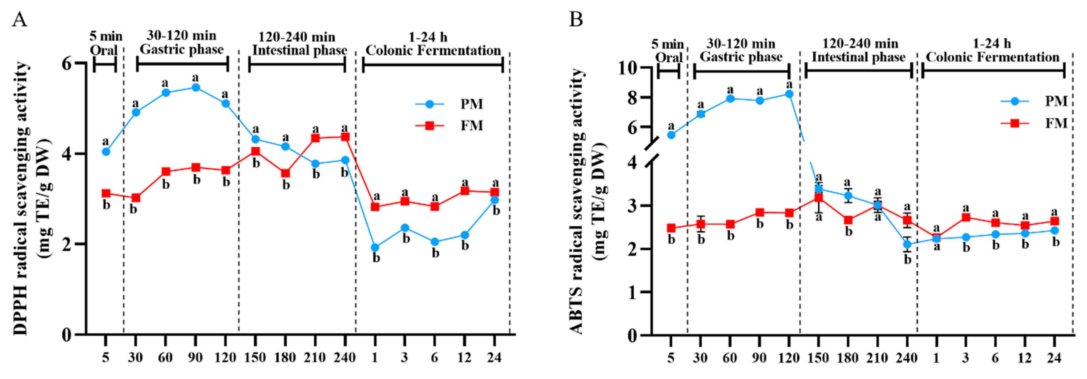
3.3. Impact of Processing on Antioxidant Activity Change During Different Digestion Stages

DPPH and ABTS assays were carried out to assess the antioxidant capacities of released phenolics during different digestion and colonic fermentation stage (Figure 2A,B). Similar to the results of TPC, DPPH and ABTS radical scavenging capacity of PM gradually increased during gastric digestion phase, reaching the maximum values of 5.46 ± 0.02 mg TE/g DW (90 min) and 8.23 ± 0.06 mg TE/g DW (120 min), respectively, which were significantly higher than those of FM (4.05 ± 0.01 mg TE/g DW and 3.18 ± 0.35 mg TE/g DW, respectively) at 150 min. However, the values of DPPH and ABTS decreased during the small intestine digestion, with the antioxidant activity of PM being lower by 13.50% and 30.23%, respectively, compared to FM. Different trends in antioxidant capacities were observed, which could be attributed to the diverse compositions of polyphenols from PM and FM. As mentioned earlier, the content of catechins and epicatechins in PM were more prominent during the gastric phase, and previous research has confirmed that these compounds readily interacted with salivary proteins during oral digestion, making them less likely to be released into the digestive juices [35]. This suggests that they were released from the flavanol polymer by the action of gastric juice and proteases. In contrast, the DPPH radical scavenging capacity of FM was higher than the DPPH in PM during the intestinal phase (210–240 min), indicating that the polyphenols from FM were more easily released in alkaline intestinal fluid environment. Additionally, the content of p-coumaric acid and naringenin from FM was increased from the gastric phase (0.99 ± 0.08 mg/100 g DW and 5.60 ± 0.27 mg/100 g DW, respectively) to the intestinal phase (10.89 ± 0.61 mg/100 g DW and 9.41 ± 0.20 mg/100 g DW, respectively). Therefore, the scavenging ability of DPPH might be affected by the high content of p-coumaric acid and naringenin. In addition, the antioxidant capacities of released phenolics from FM increased at 3 h colonic fermentation, similar to the results of TPC, possibly associated with the increase in benzoic acid, chlorogenic acid and neochlorogenic acid.

3.4. Impact of Processing on Gut Microbiota Regulation Activity

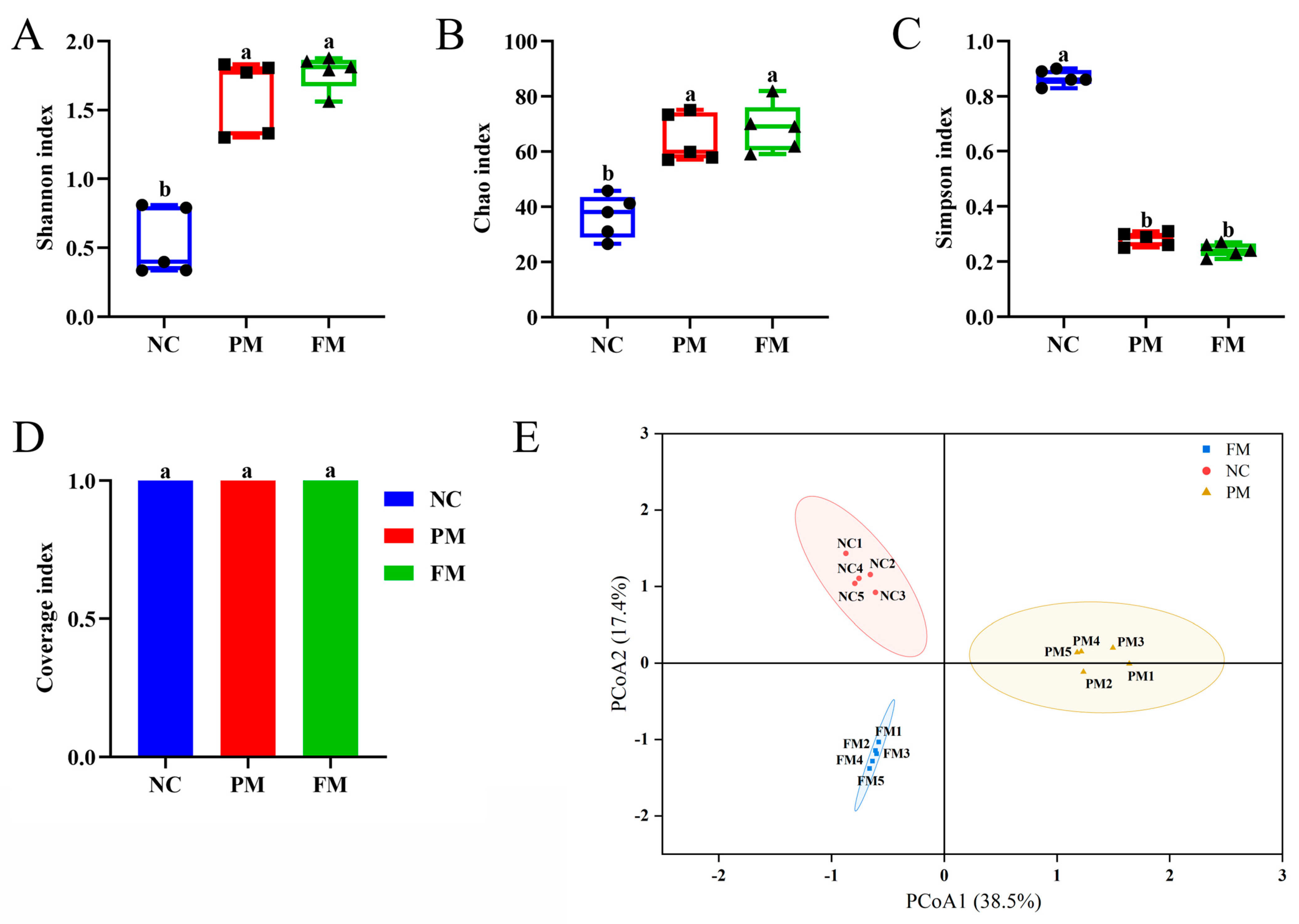
To further analyze the changes of fecal microbiota community structure from PM and FM, we measured the bacterial composition of the samples collected at 24 of colonic fermentation via high-throughput sequencing of the bacterial 16S rRNA gene. Alpha diversity reflected the richness and diversity of intestinal flora. Good’s coverages index was used to characterize the species coverage of the community during 16S rRNA gene sequencing [36]. As shown in Figure 3, compared to the negative control (NC) group, the Shannon index (Figure 3A) and Chao index (Figure 3B) of PM and FM increased, while the Simpson index (Figure 3C) decreased (p < 0.05). The Good’s coverages index (Figure 3D) of all samples in each group was greater than 0.999, indicating that the sequencing data was reliable. These results suggested that both PM and FM had a certain effect on the diversity of the gut microbiota compared to NC group, although the diversity difference between the PM and FM groups was not statistically significant. Beta diversity between different groups was analyzed by principal coordinate analysis (PCoA) [37]. The PCoA score plot showed that the three groups had distinct bacterial communities (Figure 3E).
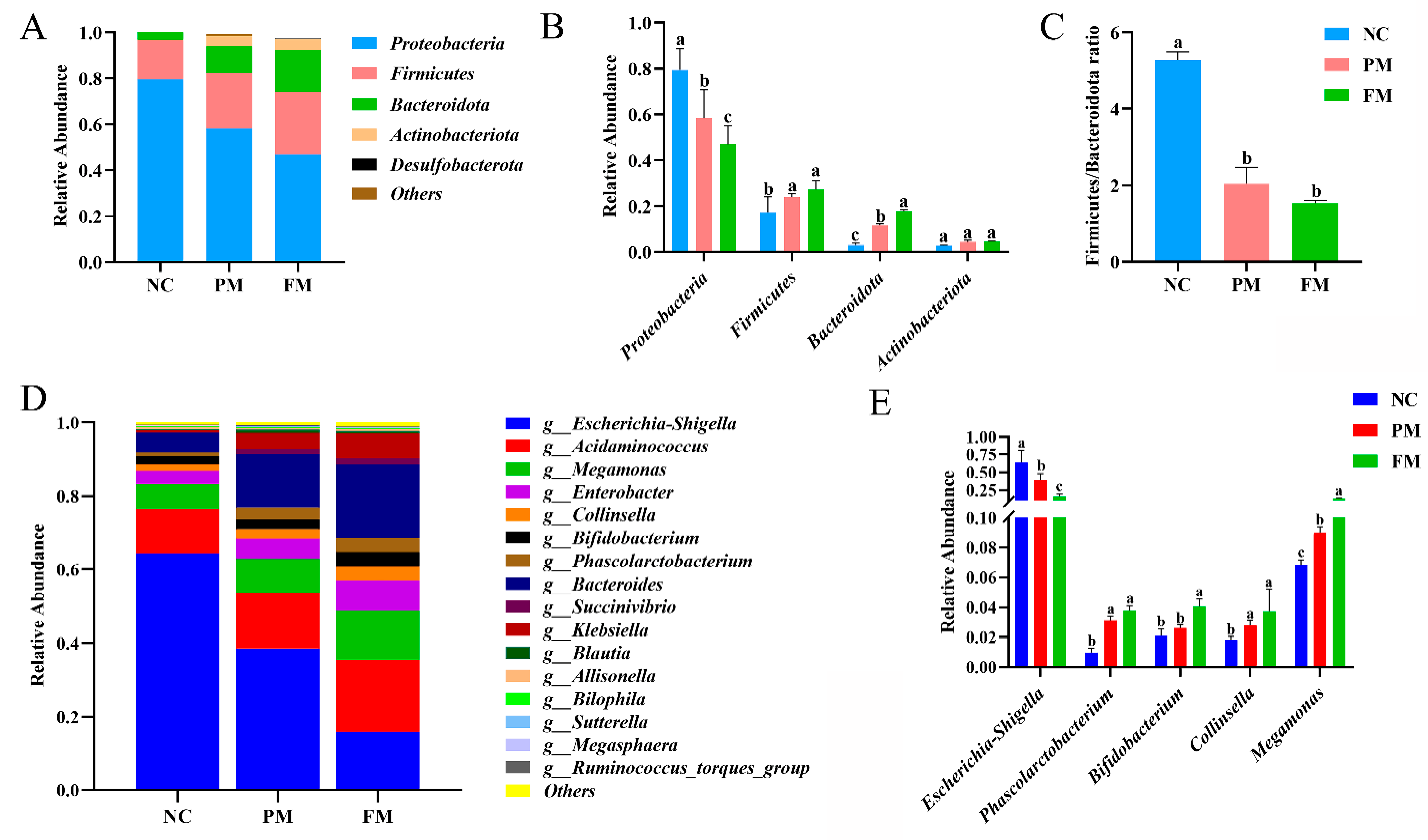
To evaluate the effects of processing on specific microbiota composition, bacterial taxa in all groups were analyzed at the phylum and genus level. At the phylum level, Proteobacteria, Firmicutes, Actinobacteria, and Bacteroidetes accounted for the majority of total microbiota in all groups (Figure 4A,B). The Firmicutes/Bacteroidetes (F/B) ratio is considered to have an important influence in maintaining intestinal homeostasis, and is associated with obesity and inflammatory bowel disease [38]. Compared with NC group, F/B values in the PM and FM groups were significantly decreased by 61.10% and 71.16% (Figure 4C), respectively, during in vitro colonic fermentation, indicating that PM and FM had potential effects in reducing weight and improving inflammatory responses and ulcers.
The relative abundance of the dominant microbiota at the genus level is shown in Figure 4D. The RA of Escherichia–Shigella in PM and FM significantly decreased by 39.95% and 75.28% compared with NC group, respectively (Figure 4E). This reduction could be attributed to higher chlorogenic acid released from FM than PM. It was consistent with the results of mung bean coat [39], among which the abundance of Escherichia–Shigella was found to be significantly negatively correlated with chlorogenic acid [40]. Notably, the relative abundance of Megamonas (increased by 33.13% and 95.26% in PM and FM group, respectively), Collinsella (increased by 51.62% and 104.93% in PM and FM group, respectively) and Phascolarctobacterium (increased by 216.66% and 280.64% in PM and FM group, respectively) significantly increased (p < 0.05). These bacteria were reported to be beneficial to keep colon healthy. Megamonas was found to be negatively associated with the increased risk of colorectal polyps [41]. Collinsella, an important bacterium in gut microbiota development, has been linked to a low risk of colon cancer and irritable bowel syndrome [42]. Phascolarctobacterium can produce short-chain fatty acids, including acetate and propionate [43,44]. Furthermore, compared with NC group, an increase in relative abundance for Bifidobacterium in both PM (23.41%) and FM (92.78%) was observed. Bifidobacterium plays a role in boosting overall immunity, including reducing and treating gastrointestinal infections, as well as improving diarrhea, constipation and eczema [45,46,47,48]. Consequently, PM, and especially FM, had potential effects in optimizing the intestinal microbiota structure, which might be the reason that FM has been used as a folk medication for gastrointestinal disorders, e.g., diarrhea, stomach ache and ulceration [49].
Table 1. Identification of polyphenols and their metabolites of PM and FM during in vitro digestion and colonic fermentation.
Peak.RT (min)PhenolicsFormulaIon Peak (m/z)Fragment (m/z) [Relative Abundance, %]SourceReferences
14.94Protocatechuic acidC7H6O4153.01839109.02888 [100]PM: IP, F1, F3, F6, F12, F24
FM: OP, GP, IP, F1, F3, F6, F12, F24
[50]
25.22Neochlorogenic acidC16H18O9353.08844191.05551 [91], 179.03433 [68],
135.04401 [12]
PM: OP, GP, IP, F1, F3, F6, F12, F24
FM: OP, GP, IP, F1, F3, F6, F12, F24
[51]
35.53Procyanidin B1C30H26O12577.13623289.07211 [100], 407.07773 [64],
125.02317 [75], 425.08859 [40],
451.10410 [10]
PM: OP, GP, IP
FM: IP
[52]
46.14Chlorogenic acidC16H18O9353.08823191.05548 [100]PM: OP, GP, IP, F1, F3, F6, F12, F24
FM: OP, GP, IP, F1, F3, F6, F12, F24
[53]
56.21CatechinC15H14O6289.07220245.08144 [30]PM: OP, GP, IP, F1, F3, F6, F12, F24
FM: F1, F3, F6, F12, F24
[39]
66.383-(3,4-Dihydroxyphenyl) propionic acidC9H10O4181.04991137.05969 [100]PM: F3, F6, F12, F24
FM: F12, F24
[54]
76.49Cryptochlorogenic acidC16H18O9353.0833173.04472 [100], 179.03421 [67], 191.05536 [31], 135.04398 [15]PM: GP, IP, F1, F3, F6, F12, F24
FM: OP, GP, IP, F1, F3, F6, F12, F24
[51]
86.45p-Hydroxybenzoic acidC7H5O3137.0233993.03323 [100]PM: GP, IP, F1, F3, F6, F12, F24
FM: OP, GP, IP, F1, F3, F6, F12, F24
[55]
96.573,4-Dihydroxyphenylacetic acidC8H8O4167.03420123.04397 [54]PM: OP, GP, IP, F1, F3, F6, F12, F24
FM: OP, GP, IP, F1, F3, F6, F12, F24
[56]
107.08EpicatechinC15H14O6289.07224245.08212 [30], 125.02299 [9]PM: OP, GP, IP, F1, F3, F6, F12, F24
FM: OP, GP, IP, F1, F3, F6, F12, F24
[15]
117.12Caffeic acidC9H8O4179.03423135.04404 [100]PM: IP, F1, F3, F6, F12, F24
FM: OP, GP, IP, F1, F3, F6, F12
[15]
127.22Syringic acidC9H10O5197.04510182.02152 [100], 153.05495 [14]PM: OP, GP, IP, F1, F3, F6, F12, F24
FM: OP, GP, IP, F1, F3, F6, F12, F24
[39]
137.27Vanillic acidC8H8O4167.03427152.01057 [100]PM: GP, F1, F3, F6, F12, F24
FM: OP, F1, F3, F6, F12, F24
[39]
148.05Benzoic acidC7H6O2121.0282277.03805 [11]PM: IP, F1, F3, F6, F12, F24
FM: OP, GP, IP, F1, F3, F6, F12, F24
[39]
158.732-O-RhamnosylvitexinC27H30O14577.15686413.09126 [48]; 293.04826 [69]PM: F1, F3, F6, F12, F24
FM: F1, F3, F6, F12, F24
[57]
168.84RutinC27H30O16609.14771300.02783 [100]PM: OP, GP, IP, F1, F3, F6, F12, F24
FM: OP, GP, IP, F1, F3, F6, F12, F24
[53]
178.93p-Coumaric acidC9H8O3163.03915119.04906 [100]PM: IP, F1, F3, F6, F12
FM: OP, GP, IP, F1, F3, F6, F12
[53]
189.19HyperosideC21H20O12463.08923300.02783 [58], 301.03546 [34]PM: OP, GP, IP, F1, F3, F6, F12, F24
FM: OP, GP, IP, F1, F3, F6, F12, F24
[58]
199.36IsoquercetinC21H20O12463.08932300.02779 [100]PM: OP, GP, IP, F1, F3, F6, F12, F24
FM: OP, GP, IP, F1, F3, F6, F12, F24
[59]
209.57Ferulic acidC10H10O4193.04988178.02635 [90], 134.03616 [100], 149.05968 [35]PM: IP, F1, F3, F6, F12, F24
FM: OP, GP, IP, F1, F3, F6, F12, F24
[15]
219.66Isoferulic acidC10H10O4193.04984178.02621 [100], 134.03612 [65]PM: F1, F3, F6, F12, F24
FM: F1, F3, F6, F12, F24
[60]
229.87Kaempferol-3-O-rutinosideC27H30O15593.15247285.04080 [62]PM: F1, F3, F6, F12, F24
FM: F1, F3, F6, F12, F24
[61]
2314.63QuercetinC15H10O7301.03583151.00272 [21], 178.99788 [15]PM: GP, F1, F3, F6, F12, F24
FM: OP, GP, IP, F1, F3, F6, F12, F24
[15]
2416.17NaringeninC15H12O5271.06155151.00255 [43], 119.04892 [14], 177.01808 [6]PM: IP, F1, F3, F6, F12, F24
FM: OP, GP, IP, F1, F3, F6, F12, F24
[15]
PM, Prunus mume; FM, Fructus mume; OP, oral phase; GP, gastric phase; IP, intestinal phase. Fermentation broth in different times: F1, 1 h; F3, 3 h; F6, 6 h; F12, 12 h; F24, 24 h.
Table 2. Quantitative results and bioaccessibility of released phenolics and metabolites from PM and FM during in vitro digestion (mg/100 g DW).
NO.PhenolicsSampleOPGPIP (PCafter)PCbeforeIntestinal (Bioaccessibility)
1Protocatechuic acidPMNdNd0.04 ± 0.03 a0.67 ± 0.04 a5.97% a
FM0.08 ± 0.02 B0.08 ± 0.01 B0.24 ± 0.04 bA2.84 ± 0.07 b8.45% b
2Neochlorogenic acidPM300.23 ± 24.17 aA325.68 ± 13.74 aA22.35 ± 0.42 aB200.91 ± 42.38 a11.12% a
FM231.63 ± 25.03 bA220.22 ± 1.40 bA12.57 ± 1.17 bB195.34 ± 6.25 a6.43% b
3Procyanidin B1PM2.79 ± 0.05 B3.88 ± 0.12 A0.03 ± 0.00 aC11.44 ± 1.38 a0.26% a
FMNdNd0.06 ± 0.00 b2.92 ± 0.15 b2.05% b
4Chlorogenic acidPM17.94 ± 0.34 aA2.48 ± 0.39 aB2.80 ± 0.10 aB18.25 ± 1.67 a15.34% a
FM14.66 ± 0.67 bB23.23 ± 0.78 bA2.22 ± 0.09 aC14.57 ± 0.55 b15.24% a
5CatechinPM9.18 ± 0.16 B12.58 ± 0.38 A0.24 ± 0.05 C34.60 ± 2.12 a0.69%
FMNdNdNd12.99 ± 1.00 bNd
63-(3,4-Dihydroxyphenyl)
propionic acid
PMNdNdNdNdNd
FMNdNdNdNdNd
7Cryptochlorogenic acidPMNd0.23 ± 0.09 aA0.41 ± 0.00 aA2.92 ± 0.39 a14.04% a
FM5.12 ± 0.20 A5.01 ± 0.18 bA0.17 ± 0.02 bB22.60 ± 0.46 b0.75% b
8p-Hydroxybenzoic acidPMNd0.02 ± 0.00 aB1.39 ± 0.01 aA6.79 ± 0.23 a20.47% a
FM1.31 ± 0.06 A1.22 ± 0.02 bA2.50 ± 0.04 bA13.69 ± 0.74 b18.26% b
93,4-Dihydroxyphenylacetic acidPM323.95 ± 20.39 aB535.14 ± 17.56 aA250.13 ± 48.81 aBNdNd
FM205.53 ± 0.46 bB257.18 ± 8.29 bA111.18 ± 3.92 bABNdNd
10EpicatechinPM45.76 ± 1.60 aB66.86 ± 1.92 aA0.98 ± 0.12 aC45.09 ± 2.12 a2.17% a
FM0.63 ± 0.00 bB0.88 ± 0.02 bA0.45 ± 0.03 bC4.90 ± 0.38 b9.18% b
11Caffeic acidPMNdNd0.61 ± 0.10 a67.40 ± 5.26 a0.90% a
FM0.68 ± 0.04 B0.72 ± 0.03 B3.25 ± 0.08 bA50.26 ± 4.59 b6.47% b
12Syringic acidPM0.07 ± 0.02 aAB0.10 ± 0.01 aB0.36 ± 0.02 aANdNd
FM0.35 ± 0.06 bB0.38 ± 0.01 bB0.47 ± 0.01 bANdNd
13Vanillic acidPMNd0.38 ± 0.05NdNdNd
FM0.21 ± 0.00NdNdNdNd
14Benzoic acidPMNdNd53.74 ± 54.05 aNdNd
FM147.53 ± 6.19 bB179.20 ± 3.02 bA98.10 ± 1.54 bCNdNd
152-O-RhamnosylvitexinPMNdNdNdNdNd
FMNdNdNdNdNd
16RutinPM1.91 ± 0.13 aA2.33 ± 0.04 aA2.56 ± 0.34 aA4.34 ± 0.97 a58.99% a
FM2.95 ± 0.06 bA3.28 ± 0.00 bA2.64 ± 0.23 aA4.18 ± 0.39 a63.16% a
17p-Coumaric acidPMNdNd3.88 ± 0.66 a33.93 ± 3.95 a11.44% a
FM0.92 ± 0.08 B0.99 ± 0.08 B10.89 ± 0.61 bA28.40 ± 3.88 b38.34% b
18HyperosidePM1.10 ± 0.09 aA1.59 ± 0.12 aA1.33 ± 0.13 aA0.84 ± 0.11 a158.33% a
FM1.42 ± 0.00 bA1.45 ± 0.05 aA1.28 ± 0.02 aB0.91 ± 0.44 a140.66% a
19IsoquercetinPM0.84 ± 0.07 aA1.21 ± 0.08 aA1.02 ± 0.08 aA1.90 ± 0.32 a53.68% a
FM1.08 ± 0.00 bA1.10 ± 0.04 aA0.98 ± 0.00 aB1.14 ± 0.37 b85.96% a
20Ferulic acidPMNdNd0.20 ± 0.01 a6.97 ± 0.80 a2.87% a
FM0.12 ± 0.02 A0.11 ± 0.01 A0.67 ± 0.04 bA6.47 ± 0.74 a10.36% b
21Isoferulic acidPMNdNdNd40.72 ± 4.77 aNd
FMNdNdNd26.86 ± 2.34 bNd
22Kaempferol-3-O-rutinosidePMNdNdNdNdNd
FMNdNdNdNdNd
23QuercetinPMNd0.04 ± 0.01 aNd0.18 ± 0.06 aNd
FM0.18 ± 0.02 B0.36 ± 0.03 bA0.12 ± 0.00 C0.48 ± 0.06 b25.00%
24NaringeninPMNdNd7.71 ± 0.33 a2.31 ± 0.20 a333.77% a
FM4.41 ± 1.00 B5.60 ± 0.27 B9.41 ± 0.20 bA10.39 ± 0.39 b90.57% b
Nd, not detected; PM, Prunus mume; FM, Fructus mume; OP, oral phase; GP, gastric phase; IP, intestinal phase; PCbefore, content of polyphenols before digestion; PCafter, content of polyphenols released after intestinal phase digestion; Bioaccessibility (%) = PCafter/PCbefore × 100%. 3-(3,4-dihydroxyphenyl) propionic acid, 3,4-dihydroxyphenylacetic acid and benzoic acid were quantified in p-hydroxybenzoic acid equivalents. Data represent mean ± standard deviation (n = 3). Different lowercase letters represent a significant difference between PM and FM at same phase (p < 0.05). Different capital letters represent a significant difference at different phases (p < 0.05).
Table 3. Quantitative results of released phenolics and metabolites from PM and FM during colonic fermentation (mg/100 g DW).
NO.PhenolicsSampleF1F3F6F12F24
1Protocatechuic acidPM0.13 ± 0.01 aC0.15 ± 0.00 aC0.18 ± 0.02 aB0.24 ± 0.01 aA0.01 ± 0.00 aD
FM0.49 ± 0.04 bB0.65 ± 0.06 bA0.70 ± 0.07 bA0.46 ± 0.05 bB0.04 ± 0.01 bC
2Neochlorogenic acidPM215.69 ± 22.41 aA186.65 ± 17.14 aA202.25 ± 16.86 aA210.26 ± 6.87 aA180.12 ± 11.52 aA
FM209.95 ± 19.78 aB270.42 ± 17.58 bA188.76 ± 20.15 aC244.86 ± 24.17 aAB254.70 ± 15.23 bAB
3Procyanidin B1PMNdNdNdNdNd
FMNdNdNdNdNd
4Chlorogenic acidPM44.31 ± 4.26 aB39.88 ± 3.70 aC53.33 ± 5.89 aAB62.17 ± 6.17 aA54.14 ± 5.37 aAB
FM68.15 ± 5.05 bB94.00 ± 9.27 bA67.94 ± 5.29 bB90.07 ± 8.34 bA95.48 ± 8.90 bA
5CatechinPM0.58 ± 0.07 aB0.16 ± 0.01 aD0.30 ± 0.02 aC0.21 ± 0.01 aCD0.78 ± 0.05 aA
FM0.09 ± 0.01 bB0.08 ± 0.01 bB0.15 ± 0.03 bA0.04 ± 0.00 bC0.02 ± 0.00 bC
63-(3,4-Dihydroxyphenyl)
propionic acid
PMNd19.93 ± 1.29 D32.35 ± 1.55 C75.20 ± 0.81 aB191.25 ± 11.83 aA
FMNdNdNd17.50 ± 6.61 bB105.15 ± 8.25 bA
7Cryptochlorogenic acidPM38.97 ± 3.37 aB38.76 ± 3.62 aB48.12 ± 4.41 aAB59.02 ± 5.47 aA58.66 ± 5.98 aA
FM77.42 ± 6.99 bB101.06 ± 8.83 bA76.16 ± 6.18 bB103.66 ± 1.18 bA103.97 ± 2.99 bA
8p-Hydroxybenzoic acidPM2.93 ± 0.21 aB3.91 ± 0.22 aA3.70 ± 0.34 aA1.68 ± 0.11 aC0.26 ± 0.02 aD
FM4.08 ± 0.49 bB4.78 ± 0.43 aAB5.42 ± 0.53 bA0.35 ± 0.02 bC0.25 ± 0.05 aC
93,4-Dihydroxyphenylacetic acidPM522.38 ± 19.13 aA462.38 ± 92.82 aA529.90 ± 9.62 aA494.60 ± 17.64 aA477.02 ± 6.48 aA
FM312.20 ± 4.91 bB380.69 ± 8.76 bA354.27 ± 17.77 bA318.83 ± 11.23 bB177.24 ± 4.55 bC
10EpicatechinPM0.96 ± 0.02 aB0.62 ± 0.04 aC0.62 ± 0.06 aC0.51 ± 0.04 aC1.54 ± 0.17 aA
FM0.52 ± 0.08 bBC0.42 ± 0.06 bD0.65 ± 0.07 aA0.57 ± 0.05 aAB0.45 ± 0.07 bCD
11Caffeic acidPM0.41 ± 0.03 aC0.94 ± 0.07 aB1.10 ± 0.12 aB1.40 ± 0.16 aA0.03 ± 0.00 aD
FM1.70 ± 0.27 bB2.67 ± 0.18 bA2.92 ± 0.24 bA2.45 ± 0.28 bANd
12Syringic acidPM0.22 ± 0.01 aC5.46 ± 0.57 aA2.57 ± 0.22 aB0.42 ± 0.01 aC0.60 ± 0.08 aC
FM5.00 ± 0.59 bB2.90 ± 0.21 bC8.06 ± 0.77 bA5.63 ± 0.51 bB0.60 ± 0.01 aD
13Vanillic acidPM0.84 ± 0.09 aBC0.78 ± 0.05 aBC1.15 ± 0.17 aA0.75 ± 0.07 aC1.02 ± 0.04 aAB
FM1.10 ± 0.15 aAB0.94 ± 0.05 bBC1.06 ± 0.06 aBC0.80 ± 0.17 aC1.35 ± 0.03 bA
14Benzoic acidPM205.28 ± 23.70 aD1874.22± 115.46 aB2826.56 ± 1.73 aA586.49 ± 22.32 aC94.96 ± 18.70 aE
FM895.92 ± 70.05 bB2920.44 ± 50.39 bA3118.91 ± 53.83 bA471.66 ± 18.92 bC271.28 ± 17.98 bD
152-O-RhamnosylvitexinPM0.26 ± 0.06 aB0.45 ± 0.00 aA0.34 ± 0.05 aB0.28 ± 0.02 aB0.32 ± 0.05 aB
FM0.29 ± 0.02 aAB0.27 ± 0.02 bAB0.33 ± 0.01 aA0.25 ± 0.04 aB0.26 ± 0.02 aB
16RutinPM4.75 ± 0.41 aAB5.32 ± 0.53 aA5.82 ± 0.47 aA5.14 ± 0.43 aA4.43 ± 0.57 aB
FM5.34 ± 0.53 aA6.67 ± 0.52 bA5.98 ± 0.62 aA6.16 ± 0.61 aA6.06 ± 0.65 bA
17p-Coumaric acidPM2.27 ± 0.28 aB5.28 ± 0.56 aA5.68 ± 0.17 aA1.92 ± 0.18 aBNd
FM5.01 ± 0.57 bC6.89 ± 0.56 bB8.65 ± 0.88 bA1.35 ± 0.12 bDNd
18HyperosidePM3.19 ± 0.25 aA3.21 ± 0.33 aA3.62 ± 0.36 aA2.89 ± 0.26 aA1.82 ± 0.19 aB
FM2.84 ± 0.22 aB3.64 ± 0.36 aA3.40 ± 0.29 aA2.96 ± 0.25 aAB2.41 ± 0.14 bB
19IsoquercetinPM2.52 ± 0.09 aAB2.63 ± 0.12 aA2.86 ± 0.11 aA2.37 ± 0.26 aB1.42 ± 0.10 aC
FM2.31 ± 0.27 aAB2.90 ± 0.21 aA2.67 ± 0.29 aA2.37 ± 0.22 aAB1.90 ± 0.13 bB
20Ferulic acidPM0.71 ± 0.01 aB0.80 ± 0.07 aAB0.87 ± 0.03 aA0.87 ± 0.06 aA0.88 ± 0.05 aA
FM0.34 ± 0.03 bC0.46 ± 0.01 bBC0.55 ± 0.06 bB0.66 ± 0.06 bA0.35 ± 0.02 bC
21Isoferulic acidPM11.7 ± 1.12 aA12.63 ± 0.72 aA14.13 ± 1.38 aA8.53 ± 0.76 aB6.99 ± 0.72 aB
FM2.08 ± 0.19 bC2.88 ± 0.26 bB3.71 ± 0.35 bA3.99 ± 0.31 bA3.99 ± 0.32 bA
22Kaempferol-3-O-rutinosidePM0.17 ± 0.03 aA0.16 ± 0.01 aA0.20 ± 0.03 aA0.18 ± 0.02 aA0.12 ± 0.01 aB
FM0.16 ± 0.02 aB0.24 ± 0.02 bA0.21 ± 0.01 aA0.22 ± 0.03 aA0.18 ± 0.02 bB
23QuercetinPM0.66 ± 0.05 aC0.76 ± 0.02 aBC0.80 ± 0.06 aAB0.92 ± 0.09 aA0.84 ± 0.05 aAB
FM1.55 ± 0.19 bB1.62 ± 0.13 bAB1.79 ± 0.14 bA1.95 ± 0.13 bA1.91 ± 0.17 bA
24NaringeninPM30.21 ± 3.52 aB33.81 ± 2.86 aB37.76 ± 3.52 aAB44.01 ± 4.34 aA44.20 ± 4.01 aA
FM49.82 ± 4.81 bA47.00 ± 4.77 bAB41.40 ± 4.80 bB45.47 ± 5.59 aAB53.23 ± 4.08 bA
Nd, not detected; PM, Prunus mume; FM, Fructus mume. Fermentation broth in different times: F1, 1 h; F3, 3 h; F6, 6 h; F12, 12 h; F24, 24 h. 3-(3,4-dihydroxyphenyl) propionic acid, 3,4-dihydroxyphenylacetic acid and benzoic acid were quantified in p-hydroxybenzoic acid equivalents. Data represent mean ± standard deviation (n = 3). Different lowercase letters represent a significant difference between PM and FM at same phase (p < 0.05). Different capital letters represent a significant difference at different phases (p < 0.05).

3.5. Correlation Between Phenolics Compounds and Gut Microbiota

The correlation between the content of major phenolic compounds released after 24 h of fermentation and the relative abundance of gut microbiota was examined using the Spearman correlation analysis (Figure 5). The protocatechuic acid, chlorogenic acid, vanillic acid, benzoic acid, rutin, hyperoside and kaempferol-3-O-rutinoside had a significantly negative correlation with Proteobacteria and Escherichia–Shigella (p < 0.01), and a significantly positive relationship with Bacteroidota (p < 0.01), Phascolarctobacterium (p < 0.05) and Bifidobacterium (p < 0.05). The stronger inhibitory activity of FM against Proteobacteria and Escherichia–Shigella might be attributed to the higher release of chlorogenic acid and benzoic acid in FM compared to PM (p < 0.05). Moreover, FM’s greater promotion of beneficial gut microbes such as Bifidobacterium and Phascolarctobacterium might be related to its higher released content of neochlorogenic acid, cryptochlorogenic acid, chlorogenic acid and benzoic acid relative to PM (p < 0.05). These results demonstrated that PM and FM possessed prebiotic potentials, which could be further enhanced after PM-processing (paozi).

4. Conclusions

The release characteristics and antioxidant activity change of phenolics from PM and FM during in vitro simulated digestion as well as gut microbiota modulation effect were firstly determined and compared in the present study. Both the total polyphenols content released and antioxidant activities of FM were much higher than those of PM during intestinal digestion and colonic fermentation. Importantly, compared with PM, FM demonstrates superior effects on colonic health by increasing the relative abundance of probiotic bacteria such as Bifidobacterium to a greater extent and exerting stronger inhibitory effects against Escherichia–Shigella. These results provided insights into the digestive properties of polyphenols of PM and FM after oral intake, indicating that FM processing potentially increased PM’s health effect on the colon. The study also provides a theoretical basis for FM used as a traditional medicine for gastrointestinal disorders. However, considering processing-induced changes in non-phenolic components (e.g., polysaccharide degradation, Maillard reaction products during heating), the differential effects of PM vs. FM on regulating the gut microbiota composition could not be ascribed solely to processing-induced alterations in phenolic compounds. Further exploration is required to determine how FM processing-induced changes in non-phenolic components affect the prebiotic potential of PM.

Supplementary Materials

The following supporting information can be downloaded at: https://www.mdpi.com/article/10.3390/foods14234067/s1, Figure S1: The primary and secondary mass spectra of vanillic acid (A) and syringic acid (B); Figure S2: The primary and secondary mass spectra of kaempferol-3-O-rutinoside (A) and 2-O-Rhamnosylvitexin (B); Table S1: Configuration of simulated saliva, gastric and intestinal fluids (SSF, SGF, SIF) and preheat to 37 °C before use; Table S2: Calibration curves used for UPLC-MS/MS quantification of polyphenols.

Author Contributions

Q.X.: Conceptualization, methodology, formal analysis, investigation, writing—original draft. Z.T.: Methodology, investigation. B.Y.: Formal analysis, validation, writing—review and editing. J.L.: Methodology, data curation, visualization. W.H.: Resources, investigation. W.L.: Writing—review and editing, conceptualization, methodology. R.Y.: Project administration, writing—review and editing, funding acquisition. All authors have read and agreed to the published version of the manuscript.

Funding

This work was financially supported by the National Key Research and Development Program of China (Grant No.2023YFF1104204), the Guangdong Basic and Applied Basic Research Foundation of the Natural Science Foundation of Guangdong Province (Grant No. 2023A1515011151, 2023A1515012801), the Special projects in key areas of general colleges and universities in Guangdong Province (No. 2023ZDZX2064), the Guangdong Provincial Key Construction Discipline Research Ability Enhancement Project (No. 2022ZDJS026), and the Key Research and Development Program of Guangdong Province (Grant No. 2023B0202050001).

Institutional Review Board Statement

Not applicable.

Informed Consent Statement

Informed consent forms have been obtained from the participants.

Data Availability Statement

The original contributions presented in the study are included in the article/Supplementary Material, further inquiries can be directed to the corresponding authors.

Acknowledgments

We thank the volunteers who participated in this study.

Conflicts of Interest

The authors declare no conflicts of interest.

References

  1. Shi, T.; Luo, W.; Li, H.; Huang, X.; Ni, Z.; Gao, H.; Iqbal, S.; Gao, Z. Association between Blooming Time and Climatic Adaptation in Prunus mume. Ecol. Evol. 2020, 10, 292–306. [Google Scholar] [CrossRef]
  2. Ye, J. Study on Preparation Technology and Quality Standard of Dark Plum Standard Slices. Master’s Thesis, Hubei University of Chinese Medicine, Wuhan, China, 2018. [Google Scholar]
  3. Wang, R.; Cheng, H.; Yang, Y.; Ou, J.; Song, Q.; Zhou, H.; Peng, H.; Wang, J.; Fang, C.W. Ultra High Performance Liquid Chromatography with Quadrupole Time-of-flight Tandem Mass Spectrometry and Liquid Chromatography with Tandem Mass Spectrometry Combined with Chemometric Analysis as an Approach for the Quality Evaluation of Mume fructus. J. Sep. Sci. 2022, 45, 1884–1893. [Google Scholar] [CrossRef]
  4. Chen, Y.; Wong, R.W.K.; Seneviratne, C.J.; Hägg, U.; Mcgrath, C.; Samaranayake, L.P.; Kao, R. The Antimicrobial Efficacy of Fructus mume Extract on Orthodontic Bracket: A Monospecies-Biofilm Model Study in Vitro. Arch. Oral Biol. 2011, 56, 16–21. [Google Scholar] [CrossRef]
  5. Xu, Z.; Zhang, X.; Wang, W.; Zhang, D.; Ma, Y.; Zhang, D.; Chen, M. Fructus mume (Wu Mei) Attenuates Acetic Acid-Induced Ulcerative Colitis by Regulating Inflammatory Cytokine, Reactive Oxygen Species, and Neuropeptide Levels in Model Rats. J. Med. Food 2022, 25, 389–401. [Google Scholar] [CrossRef]
  6. Yan, S.; Wang, P.; Wei, H.; Jia, R.; Zhen, M.; Li, Q.; Xue, C.; Li, J. Treatment of Ulcerative Colitis with Wu-Mei-Wan by Inhibiting Intestinal Inflammatory Response and Repairing Damaged Intestinal Mucosa. Phytomedicine 2022, 105, 154362. [Google Scholar] [CrossRef] [PubMed]
  7. Wu, X.; Chen, H.; Gao, X.; Gao, H.; He, Q.; Li, G.; Yao, J.; Liu, Z. Natural Herbal Remedy Wumei Decoction Ameliorates Intestinal Mucosal Inflammation by Inhibiting Th1/Th17 Cell Differentiation and Maintaining Microbial Homeostasis. Inflamm. Bowel Dis. 2022, 28, 1061–1071. [Google Scholar] [CrossRef] [PubMed]
  8. Chen, G.-L.; Chen, S.-G.; Zhao, Y.-Y.; Luo, C.-X.; Li, J.; Gao, Y.-Q. Total Phenolic Contents of 33 Fruits and Their Antioxidant Capacities before and after in Vitro Digestion. Ind. Crops Prod. 2014, 57, 150–157. [Google Scholar] [CrossRef]
  9. Kaulmann, A.; André, C.M.; Schneider, Y.-J.; Hoffmann, L.; Bohn, T. Carotenoid and Polyphenol Bioaccessibility and Cellular Uptake from Plum and Cabbage Varieties. Food Chem. 2016, 197, 325–332. [Google Scholar] [CrossRef]
  10. Mubarak, A.; Swinny, E.E.; Ching, S.Y.L.; Jacob, S.R.; Lacey, K.; Hodgson, J.M.; Croft, K.D.; Considine, M.J. Polyphenol Composition of Plum Selections in Relation to Total Antioxidant Capacity. J. Agric. Food Chem. 2012, 60, 10256–10262. [Google Scholar] [CrossRef]
  11. You, B.; Yang, S.; Yu, J.; Xian, W.; Deng, Y.; Huang, W.; Li, W.; Yang, R. Effect of Thermal and Dry Salt-Curing Processing on Free and Bound Phenolics and Antioxidant Activity in Prunus mume Fruits Together with the Phenolic Bioaccessibility. LWT 2021, 145, 111355. [Google Scholar] [CrossRef]
  12. Tomas, M.; Zhang, L.; Zengin, G.; Rocchetti, G.; Capanoglu, E.; Lucini, L. Metabolomic Insight into the Profile, in Vitro Bioaccessibility and Bioactive Properties of Polyphenols and Glucosinolates from Four Brassicaceae Microgreens. Food Res. Int. 2021, 140, 110039. [Google Scholar] [CrossRef]
  13. Hao, Y.; Yang, J.; Cui, J.; Fan, Y.; Li, N.; Wang, C.; Liu, Y.; Dong, Y. Stability and Mechanism of Phenolic Compounds from Raspberry Extract under in Vitro Gastrointestinal Digestion. LWT 2021, 139, 110552. [Google Scholar] [CrossRef]
  14. Cortés-Martín, A.; Selma, M.V.; Tomás-Barberán, F.A.; González-Sarrías, A.; Espín, J.C. Where to Look into the Puzzle of Polyphenols and Health? The Postbiotics and Gut Microbiota Associated with Human Metabotypes. Mol. Nutr. Food Res. 2020, 64, 1900952. [Google Scholar] [CrossRef]
  15. Zhao, Q.; Wang, Z.; Wang, X.; Yan, X.; Guo, Q.; Yue, Y.; Yue, T.; Yuan, Y. The Bioaccessibility, Bioavailability, Bioactivity, and Prebiotic Effects of Phenolic Compounds from Raw and Solid-Fermented Mulberry Leaves during in Vitro Digestion and Colonic Fermentation. Food Res. Int. 2023, 165, 112493. [Google Scholar] [CrossRef] [PubMed]
  16. Brodkorb, A.; Egger, L.; Alminger, M.; Alvito, P.; Assunção, R.; Ballance, S.; Bohn, T.; Bourlieu-Lacanal, C.; Boutrou, R.; Carrière, F.; et al. INFOGEST Static in Vitro Simulation of Gastrointestinal Food Digestion. Nat. Protoc. 2019, 14, 991–1014. [Google Scholar] [CrossRef] [PubMed]
  17. Liu, S.; Yu, Q.; Huang, H.; Hou, K.; Dong, R.; Chen, Y.; Xie, J.; Nie, S.; Xie, M. The Effect of Bound Polyphenols on the Fermentation and Antioxidant Properties of Carrot Dietary Fiber in Vivo and in Vitro. Food Funct. 2020, 11, 748–758. [Google Scholar] [CrossRef]
  18. Di Naso, F.C.; Simões Dias, A.; Porawski, M.; Marroni, N.A.P. Exogenous Superoxide Dismutase: Action on Liver Oxidative Stress in Animals with Streptozotocin-Induced Diabetes. Exp. Diabetes Res. 2011, 2011, 1–6. [Google Scholar] [CrossRef] [PubMed]
  19. Li, W.; Yang, R.; Ying, D.; Yu, J.; Sanguansri, L.; Augustin, M.A. Analysis of Polyphenols in Apple Pomace: A Comparative Study of Different Extraction and Hydrolysis Procedures. Ind. Crops Prod. 2020, 147, 112250. [Google Scholar] [CrossRef]
  20. He, Z.; Tao, Y.; Zeng, M.; Zhang, S.; Tao, G.; Qin, F.; Chen, J. High Pressure Homogenization Processing, Thermal Treatment and Milk Matrix Affect in Vitro Bioaccessibility of Phenolics in Apple, Grape and Orange Juice to Different Extents. Food Chem. 2016, 200, 107–116. [Google Scholar] [CrossRef]
  21. Velázquez, E.; Tournier, H.A.; Mordujovich De Buschiazzo, P.; Saavedra, G.; Schinella, G.R. Antioxidant Activity of Paraguayan Plant Extracts. Fitoterapia 2003, 74, 91–97. [Google Scholar] [CrossRef]
  22. Xian, W.; Yang, S.; Deng, Y.; Yang, Y.; Tan, Z.; Li, W.; Yang, R. Potential of Establishing the Corresponding Human Microbial Community in Pseudo Germ-Free Mice through Fecal Microbe Transfer from Three Urolithin Metabotypes. J. Agric. Food Chem. 2022, 70, 9388–9398. [Google Scholar] [CrossRef] [PubMed]
  23. Tagliazucchi, D.; Verzelloni, E.; Bertolini, D.; Conte, A. In Vitro Bio-Accessibility and Antioxidant Activity of Grape Polyphenols. Food Chem. 2010, 120, 599–606. [Google Scholar] [CrossRef]
  24. Bazzocco, S.; Mattila, I.; Guyot, S.; Renard, C.M.G.C.; Aura, A.-M. Factors Affecting the Conversion of Apple Polyphenols to Phenolic Acids and Fruit Matrix to Short-Chain Fatty Acids by Human Faecal Microbiota in Vitro. Eur. J. Nutr. 2008, 47, 442–452. [Google Scholar] [CrossRef]
  25. Aura, A.-M.; Mattila, I.; Seppänen-Laakso, T.; Miettinen, J.; Oksman-Caldentey, K.-M.; Orešič, M. Microbial Metabolism of Catechin Stereoisomers by Human Faecal Microbiota: Comparison of Targeted Analysis and a Non-Targeted Metabolomics Method. Phytochem. Lett. 2008, 1, 18–22. [Google Scholar] [CrossRef]
  26. Parada, J.; Aguilera, J.M. Food Microstructure Affects the Bioavailability of Several Nutrients. J. Food Sci. 2007, 72, R21–R32. [Google Scholar] [CrossRef] [PubMed]
  27. Morzel, M.; Canon, F.; Guyot, S. Interactions between Salivary Proteins and Dietary Polyphenols: Potential Consequences on Gastrointestinal Digestive Events. J. Agric. Food Chem. 2022, 70, 6317–6327. [Google Scholar] [CrossRef] [PubMed]
  28. Sánchez-Patán, F.; Cueva, C.; Monagas, M.; Walton, G.E.; Gibson, G.R.; Martín-Álvarez, P.J.; Victoria Moreno-Arribas, M.; Bartolomé, B. Gut Microbial Catabolism of Grape Seed Flavan-3-Ols by Human Faecal Microbiota. Targetted Analysis of Precursor Compounds, Intermediate Metabolites and End-Products. Food Chem. 2012, 131, 337–347. [Google Scholar] [CrossRef]
  29. Eran Nagar, E.; Okun, Z.; Shpigelman, A. Digestive Fate of Polyphenols: Updated View of the Influence of Chemical Structure and the Presence of Cell Wall Material. Curr. Opin. Food Sci. 2020, 31, 38–46. [Google Scholar] [CrossRef]
  30. Su, D.; Zhang, R.; Hou, F.; Zhang, M.; Guo, J.; Huang, F.; Deng, Y.; Wei, Z. Comparison of the Free and Bound Phenolic Profiles and Cellular Antioxidant Activities of Litchi Pulp Extracts from Different Solvents. BMC Complement. Altern. Med. 2014, 14, 9. [Google Scholar] [CrossRef]
  31. Gonthier, M.-P.; Verny, M.-A.; Besson, C.; Rémésy, C.; Scalbert, A. Chlorogenic Acid Bioavailability Largely Depends on Its Metabolism by the Gut Microflora in Rats. J. Nutr. 2003, 133, 1853–1859. [Google Scholar] [CrossRef]
  32. Bokkenheuser, V.D.; Shackleton, C.H.; Winter, J. Hydrolysis of Dietary Flavonoid Glycosides by Strains of Intestinal Bacteroides from Humans. Biochem. J. 1987, 248, 953–956. [Google Scholar] [CrossRef]
  33. Shim, S.-M.; Yoo, S.-H.; Ra, C.-S.; Kim, Y.-K.; Chung, J.-O.; Lee, S.-J. Digestive Stability and Absorption of Green Tea Polyphenols: Influence of Acid and Xylitol Addition. Food Res. Int. 2012, 45, 204–210. [Google Scholar] [CrossRef]
  34. Rechner, A.R.; Spencer, J.P.E.; Kuhnle, G.; Hahn, U.; Rice-Evans, C.A. Novel Biomarkers of the Metabolism of Caffeic Acid Derivatives in Vivo. Free Radic. Biol. Med. 2001, 30, 1213–1222. [Google Scholar] [CrossRef]
  35. De Freitas, V.; Mateus, N. Structural Features of Procyanidin Interactions with Salivary Proteins. J. Agric. Food Chem. 2001, 49, 940–945. [Google Scholar] [CrossRef]
  36. Dou, Z.; Chen, C.; Huang, Q.; Fu, X. In Vitro Digestion of the Whole Blackberry Fruit: Bioaccessibility, Bioactive Variation of Active Ingredients and Impacts on Human Gut Microbiota. Food Chem. 2022, 370, 131001. [Google Scholar] [CrossRef]
  37. Li, Y.; Rahman, S.U.; Huang, Y.; Zhang, Y.; Ming, P.; Zhu, L.; Chu, X.; Li, J.; Feng, S.; Wang, X.; et al. Green Tea Polyphenols Decrease Weight Gain, Ameliorate Alteration of Gut Microbiota, and Mitigate Intestinal Inflammation in Canines with High-Fat-Diet-Induced Obesity. J. Nutr. Biochem. 2020, 78, 108324. [Google Scholar] [CrossRef] [PubMed]
  38. Stojanov, S.; Berlec, A.; Štrukelj, B. The Influence of Probiotics on the Firmicutes/Bacteroidetes Ratio in the Treatment of Obesity and Inflammatory Bowel Disease. Microorganisms 2020, 8, 1715. [Google Scholar] [CrossRef] [PubMed]
  39. Xie, J.; Sun, N.; Huang, H.; Xie, J.; Chen, Y.; Hu, X.; Hu, X.; Dong, R.; Yu, Q. Catabolism of Polyphenols Released from Mung Bean Coat and Its Effects on Gut Microbiota during in Vitro Simulated Digestion and Colonic Fermentation. Food Chem. 2022, 396, 133719. [Google Scholar] [CrossRef] [PubMed]
  40. Jia, M.; Li, D.; Wang, R.; Wang, A.; Strappe, P.; Wu, Q.; Shang, W.; Wang, X.; Zhuang, M.; Blanchard, C.; et al. Gut Microbiota Derived Structural Changes of Phenolic Compounds from Colored Rice and Its Corresponding Fermentation Property. Food Funct. 2022, 13, 10759–10768. [Google Scholar] [CrossRef]
  41. Ren, X.; Xu, J.; Zhang, Y.; Chen, G.; Zhang, Y.; Huang, Q.; Liu, Y. Bacterial Alterations in Post-Cholecystectomy Patients Are Associated With Colorectal Cancer. Front. Oncol. 2020, 10, 1418. [Google Scholar] [CrossRef]
  42. Mottawea, W.; Sultan, S.; Landau, K.; Bordenave, N.; Hammami, R. Evaluation of the Prebiotic Potential of a Commercial Synbiotic Food Ingredient on Gut Microbiota in an Ex Vivo Model of the Human Colon. Nutrients 2020, 12, 2669. [Google Scholar] [CrossRef]
  43. Wu, F.; Guo, X.; Zhang, J.; Zhang, M.; Ou, Z.; Peng, Y. Phascolarctobacterium Faecium Abundant Colonization in Human Gastrointestinal Tract. Exp. Ther. Med. 2017, 14, 3122–3126. [Google Scholar] [CrossRef]
  44. Deleu, S.; Machiels, K.; Raes, J.; Verbeke, K.; Vermeire, S. Short Chain Fatty Acids and Its Producing Organisms: An Overlooked Therapy for IBD? eBioMedicine 2021, 66, 103293. [Google Scholar] [CrossRef]
  45. Sivan, A.; Corrales, L.; Hubert, N.; Williams, J.B.; Aquino-Michaels, K.; Earley, Z.M.; Benyamin, F.W.; Man Lei, Y.; Jabri, B.; Alegre, M.-L.; et al. Commensal Bifidobacterium Promotes Antitumor Immunity and Facilitates Anti–PD-L1 Efficacy. Science 2015, 350, 1084–1089. [Google Scholar] [CrossRef]
  46. Wang, R.; Sun, J.; Li, G.; Zhang, M.; Niu, T.; Kang, X.; Zhao, H.; Chen, J.; Sun, E.; Li, Y. Effect of Bifidobacterium animalis Subsp. Lactis MN-Gup on Constipation and the Composition of Gut Microbiota. BM 2021, 12, 31–42. [Google Scholar] [CrossRef] [PubMed]
  47. Kim, J.Y.; Kwon, J.H.; Ahn, S.H.; Lee, S.I.; Han, Y.S.; Choi, Y.O.; Lee, S.Y.; Ahn, K.M.; Ji, G.E. Effect of Probiotic Mix (Bifidobacterium Bifidum, Bifidobacterium Lactis, Lactobacillus Acidophilus) in the Primary Prevention of Eczema: A Double-blind, Randomized, Placebo-controlled Trial. Pediatr. Allergy Immunol. 2010, 21, e386–e393. [Google Scholar] [CrossRef] [PubMed]
  48. Abrantes, F.A.; Nascimento, B.B.; Andrade, M.E.R.; De Barros, P.A.V.; Cartelle, C.T.; Martins, F.S.; Nicoli, J.R.; Arantes, R.M.E.; Generoso, S.V.; Fernandes, S.O.A.; et al. Treatment with Bifidobacterium Longum 51A Attenuates Intestinal Damage and Inflammatory Response in Experimental Colitis. Benef. Microbes 2020, 11, 47–58. [Google Scholar] [CrossRef] [PubMed]
  49. Kim, J.-H.; Won, Y.-S.; Cho, H.-D.; Hong, S.-M.; Moon, K.-D.; Seo, K.-I. Protective Effect of Prunus mume Fermented with Mixed Lactic Acid Bacteria in Dextran Sodium Sulfate-Induced Colitis. Foods 2020, 10, 58. [Google Scholar] [CrossRef]
  50. Zhao, Y.; Leng, F.; Fan, S.; Ding, Y.; Chen, T.; Zhou, H.; Xiao, X. Changes of Barley Bound Phenolics and Their Characteristics During Simulated Gastrointestinal Digestion and Colonic Fermentation In Vitro. Foods 2025, 14, 1114. [Google Scholar] [CrossRef]
  51. Jakobek, L.; Pöc, K.; Valenteković, M.; Matić, P. The Behavior of Phenolic Compounds from Apples during Simulated Gastrointestinal Digestion with Focus on Chlorogenic Acid. Foods 2024, 13, 693. [Google Scholar] [CrossRef]
  52. Sirisena, S.; Ajlouni, S.; Ng, K. Simulated Gastrointestinal Digestion and in Vitro Colonic Fermentation of Date (Phoenix dactylifera L.) Seed Polyphenols. Int. J. Food Sci. Tech. 2018, 53, 412–422. [Google Scholar] [CrossRef]
  53. Chait, Y.A.; Gunenc, A.; Bendali, F.; Hosseinian, F. Simulated Gastrointestinal Digestion and in Vitro Colonic Fermentation of Carob Polyphenols: Bioaccessibility and Bioactivity. LWT 2020, 117, 108623. [Google Scholar] [CrossRef]
  54. Loo, Y.T.; Howell, K.; Suleria, H.; Zhang, P.; Gu, C.; Ng, K. Sugarcane Polyphenol and Fiber to Affect Production of Short-Chain Fatty Acids and Microbiota Composition Using in Vitro Digestion and Pig Faecal Fermentation Model. Food Chem. 2022, 385, 132665. [Google Scholar] [CrossRef] [PubMed]
  55. Li, R.; Wang, Z.; Kong, K.W.; Xiang, P.; He, X.; Zhang, X. Probiotic Fermentation Improves the Bioactivities and Bioaccessibility of Polyphenols in Dendrobium officinale under in Vitro Simulated Gastrointestinal Digestion and Fecal Fermentation. Front. Nutr. 2022, 9, 1005912. [Google Scholar] [CrossRef] [PubMed]
  56. Rocchetti, G.; Bhumireddy, S.R.; Giuberti, G.; Mandal, R.; Lucini, L.; Wishart, D.S. Edible Nuts Deliver Polyphenols and Their Transformation Products to the Large Intestine: An in Vitro Fermentation Model Combining Targeted/Untargeted Metabolomics. Food Res. Int. 2019, 116, 786–794. [Google Scholar] [CrossRef]
  57. Chow, J.; Yang, X.; Hu, J.; Yu, Q.; Zhong, Y.; Hu, X.; Liang, J.; Zhu, C.; Yan, S.; Li, L.; et al. Gastrointestinal Absorption and Its Regulation of Hawthorn Leaves Flavonoids. Sci. Rep. 2025, 15, 658. [Google Scholar] [CrossRef] [PubMed]
  58. Li, B.; Xiang, T.; Bindawa Isah, M.; Chen, C.; Zhang, X. In Vitro Simulated Saliva, Gastric, and Intestinal Digestion Followed by Faecal Fermentation Reveals a Potential Modulatory Activity of Epimedium on Human Gut Microbiota. J. Pharm. Biomed. Anal. 2024, 245, 116151. [Google Scholar] [CrossRef]
  59. Cañas, S.; Rebollo-Hernanz, M.; Braojos, C.; Benítez, V.; Ferreras-Charro, R.; Dueñas, M.; Aguilera, Y.; Martín-Cabrejas, M.A. Understanding the Gastrointestinal Behavior of the Coffee Pulp Phenolic Compounds under Simulated Conditions. Antioxidants 2022, 11, 1818. [Google Scholar] [CrossRef]
  60. Obayiuwana, O.A.; Behrends, V.; Calle-Patino, Y.; Barone, M.; Turroni, S.; Brigidi, P.; Costabile, A.; Corona, G. Cooking, Digestion, and In Vitro Colonic Fermentation of Nigerian Wholegrains Affect Phenolic Acid Metabolism and Gut Microbiota Composition. Int. J. Mol. Sci. 2023, 24, 14111. [Google Scholar] [CrossRef]
  61. Sun, Y.; Guo, F.; Peng, X.; Cheng, K.; Xiao, L.; Zhang, H.; Li, H.; Jiang, L.; Deng, Z. Metabolism of Phenolics of Tetrastigma Hemsleyanum Roots under In Vitro Digestion and Colonic Fermentation as Well as Their In Vivo Antioxidant Activity in Rats. Foods 2021, 10, 2123. [Google Scholar] [CrossRef]
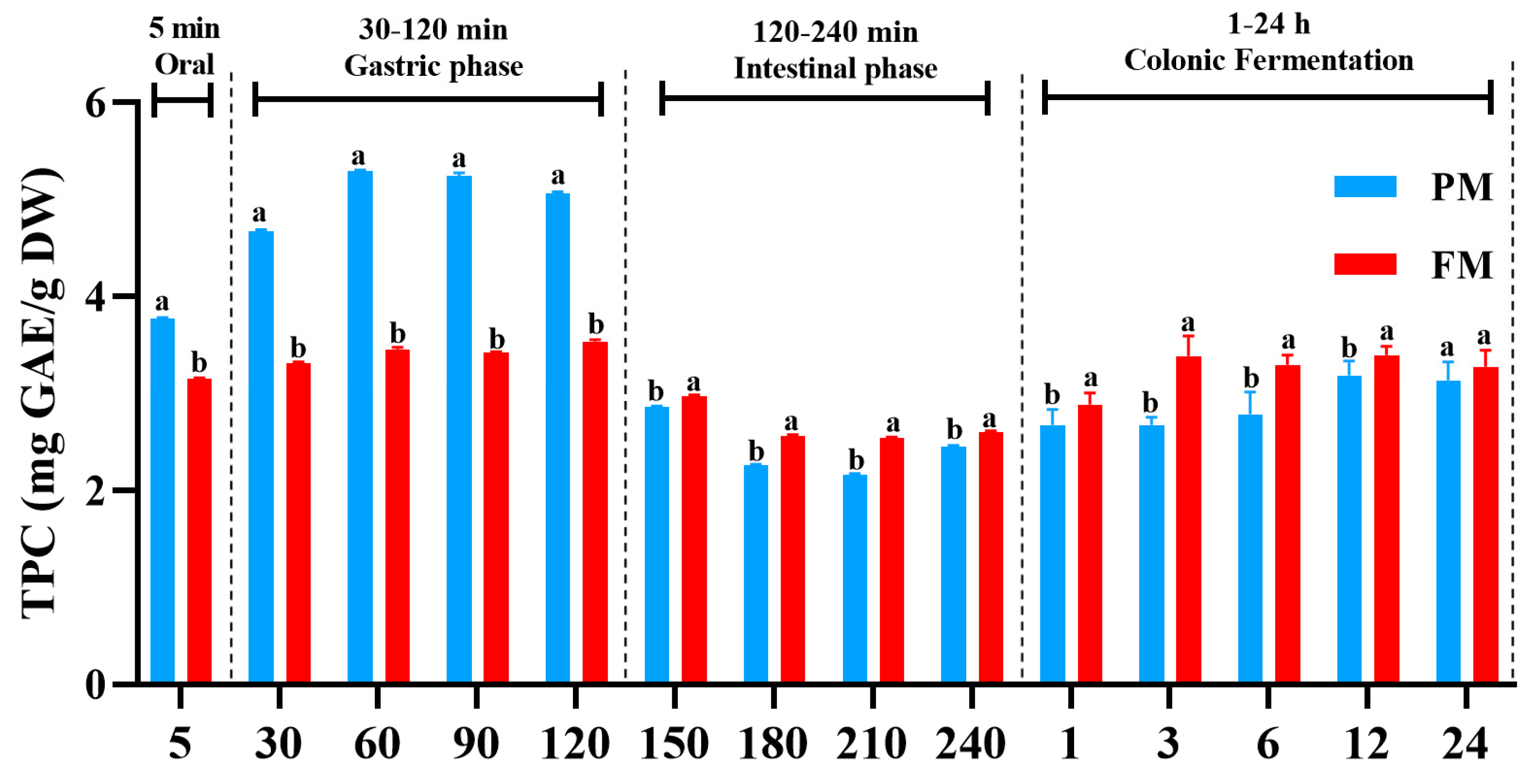
Figure 1. Total phenolic content (TPC) from PM and FM during different digestion stages and colonic fermentation. PM, Prunus mume; FM, Fructus mume. Data represent mean values ± SD (n = 3). For each time point, different letters indicate significant differences (p < 0.05).
Foods 14 04067 g001
Figure 2. Effect of simulated digestion on DPPH (A) and ABTS (B) radical scavenging activity in vitro gastrointestinal digestion and colonic fermentation. PM, Prunus mume; FM, Fructus mume. Data represent mean values ± SD (n = 3). For each time point, different letters indicate significant differences (p < 0.05).
Foods 14 04067 g002
Figure 3. Overall structural changes in gut microbiota after 24 h of colonic fermentation in Prunus mume (PM) group, Fructus mume (FM) group and negative control (NC; only feces fermentation) group. Shannon index (A), Chao1 index (B) and Simpson index (C) were used as estimators of alpha diversity of gut microbiota. Coverage index (D) was used to verify reasonability of sequencing data. Principal coordinate analysis (PCoA) (E) was based on relative abundance of OTUs of all samples. Means with no letter (a, b) in common are significantly different (p < 0.05).
Foods 14 04067 g003
Figure 4. Comparisons of microbiota composition after 24 h of in vitro colonic fermentation in PM, FM and negative control (NC; only feces fermentation) groups. (A,B) Relative abundance of microbiota at phylum level. (C) Ratio of Firmicutes/Bacteroidetes (F/B). (D,E) Relative abundance of microbiota at genus level. Data represent mean values ± SD (n = 5). Bars with no letter (a, b, c) in common are significantly different (p < 0.05) among different groups.
Foods 14 04067 g004
Figure 5. Heatmap of correlation analysis between gut microbiota and individual phenolics. Red squares represent positive correlations, blue squares represent negative correlations and white squares represent no correlation. * p < 0.05, ** p < 0.01.
Foods 14 04067 g005
Disclaimer/Publisher’s Note: The statements, opinions and data contained in all publications are solely those of the individual author(s) and contributor(s) and not of MDPI and/or the editor(s). MDPI and/or the editor(s) disclaim responsibility for any injury to people or property resulting from any ideas, methods, instructions or products referred to in the content.