Abstract
Chronic stress is a key risk factor for depression and metabolic dysfunction, widely mediated through oxidative stress and inflammatory pathways. Natural products such as honey are increasingly investigated for their potential to attenuate stress-induced pathophysiological changes. This study evaluated the protective effects of Malaysian Acacia honey (AH) on glucose regulation, oxidative damage, histopathological alterations, and pro-inflammatory cytokine expression in stress-induced rats. Male Sprague–Dawley rats (n = 42) were subjected to chronic unpredictable mild stress and supplemented with AH, amitriptyline (AMT), or their combination for 28 days. Blood glucose levels, erythrocyte hemolysis, histological changes in liver and kidney, and expression of IL-1β and TGF-β1 in ileum, caecum, and hypothalamus were assessed. Data were reported as mean and standard error of mean (SEM) after three or more independent experiments had been conducted. The data were analyzed using a paired-t-test or a one-way or two-way analysis of variance (ANOVA) and considered significant if p < 0.05. Stress markedly elevated glucose levels (7.97 ± 0.20 mmol/L), increased hemolysis (14.30% ± 2.96), and induced hepatic (cytoplasmic vacuolation, 1.40 ± 0.25; cell lining absent, 1.20 ± 0.37) and renal lesions (dilated intertubular capillaries, 1.40 ± 0.51; inflammation, 2.20 ± 0.20), accompanied by upregulation of IL-1β (1.27-fold ± 0.20) and TGF-β1 (1.00-fold ± 0.08). Supplementation with AH significantly reduced hyperglycemia, inhibited hemolysis, ameliorated tissue damage, and downregulated pro-inflammatory cytokines. Combination therapy with AH and AMT produced the most significant improvements near to normal level, suggesting synergistic benefits. These findings highlight the therapeutic potential of AH as a natural adjunct in managing stress-related metabolic and inflammatory disturbances.
1. Introduction
Chronic stress is a major contributor to the onset and progression of depression and its associated comorbidities, including metabolic disorders, cardiovascular disease, and neurodegeneration. Stress exerts systemic effects primarily through activation of the hypothalamic–pituitary–adrenal (HPA) axis, resulting in hypercortisolemia, impaired glucose homeostasis, and immune dysregulation []. Long-term stress has been shown to increase oxidative stress, promote inflammatory responses, and trigger histopathological changes in vital organs such as the liver, kidney, and brain, thereby exacerbating disease risk [,].
Oxidative stress plays a central role in the pathophysiology of stress-induced disorders. Overproduction of reactive oxygen species (ROS) and impaired antioxidant defenses lead to lipid peroxidation, protein oxidation, and DNA damage []. Similarly, chronic stress promotes inflammation, often characterized by upregulation of pro-inflammatory cytokines such as interleukin-1 beta (IL-1β) and transforming growth factor beta 1 (TGF-β1) []. These cytokines have been implicated in both peripheral organ dysfunction and central nervous system alterations, contributing to behavioral and physiological manifestations of stress-related depression [,].
Current pharmacological treatments for depression, such as tricyclic antidepressants including amitriptyline (AMT), primarily target monoaminergic pathways []. While effective for mood regulation, these drugs often fail to fully address stress-induced oxidative and inflammatory processes and are associated with side effects. This gap highlights the need for adjunctive strategies that target broader pathophysiological mechanisms.
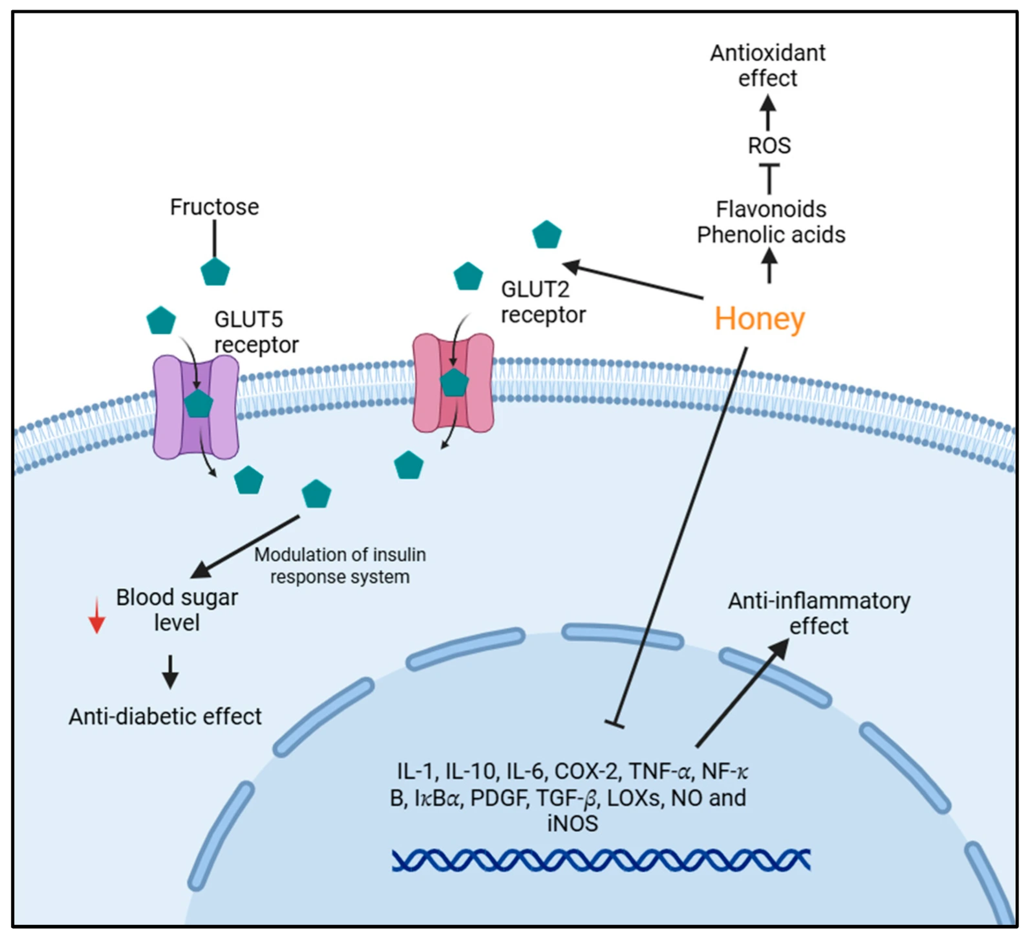
Honey, a natural substance produced by bees, is rich in phenolic compounds, flavonoids, and sugars (fructose, glucose, sucrose, maltose and others) that have strong antioxidants and anti-inflammatory properties (Figure 1). Several studies have reported the beneficial effects of honey in mitigating oxidative stress, improving glycemic control, and modulating immune responses [,,]. In animal models, honey supplementation has been shown to lower fasting blood glucose, enhance antioxidant enzyme activities, and protect against oxidative tissue injury [,]. Clinical studies further suggest that honey improves antioxidant biomarkers and reduces fasting blood sugar in humans [].
Malaysian Acacia honey (AH) is a unifloral honey predominantly derived from the nectar of Acacia mangium or Robinia pseudoacacia trees, known for its distinctive chemical composition as well as high antioxidant capacity due to its flavonoid and phenolic content []. A physicochemical analysis revealed that AH contains approximately 39.9% fructose, 30.7% glucose, 4.7% sucrose, and 3.1% maltose, resulting in a fructose-to-glucose ratio of 1.3, which contributes to its relatively low glycemic index and slower crystallization rate []. Fructose intake from honey has been reported to attenuate hyperglycemia in various experimental and clinical models, including rodents, healthy individuals, and patients with diabetes []. This effect is primarily attributed to delayed gastric emptying, which reduces gastric motility and slows the rate of intestinal glucose absorption, thereby improving postprandial glycemic response [].
Several studies have investigated the effects of AH intake on blood glucose regulation. For example, Sukri et al. [] demonstrated that consumption of AH before and during exercise did not significantly alter plasma glucose or insulin levels compared to a commercial sports drink, but it did enhance antioxidant status. Similarly, Ahmad et al. [] reported that a sodium-enriched AH drink consumed during rehydration after exercise maintained stable blood glucose levels and improved plasma volume recovery. Previous research demonstrated that AH supplementation improves oxidative balance, reduces lipid peroxidation, and enhances immune function [,]. However, its effects on stress-induced metabolic, inflammatory, and histopathological changes remain underexplored.
Figure 1.
Summary of mechanisms of honey in modulating glucose metabolism and inflammation as reported by Ahmed et al. []. Honey contains bioactive compounds such as flavonoids and phenolic acids that exert antioxidant effects by reducing reactive oxygen species (ROS). These compounds also contribute to anti-inflammatory activity by downregulating pro-inflammatory mediators (IL-1, IL-10, IL-6, COX-2, TNF-α, NF-κB, IκBα, PDGF, TGF-β, LOXs, NO, and iNOS). Fructose in honey is transported into cells via GLUT5 and GLUT2 receptors, influencing insulin response and lowering blood glucose levels (red arrow), which may lead to anti-diabetic effects.
The chronic unpredictable mild stress (CUMS) model employed in this study is a widely accepted paradigm for inducing depression-like behaviors in rodents, including anhedonia, behavioral despair, and neuroinflammatory changes, thereby closely mimicking the core symptoms and pathophysiology of human major depressive disorder (MDD) []. The model demonstrates strong face, construct, and predictive validity, making it a valuable tool for investigating the biological mechanisms underlying stress-induced depression and for evaluating potential therapeutic interventions []. Sprague-Dawley (SD) rats were selected for this study due to their well-characterized physiology, docile temperament, and widespread use in neurobehavioral and stress-related research, particularly in modeling depressive disorders [].
In this study, we investigated the potential of AH to modulate stress-induced alterations in glucose regulation, oxidative damage, histopathology of the liver and kidney, and expression of IL-1β and TGF-β1 in vital tissues. We hypothesized that AH supplementation would attenuate hyperglycemia, protect erythrocytes against oxidative hemolysis, ameliorate organ damage, and downregulate inflammatory cytokine expression. Furthermore, we examined whether combining AH with AMT would produce additive or synergistic effects. Our findings aim to expand current knowledge on the therapeutic role of AH as a natural adjunctive intervention for stress-related metabolic and inflammatory dysfunction.
2. Methodology
2.1. Chemicals
Chemicals were obtained from Sigma-Aldrich Company Ltd., Gillingham, UK, unless otherwise stated.
2.2. Honey Collection
AH was collected in Kluang, Johor, Malaysia, in July 2017, corresponding to the dry season when Acacia species are in full bloom and widely maintained by local beekeepers. The honey was freshly obtained from local apiaries within one to three days after harvesting and stored at 4 °C until further use. The honey was produced by the domesticated honeybee species Apis mellifera. Our study shows that AH contained Robinia pseudoacacia as predominant pollen []. The AH sample was not commercially purchased but collected directly from beekeepers to ensure authenticity and freshness.
2.3. Housing and Husbandry of Rats
The in vivo study was conducted with slight modifications based on the protocol described by Abidin et al. []. All animals used in this study were male Sprague-Dawley (SD) rats bred in-house at the Laboratory Animal Facility and Management (LAFAM), Universiti Teknologi MARA (UiTM). Male rats aged 8 weeks (150–170 g) were used to minimize hormonal variability associated with the estrous cycle in females, which can influence stress responses and behavioral outcomes []. The rats were healthy, immunocompetent, and non-genetically modified, with no prior exposure to experimental procedures before the study.
All experimental procedures were approved by the Animal Research and Ethics Committee of Universiti Teknologi MARA (UiTM), Puncak Alam Campus, Faculty of Pharmacy [UiTM CARE:237/2/2018 (6 April 2018)]. Prior to the experiment, the rats underwent a 7-day acclimatization period (3rd-floor LAFAM). Before the experiments, rats were individually caged and fed rat chow and reverse osmosis water (RO) ad libitum while kept under standard laboratory conditions (24 °C ± 1, 45% ± 15 relative humidity and 12 h light/dark cycle). To minimize pain, suffering, and distress, all animals were housed in standard cages with environmental enrichment, including nesting materials and tunnels (tissue roll). Gentle handling techniques were used throughout the study.
2.4. Stress-Depression Model
The sample size for the animal study was determined using G*Power software (version 3.1.9.2) based on a one-way ANOVA (fixed effects, omnibus test). The parameters used were an effect size (f) of 0.8329931, significance level (α) of 0.05, statistical power (1–β) of 0.95, and seven experimental groups. Based on these inputs, the total required sample size was calculated to be 42 animals, ensuring an actual power of 0.9767. All animals were retained throughout the experimental procedures and included in the final data analysis.
Forty-two male Sprague-Dawley rats were randomly assigned into seven groups with six rats in each group: (1) normal control (NC), (2) AH (2.25 mg/kg), (3) amitriptyline (AMT, 0.1 mg/kg), (4) stress-induced (S), (5) stress-induced with AH (S + AH), (6) stress-induced with AMT (S + AMT, positive control), and (7) stress-induced with a combination of AH and AMT (S + AH + AMT). All doses were adapted from our preliminary studies by Abidin [] and Samat []. AH and AMT were freshly dissolved in RO ad libitum and administered to the rats.
The experimental timeline was structured to evaluate the glucose level in rats at day 0 and day 30. From day 1 to day 28, rats received daily oral gavage of the respective treatments (AH, AMT, or combination) while being concurrently exposed to CUMS. The CUMS protocol included a randomized schedule of stressors such as overnight illumination, cage tilting, forced swimming, white noise, damp bedding, and restricted movement and rest (Table 1). On day 29, the rats undergone open field test. On day 30, all animals were euthanized via intraperitoneal injection of sodium pentobarbital overdose. Blood samples were collected through cardiac puncture for analysis of blood glucose levels and erythrocyte hemolysis.
Table 1.
Chronic unpredictable mild stress procedure.
2.5. Hemolysis Assay
The isolation of erythrocytes for hemolysis assay using hydrogen peroxide (H2O2) was conducted per Okoko and Ere [] with slight modification. Briefly, blood was collected into EDTA tubes and centrifuged at 4000 rpm for 10 min at 4 °C. The plasma was discarded, and the erythrocytes were washed three times with phosphate-buffered saline (PBS, 0.2 M, pH 7.4). The resulting packed cells were either stored at −80 °C or immediately used to prepare a 5% erythrocyte suspension (ES) by mixing 50 μL of packed cells with 950 μL of PBS. For the assay, 1 mL of ES was mixed with 1 mL of PBS, with or without 0.5 mL of 10 mM H2O2, and incubated at 37 °C for 3 h. After incubation, 6 mL of PBS was added to each tube, followed by centrifugation at 2000× g for 10 min at 4 °C. The absorbance of the mixture was analyzed at 540 nm using a SmartSpecTM Plus spectrophotometer (Bio-Rad, Hercules, CA, USA).
2.6. Histopathology
All animals underwent gross necropsy, followed by histopathological examination of the kidney and liver tissues. Briefly, fixed liver and kidney tissues (10% buffered formalin for 24 hrs) were embedded in paraffin, sectioned (Leica, Wetzler, Germany), dried, and stained with hematoxylin-eosin (H & E) using an automatic tissue stainer (Leica, Wetzler, Germany). After overnight incubation, the stained slides were mounted using DPX (Sigma-Aldrich, St. Louis, MO, USA) and viewed under a light microscope (Leica, Wetzler, Germany) using randomization method. Changes observed in liver and kidney tissue sections were graded using a semiquantitative lesion scoring system. Scores were assigned as follows: 0 for no observable changes, 1 for mild changes, 2 for moderate changes, and 3 for severe histopathological alterations. For each group, the mean lesion score was calculated from six animals, with one representative tissue section analysed per organ per animal. All assessments were performed in a blinded manner by an experienced histopathologist to ensure consistency and minimize bias.
2.7. Quantitative Real-Time PCR
Total RNA was extracted from approximately 30 mg of tissue samples (ileum, caecum and hypothalamus) using the SV Total RNA Isolation System (Promega, Madison, WI, USA, Cat # Z3105). RNA purity and quantity were measured by NanoDrop Lite Spectrophotometer (Thermo Scientific, Waltham, MA, USA) (A260/280, Eppendorf; 1.8–2.2). cDNA (2 µL) was synthesized with a GoScriptTM Reverse Transcription Mix, Oligo (dT) (Promega, USA, Cat # A2791). Quantitative real-time PCR was performed using a GoTaq® qPCR Master Mix (Promega, USA, Cat # A6001). RT-qPCR assays were run on a Rotor Gen Q 5 plex HRM platform (Qiagen, Hilden, Germany) in 36-tube over 40 cycles of 95 °C for 15 s and 60 °C for 1 min by holding temperature of 95 °C for 2 min before cycle starts. Rotor gene Q series software v 2.3.5 was used to acquire the threshold cycle (Ct) values and normalized to those of GAPDH. Fold differences were determined using the 2−ΔΔCT. The primer sequences utilized in this study are presented in Table 2.
Table 2.
Primer Sequences.
2.8. Statistical Analysis
Data were presented as mean ± standard error of the mean (SEM) from at least three independent experiments. Data normality was assessed using the Shapiro–Wilk test. When assumptions of normality were violated, appropriate non-parametric tests were applied. However, as all data were normally distributed, parametric test were used throughout the study. Blood glucose levels were analyzed using both paired-t-test and one-way analysis of variance (ANOVA), while lesion scores were analyzed using two-way ANOVA. Data for hemolysis and gene expression were analyzed using one-way ANOVA with Bonferroni post hoc test. Differences between group means were considered statistically significant at p < 0.05. All statistical analyses were performed using GraphPad Prism version 8.0 (GraphPad Software, San Diego, CA, USA).
3. Results and Discussion
Stress-related depression is a multifactorial disorder involving neuroendocrine and metabolic dysregulation. The chronic unpredictable mild stress (CUMS) model is widely used to mimic human depression. The successful establishment of the CUMS model is typically validated through behavioral and biochemical markers that reflect depressive-like states. In our preliminary study [,], two core indicators were employed: the sucrose preference test (SPT) and serum cortisol levels. The SPT is a widely recognized behavioral assay for assessing anhedonia, a key symptom of depression, where reduced sucrose consumption indicates diminished reward sensitivity [,]. Concurrently, elevated cortisol levels serve as a biochemical marker of HPA axis hyperactivity, which is commonly observed in stress-induced depression []. These indicators are consistent with established protocols for validating CUMS models, which often include behavioral tests such as the open field test, alongside biochemical assays.
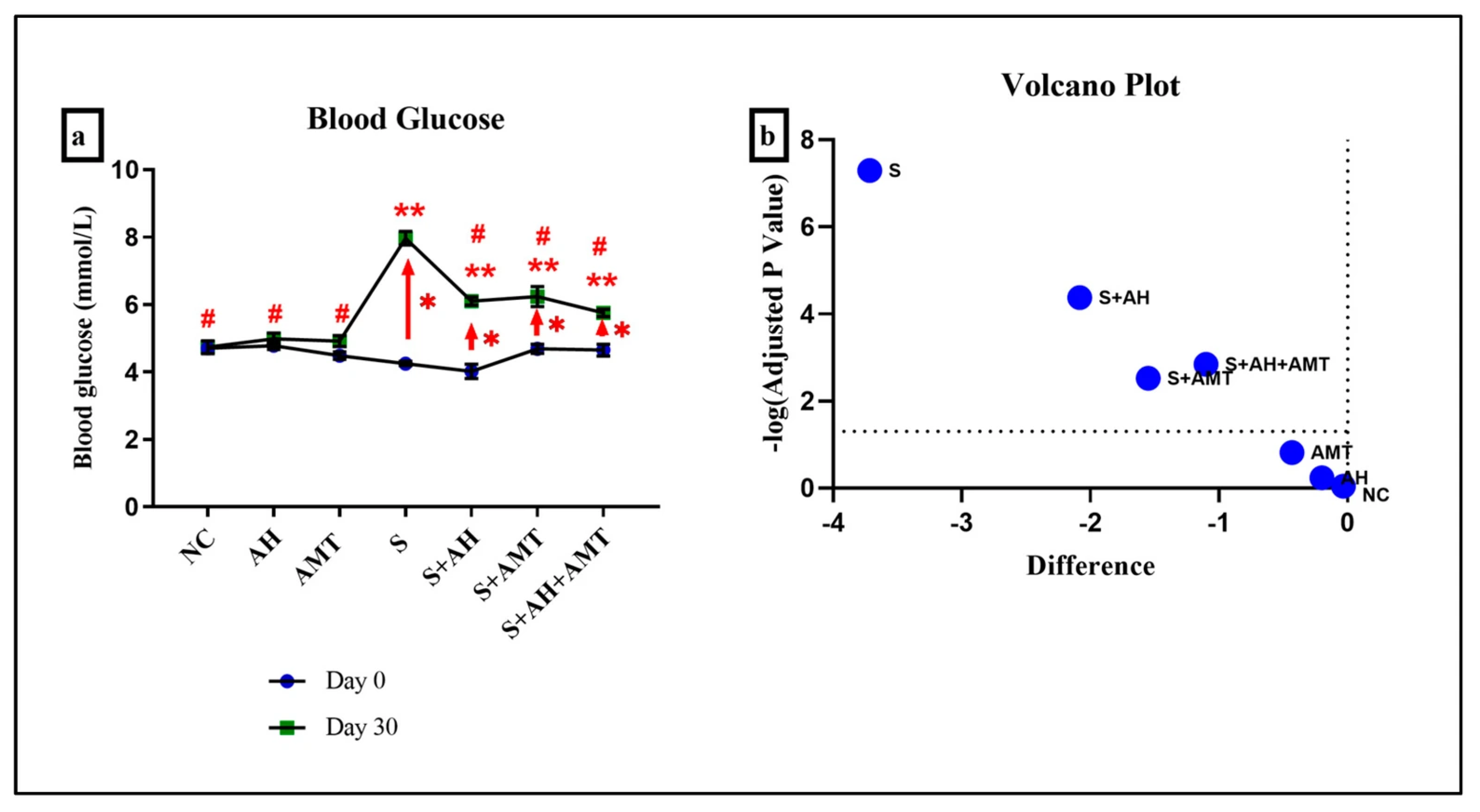
CUMS activates the HPA axis, leading to elevated blood glucose levels due to impaired glucose homeostasis and stress-induced hyperglycemia []. Figure 2 illustrates the blood glucose levels in rats on day 0 and day 30 across all experimental groups. Since AH contains natural sugars that could potentially influence blood glucose levels, this parameter was monitored to evaluate its metabolic impact. The data were collected to determine whether AH supplementation, particularly under stress conditions, contributes to hyperglycemia or exhibits a regulatory effect on glucose homeostasis.
Figure 2.
(a) Blood glucose level in rats were measured on day 0 and day 30 in all groups. Comparison was performed using Paired-t-test. (b) The results were visualized in a volcano plot. The higher plot values observed in the S group compared to other groups indicate the most significant changes occured in the S group. * indicates statistical significance (p < 0.05) comparison between day 0 and day 30 within the same group (red arrow, Paired-t-test). ** indicates statistical significance (p < 0.05) comparison with NC group on Day 30 (one-way ANOVA), # indicates statistical significance (p < 0.05) compared with S group comparison using one-way ANOVA, n = 6 independent observations.
Before the experiment (day 0), no significant differences in blood glucose were observed among the group (p > 0.05), indicating comparable baseline metabolic states. Interestingly, on day 30, rats supplemented with AH alone (AH group) showed no significant change in blood levels (p = 0.58), suggesting that AH does not induce hyperglycemia and may be metabolically safe. This finding is consistent with previous research showing that honey, particularly Acacia honey, has a low glycemic index and may improve glycemic control due to its high fructose content and antioxidant properties [,].
In contrast, the stress-induced group (S) demonstrated a significant elevation in blood glucose at the end of experiment (7.97 ± 0.20 mmol/L) compared with day 0 (4.25 mmol/L ± 0.07, p = 0.00) (Figure 2, red arrow). This confirms that CUMS model disrupts glucose homeostasis, leading to a hyperglycaemic state. The rise in blood glucose is consistent with the well-documented role of stress in activating the HPA axis, resulting in increased glucocorticoid secretion, enhanced hepatic gluconeogenesis, and reduced insulin sensitivity []. These metabolic alterations explain the significant difference between pre- and post-supplementation values observed in the stress group.
Interestingly, on day 30, rats supplemented with Malaysian AH (S + AH) showed a reduction in blood glucose levels to 6.10 mmol/L ± 0.12, while AMT treatment (S + AMT) produced a comparable reduction to 6.23 mmol/L ± 0.30, p = 0.00. Both interventions significantly mitigated the hyperglycaemic response associated with stress exposure. Importantly, the combined supplementation of AH and AMT (S + AH + AMT) resulted in the greatest reduction (5.75 mmol/L ± 0.11, p = 0.00). The values approaching closer to those of non-stressed controls. This suggests a potential synergistic effect between natural supplementation and pharmacological treatment in improving metabolic outcomes under stress conditions.
The glucose-lowering effect of AH may be attributed to its high content of phenolic compounds and flavonoids, which are known to enhance antioxidant defenses, protect pancreatic β-cell integrity, and improve peripheral insulin sensitivity [,,]. Previous in vivo studies have shown that honey supplementation attenuates oxidative stress and hyperglycaemia in diabetic models, supporting its potential therapeutic role beyond basic nutrition [,]. In addition, a clinical trial in humans demonstrated that honey consumption stabilized fasting blood sugar and enhanced antioxidant status, further reinforcing its beneficial metabolic properties [].
On the other hand, AMT, a tricyclic antidepressant, exerts its effect mainly through modulation of serotonergic and noradrenergic neurotransmission. Its impact on glucose regulation is likely indirect, mediated by a reduction in the neuroendocrine burden of chronic stress rather than direct action on glucose metabolism []. However, when combined with AH, the overall improvement in blood glucose suggests complementary pathways. Honey may reduce oxidative stress and inflammation [,], while AMT alleviates stress-related neurochemical changes via TrkA signaling pathways [] and enhancing glycaemic control [,].
Previous studies have demonstrated a link between blood glucose dysregulation and hemolysis []. Stress-induced hyperglycemia elevates circulating glucose, facilitating erythrocyte glycation and oxidative stress. These processes compromise membrane integrity, reduce cellular deformability, and promote eryptosis, ultimately leading to hemolysis under stress conditions [].
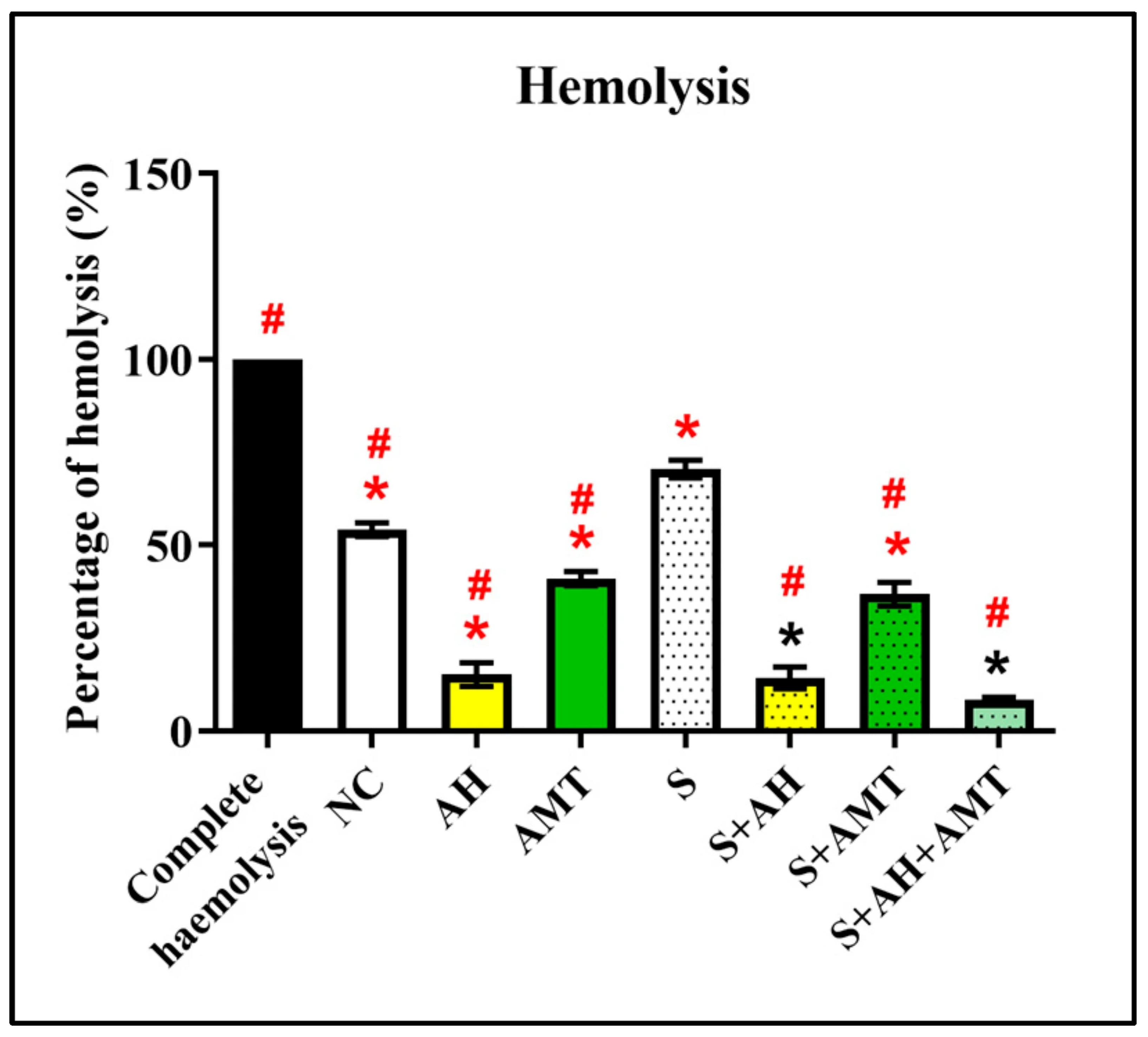
An in vitro blood hemolysis assay was conducted at the end of the in vivo study to evaluate the protective effect of AH against oxidative stress-induced hemolysis (Figure 3). All treatment groups [NC (81.00% ± 4.64), AH (15.15% ± 3.21), AMT (40.95% ± 1.91), S (70.48% ± 2.41), S + AH (14.30% ± 2.96), S + AMT (36.80% ± 3.18), and S + AH + AMT (8.45% ± 0.62)] showed a significant reduction in hemolysis compared to the complete hemolysis control group (100% ± 0.00, p = 0.00). Particularly, the S + AH, S + AMT, and S + AH + AMT groups exhibited significantly lower hemolysis compared to the S group (p = 0.00), indicating that AH, alone or in combination with AMT, effectively mitigates H2O2-induced erythrocyte damage under stress conditions. This is due to high phenolic and flavonoid content in AH, as demonstrated in our previous study [].
Figure 3.
Percentage of blood hemolysis induced by H2O2 on day 30. Acacia honey protects the blood against H2O2-induced lysis, indicated by a low percentage of hemolysis in the S + AH group compared to the S group. * indicates statistical significance (p < 0.05) compared with the NC group. # indicates statistical significance (p < 0.05) compared with S group comparison using one-way ANOVA, with Bonferroni post hoc test, n = 6 independent observations.
The current study was consistent with the previous finding, in which healthy rats that supplemented with AH (AH group) were found to exhibit low blood hemolysis []. As the pattern was similar in the rats-induced stress and supplemented with AH (S + AH group), these further confirm the protective effect of AH was by inhibiting blood hemolysis-induced with H2O2. The protective effect of AH was due to the binding of the flavonoids to the blood cell membrane, preventing H2O2 from attacking the erythrocyte membrane []. Particularly, AH was demonstrated to have a high scavenging capacity []. A clinical trial involving participants supplemented with honey for two weeks reported increased levels of antioxidant biomarkers, including blood vitamin C, beta-carotene, and glutathione reductase, as well as improvements in serum iron, hematological indices, and trace elements [,].
Hemolysis during stress releases free hemoglobin and heme, overwhelming scavenger systems and promoting systemic oxidative stress []. Excess iron from hemolysis catalyzes reactive oxygen species formation, aggravating hepatocellular injury []. Histopathological findings, including vacuolar degeneration and inflammatory infiltration, reflect this oxidative burden and link erythrocyte destruction to liver pathology under stress conditions [].
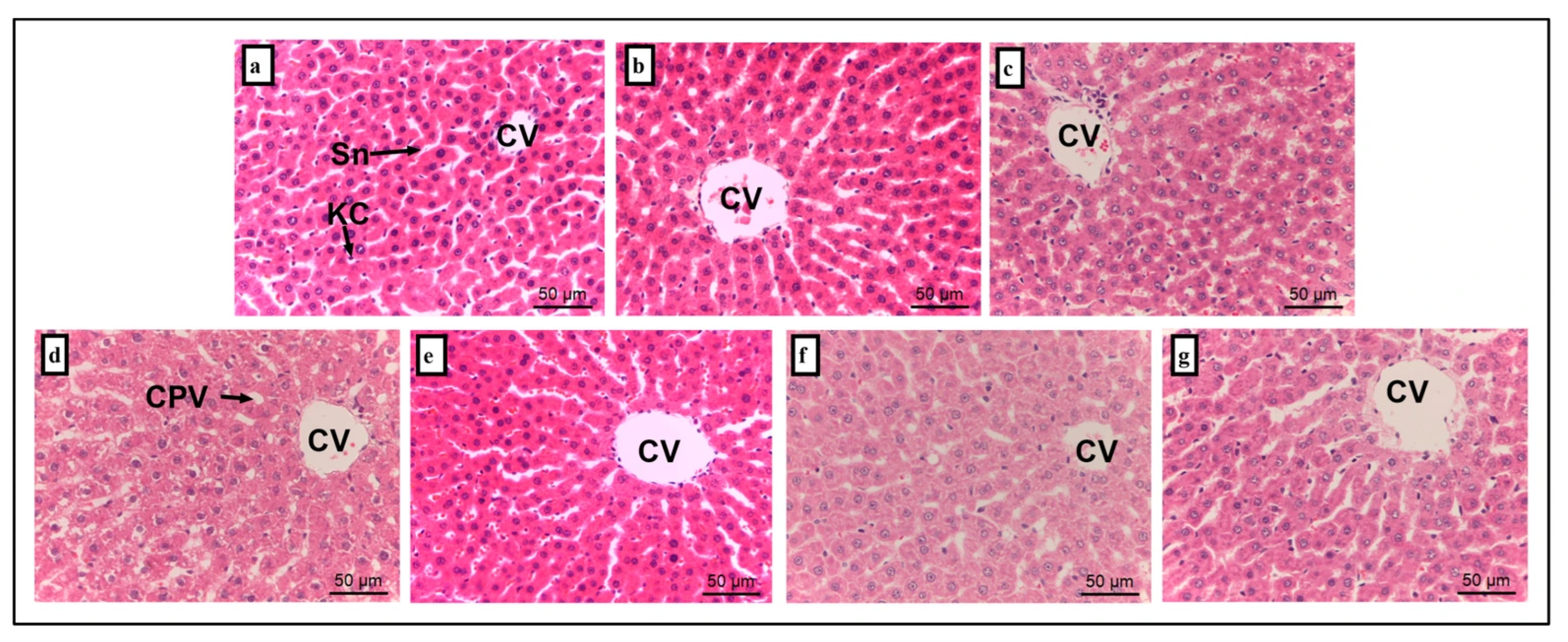
Histopathological changes in the liver and kidney tissues are summarized in Figure 4 and Figure 5, with corresponding lesion scores presented in Table 3. Liver sections from the NC (Figure 4a) and AH group (Figure 4b) exhibited normal histoarchitecture, including intact central vein (CV), sinusoidal (Sn), and Kupffer cell (KC) (lession score, 0.00 ± 0.00). In contrast, liver tissues from the AMT (Figure 4c) and S group (Figure 4d) showed cytoplasmic vacuolation with the lesion score of 1.20 ± 0.37 and 1.40 ± 0.25 when compared to the NC group (p = 0.00), respectively. Notably, the S + AH (Figure 4e; 0.40 ± 0.25) and S + AH + AMT (Figure 4g; 0.00 ± 0.00) groups demonstrated no significant histological alterations compared to the NC group (p = 0.99). A significant reduction in cytoplasmic vacuolation was observed in both S + AH and S + AH + AMT groups relative to the S group, indicating the hepatoprotective effect of AH, particularly when combined with AMT.
Figure 4.
Histological sections of liver tissue. Representative liver sections from each experimental group (n = 6): (a) normal control, (b) Acacia honey, (c) amitriptyline, (d) stress-induced, (e) stress treated with Acacia honey, (f) stress treated with amitriptyline, and (g) stress treated with a combination of Acacia honey and amitriptyline. CV, central vein; Sn, sinusoidal; KC, Kupffer cell; CPV, cytoplasmic vacuolation. Scale bar: 50 µm.
Figure 5.
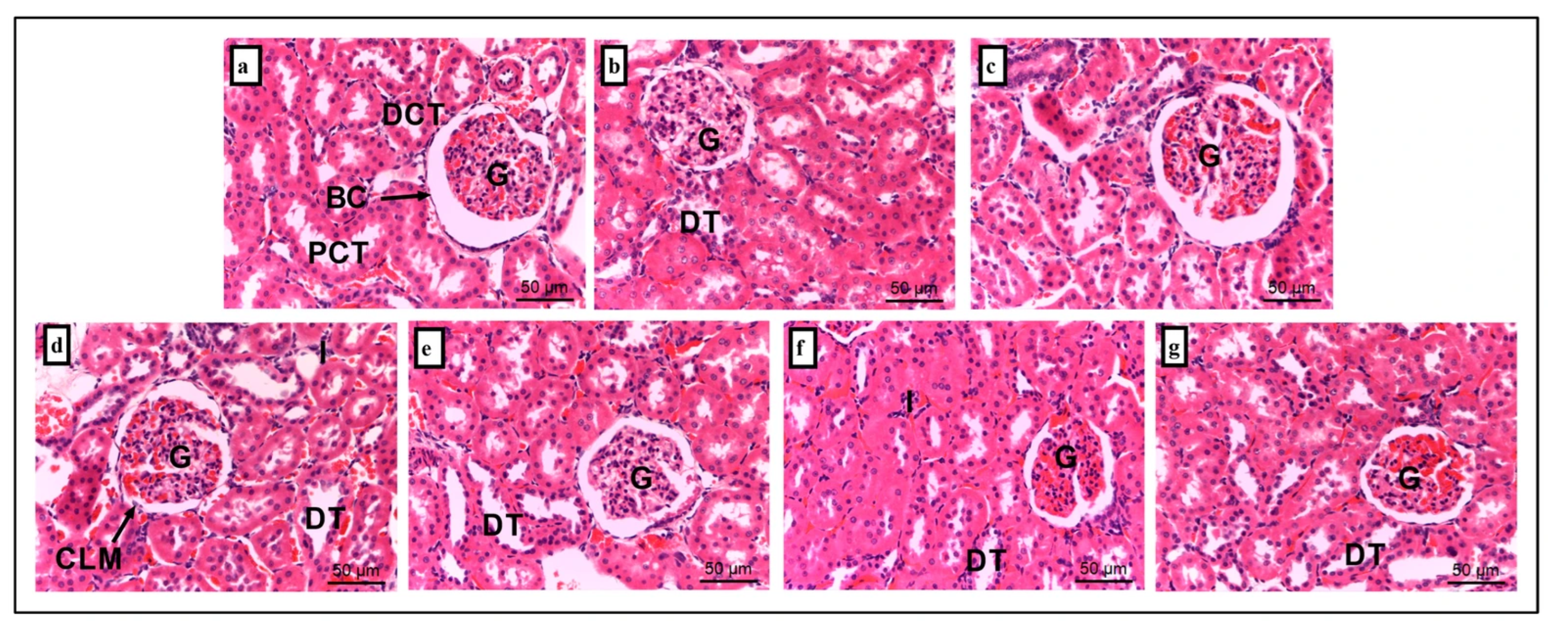
Histological section of kidney tissue. It was consists of groups (a) NC, (b) acacia honey, (c) amitriptyline, (d) stress-induced, (e) stress treated with Acacia honey, (f) stress treated with amitriptyline, and (g) stress treated with a combination of Acacia honey and amitriptyline. G, glomerulus; BC, bowman’s capsule; PCT, proximal convoluted tubules; DCT, distal convoluted tubules; CLM, cell lining missing; DT, dilated intertubular capillaries; I, inflammation. Scale bar: 50 µm. n = 6 independent observations.
Table 3.
Lesion scores of kidneys and liver tissues.
Histological liver examination of stress rats reveals prominent cytoplasmic vacuolation. A previous study has reported that an increase in serum AST [], ALT [] and GGT levels reflects the loss of structural integrity of the liver [], which corroborated the current findings. AST is released into the bloodstream due to hepatocellular degeneration and necrotic changes leading to elevated serum AST levels []. Although mild cytoplasmic vacuolation was observed in the liver tissues of stress-induced rats, no signs of hepatocellular degeneration or necrosis were detected. Cytoplasmic vacuolization in mammalian cells can be transient or irreversible. The transient vacuolization can only be observed during exposure to stressors and may be reversibly affects the cell cycle and migration. Meanwhile, cytoplasmic vacuolation was not observed in the stress rats supplemented with AH. These findings suggest AH may have protective effects towards stress-induced hepatocellular damage, likely through its antioxidant and anti-inflammatory properties.
Similarly, free hemoglobin and iron deposition in renal tissue induce oxidative stress and tubular damage, contributing to acute kidney injury []. Persistent hemolysis can further promote renal fibrosis and chronic kidney disease through iron-mediated oxidative pathways []. Subsequently, kidney sections were examined to assess the structural effects of stress on renal tissue. Histology of kidney tissue for the NC group (Figure 5a) reveals intact renal architecture, including well-preserved glomerulus (G), Bowman’s capsule (BC), proximal convoluted tubules (PCT), and distal convoluted tubules (DCT). However, the S group (Figure 5d) exhibited significant pathological changes, including decreased of tubular cell lining (1.200 ± 0.374), dilated intertubular capillaries (1.400 ± 0.510), and inflammation (2.200 ± 0.200) when compared to the NC group (0.000 ± 0.000, p = 0.00). Supplementation with AH (S + AH group; Figure 5e), AMT (S + AMT group; Figure 5f), and their combination (S + AH + AMT group; Figure 5g) significantly decreased tubular cell lining and inflammation (Table 3) when compared to S group only (p = 0.00).
In particular, the histology of kidney tissue of the stress rats demonstrated dilated intertubular capillaries, inflammation and absence of cell lining. Supplementation with AH in stress rats significantly reduced abnormal morphology characteristics. The presence of abnormal structure in stress rats was associated with the overproduction of reactive oxygen species [], leading to activation of IL-1 [], IL-6 [], COX-2 [] and TNF-α []. The protective effect of honey on renal tubular has been shown previously against cadmium through honey’s ability to reduce lipid peroxidation and increase tissue levels of glutathione and glutathione peroxidase activity []. These findings suggest that AH may have protective effects against stress-induced hepatocellular and kidney damage. The protective effect was observed to be further enhanced with the co-supplementation with both AH and AMT.
Interleukin-1β (IL-1β) plays a critical role in mediating immunological, behavioral, and neuroendocrine responses to infection and inflammation. It is also implicated in the activation of the HPA axis during stress. Therefore, this study investigated IL-1β gene expression on three HPA axis-related organs: the ileum, caecum and hypothalamus. Real-time PCR was employed to assess the transcriptional regulation of pro-inflammatory cytokines, IL-1β in the ileum, caecum, and hypothalamus.
In the ileum (Figure 6a), IL-1β expression is significantly upregulated in the S (2.79-fold ± 0.19, p = 0.01) and S + AH + AMT groups (2.55-fold ± 0.21, p = 0.03) compared to the NC group (1.00-fold ± 0.00). No significant changes were observed in the AH (1.64-fold ± 0.29, p = 0.99), AMT (0.79-fold ± 0.48, p = 0.99), S + AH (1.19.85-fold ± 0.33, p = 0.99) and S + AMT groups (0.49-fold ± 0.10, p = 0.99). In contrast, treating stress rats with AH (S + AH, p = 0.02) and AMT (S + AMT, p = 0.00) downregulated the IL-1β gene expression.
Figure 6.
IL-1β gene expressions in (a) ileum, (b) caecum, and (c) hypothalamus. *, shows the comparison between NC group and other groups indicates statistical significance (p < 0.05); #, the comparison between stress rats (S) with other groups indicates statistical significance (p < 0.05), comparison using one-way ANOVA, Bonferroni post hoc test, n = 3 independent observations.
In contrast, no significant changes were observed in the caecum sample (Figure 6b) for group of AH (1.06-fold ± 0.09, p = 1.00), AMT (1.21-fold ± 0.08, p = 0.54), S (1.21-fold ± 0.10, p = 0.54), S + AH (1.09-fold ± 0.18, p = 0.97) S + AMT (1.19-fold ± 0.09, p = 0.60) and S + AH + AMT (1.21-fold ± 0.07, p = 0.51) compared to NC group (1.00-fold ± 0.00). IL-1β was not modulated in the caecum during stress-induced depression in rats, possibly due to regional immune tolerance and lower microbial stimulation compared to the ileum. The ileum includes higher microbial density and immune cell activity, as previously reported in TH17 cell expansion that enhance IL-1β signalling via microbiota-derived cues and NF-κB activation [].
In this study, IL-1β was shown to be upregulated in the stress rats’ ileum, which is in line with other studies [,]. Intestine tissues from mice with colitis were reported to have higher amounts of IL-1β mRNA, which inhibits the development of intestinal inflammation by reducing occludin expression by enterocytes and therefore increasing tight junction permeability []. A similar report was demonstrated by Uchakin et al. [] during the academic examination that increases IFN-γ, IL-2, IL-1α and IL-1β in the serum of stress medical student and the expression reduced after 2 days post examination.
In the hypothalamus, IL-1β gene expression was significantly upregulated in the AMT (3.71-fold ± 0.18, p = 0.00), S (2.95-fold ± 0.48, p = 0.00) and S + AH + AMT (2.73-fold ± 0.41, p = 0.01) groups compared to the NC group (1.00-fold ± 0.00). Contrarily, treatment with AH (S + AH; 0.70-fold ± 0.04, p = 0.00) and AMT (S + AMT; 1.25-fold ± 0.21, p = 0.01) significantly downregulated IL-1β expression compared to the S group (Figure 6c).
In this study, IL-1β was shown to be upregulated in the stress rats’ hypothalamus, which is in line with other studies [,]. In addition, IL-1β mRNA levels in the hypothalamus in intact rats were increased by LPS injection []. c-FOS is an immediate early gene commonly used as a marker of neuronal activation in response to various stimuli. During stress, the increment of IL-1β during stress might increase action potential firing in vagal afferents, induced C-FOS expression in the nucleus tractus solitarii, the central projection area of vagal afferent nerve fibres the medullary brainstem, and sensitized vagal afferent pathways to gastric acid [].
The increase in IL-1β expression observed during stress was significantly downregulated following supplementation with AH, AMT or their combination. This suggests that both AH and AMT exert anti-inflammatory effects, potentially through modulation of the gut–brain axis. Honey has been shown to influence brain function via the gut–brain axis by promoting the growth of beneficial gut bacteria, such as Bifidobacteria, which in turn reduce oxidative stress and inflammation in the gut. These bacteria produce short-chain fatty acids [,], such as butyrate and propionate, which have been reported to enhance intestinal barrier integrity, modulate immune responses, and influence neuroinflammation through signaling pathways that affect the central nervous system []. In particular, these events result in appropriate myelination of neurons, blood–brain barrier repair, decreased production of inflammatory cytokines (IL-1β, TNF-α, and BDNF), and mediated restoration of synaptic plasticity in the brain increase in memory [].
IL-1β and TGF-β1 exhibit a synergistic role in stress-induced pathology []. IL-1β amplifies inflammation and oxidative stress, while TGF-β1 drives fibrotic remodeling []. Their combined activation promotes epithelial–mesenchymal transition and extracellular matrix deposition, accelerating organ dysfunction under chronic stress conditions [,].
Based on Figure 7, the expression of TGF-β1 gene shows distinct patterns across the ileum, caecum, and hypothalamus. TGF-β1 expression in the ileum (Figure 7a) was significantly downregulated in AH group (0.11-fold ± 0.02), followed by S + AH + AMT (0.14-fold ± 0.01) and AMT group (0.00-fold ± 0.00) compared to the NC group (1.00-fold ± 0.00, p = 0.00). Stress alone (S group; 3.42-fold ± 0.17) significantly upregulated the TGF-β1 gene expression (3.43-fold ± 0.17), while S + AH (1.57-fold ± 0.23) and S + AMT (0.77-fold ± 0.06) showed reduction in TGF-β1 expression (p = 0.00).
Figure 7.
TGF-β1 gene expressions in ileum (a), caecum (b), and hypothalamus (c). *, shows the comparison between NC group and other groups indicates statistical significance (p < 0.05); #, the comparison between stress rats (S) group with other groups indicates statistical significance (p < 0.05), comparison using one-way ANOVA, Bonferroni post hoc test, n = 3 independent observations.
The significant downregulation of TGF-β1 in the ileum following AH supplementation suggests honey anti-inflammatory potential in stress-induced depression. TGF-β1 is known to mediate mucosal immunity and fibrosis in the gut, and its overexpression under stress may exacerbate intestinal inflammation. AH appears to restore immune homeostasis, possibly via modulation of Smad-dependent pathways [,]. The combination of AH and AMT also reduced TGF-β1, indicating synergistic effects. These findings align with studies showing that natural antioxidants can suppress TGF-β1 signaling and improve gut barrier integrity [].
In the caecum (Figure 7b), no significant changes were observed in TGF-β1 expression in group of AH (1.34-fold ± 0.63, p = 0.96), AMT (1.30-fold ± 0.30, p = 0.98), S (0.84-fold ± 0.44, p = 1.00), S + AH (1.13-fold ± 0.39, p = 1.00), S + AMT (0.52-fold ± 0.09, p = 0.84), and S + AH + AMT (0.80-fold ± 0.21, p = 1.00) compared to NC group (1.00-fold ± 0.00). In contrast to ileum, TGF-β1 expression in the caecum remained relatively unchanged across all groups, suggesting regional specificity in cytokine regulation. The caecum’s microbial richness may buffer against stress-induced cytokine fluctuations, maintaining immune equilibrium []. Previous studies have shown that TGF-β1 plays a role in maintaining mucosal tolerance and preventing excessive immune activation []. The lack of significant modulation by AH or AMT implies that therapeutic effects may be more pronounced in regions with active inflammation or epithelial turnover.
Under stress, elevated TGF-β1 contributes to brain inflammation, fibrosis, and immune dysregulation. In the hypothalamus (Figure 7c), TGF-β1 expression was significantly increased in AMT (3.71-fold ± 0.19), S (2.95-fold ± 0.48), and S + AH + AMT group (2.73-fold ± 0.41) compared to NC group (1.00-fold ± 0.00, p = 0.00). This effect was consistent with its role in neuroinflammation and HPA axis dysregulation []. High level of TGF-β1 has been reported for neuroprotective and anti-inflammatory effects [] as well as involve in microglial activation and depressive-like behaviors, and its modulation may influence neuroimmune signaling [].
During stress, AH (S + AH; 0.70-fold ± 0.04, p = 0.00) and AMT (S + AMT; 1.25-fold ± 0.21, p = 0.00) had showed reduction in TGF-β1 levels. Malaysian AH contains phenolic compounds and flavonoids that exert antioxidant and anti-inflammatory effects, modulating cytokine expression. Honey downregulates TGF-β1 by modulating NF-κB and MAPK pathways [], which are upstream regulators of TGF-β1. The reduction in TGF-β1 expression by AMT was due to the inhibiting of microglial activation in the brain, which is a major source of TGF-β1 during neuroinflammation [], modulating calcium signaling and sigma-1 receptor activity, which affects cytokine release in immune cells [] and reducing oxidative stress and neuroinflammation, thereby indirectly suppressing TGF-β1 transcription [].
In addition, supplementation of AMT alone increased the IL-1β (Figure 6c) and TGF-β1 expression (Figure 7c). Although AMT is known for its anti-inflammatory effects, its impact on central cytokine expression can be context dependent. In non-stressed rats, AMT may modulate monoaminergic neurotransmission, leading to microglial activation and increased IL-1βa and TGF-β1 expression in the hypothalamus.
4. Conclusions
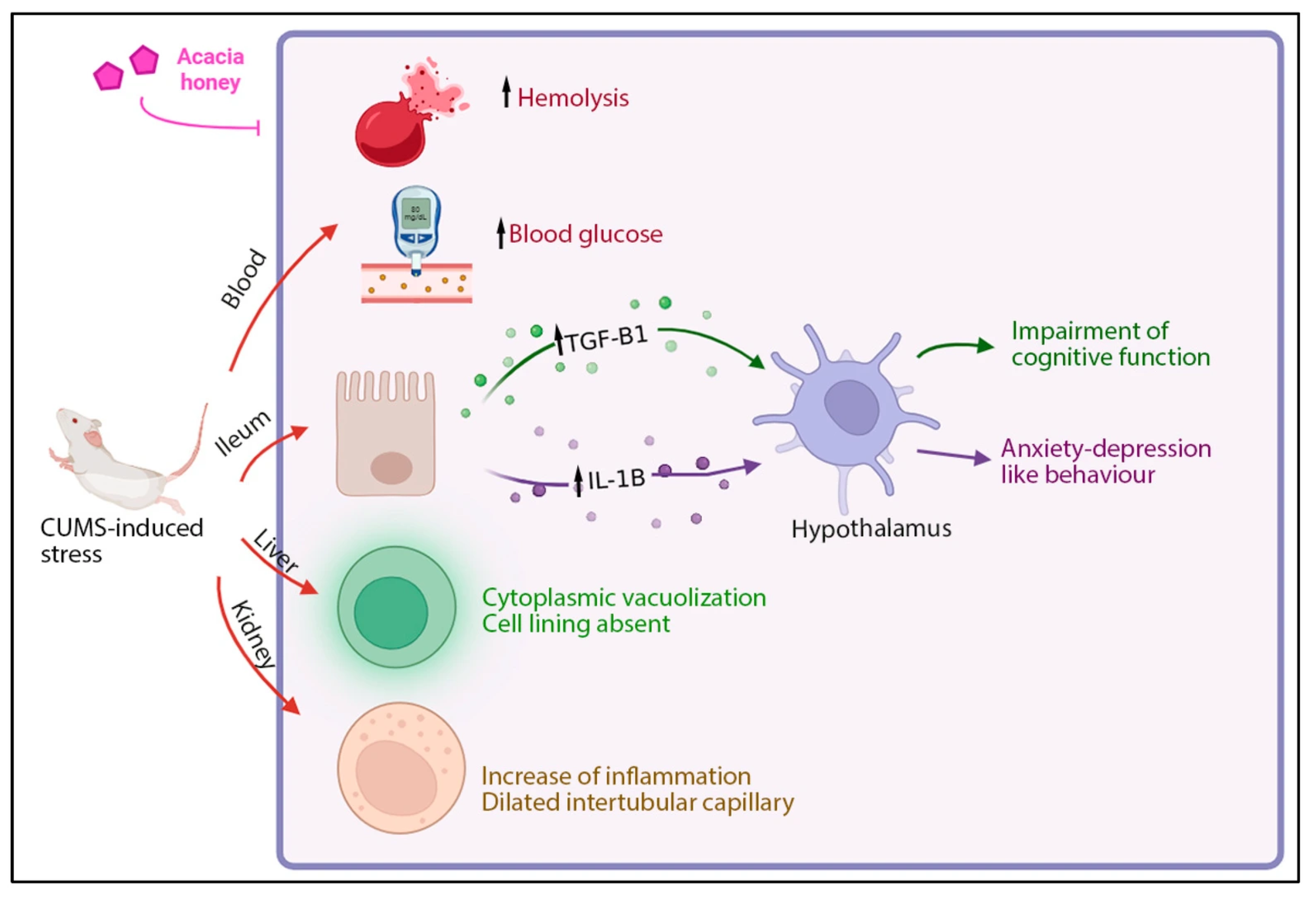
The current study demonstrates that supplementation with AH significantly modulates stress-induced metabolic, oxidative, and inflammatory disturbances (Figure 8). Chronic unpredictable mild stress induced hyperglycemia, increased erythrocyte hemolysis, hepatic vacuolation, renal tubular injury, and upregulation of pro-inflammatory cytokines (IL-1β and TGF-β1) in the ileum, caecum, and hypothalamus. Supplementation with AH markedly reduced blood glucose, protected erythrocytes against H2O2-induced hemolysis, and ameliorated histopathological changes in both liver and kidney. In parallel, AH downregulated IL-1β and TGF-β1 expression in stress-related tissues, reflecting anti-inflammatory modulation.
Figure 8.
Summary of AH’s effect on hemolysis, blood glucose, IL-1β and TGF-β1 gene expression in the ileum and hypothalamus, as well as structural alteration in kidney and liver under stress conditions. The figure illustrates that CUMS-induced stress increases hemolysis, blood glucose levels, inflammation (black arrow), and anxiety- and depression-like behaviors, while impairing cognitive function and damaging multiple organs including the liver and kidney. Supplementation with AH effectively blocks these effects, reducing inflammation and behavioral symptoms, and restoring physiological and cellular integrity. Image created using Biorender.com.
AMT, used as the pharmacological control, improved several parameters, but the combined treatment with AH and AMT produced the most profound effects, suggesting a synergistic interaction. The mechanisms underlying these protective outcomes may involve the high phenolic and flavonoid content of AH, which can enhance antioxidant defenses, reduce oxidative stress, and improve insulin sensitivity. In addition, the anti-inflammatory and membrane-stabilizing properties of honey further contribute to its protective role.
Findings from this study using male Sprague-Dawley rats in the CUMS model are relevant to human depression, as the model replicates key behavioral and neuroinflammatory features of the disorder. Despite species differences, the results offer translational insights into stress-related pathophysiology and may inform therapeutic strategies for human depressive conditions.
A key limitation of this study is the complexity of stress tolerance mechanisms in mammals which may involve adaptive responses at multiple biological levels. Beyond metabolomic shifts, stress-induced tolerance is mediated by receptor activation, protein signaling cascades, and serum biomolecule modulation. These processes orchestrate transcriptomic and proteomic reprogramming, influencing metabolic pathways such as glucose and amino acid metabolism. A systemic biology approach is essential to elucidate the interplay between stress-related transcription factors, receptor-mediated signaling, and serum biomarkers. Future studies should expand to clinical trials to confirm translational relevance, establish dose–response relationships, and explore the molecular mechanisms linking honey’s antioxidant compounds with neuroendocrine regulation.
Author Contributions
Data curation, Methodology, Writing—original draft, A.S.M. and H.M.; Methodology, Writing—Reviewing and Editing, N.A.S.N.Z., N.L.M.N., M.R.A.A.R. and L.M.S.; Writing—Reviewing and Editing, A.M.A., K.R. and M.H.H.; Conceptualization, Supervision, Writing—Reviewing and Editing, Z.E. All authors have read and agreed to the published version of the manuscript.
Funding
This research was funded by the Ministry of Higher Education Malaysia under the Fundamental Research Grant Scheme (FRGS) [Grant No: FRGS/1/2017/WAB11/UITM/03/1].
Institutional Review Board Statement
The procedures were conducted as approved by the Animal Research and Ethics Committee of University Teknologi MARA (UiTM), Puncak Alam Campus, Faculty of Pharmacy (Approval No. UiTM CARE 237/2/2018 was granted on 4 June 2018).
Informed Consent Statement
Not applicable.
Data Availability Statement
The original contributions presented in the study are included in the article, further inquiries can be directed to the corresponding author.
Acknowledgments
The author gratefully acknowledged the staff at National Poison Centre for their expert assistance in the preparation of this manuscript.
Conflicts of Interest
The authors declare no conflicts of interest.
Abbreviations
The following abbreviations are used in this manuscript:
| AH | Malaysian Acacia honey |
| ALT | Alanine aminotransferase |
| AMT | Amitriptyline |
| ANOVA | Analysis of variance |
| AST | Aspartate transaminase |
| BDNF | Brain-derived neurotrophic factor |
| BC | Bowman’s capsule |
| c-FOS | Celular-FOS |
| CLM | Cell lining missing |
| COX-2 | Cyclooxygenase-2 |
| CPV | Cytoplasmic vacuolation |
| Ct | Threshold cycle |
| CUMS | Chronic unpredictable mild stress |
| CV | Central vein |
| DCT | Distal convoluted tubules |
| DNA | Deoxyribonucleic acid |
| DT | Dilated intertubular capillaries |
| ES | Erythrocyte suspension |
| G | Glomerulus |
| GAPDH | Glyceraldehyde 3-phosphate dehydrogenase |
| GGT | Gamma-glutamyltransferase |
| GLUT2 receptor | Glucose Transporter Type 2 receptor |
| GLUT5 receptor | Glucose Transporter Type 5 receptor |
| h | Hour |
| H2O2 | Hydrogen peroxide |
| HPA | Hypothalamic–pituitary–adrenal |
| H&E | Hematoxylin-eosin |
| iNOS | Inducible Nitric Oxide Synthase |
| I | Inflammation |
| IL-1 | Interleukin-1 |
| IL-1α | Interleukin-1 alpha |
| IL-1β | Interleukin-1 beta |
| IL-2 | Interleukin-2 |
| IL-6 | Interleukin-6 |
| IL-10 | Interleukin-10 |
| IFN-γ | Interferon gamma |
| KC | Kupffer cell |
| LPS | Lipopolysaccharide |
| LOXs | Lipoxygenases |
| min | minute |
| mRNA | Messenger ribonucleic acid |
| MDD | major depressive disorder |
| NC | Normal control |
| NF-κB | Nuclear Factor kappa-light-chain-enhancer of activated B cells |
| NO | Nitric Oxide |
| IκBα | Inhibitor of kappa B alpha |
| PBS | Phosphate-buffered saline |
| PCT | Proximal convoluted tubules |
| PDGF | Platelet-Derived Growth Factor |
| RNA | Ribonucleic acid |
| RO | Reverse osmosis water |
| ROS | Reactive oxygen species |
| S | Stress-induced group |
| SEM | Standard error of mean |
| Sn | Sinusoidal |
| SD | Sprague-Dawley |
| SPT | Sucrose preference test |
| TGF-β | Transforming growth factor beta |
| TGF-β1 | Transforming growth factor beta 1 gene |
| TNF-α | Tumor necrosis factor-alpha |
References
- Joseph, J.J.; Golden, S.H. Cortisol Dysregulation: The Bidirectional Link between Stress, Depression, and Type 2 Diabetes Mellitus. Ann. N. Y. Acad. Sci. 2017, 1391, 20–34. [Google Scholar] [CrossRef]
- Maydych, V. The Interplay between Stress, Inflammation, and Emotional Attention: Relevance for Depression. Front. Neurosci. 2019, 13, 384. [Google Scholar] [CrossRef] [PubMed]
- Allameh, A.; Niayesh-Mehr, R.; Aliarab, A.; Sebastiani, G.; Pantopoulos, K. Oxidative Stress in Liver Pathophysiology and Disease. Antioxidants 2023, 12, 1653. [Google Scholar] [CrossRef]
- Atagana, O.S.; Asagba, S.O. Protective Effects of Honey against Cadmium-Induced Alteration of Some Biochemical Parameters in Rats. Toxicol. Environ. Chem. 2014, 96, 1557–1563. [Google Scholar] [CrossRef]
- Liu, Z.; Lei, M.; Bai, Y. Chronic Stress Mediates Inflammatory Cytokines Alterations and Its Role in Tumorigenesis. J. Inflamm. Res. 2025, 18, 1067–1090. [Google Scholar] [CrossRef] [PubMed]
- Turner, L.; Galante, J.; Vainre, M.; Stochl, J.; Dufour, G.; Jones, P.B. Immune Dysregulation among Students Exposed to Exam Stress and Its Mitigation by Mindfulness Training: Findings from an Exploratory Randomised Trial. Sci. Rep. 2020, 10, 5812. [Google Scholar] [CrossRef]
- Holzer, P.; Reichmann, F.; Farzi, A. Neuropeptide Y, Peptide YY and Pancreatic Polypeptide in the Gut-Brain Axis. Neuropeptides 2012, 46, 261–274. [Google Scholar] [CrossRef]
- Moraczewski, J.; Awosika, A.; Aedma, K. Tricyclic Antidepressants. In StatPearls; StatPearls Publishing: Treasure Island, FL, USA, 2025. [Google Scholar]
- Erejuwa, O.O.; Sulaiman, S.A.; Wahab, M.S.A. Fructose Might Contribute to the Hypoglycemic Effect of Honey. Molecules 2012, 17, 1900–1915. [Google Scholar] [CrossRef]
- Al-Waili, N.S. Effects of Daily Consumption of Honey Solution on Hematological Indices and Blood Levels of Minerals and Enzymes in Normal Individuals. J. Med. Food 2003, 6, 135–140. [Google Scholar] [CrossRef]
- Erejuwa, O.O.; Sulaiman, S.A.; Ab Wahab, M.S. Honey: A Novel Antioxidant. Molecules 2012, 17, 4400–4423. [Google Scholar] [CrossRef] [PubMed]
- Blasa, M.; Candiracci, M.; Accorsi, A.; Piacentini, M.P.; Albertini, M.C.; Piatti, E. Raw Millefiori Honey Is Packed Full of Antioxidants. Food Chem. 2006, 97, 217–222. [Google Scholar] [CrossRef]
- Tlak Gajger, I.; Dar, S.A.; Ahmed, M.M.M.; Aly, M.M.; Vlainić, J. Antioxidant Capacity and Therapeutic Applications of Honey: Health Benefits, Antimicrobial Activity and Food Processing Roles. Antioxidants 2025, 14, 959. [Google Scholar] [CrossRef] [PubMed]
- Mohamed, A.S.; Abdah, M.A.; Ramasamy, K.; Hasan, M.H.; Hamimi, I.A.; Zolkapli, E. Physicochemical Analysis and Sugar Profiling of Acacia Honey. Malays. J. Microsc. 2018, 14, 157–164. [Google Scholar]
- Samat, S. Physicochemical Profile of Selected Malaysian Honey and The Effect of Pineapple Honey on Obesity In Vitro and In Vivo Model. Ph.D. Thesis, Universiti Teknologi Mara, Shah Alam, Selangor, 2021. [Google Scholar]
- Jamwal, N.; Jasrotia, R.; Badyal, N.; Hajam, Y.A.; Langer, S. Honey: An Antidiabetic and Hypoglycemic Agent to Reverse Diabetes Induced Complications BT—Honey in Food Science and Physiology; Kumar, R., Hajam, Y.A., Bala Dhull, S., Giri, A., Eds.; Springer Nature: Singapore, 2024; pp. 369–388. ISBN 978-981-97-3565-5. [Google Scholar]
- Sukri, N.M.; Ooi, F.K.; Chee, K.C.; Sirajudeen, K.N.S. Effect of Acacia Honey Drink Consumed Pre and During Exercise on Glucose Metabolism, Total Antioxidant Status, and Running Performance in the Heat. IIUM Med. J. Malaysia 2022, 21, 85–95. [Google Scholar] [CrossRef]
- Ahmad, N.S.; Ismail, M.S.; Mohamed, M.; Ooi, F.K. Blood Glucose Metabolism, Serum, and Urine Osmolality in Response to Sodium-Enriched Acacia Honey Drink Consumption during Rehydration after Exercise in Hot and Humid Environment. J. Sustain. Sci. Manag. 2020, 15, 72–83. [Google Scholar] [CrossRef]
- Schell, K.R.; Fernandes, K.E.; Shanahan, E.; Wilson, I.; Blair, S.E.; Carter, D.A.; Cokcetin, N.N. The Potential of Honey as a Prebiotic Food to Re-Engineer the Gut Microbiome Toward a Healthy State. Front. Nutr. 2022, 9, 957932. [Google Scholar] [CrossRef] [PubMed]
- Ahmed, S.; Sulaiman, S.A.; Baig, A.A.; Ibrahim, M.; Liaqat, S.; Fatima, S.; Jabeen, S.; Shamim, N.; Othman, N.H. Honey as a Potential Natural Antioxidant Medicine: An Insight into Its Molecular Mechanisms of Action. Oxid. Med. Cell. Longev. 2018, 2018, 8367846. [Google Scholar] [CrossRef]
- Harro, J. Animal Models of Depression: Pros and Cons. Cell Tissue Res. 2019, 377, 5–20. [Google Scholar] [CrossRef]
- Antoniuk, S.; Bijata, M.; Ponimaskin, E.; Wlodarczyk, J. Chronic Unpredictable Mild Stress for Modeling Depression in Rodents: Meta-Analysis of Model Reliability. Neurosci. Biobehav. Rev. 2019, 99, 101–116. [Google Scholar] [CrossRef]
- van Zyl, P.J.; Dimatelis, J.J.; Russell, V.A. Behavioural and Biochemical Changes in Maternally Separated Sprague–Dawley Rats Exposed to Restraint Stress. Metab. Brain Dis. 2016, 31, 121–133. [Google Scholar] [CrossRef]
- Abidin, Q.H.Z.; Ismail, W.I.W.; Ismail, N.E.; Eshak, Z. Preliminary Study on Malaysian Honey as Anti- Stress Agent. Thai J. Pharm. Sci. 2017, 41, 133–136. [Google Scholar]
- Beery, A.K.; Zucker, I. Sex Bias in Neuroscience and Biomedical Research. Neurosci. Biobehav. Rev. 2011, 35, 565–572. [Google Scholar] [CrossRef]
- Zhang, Y.; Wang, Y.; Lei, H.; Wang, L.; Xue, L.; Wang, X.; Zhu, X. Optimized animal model to mimic the reality of stress-induced depression in the clinic. BMC Phychiatry 2017, 17, 171. [Google Scholar] [CrossRef]
- Okoko, T.; Ere, D. Reduction of Hydrogen Peroxide—Induced Erythrocyte Damage by Carica Papaya Leaf Extract. Asian Pac. J. Trop. Biomed. 2012, 2, 449–453. [Google Scholar] [CrossRef]
- Abidin, Q.H.Z.; Rujhan, N.H.M.; Wan Ismail, W.I.; Ismail, N.E.; Eshak, Z. Potential Used of Tualang and Acacia Honey in Ameliorating Stress-Depression Disorder: A Preliminary Study. In Proceedings of the Seoul International Congress of Endocrinology and Metabolism, Seoul, Republic of Korea, 4–6 November 2015; Korea Endocrine Society: Seoul, Republic of Korea, 2015; p. 309. [Google Scholar]
- Abidin, Q.H.Z. Anti Stress-Depression Effects of Acacia Honey on Chronic Unpredictable Mild Stress Rats. Ph.D. Thesis, Universiti Teknologi Mara, Shah Alam, Selangor, 2017. [Google Scholar]
- Markov, D.D. Sucrose Preference Test as a Measure of Anhedonic Behavior in a Chronic Unpredictable Mild Stress Model of Depression: Outstanding Issues. Brain Sci. 2022, 12, 1287. [Google Scholar] [CrossRef]
- He, L.W.; Zeng, L.; Tian, N.; Li, Y.; He, T.; Tan, D.M.; Zhang, Q.; Tan, Y. Optimization of Food Deprivation and Sucrose Preference Test in SD Rat Model Undergoing Chronic Unpredictable Mild Stress. Anim. Model. Exp. Med. 2020, 3, 69–78. [Google Scholar] [CrossRef] [PubMed]
- Duan, L.; Fan, R.; Li, T.; Yang, Z.; Hu, E.; Yu, Z.; Tian, J.; Luo, W.; Zhang, C. Metabolomics Analysis of the Prefrontal Cortex in a Rat Chronic Unpredictable Mild Stress Model of Depression. Front. Psychiatry 2022, 13, 815211. [Google Scholar] [CrossRef] [PubMed]
- MacDonald, A.J.; Yang, Y.H.C.; Cruz, A.M.; Beall, C.; Ellacott, K.L.J. Brain-Body Control of Glucose Homeostasis—Insights From Model Organisms. Front. Endocrinol. 2021, 12, 662769. [Google Scholar] [CrossRef] [PubMed]
- Zamanian, M.; Azizi-soleiman, F. Honey and Glycemic Control: A Systematic Review. PharmaNutrition 2020, 11, 100180. [Google Scholar] [CrossRef]
- Ayoub, S.; Javed, M.; Sattar, K.; Ullah, H.; Hajjar, W.; Alasiri, S. Honey and Diabetes Mellitus: Obstacles and Challenges—Road to Be Repaired. Saudi J. Biol. Sci. 2017, 24, 1030–1033. [Google Scholar] [CrossRef]
- Mushtaq, S.; Imtiyaz, Z.; Wali, A.F.; Khan, A.; Rashid, S.M.; Amin, I.; Ali, A.; Rehman, M.U.; Arafah, A. Honey: A Powerful Natural Antioxidant and Its Possible Mechanism of Action BT—Therapeutic Applications of Honey and Its Phytochemicals; Rehman, M.U., Majid, S., Eds.; Springer: Singapore, 2020; Volume 1, pp. 11–29. ISBN 978-981-15-6799-5. [Google Scholar]
- Martinez-Armenta, C.; Camacho-Rea, M.C.; Martínez-Nava, G.A.; Espinosa-Velázquez, R.; Pineda, C.; Gomez-Quiroz, L.E.; López-Reyes, A. Therapeutic Potential of Bioactive Compounds in Honey for Treating Osteoarthritis. Front. Pharmacol. 2021, 12, 642836. [Google Scholar] [CrossRef]
- Zhang, L.; Peng, X.; Ai, Y.; Li, L.; Zhao, S.; Liu, Z.; Peng, Q.; Deng, S.; Huang, Y.; Mo, Y.; et al. Amitriptyline Reduces Sepsis-Induced Brain Damage Through TrkA Signaling Pathway. J. Mol. Neurosci. 2020, 70, 2049–2057. [Google Scholar] [CrossRef] [PubMed]
- Milenina, L.S.; Krutetskaya, Z.I.; Antonov, V.G.; Krutetskaya, N.I. Tricyclic Antidepressant Amitriptyline Suppresses Ca2+ Responses in Rat Peritoneal Macrophages. Cell Tissue Biol. 2024, 18, 439–450. [Google Scholar] [CrossRef]
- Turpin, C.; Catan, A.; Guerin-Dubourg, A.; Debussche, X.; Bravo, S.B.; Álvarez, E.; van Den Elsen, J.; Meilhac, O.; Rondeau, P.; Bourdon, E. Enhanced Oxidative Stress and Damage in Glycated Erythrocytes. PLoS ONE 2020, 15, e0235335. [Google Scholar] [CrossRef] [PubMed]
- Blasa, M.; Candiracci, M.; Accorsi, A.; Piacentini, M.; Piatti, E. Honey Flavonoids as Protection Agents against Oxidative Damage to Human Red Blood Cells. Food Chem. 2007, 104, 1635–1640. [Google Scholar] [CrossRef]
- Vallelian, F.; Buehler, P.W.; Schaer, D.J. Hemolysis, Free Hemoglobin Toxicity, and Scavenger Protein Therapeutics. Blood 2022, 140, 1837–1844. [Google Scholar] [CrossRef]
- Pelusi, S.; Valenti, L.; Fargion, S. Oxidative Stress and Hepatic Iron Overload BT—Studies on Hepatic Disorders; Albano, E., Parola, M., Eds.; Springer International Publishing: Cham, Switzerland, 2015; pp. 345–356. ISBN 978-3-319-15539-5. [Google Scholar]
- Greenhalgh, S.N.; Thompson, A.I.; Henderson, N.C.; Iredale, J.P. Oxidative Stress and Liver Inflammation BT—Studies on Hepatic Disorders; Albano, E., Parola, M., Eds.; Springer International Publishing: Cham, Switzerland, 2015; pp. 123–147. ISBN 978-3-319-15539-5. [Google Scholar]
- Wang, X.; Ling, W.; Zhu, Y.; Ji, C.; An, X.; Qi, Y.; Li, S.; Zhang, C.; Tong, R.; Jiang, D.; et al. Spermidine Alleviates Copper-Induced Oxidative Stress, Inflammation and Cuproptosis in the Liver. FASEB J. 2025, 39, e70453. [Google Scholar] [CrossRef]
- Yaman, T.; Yener, Z.; Celik, I. Histopathological and Biochemical Investigations of Protective Role of Honey in Rats with Experimental Aflatoxicosis. BMC Complement. Altern. Med. 2016, 16, 232. [Google Scholar] [CrossRef]
- Van Avondt, K.; Nur, E.; Zeerleder, S. Mechanisms of Haemolysis-Induced Kidney Injury. Nat. Rev. Nephrol. 2019, 15, 671–692. [Google Scholar] [CrossRef]
- Xie, T.; Yao, L.; Li, X. Advance in Iron Metabolism, Oxidative Stress and Cellular Dysfunction in Experimental and Human Kidney Diseases. Antioxidants 2024, 13, 659. [Google Scholar] [CrossRef]
- Abdel-moneim, W.M.; Ghafeer, H.H. The Potential Protective Effect of Natural Honey against Cadmium-Induced Hepatotoxicity and Nephrotoxicity. Mansoura J. Forensic Med. Clin. Toxicol. 2007, XV, 75–98. [Google Scholar] [CrossRef]
- Yaacob, W.M.H.W.; Long, I.; Zakaria, R.; Othman, Z. Tualang Honey and Its Methanolic Fraction Protect against LPS-Induced Neuroinflammation and Amyloid Deposition in Male Rats. Asian J. Med. Biomed. 2018, 59. [Google Scholar]
- Bird, L. IL-22 Protects against Stress-Induced Anxiety. Nat. Rev. Immunol. 2025, 25, 75. [Google Scholar] [CrossRef]
- Uchakin, P.N.; Tobin, B.; Cubbage, M.; Marshall, G.J.; Sams, C. Immune Responsiveness Following Academic Stress in First-Year Medical Students. J. Interf. Cytokine Res. Off. J. Int. Soc. Interf. Cytokine Res. 2001, 21, 687–694. [Google Scholar] [CrossRef]
- Rawat, M.; Nighot, M.; Al-Sadi, R.; Gupta, Y.; Viszwapriya, D.; Yochum, G.; Koltun, W.; Ma, T.Y. IL1B Increases Intestinal Tight Junction Permeability by Up-Regulation of MIR200C-3p, Which Degrades Occludin MRNA. Gastroenterology 2020, 159, 1375–1389. [Google Scholar] [CrossRef]
- Ma, Y.; Matsuwaki, T.; Yamanouchi, K.; Nishihara, M. Cyclooxygenase-2-Related Signaling in the Hypothalamus Plays Differential Roles in Response to Various Acute Stresses. Brain Res. 2013, 1508, 23–33. [Google Scholar] [CrossRef] [PubMed]
- Holzer, P.; Farzi, A.; Hassan, A.M.; Zenz, G.; Jacan, A.; Reichmann, F. Visceral Inflammation and Immune Activation Stress the Brain. Front. Immunol. 2017, 8, 1613. [Google Scholar] [CrossRef] [PubMed]
- Silva, Y.P.; Bernardi, A.; Frozza, R.L. The Role of Short-Chain Fatty Acids From Gut Microbiota in Gut-Brain Communication. Front. Endocrinol. 2020, 11, 25. [Google Scholar] [CrossRef]
- Li, C.; Yao, J.; Yang, C.; Yu, S.; Yang, Z.; Wang, L.; Li, S.; He, N. Gut Microbiota-Derived Short Chain Fatty Acids Act as Mediators of the Gut-Liver-Brain Axis. Metab. Brain Dis. 2025, 40, 122. [Google Scholar] [CrossRef] [PubMed]
- Farhana, Z.; Alhewairini, S.; Mahamood, M. The Gut–Brain Axis, Cognition and Honey. In Therapeutic Applications of Honey and Its Phytochemicals; Springer: Singapore, 2020; pp. 331–343. [Google Scholar] [CrossRef]
- Liu, C.; Zhang, Q.; Zhou, H.; Jin, L.; Liu, C.; Yang, M.; Zhao, X.; Ding, W.; Xie, W.; Kong, H. GLP-1R Activation Attenuates the Progression of Pulmonary Fibrosis via Disrupting NLRP3 Inflammasome/PFKFB3-Driven Glycolysis Interaction and Histone Lactylation. J. Transl. Med. 2024, 22, 954. [Google Scholar] [CrossRef]
- Kolb, M.; Margetts, P.J.; Anthony, D.C.; Pitossi, F.; Gauldie, J. Transient Expression of IL-1β Induces Acute Lung Injury and Chronic Repair Leading to Pulmonary Fibrosis. J. Clin. Investig. 2001, 107, 1529–1536. [Google Scholar] [CrossRef]
- Troncone, E.; Marafini, I.; Stolfi, C.; Monteleone, G. Transforming Growth Factor-Β1/Smad7 in Intestinal Immunity, Inflammation, and Cancer. Front. Immunol. 2018, 9, 4–9. [Google Scholar] [CrossRef] [PubMed]
- Yamada, Y.; Mashima, H.; Sakai, T.; Matsuhashi, T.; Jin, M.; Ohnishi, H. Functional Roles of TGF-Β1 in Intestinal Epithelial Cells through Smad-Dependent and Non-Smad Pathways. Dig. Dis. Sci. 2013, 58, 1207–1217. [Google Scholar] [CrossRef] [PubMed]
- Yun, S.M.; Kim, S.H.; Kim, E.H. The Molecular Mechanism of Transforming Growth Factor-β Signaling for Intestinal Fibrosis: A Mini-Review. Front. Pharmacol. 2019, 10, 162. [Google Scholar] [CrossRef] [PubMed]
- Tie, Y.; Tang, F.; Peng, D.; Zhang, Y.; Shi, H. TGF-Beta Signal Transduction: Biology, Function and Therapy for Diseases. Mol. Biomed. 2022, 3, 45. [Google Scholar] [CrossRef]
- Rodari, M.M.; Cerf-Bensussan, N.; Parlato, M. Dysregulation of the Immune Response in TGF-β Signalopathies. Front. Immunol. 2022, 13, 1066375. [Google Scholar] [CrossRef]
- Kajumba, M.M.; Kakooza-Mwesige, A.; Nakasujja, N.; Koltai, D.; Canli, T. Treatment-Resistant Depression: Molecular Mechanisms and Management. Mol. Biomed. 2024, 5, 43. [Google Scholar] [CrossRef]
- Cao, B.B.; Zhang, X.X.; Du, C.Y.; Liu, Z.; Qiu, Y.H.; Peng, Y.P. TGF-Β1 Provides Neuroprotection via Inhibition of Microglial Activation in 3-Acetylpyridine-Induced Cerebellar Ataxia Model Rats. Front. Neurosci. 2020, 14, 187. [Google Scholar] [CrossRef]
- Xie, J.; Xiong, S.; Li, Y.; Xia, B.; Li, M.; Zhang, Z.; Shi, Z.; Peng, Q.; Li, C.; Lin, L.; et al. Phenolic Acids from Medicinal and Edible Homologous Plants: A Potential Anti-Inflammatory Agent for Inflammatory Diseases. Front. Immunol. 2024, 15, 1345002. [Google Scholar] [CrossRef]
- Bang, E.; Tobery, A.; Montgomery, K.S.; Fincher, A.S.; Earnest, D.J.; Murchison, D.A.; Griffith, W.H. Amitriptyline Decreases GABAergic Transmission in Basal Forebrain Neurons Using an Optogenetic Model of Aging. Front. Aging Neurosci. 2021, 13, 673155. [Google Scholar] [CrossRef]
Disclaimer/Publisher’s Note: The statements, opinions and data contained in all publications are solely those of the individual author(s) and contributor(s) and not of MDPI and/or the editor(s). MDPI and/or the editor(s) disclaim responsibility for any injury to people or property resulting from any ideas, methods, instructions or products referred to in the content. |
© 2025 by the authors. Licensee MDPI, Basel, Switzerland. This article is an open access article distributed under the terms and conditions of the Creative Commons Attribution (CC BY) license (https://creativecommons.org/licenses/by/4.0/).