Article

Development of Multiplex qPCR Method for Accurate Detection of Enzyme-Producing Psychrotrophic Bacteria

by
Kidane Yalew
1,2,†,
Shuwen Zhang
1,
Solomon Gebreyowhans
3,
Ning Xie
1,
Yunna Wang
1,
Jiaping Lv
1,
Xu Li
1,* and
Xiaoyang Pang
1,*
1
Key Laboratory of Agro-products Quality and Safety Control in Storage and Transport Process, Ministry of Agriculture and Rural Affairs, Institution of Food Science and Technology, Chinese Academy of Agricultural Sciences, Beijing 100193, China
2
Department of Vet. Public Health and Food Safety, College of Veterinary Science, Mekelle University, Mekelle 0231, Ethiopia
3
Tigray Agricultural Research Institution, Mekelle 0492, Ethiopia
*
Authors to whom correspondence should be addressed.
This author is the first author.
Foods 2025, 14(11), 1975; https://doi.org/10.3390/foods14111975
Submission received: 7 April 2025 / Revised: 7 May 2025 / Accepted: 19 May 2025 / Published: 3 June 2025

Abstract

:
Microbial detection in milk is crucial for food safety and quality, as beneficial and harmful microorganisms can affect consumer health and dairy product integrity. Identifying and quantifying these microorganisms helps prevent contamination and spoilage. The study employs advanced molecular techniques to detect and quantify the genomic DNA for the target hydrolytic enzyme coding genes lipA and aprX based on the multi-align sequence conserved region, specific primer pair, and hydrolysis probes designed using the singleplex qPCR and multiplex qPCR. Cultured isolates and artificially contaminated sterilized ultra-high-temperature (UHT) milk were analyzed for their specificity, cross-reactivity, and sensitivity. The finding indicated that strains with lipA and aprX genes were amplified while the other strains were not amplified. This indicated that the designed primer pairs/probes were very specific to the target gene of interest. The specificity of each design primer pair was checked using SYBR Green qPCR using 16 different isolate strains from the milk sample. The quantification specificity of each strain target gene was deemed to be with a mean Ct value for positive pseudomonas strain > 16.98 ± 1.76 (p < 0.0001), non-pseudomonas positive strain ≥ 27.47 ± 1.25 (p < 0.0001), no Ct for the negative control and molecular grade water. The sensitivity limit of detection (LOD) analyzed based on culture broth and milk sample was >105 and >104 in PCR amplification while it was >104 and >103 in real-time qPCR, respectively. At the same time, the correlation regression coefficient of the standard curve based on the pure culture cell DNA as the DNA concentration serially diluted (20 ng/µL to 0.0002 ng/µL) was obtained in multiplex without interference and cross-reactivity, yielding R2 ≥ 0.9908 slope (−3.2591) and intercepting with a value of 37, where the efficiency reached the level of 95–102% sensitivity reached up to 0.0002 ng/µL concentration of DNA, and sensitivity of microbial load was up to 1.2 × 10² CFU/mL. Therefore, multiplex TaqMan qPCR simultaneous amplification was considered the best method developed for the detection of the lipA and aprX genes in a single tube. This will result in developing future simultaneous (three- to four-gene) detection of spoilage psychrotrophic bacteria in raw milk.

1. Introduction

Microbial detection in milk is crucial for ensuring both food safety and quality [1,2,3]. The presence of microorganisms in milk, including both beneficial and harmful species, can affect consumer health and the integrity of dairy products [4]. Ensuring the microbiological quality of milk involves detecting and quantifying these microorganisms to prevent contamination and spoilage [5].
Detection of psychrotrophic bacteria is critical for assessing the quality of raw and pasteurized milk, as their presence can significantly affect shelf life and spoilage [6,7,8]. These bacteria are significant in various industries for their capability of extracellular enzyme production that can resist heat, including lipases and proteases post-pasteurization, which are essential for the hydrolysis of fats and proteins, leading to spoilage and quality degradation. Various genera, such as Pseudomonas, Acinetobacter, and Lactobacillus, have been documented as enzyme producers at low temperatures and are often associated with shelf life reduction through continuous enzymatic activity during storage and economic loss leading to product recalls, consumer complaints, and brand reputation damage. Process disruption in cheese production can also occur sue to uncontrolled proteolysis crud formation and ageing kinetics, while lipolysis can affect flavor profile defects and other quality issues in dairy products [9,10,11].
Quantitative real-time PCR (qPCR) and multiplex qPCR have emerged as rapid, sensitive, and specific tools for identifying and quantifying spoilage microbes, enabling proactive quality control in the dairy supply chain superior to traditional culture-based detection methods which are time-consuming (24–72 h) and often fail to detect low-abundance or non-culturable microbes [12,13,14]. The molecular identification of spoilage psychrotrophic bacteria has gained prominence as it allows precise detection and variation of species present in milk and dairy products [14,15,16], packed meat [17,18], seafood [19], and yogurt [13].
According to recent research, qPCR and multiplex qPCR are extremely sensitive methods for identifying pathogen bacteria and food spoilage, with detection limits that vary from 10 to 103 CFU/g depending on the target and food matrix. Using qPCR, food pathogens such as Salmonella enterica and Escherichia coli O157:H7 were detected 10 to 102 CFU/mL in contaminated foods following enrichment, and the limit of detection for the spoilage bacteria Pseudomonas spp., which is common in milk was 102 CFU/mL. The sensitivity simultaneous multiplex qPCR of pathogens for Listeria monocytogenes, Salmonella spp., and Escherichia coli O157:H7 in food achieved 102–103 CFU/g for each pathogen in spiked food samples and spoilage bacteria 103 CFU/g pseudomonas spp. in seafood [20,21,22]. The future of microbial detection in milk is likely to see advancements in rapid testing technologies and automation. New methodologies, such as automated digital PCR (dPCR) systems for real-time detection of microorganisms to enhance the sensitivity and efficiency of microbial analysis in the dairy industry, have been developed [23,24].
Therefore, as consumer awareness around food safety increases, the demand for high-quality, microbiologically safe dairy products is expected to grow, driving continued innovation in simultaneous detection psychrotrophic bacteria producing hydrolytic enzyme in milk. It is vital, time-saving, cost-effective, and more specific than uniplex fashion. This will improve quality control and minimize economic losses in the dairy industry.

2. Methods and Materials

2.1. Bacteria Culture Growth

From the total of 33 isolate bacteria listed in Table S1, the selected group of bacteria for this research study are listed in Table 1. All of them were isolated from 15 chilled raw milk samples of 50 mL collected in a sterile glass and transported in ice to our laboratory from 5 different dairy farms in the Hebei province of China. Psychrotrophic bacteria species were cultivated under aerobic conditions on LB nutrient agar (Qingdao Bio-Tech Co., Ltd., Qingdao, China) incubated at 4 °C for 10 days. Cell counts were determined on LB plate count agar after incubation in 28 °C orbital shaker with 150 rpm for 24–48 h.

2.2. Screening of Target Gene

Genomic sequence of different bacterial species and Pseudomonas strains with a target gene fasta was downloaded from the NCBI gene bank using the keywords triacylglycerol lipase and aprX gene. Hundreds of strains were available, but only eight strains with lipA gene and seven with aprX gene, including the noble strain P. fluorescens and non-Pseudomonas lipase positive, were selected for this research. Then, the selected strain’s sequence fasta was aligned using the Ugene software (Version 1.29.0) [25] for multiple sequence alignment; the candidate target conserved region reference was used, with the noble strain P. fluorescens as a target.

2.3. Genomic DNA (gDNA)

Genomic DNA was extracted from a broth culture bacteria revived in LB broth (Qingdao Bio-Tech Co., Ltd., Qingdao, China) centrifuge using 3K15 Benchtop (Sigma, Ontario, CA, USA) at room temperature (RT) at 10,000 rpm for 1 min, while the milk sample was centrifuged on RT at 6000 rpm for 30 min to remove the upper layer of fat with the supernatant. gDNA from the residual sediment cell was extracted and eluted based on the protocol using a TIANGEN bacteria gDNA Kit® (Tiangen Bio-Technology, Co., Ltd., Beijing, China). Eluted gDNA with a volume of 100 µL molecular-grade water (Biorigin, Beijing, China) was checked for purity, and its concentration ratio @260/280 was determined using the Nucleic Acid Micro spectrophotometer K5500 Plus (Xi’an Hub Biotechnology Co., Ltd., Xi’an, China); it was stored at −20 °C until use to prevent DNA lysis. The gDNA of pure culture and artificially contaminated milk samples were further diluted to a working concentration of 50 ng/µL and 20 ng/µL for the PCR and qPCR, respectively, to verify our estimated result quantification. The limit of detection was set based on the plate count CFU/µL.

2.4. Primer Pair and Hydrolysis Probe Designing

Different online applications and primer design software were used to design the intended primer pair amplification of the candidate target genes, such as Primer3, NCBI primer pick, SnapGene (version 6.0.2), Primer Premier 6, and Genescript (https://www.genscript.com/) [26]. The designed primer pairs were selected from different regions of the candidate target gene multi-align sequence conserved region, focusing on the triacylglycerol lipase for lipA and aprX for the protease enzyme produced targeting the noble strain P. fluorescens. The primer pair and hydrolysis probe design listed in Table S2, Table 2 were synthesized at the Beijing genomic institution (BGI, Beijing, China).

2.5. PCR and qPCR Condition Setup

2.5.1. PCR Base

The PCR mix was prepared using the TianGen 2xTaq PCR Master Mix kit (Tiangen, Beijing, China); the mix reaction volume was 20 µL with 2xTaq PCR MasterMix 10 µL, 1 µL each primer (F&R), 7 µL molecular-grade water, and 1 µL templet DNA of 50 ng/µL on a PCR system Applied Biosystems 96 well (VeritiTM Thermal Cycler, Milford, CT, USA) Thermo Fisher Scientific, program: 95 °C: 5 min, 30 cycles (95 °C: 30 s, 60 °C: 30 s, 72 °C:1 min). The final step was performed at 72 °C: 10 min. The mixture was verified on 2% agarose gel (Biowest, Madrid, Spain) electrophoresis (AGE) and visualized using 10,000× gene green nucleic acid dye (Tiangen, Beijing, China) for the visibility of clear band based on the DNA marker (Tiangen, Beijing, China) using a Gel-image scanner (Clinx®, Shanghai, China).

2.5.2. TaqMan Probe Base

For amplification of single and multiplex qPCR, the total reaction volume was used as per the kit protocol 20 μL, with 10 μL of Super Real TaqMan mix (TianGen®,Beijing, China), with primer final optimized concentration of 200 nM, probe final optimized concentration of 150 nM, 2.5 μL template DNA of 20 ng/µL and molecular-grade water up to the final volume. ABI7500 real-time PCR 96 Systems (Applied Biosystems) thermal cycling was performed, with the following protocol: Step one—holding stage at 50 °C: 2 min, second holding stage 95 °C: 10 min, then a 40-cycle denaturation at 95 °C: 15 s, annealing optimized at 60 °C: 1 min, and extension at 72 °C:1 min. The qPCR assays were performed in triplicate, and water was used to ensure the absence of contamination as a no template (NT) and UHT milk DNA was used as negative control.

2.6. Primer Pair and Hydrolysis Probe Specificity

A total of 16 different bacterial species (Table S3) were used to determine the specificity of the design primer pair assay with water as no template to ensure the absence contamination using the SYBR Green assay. A 20 ng/µL gDNA from each isolate was amplified, and the mean CT value was obtained. Following the primer specificity amplification, the singleplex and multiplex amplification for the probe designed was checked for its specificity, targeting sample positive control, negative control, and water for each designed probe in triplicate, and the mean CT value was obtained.

2.7. Broth Sample Detection Limit Evaluation

A fresh culture of P. fluorescens with a known concentration of 1.2 × 10⁷ CFU/mL was serially diluted 107 to 100 in a 0.85% saline solution (Phytotech, Beijing, China) 10-fold series. A total of 1 μL of diluted gDNA extract was taken as a template in a PCR volume mix of 20 μL, 1 μL water was used in one tube PCR mixture as a no template DNA, and 2.5 μL template DNA with each dilution series extracted was used for detection limit quantification qPCR in triplicate. Standard curves were developed by plotting CFU/mL versus the threshold cycle (CT) produced for the target gene. The data were analyzed using the built-in ABI 7500 software.

2.8. Artificial Contamination and Natural Milk Sample Experiments

A fresh culture of P. fluorescens with a concentration of 1.2 × 10⁷ CFU/mL estimated by plate counting was injected into the sterile UHT milk purchased from the supermarket and serially diluted about 107 to 100 with no incubation time. The sample was mixed and pipetted in 1 mL doses from each artificially contaminated UHT milk into a new clean 1.5 mL eppendorf tube, centrifuged at 6000 rpm for 30 min, after which the upper layer of fat was removed, the supernatant was discarded, and then the gDNA was extracted. Randomly selected 12 natural raw milk samples from the Sanxi, dairy industry in Beijing were transported to our lab at +4 °C and kept in +4 °C for 2–3 days until processed. Then, their gDNA was extracted for use and their microbial load in the milk in terms of CFU/mL was determined.

2.9. Data Analysis

Data analysis was conducted using qPCR soft V2.3 built-in software ABI7500 machine. For each amplification cycle denaturation step, the real-time qPCR measures the reaction of fluorescence mixture and logs the cycle number (referred to as the threshold cycle [CT]) where the fluorescence reaches a specific value of threshold during exponential amplification [27]. Thus, the Ct represents the amount of relevant transcript, where the target gDNA copy with the Ct value is correlated inversely. The defined Ct values were exported into Microsoft Excel for additional statistical analysis as part of the data analysis process. Student’s t-test was utilized for statistical significance of mean Ct value with a p-value < 0.05 considered statistically significant.

3. Result

3.1. Screening of Target Gene

Both the lipA and aprX genes were screened based on the multi-align sequence for the eight bacterial species of lipA and seven Pseudomonas strains of aprX fasta downloaded from the NCBI 16SrRNA partial sequence of the target and highly conserved regions with very similar sequences. Many variable regions with a total length of 362 to 442 bp were chosen with variable regions accounting for 40.62% of the whole length of the target gene each sequence. Therefore, the aligned region conserved was selected for each gene to design further primer pairs and hydrolysis probes (Figure S1).

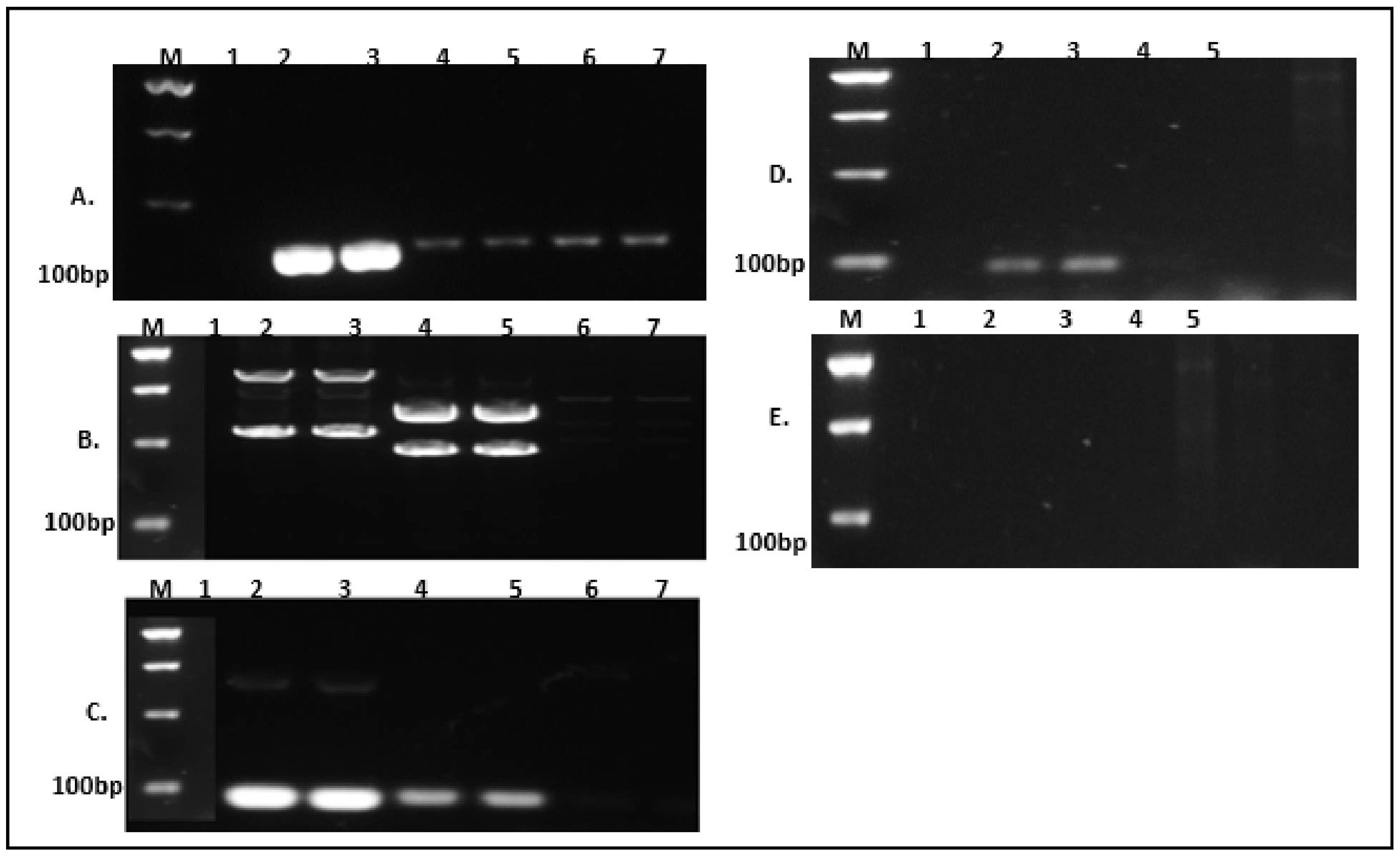
3.2. Primer Pair Design and Verification

The primer pair was designed based on the genomic context of the target region location chromosome I, description of each target using multiple alignment sequences of the target gene conserved region of the lipA gene and aprX targeting the noble strain P. fluorescens of the variable region primer (Figure S1) picked, respectively. The design primer pair was checked for specificity using different target strain pure culture genomic extracted DNA. Only strains with the lipA/aprX gene were amplified. In contrast, the other non-lipA/aprX gene strains and the negative control were not amplified, as shown in Figure 1C for the lipA primer, in Figure 1D for the aprX primer. At the same time, as shown in Figure 1A, there was still amplification for the negative control which means that the lipA1 primer was not specific to the target gene. However, the primer lipA2 (Figure 1B) was specific, but it still had the issue of primer dimmer while in Figure 1E, it is seen that aprX2 had no amplified products. Thus, the AGE PCR amplified result was shown in Figure 1.

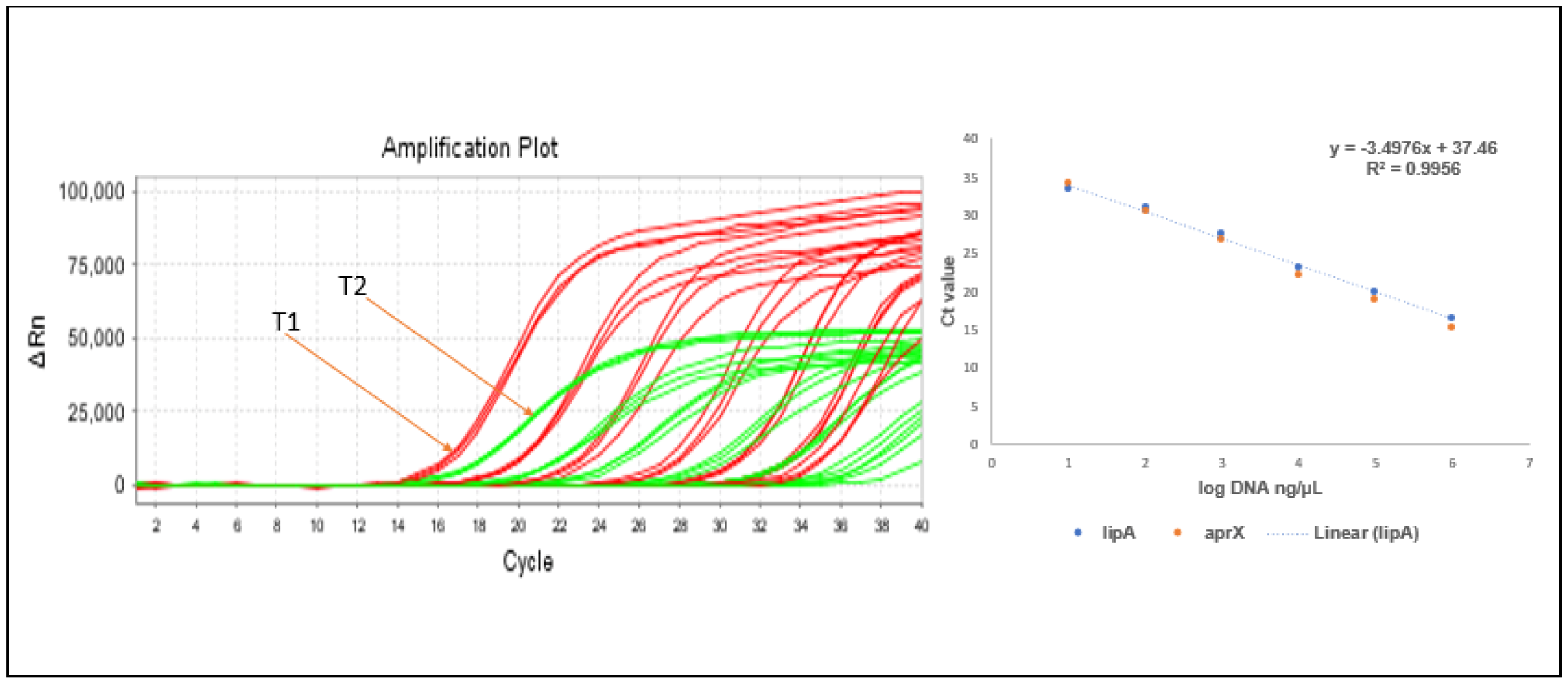
3.3. qPCR Primer Verification

The primer pair was selected based on the amplified lower mean Ct value, where the primer with no amplification was rejected. The specificity of the selected design primer for the target gene of interest was verified using positive and negative controls as well as no template (water) in triplicate for each (Figure S2). The sensitivity of the Sybr Green qPCR assay for the target gene was evaluated using genomic DNA (gDNA) dilutions across a concentration range of 20 ng/µL to 0.0002 ng/µL. The assay demonstrated a detection limit as low as 0.0002 ng/µL, with amplification efficiency exceeding 95%.
The mean cycle threshold (Ct) values for the 16 selected target bacterial species strains are detailed in Table S3. The specificity of the qPCR primers was confirmed by testing positive strains, including the specific strain Pseudomonas fluorescens, using design-specific primers. For non-Pseudomonas lipase-producing DNA amplification products, the average Ct value was significantly higher (27.47 ± 1.25; p < 0.0001) compared to the target strains, which showed a mean Ct of 16.98 ± 1.76 (p < 0.0001). Negative controls produced Ct values > 35, while no-template controls (NTCs) yielded no detectable Ct values. The specificity of Sybr Green qPCR amplification was further verified by analyzing melting curves. All results exhibited a unique, reproducible peak melting temperature (Tm), confirming the absence of non-specific products or primer dimers.
Detection of pure culture diluted in a 10-fold mixture, ranging from 106 to 10−1 CFU/mL in pure broth culture and artificially contaminated UHT milk sample, was performed, where the standard curve was developed targeting the CT mean value at each dilution of concentration for the noble strain P. fluorescens and its linear relationship between the CT and log input DNA was calculated. The singleplex correlation coefficients of standard curves based on pure culture cells and the milk sample were R21 = 0.9982 (slope = 3.16) and R22 = 0.9991 (slope = 3.18) (Figure S3), and the minimum levels of detection were >104 and >103 in PCR amplification and >103 and >102 in real time, respectively, with the concentration series of Lanes 1–8 (lane 1: 1.2 × 10⁶ to lane 8: 1.2 × 10⁻¹ CFU/mL) (Figure 2). The microbial load detection of the natural milk sample was calculated as 1.2 × 101 CFU/mL, which was in the acceptable range for the sterile milk sample microbial quality, showing the detection presence of the enzyme protease and lipase in all the natural raw milk samples.

3.4. Taqman Probe-Based Quantification

Based on the endpoint PCR and SybrGreen primer pair verification findings, we came up with a design for a hydrolysis probe for the selected best primer targeting our gene of interest for lipA and aprX genes, which is aimed to amplify simultaneous detection in a single tube reaction using multiplex qPCR for the two target genes. For the verification of the hydrolysis probe assay fluorescence-based measurements, we used a reporter dye at 5′end fluorophores attached (FAM, VIC, Cy5) and a quencher at 3′ end attached (BHQ1, BHQ2, TAMRA, and MGB) [28] in (Table 2).
The optimum gradient temperature for quantification of the target gene based on the designed primer pair and hydrolysis probe annealing temperature for the qPCR ranges from 62 °C to 58.5 °C with an increment of −0.5 °C, and it was set to be 60 °C. Then, we quantified each representative positive, negative, and water with a probe concentration (150 nM).
The lipA and aprX genes are commonly used as genetic markers to detect spoilage bacteria in milk and dairy products, particularly those belonging to the Pseudomonas genus [29,30]. These bacteria are the major concerns in milk spoilage since they release heat-resistant extracellular hydrolysis enzymes lipases and proteases that can spoil milk and dairy products even after pasteurization [31]. Here, in Figure S4, a comparison was presented of the efficiency of the lipA and aprX genes in detecting spoilage microbes in raw milk with a mean Ct of each amplification based on the target probe design, where Target 1 shown in Figure S4A,B was for lipA, Target 1A was lipA6 with low mean Ct, Target 2 in Figure S4C,D was for aprX, Target 2C was aprX1 with lower Ct. These values were taken as quantification primers and probes chosen for the intended experiment based on the criteria of the lower mean Ct value and good amplification efficiency. The executed mean Ct value of each target was compared, and the lowest Ct value was considered for qPCR quantification, for use as a probe of choice for the experiment based on one positive control pseudomonas, two positive non-pseudomonas, negative control, and no template for each gene of interest in triplicate. Each sample’s result was interpreted in terms of a singleplex with target and sample-based quantifications.
Then, finally, we decided to compare and offer justification for the multiplex qPCR findings with two positive pseudomonas, two positive non-pseudomonas, negative control milk DNA, and no template in a single PCR tube in triplicate to make sure there is no interference and cross-reactivity compared to the singleplex qPCR result. Multiplex qPCR appeared a promising method for the simultaneous detection of the two target genes and was effective in amplifying each target gene in a single tube. Therefore, as per the findings, multiplex qPCR was more sensitive, cost-effective, and time-saving to detect and quantify than a singleplex qPCR.
The sensitivity comparison between the two target probes reveals that Target 1 (lipA) exhibits greater sensitivity than Target 2 (aprX), as demonstrated in Figure 3; however, this finding contrasts with conclusions from some researchers [32,33], who reported that aprX shows higher sensitivity for microbial detection in raw milk compared to lipA in their studies. This discrepancy highlights potential context-dependent variability in assay performance, possibly influenced by sample type or experimental conditions.
The sensitivity detection of singleplex qPCR for the target template DNA was performed, with the DNA concentration increasing 10-fold in a dilution series from 20 ng/µL to 0.0002 ng/µL. The coefficient of regression value R21 for Targets 1 (lipA) and 2 (aprX) (0.9908) was on a slope (−3.2591) and intercepted with (36), where the percent of efficiency (%E) of the single plex qPCR reached the level of 95.80%. The sensitivity detection of multiplex regression value R22 for Targets 1,2 (0.9956) was on a slope (−3.4974) and intercepted with (37). The percent of efficiency (%E) reached the level of 102.69%, and both could detect up to 0.0002 ng/µL. Amplification efficiency (E) of the qPCR was calculated as E = 10(−1/slope) and the percent of efficiency (%E) was calculated as %E= (E − 1) × 100% [34]. Therefore, here, we confirm that there is no cross-reaction sensitivity and no interference for the multiplex qPCR, though the two target probes combine to quantify the target gene of interest in a single tube PCR as we see in Figure 4. The duplex amplification curve ΔRn of aprX is higher than that of lipA. As the concentration of the DNA decreases, the fluorescence signal decreases in both cases.

4. Discussion

Phenotypic and biochemical approaches have been the standard for identifying milk microbial composition [35]. These procedures are labor-intensive and need a technician to spend a lot of time handling them during setup and continuous quality control [36]. To enable more accurate strain-to-species identification, genotype-based identification techniques extend beyond the issue of variable phenotypes [37]. Recently, methods for identifying microbials based on their molecular characteristics have been devised, targeting the specific gene. However, the deficiency and mutation of some gene factors in strains can lead to a false positive and even present a risk of food spoilage [38]. With the advancement of sequencing technology and bioinformatics, a huge number of microbial genome sequences have been described and published [39]. As a result, many researchers have concentrated on investigating and screening novel specific target markers that may be able to substitute some target genes with low specificity [38,40].
In this research study, we develop a real-time multiplex qPCR method to detect hydrolytic enzyme-producing strains targeting the noble strain P. fluorescens in raw milk. The technique is designed to detect the two-target enzyme coding lipase (lipA) and alkaline metalloprotease (aprX) genes which are important in milk and dairy products because they encode enzymes that can lead to spoilage [41]. Lipase, a lipolytic enzyme, can break down milk fat (triglycerides), producing free fatty acids and glycerol that contribute to off-flavors and rancidity [42]. AprX, an alkaline metalloprotease, can degrade various proteins, including casein in milk, leading to bitter flavors and changes in texture [43]. These enzymes are often produced by Psychrotrophic bacteria, which can survive and grow in refrigerated conditions, thus contributing to the spoilage of dairy products with long shelf lives [44]. In milk bacteria, especially strains of Pseudomonas, the lipA gene plays a significant part in the milk spoilage potential of refrigerated by-products since the lipolytic activity of these enzymes [44,45]. The triacylglycerol lipase gene (lipA) is the preferred target for primer pair and hydrolysis probe design, used for PCR and real-time PCR. While the aprX gene encodes alkaline metalloproteases, it is a key marker primarily associated with Pseudomonas fluorescens which are commonly found in milk and dairy products [43]. These bacteria thrive at refrigeration temperatures and produce heat-stable enzymes that can degrade proteins and lipids, leading to bitterness and coagulation, causing gelation, off-flavors, and texture defects in dairy products [29].
The designed primer pair specificity is confirmed by evaluating different strains of bacteria gDNA extracted and amplifying them using end-point PCR and qPCR applications. All Pseudomonas and non-Pseudomonas enzyme producing strains with both target genes have positive PCR amplification products, while all non-enzyme content strains provide negative amplification for the agarose gel electrophoresis PCR. The positive strains quantified in this research method, including the P. fluorescens strain, show a constant peak Tm of 86 °C with a mean Ct value of 16.98 ± 1.76, 27.47 ± 1.25 for enzyme-producing and no Ct for the negative control, respectively, with a standard curve showing a strict inverse correlation in broth and artificially contaminated UHT, where the sensitivity reaches >103 and >102 CFU/mL, respectively, where the sensitivity of the natural milk sample is deemed in the acceptable range of 1.2 × 101 CFU/mL, and with a gDNA sensitivity up to 0.0002 ng/µL. These are the same findings as previously reported by Liang et al., a study performed on detection and quantification of Bacillus cereus and its spores in raw milk [46,47].
The TaqMan probe method is applied in this research where each quencher’s function is to absorb the fluorescence emitted by the reporter dye, preventing it from being detected when the probe is intact and thus suppressing the fluorescent signal [48,49]. This results in a low background signal during the initial stages of a PCR, enhancing the clarity of the data as the amplification progresses for comparison of gene detection sensitivity of the lipA and aprX. Primer and hydrolysis probes are designed for each target base real-time qPCR with a mean Ct value for the selected strains of both pseudomonas and non-pseudomonas enzyme producing strains and negative control quantification with consistent values of 16, 28, and 36, respectively, and no Ct for the no template sample. This shows that Taqman probe detection is more sensitive than the Sybr green compared to each Ct value. Comparing the specificity and efficiency of each mean Ct value for each singleplex with the multiplex real-time qPCR for Target 1 and Target 2, their correlation standard curve values show R21: 0.9908 and R22: 0.9956 with a significance efficiency > 95%, indicating that there was no interference for combining the two targets in a single PCR tube. Thus, multiplex qPCR is more specific, sensitive, time-saving, and cost-effective in determining the detection of the target spoilage bacteria in milk and dairy products. This method was affirmed as a promising approach by Wu et al. [50] and the comprehensive approach in that study for monitoring spoilage potential in milk. Another method developed by Maier et al. [51] describes well the powerful use of multiplex quantification, thereby furthering its future application to predict spoilage risk and shelf life of raw milk at an early stage.

5. Conclusions

In general, with the advancement in detection technology, conventional PCR using gel electrophoresis to separate the amplified product and then hybridizing it with a probe are standard PCR procedures. Nowadays, the detection technology of milk microbes is becoming accurate, quicker, and more convenient; thus, qPCR quantification assays are taking the place of these laborious methods to identify pathogen and spoilage microorganisms in milk samples; the real-time multiplex qPCR method outlined in this research study is straightforward, accurate, specific, and helps in early detection, which could help improve and milk product quality.
The limitation of this study is the use of a limited number of bacterial strains. While the primary objective is to develop a multiplex qPCR assay for simultaneous detection of enzyme coding gene markers responsible for milk spoilage, the enzymatic activity of psychrotrophic bacteria critical to understanding spoilage dynamics is not analyzed. The multiplex qPCR method shows promise as a rapid, cost-effective tool for detecting spoilage-related psychrotrophic bacteria in raw milk. By enabling simultaneous identification of target enzyme coding gene markers, this approach could streamline monitoring of spoilage potential, reduce processing time, and enhance quality control in the dairy industry. Future studies should integrate enzymatic activity assessments to further refine the correlation between enzyme-encoding genes and functional spoilage outcomes for improving milk quality and monitoring.

Supplementary Materials

The following supporting information can be downloaded at: https://www.mdpi.com/article/10.3390/foods14111975/s1, Table S1. List of isolate strains amplified using 16S gene sequence with their extracellular hydrolysis profile and accession number. Table S2. List of primer pair designs for the intended qPCR amplification of the target genes lipA and aprX with their annealed basses and bp size. Table S3. Primer specificity and cross-reactivity verification. Figure S1. (A) Multiple align sequences of the target gene fasta 16s rRNa partial sequence of selected strains for both lipA & aprX gene, (B) Central Chromosome I location, and primer pair selection. Figure S2. A.verification of lipA primer B. Verification of aprX primer C. Specificity of selected working primer verification D. Sensitivity of the primer pair with gDNA concentration 20 ng/µL to 0.0002 ng/µL. Figure S3. The coefficient of efficiency for each singleplex target gene of lipA (A) and aprX (B). Figure S4. Each amplification result shows the best probe selection among each design for each target gene Ct value lipA (A: probe 6, B: probe 3) and aprX (C: probe 7, D: probe 8).

Author Contributions

K.Y.: Writing—original draft, writing—review and editing, software; X.L.: writing—review and editing, supervision, investigation; S.Z., S.G., N.X. and Y.W.: review and commenting; J.L.: conceptualization; X.P.: Conceptualization, Writing—review and editing, Supervision, Funding acquisition. All authors have read and agreed to the published version of the manuscript.

Funding

This work was supported by National Natural Science Foundation of China (No. 32172190), China Agriculture Research System the National Dairy Industry and Technology System (CARS-36), National Center of Technology Innovation for Dairy (2022-10, 2023-KFKT-21), Shanghai Dairy Applied Technology Development Program, China (No. 2023) and Shijiazhuang Agricultural Technology Program (No. 24031).

Institutional Review Board Statement

Not applicable.

Informed Consent Statement

Not applicable.

Data Availability Statement

The original contributions presented in this study are included in the article/Supplementary Materials. Further inquiries can be directed to the corresponding author.

Conflicts of Interest

The authors declare no conflicts of interest.

References

  1. Agrimonti, C.; Bottari, B.; Sardaro, M.L.S.; Marmiroli, N. Application of Real-Time PCR (QPCR) for Characterization of Microbial Populations and Type of Milk in Dairy Food Products. Crit. Rev. Food Sci. Nutr. 2019, 59, 423–442. [Google Scholar] [CrossRef] [PubMed]
  2. Martínez, N.; Martín, M.C.; Herrero, A.; Fernández, M.; Alvarez, M.A.; Ladero, V. QPCR as a Powerful Tool for Microbial Food Spoilage Quantification: Significance for Food Quality. Trends Food Sci. Technol. 2011, 22, 367–376. [Google Scholar] [CrossRef]
  3. Dash, K.K.; Fayaz, U.; Dar, A.H.; Shams, R.; Manzoor, S.; Sundarsingh, A.; Deka, P.; Khan, S.A. A Comprehensive Review on Heat Treatments and Related Impact on the Quality and Microbial Safety of Milk and Milk-Based Products. Food Chem. Adv. 2022, 1, 100041. [Google Scholar] [CrossRef]
  4. Mangal, M.; Bansal, S.; Sharma, S.K.; Gupta, R.K. Molecular Detection of Foodborne Pathogens: A Rapid and Accurate Answer to Food Safety. Crit. Rev. Food Sci. Nutr. 2016, 56, 1568–1584. [Google Scholar] [CrossRef]
  5. Ziyaina, M.; Rasco, B.; Sablani, S.S. Rapid Methods of Microbial Detection in Dairy Products. Food Control. 2020, 110, 107008. [Google Scholar] [CrossRef]
  6. Brasca, M.; Decimo, M.; Morandi, S.; Machado, S.G.; Bagliniére, F.; Vanetti, M.C.D. Psychrotrophic Bacteria. In Microbiology in Dairy Processing: Challenges and Opportunities; John Wiley & Sons, Inc.: Hoboken, NJ, USA, 2017; pp. 37–61. [Google Scholar] [CrossRef]
  7. Thomas, S.B. Methods of Assessing the Psychrotrophic Bacterial Content of Milk. J. Appl. Bacteriol. 1969, 32, 269–296. [Google Scholar] [CrossRef]
  8. Ingram, M. Psychophilic and Psychrotrophic Microorganisms. Ann. Inst. Pasteur Lille 1965, 16, 111–118. [Google Scholar] [PubMed]
  9. Zhang, D.; Palmer, J.; Teh, K.H.; Flint, S. Identification and Selection of Heat-Stable Protease and Lipase-Producing Psychrotrophic Bacteria from Fresh and Chilled Raw Milk during up to Five Days Storage. LWT 2020, 134, 110165. [Google Scholar] [CrossRef]
  10. Zheng, C. Screening and Identification of Lipase Producing Bacterium. In IOP Conference Series: Earth and Environmental Science; Institute of Physics Publishing: Bristol, UK, 2018; Volume 108. [Google Scholar] [CrossRef]
  11. Cousin, M.A. Presence and Activity of Psychrotrophic Microorganisms in Milk and Dairy Products: A Review. J. Food Prot. 1982, 45, 172–207. [Google Scholar] [CrossRef]
  12. Omiccioli, E.; Amagliani, G.; Brandi, G.; Magnani, M. A New Platform for Real-Time PCR Detection of Salmonella Spp., Listeria Monocytogenes and Escherichia Coli O157 in Milk. Food Microbiol. 2009, 26, 615–622. [Google Scholar] [CrossRef]
  13. Postollec, F.; Falentin, H.; Pavan, S.; Combrisson, J.; Sohier, D. Recent Advances in Quantitative PCR (QPCR) Applications in Food Microbiology. Food Microbiol. 2011, 28, 848–861. [Google Scholar] [CrossRef] [PubMed]
  14. Maier, C.; Hofmann, K.; Huptas, C.; Scherer, S.; Wenning, M.; Lücking, G. Simultaneous Quantification of the Most Common and Proteolytic Pseudomonas Species in Raw Milk by Multiplex QPCR. Appl. Microbiol. Biotechnol. 2021, 105, 1693. [Google Scholar] [CrossRef] [PubMed]
  15. Al Rodhan, A.M.; Nasear, H.A. Pcr-Based Detection of Pseudomonas Fluorescens in Cows and Buffaloes. Bas. J. Vet. Res. 2016, 1, 194–208. [Google Scholar]
  16. Aparna Sudhakaran, V.; Anand, S. Approaches for Detection of Dairy Microorganisms: An Update. In Dairy Processing: Advanced Research to Applications; Springer: Berlin/Heidelberg, Germany, 2020; pp. 217–241. [Google Scholar] [CrossRef]
  17. Bahlinger, E.; Dorn-In, S.; Beindorf, P.M.; Mang, S.; Kaltner, F.; Gottschalk, C.; Gareis, M.; Schwaiger, K. Development of Two Specific Multiplex QPCRs to Determine Amounts of Pseudomonas, Enterobacteriaceae, Brochothrix Thermosphacta and Staphylococcus in Meat and Heat-Treated Meat Products. Int. J. Food Microbiol. 2021, 337, 108932. [Google Scholar] [CrossRef]
  18. Mamlouk, K.; Macé, S.; Guilbaud, M.; Jaffrès, E.; Ferchichi, M.; Prévost, H.; Pilet, M.F.; Dousset, X. Quantification of Viable Brochothrix Thermosphacta in Cooked Shrimp and Salmon by Real-Time PCR. Food Microbiol. 2012, 30, 173–179. [Google Scholar] [CrossRef] [PubMed]
  19. Odeyemi, O.A.; Burke, C.M.; Bolch, C.C.J.; Stanley, R. Seafood Spoilage Microbiota and Associated Volatile Organic Compounds at Different Storage Temperatures and Packaging Conditions. Int. J. Food Microbiol. 2018, 280, 87–99. [Google Scholar] [CrossRef]
  20. Azinheiro, S.; Carvalho, J.; Prado, M.; Garrido-Maestu, A. Multiplex Detection of Salmonella spp., E. coli O157 and L. monocytogenes by QPCR Melt Curve Analysis in Spiked Infant Formula. Microorganisms 2020, 8, 1359. [Google Scholar] [CrossRef]
  21. Botaro, B.; Cortinhas, C.; MarÃ, L.; Moreno, J.; Silva, L.; Benites, N.; Santos, M. Detection and Enumeration of Staphylococcus Aureus from Bovine Milk Samples by Real-Time Polymerase Chain Reaction. J. Dairy. Sci. 2013, 96, 6955–6964. [Google Scholar] [CrossRef]
  22. Garrido, A.; Chapela, M.J.; Román, B.; Fajardo, P.; Lago, J.; Vieites, J.M.; Cabado, A.G. A New Multiplex Real-Time PCR Developed Method for Salmonella Spp. and Listeria Monocytogenes Detection in Food and Environmental Samples. Food Control. 2013, 30, 76–85. [Google Scholar] [CrossRef]
  23. O’Grady, J.; Cronin, U.; Tierney, J.; Piterina, A.V.; O’Meara, E.; Wilkinson, M.G. Gaps in the Assortment of Rapid Assays for Microorganisms of Interest to the Dairy Industry. Adv. Appl. Microbiol. 2020, 113, 1. [Google Scholar] [CrossRef]
  24. Mortari, A.; Lorenzelli, L. Recent Sensing Technologies for Pathogen Detection in Milk: A Review. Biosens. Bioelectron. 2014, 60, 8–21. [Google Scholar] [CrossRef] [PubMed]
  25. Okonechnikov, K.; Golosova, O.; Fursov, M.; Varlamov, A.; Vaskin, Y.; Efremov, I.; German Grehov, O.G.; Kandrov, D.; Rasputin, K.; Syabro, M.; et al. Unipro UGENE: A Unified Bioinformatics Toolkit. Bioinformatics 2012, 28, 1166–1167. [Google Scholar] [CrossRef] [PubMed]
  26. Genscript Advanced Primer Design. Available online: https://www.genscript.com.cn/tools/pcr-primers-designer/advanced (accessed on 30 April 2025).
  27. Applications Guide Real-Time PCR Applications Guide. Available online: https://www.bio-rad.com/webroot/web/pdf/lsr/literature/Bulletin_5279.pdf (accessed on 6 May 2025).
  28. Chapela, M.J.; Garrido-Maestu, A.; Cabado, A.G. Detection of Foodborne Pathogens by QPCR: A Practical Approach for Food Industry Applications. Cogent Food Agric. 2015, 1, 1013771. [Google Scholar] [CrossRef]
  29. Zhang, C.; Bijl, E.; Svensson, B.; Hettinga, K. The Extracellular Protease AprX from Pseudomonas and Its Spoilage Potential for UHT Milk: A Review. In Comprehensive Reviews in Food Science and Food Safety; Blackwell Publishing Inc.: Oxford, UK, 2019. [Google Scholar] [CrossRef]
  30. Woods, R.G.; Burger, M.; Beven, C.A.; Beacham, I.R. The AprX-LipA Operon of Pseudomonas Fluorescens B52: A Molecular Analysis of Metalloprotease and Lipase Production. Microbiology 2001, 147 Pt 2, 345–354. [Google Scholar] [CrossRef]
  31. Martins, M.L.; De Araújo, E.F.; Mantovani, H.C.; Moraes, C.A.; Vanetti, M.C.D. Detection of the Apr Gene in Proteolytic Psychrotrophic Bacteria Isolated from Refrigerated Raw Milk. Int. J. Food Microbiol. 2005, 102, 203–211. [Google Scholar] [CrossRef]
  32. Meng, L.; Zhang, Y.; Liu, H.; Zhao, S.; Wang, J.; Zheng, N. Characterization of Pseudomonas Spp. and Associated Proteolytic Properties in Raw Milk Stored at Low Temperatures. Front. Microbiol. 2017, 8, 2158. [Google Scholar] [CrossRef]
  33. Machado, S.G.; Baglinière, F.; Marchand, S.; Van Coillie, E.; Vanetti, M.C.D.; De Block, J.; Heyndrickx, M. The Biodiversity of the Microbiota Producing Heat-Resistant Enzymes Responsible for Spoilage in Processed Bovine Milk and Dairy Products. Front. Microbiol. 2017, 8, 302. [Google Scholar] [CrossRef]
  34. Bustin, S.A.; Benes, V.; Garson, J.A.; Hellemans, J.; Huggett, J.; Kubista, M.; Mueller, R.; Nolan, T.; Pfaffl, M.W.; Shipley, G.L.; et al. The MIQE Guidelines: Minimum Information for Publication of Quantitative Real-Time PCR Experiments. Clin. Chem. 2009, 55, 611–622. [Google Scholar] [CrossRef] [PubMed]
  35. Wiedmann, M.; Weilmeier, D.; Dineen, S.S.; Ralyea, R.; Boor, K.J. Molecular and Phenotypic Characterization of Pseudomonas Spp. Isolated from Milk. Appl. Environ. Microbiol. 2000, 66, 2085–2095. [Google Scholar] [CrossRef]
  36. Du, B.; Meng, L.; Liu, H.; Zheng, N.; Zhang, Y.; Zhao, S.; Wang, J. Single Molecule Real-Time Sequencing and Traditional Cultivation Techniques Reveal Complex Community Structures and Regional Variations of Psychrotrophic Bacteria in Raw Milk. Front. Microbiol. 2022, 13, 853263. [Google Scholar] [CrossRef]
  37. Ercolini, D.; Russo, F.; Ferrocino, I.; Villani, F. Molecular Identification of Mesophilic and Psychrotrophic Bacteria from Raw Cow’s Milk. Food Microbiol. 2009, 26, 228–231. [Google Scholar] [CrossRef] [PubMed]
  38. Wang, C.; Ye, Q.; Jiang, A.; Zhang, J.; Shang, Y.; Li, F.; Zhou, B.; Xiang, X.; Gu, Q.; Pang, R.; et al. Pseudomonas Aeruginosa Detection Using Conventional PCR and Quantitative Real-Time PCR Based on Species-Specific Novel Gene Targets Identified by Pangenome Analysis. Front. Microbiol. 2022, 13, 820431. [Google Scholar] [CrossRef]
  39. Besser, J.; Carleton, H.A.; Gerner-Smidt, P.; Lindsey, R.L.; Trees, E. Next-Generation Sequencing Technologies and Their Application to the Study and Control of Bacterial Infections. Clin. Microbiol. Infect. 2017, 24, 335. [Google Scholar] [CrossRef]
  40. Nam, H.M.; Srinivasan, V.; Gillespie, B.E.; Murinda, S.E.; Oliver, S.P. Application of SYBR Green Real-Time PCR Assay for Specific Detection of Salmonella Spp. in Dairy Farm Environmental Samples. Int. J. Food Microbiol. 2005, 102, 161–171. [Google Scholar] [CrossRef] [PubMed]
  41. McCarthy, C.N.; Woods, R.G.; Beacham, I.R. Regulation of the AprX–LipA Operon of Pseudomonas Fluorescens B52: Differential Regulation of the Proximal and Distal Genes, Encoding Protease and Lipase, by OmpR–EnvZ. FEMS Microbiol. Lett. 2004, 241, 243–248. [Google Scholar] [CrossRef]
  42. Dorothy, B.; Priya, S. Characterization of Lipase from Lipase-Producing Bacteria and Its Application in the Reduction of Triglyceride Content in Milk Sample. Res. J. Biotechnol. 2022, 17, 118–126. [Google Scholar] [CrossRef]
  43. Xu, J.; Wang, X.; Zhao, X.; Cao, H.; Wang, Y.; Xie, N.; Li, X.; Pang, X.; Lv, J.; Zhang, S. Evaluation of UHT Milk Spoilage Caused by Proteases from Psychrophilic Bacteria Based on Peptidomics. Food Chem. X 2024, 24, 102059. [Google Scholar] [CrossRef]
  44. Johnson, L.A.; Beacham, I.R.; MacRae, I.C.; Free, M.L. Degradation of Triglycerides by a Pseudomonad Isolated from Milk: Molecular Analysis of a Lipase-Encoding Gene and Its Expression in Escherichia Coli. Appl. Environ. Microbiol. 1992, 58, 1776–1779. [Google Scholar] [CrossRef]
  45. Yuan, L.; Sadiq, F.A.; Burmølle, M.; Liu, T.; He, G. Insights into Bacterial Milk Spoilage with Particular Emphasis on the Roles of Heat-Stable Enzymes, Biofilms, and Quorum Sensing. J. Food Prot. 2018, 81, 1651–1660. [Google Scholar] [CrossRef]
  46. Liang, L.; Wang, P.; Qu, T.; Zhao, X.; Ge, Y.; Chen, Y. Detection and Quantification of Bacillus Cereus and Its Spores in Raw Milk by QPCR, and Distinguish Bacillus Cereus from Other Bacteria of the Genus Bacillus. Food Qual. Saf. 2022, 6, fyab035. [Google Scholar] [CrossRef]
  47. Fan, X.; Li, X.; Zhang, T.; Xu, J.; Shi, Z.; Wu, Z.; Wu, J.; Pan, D.; Du, L. A Novel QPCR Method for the Detection of Lactic Acid Bacteria in Fermented Milk. Foods 2021, 10, 3066. [Google Scholar] [CrossRef] [PubMed]
  48. Arya, M.; Shergill, I.S.; Williamson, M.; Gommersall, L.; Arya, N.; Patel, H.R. Basic Principles of Real-Time Quantitative PCR. Expert. Rev. Mol. Diagn. 2005, 5, 209–219. [Google Scholar] [CrossRef] [PubMed]
  49. Peirson, S.N.; Butler, J.N. Quantitative Polymerase Chain Reaction. Circadian Rhythm. Methods Protoc. 2007, 362, 349–362. [Google Scholar] [CrossRef]
  50. Wu, J.; Meng, L.; Xu, W.; Zhao, Y.; Liu, H.; Zheng, N.; Rahman, A.; Wang, J. Multiplex QPCR for Simultaneous Quantification of the Main Psychrotrophic Bacteria and Their Enzymes in Raw Milk. LWT 2025, 222, 117628. [Google Scholar] [CrossRef]
  51. Maier, C.M. Quantification of Pseudomonas Spp. in Raw Milk and Molecular Analysis of Their Proteolytic Potential. Doctoral Dissertation, Technische Universität München, München, Germany, 2021. [Google Scholar]
Figure 1. Primer pair PCR verification of the designed primer for the qPCR targeting lipA (AC) Lanes 1 (water), 2–5 (positive), and Lanes 6,7 (negative); aprX (D,E) Lane 1 (water), Lanes 2,3 (positive), Lanes 4,5 (negative). Water: molecular-grade water, RNAse- and DNA-free.
Foods 14 01975 g001
Figure 2. (A) Pure culture PCR detection, (B) spike milk sample PCR detection, where M is the gDNA marker, Lanes 1–8 (Lane1 1.2 × 10⁶ CFU/mL to Lane8: 1.2 × 10⁻¹ CFU/mL), (C) pure culture establishment of a standard curve (R21), and (D) spike milk sample established of a standard curve (R22).
Foods 14 01975 g002
Figure 3. Development of simultaneous detection using multiplex qPCR amplification for the two target genes in a single tube PCR, targeting Pseudomonas fluorescens (T1: Target1 (lipA), T2: Target2 (aprX)).
Foods 14 01975 g003
Figure 4. The simultaneous detection limit of multiplex qPCR for Target 1 (lipA) and Target 2 (aprX) gene, with an average Ct value and the logarithm of the concentration of gDNA of target genes (T1: Target 1 (lipA), T2: Target 2 (aprX)).
Foods 14 01975 g004
Table 1. List of selected bacterial strains for the experiment. (+ve: enzyme hydrolysis positive, −ve: enzyme hydrolysis negative).
NoBacteriaNCBI Acc. NoLipase/Protease (+Ve/−Ve)
1Enterococcus hiraeNR_037082−ve
2Acinetobacter indicusNZ_CP045198+ve
3Pseudomonas fluorescensNZ_LT907842+ve
4Serratia liquefacientsNZ_MQRG01000035+ve
5Pseudomonas azotoformansNZ_LT629702+ve
6Pseudomonas paralactisNR_156987+ve
7Exiguobacterium indicumNR_042347−ve
8Pseudomonas putidaNC_021505+ve
Table 2. Primer pair list with their hydrolysis probe design specific to 5′ reporter and 3′ quencher attached with fluorescence signals. (NB:lipA3 and lipA6 are among the best selected primer designs from the total of 6 lipase gene primers, while aprX1 and aprX2 are also among the best selected designed primers for the alkaline metalloprotease aprX gene).
Primer5′ to 3′Annealed Basesbp
lipA 3F- ACGTGGTGATCACTTCGGTA2073
R-CGAATGCAGTCGGCAAAGTG20
P-FAM-CCGTCACGCAGGTCGTCGCG-TAMRA20
lipA 6F-CTCAGCACTTTGCCGACTG1972
R-GAACCAGGGTTTCGAGCATC20
P-FAM-CCGCAAGCTGTCGCCGAACGT-MGB21
aprX 1F-AACGGCAACCCGACCTATAA2085
R-TGTTGCTCTCGCTCCAGTAA20
P-Cy5-CGCAACCTATGGACAGGACACGCG-BHQ224
aprX 2F-CGGACCTGAACAACTATGGC20109
R-TTATAGGTCGGGTTGCCGTT20
P-Cy5-TCGGCCACACCCTGGGCCTG-BHQ120
Disclaimer/Publisher’s Note: The statements, opinions and data contained in all publications are solely those of the individual author(s) and contributor(s) and not of MDPI and/or the editor(s). MDPI and/or the editor(s) disclaim responsibility for any injury to people or property resulting from any ideas, methods, instructions or products referred to in the content.

