

Characterization of Okra Seed Protein/Rutin Covalent Complex and Its Application in Nanoemulsions

Abstract
1. Introduction
2. Materials and Methods
2.1. Materials and Chemicals
2.2. Extraction of OSP
2.3. Preparation of OSP/Rutin Covalent Complex
2.4. Determination of Ultraviolet Spectrum (UV)
2.5. Measurement of Fourier Transform Infrared Spectra (FT-IR)
2.6. Determination of Circular Dichroism (CD)
2.7. Measurement of Endogenous Fluorescence (FS)
2.8. Determination of Total Phenolic Content (TPC)
2.9. Evaluation of Surface Hydrophobicity
2.10. ABTS Free Radical Scavenging Assay
2.11. Reducing Power Assay
2.12. Preparation of Nanoemulsions
2.13. Storage Stability Experiment
2.14. Evaluation of Anti-Lipid Oxidation Capacity
2.15. Determination of Protective Effect of Lutein
2.16. Statistical Analysis
3. Results and Discussion
3.1. UV Analysis
3.2. FT-IR Analysis
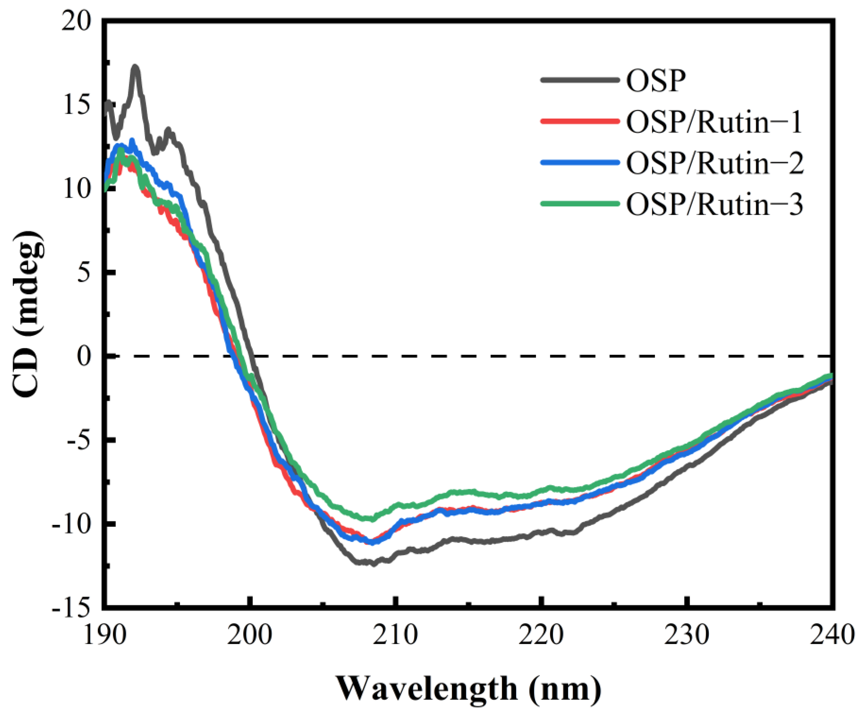
3.3. CD Analysis
3.4. FS Analysis
3.5. Total Phenolic Content
3.6. Surface Hydrophobicity Analysis
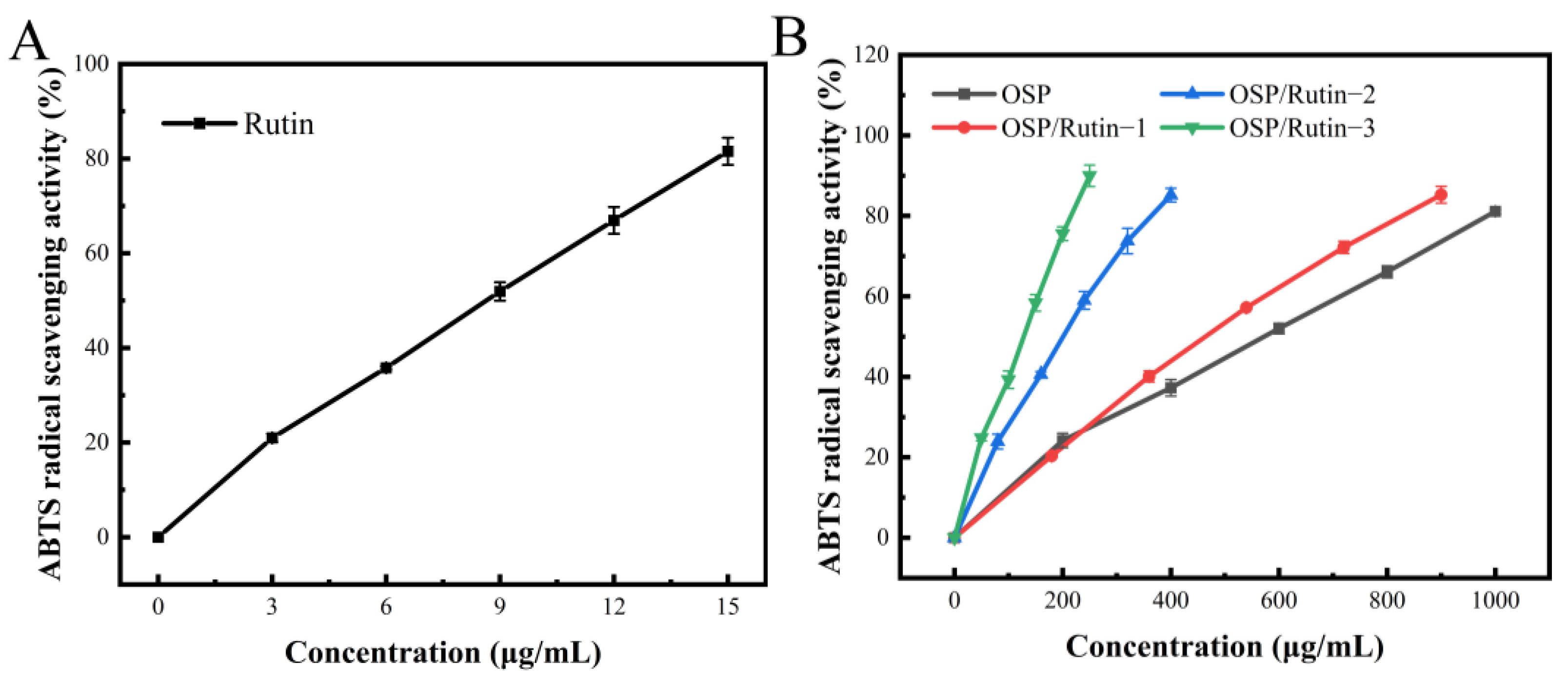
3.7. ABTS Free Radical Scavenging Ability
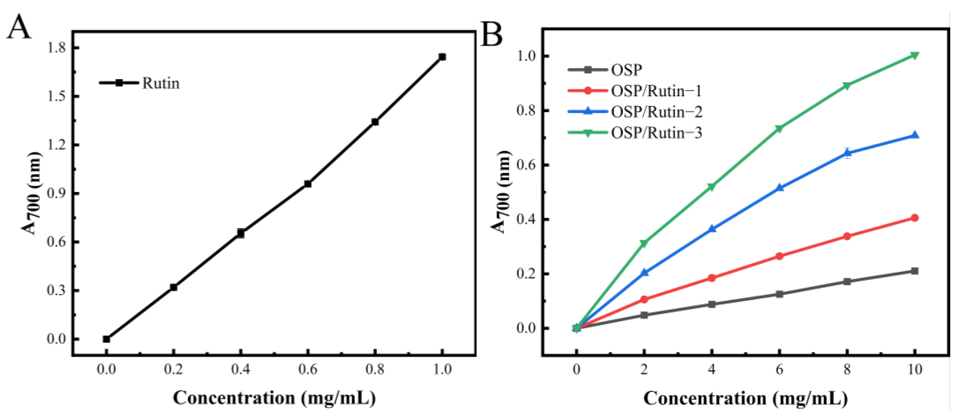
3.8. Reducing Power
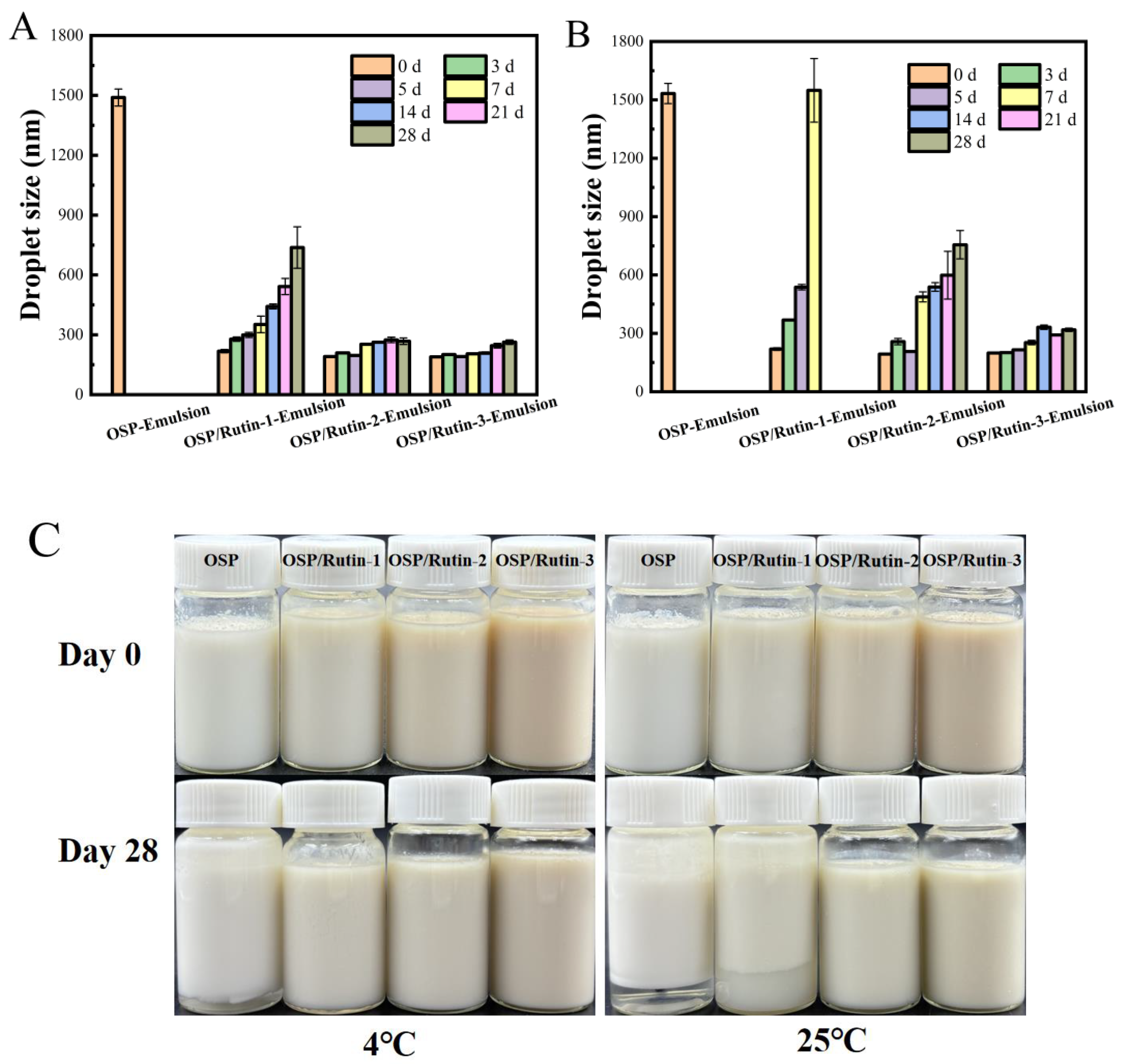
3.9. Storage Stability Analysis
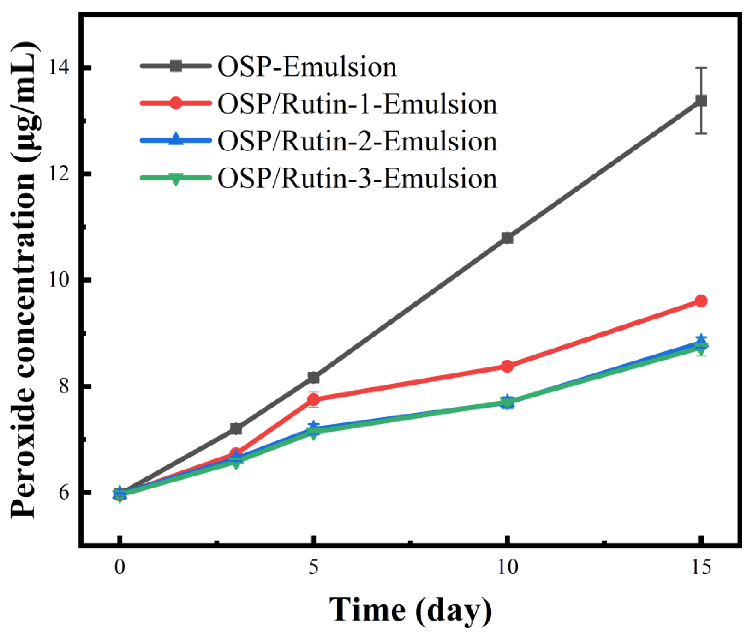
3.10. Anti-Lipid Oxidation Capacity
3.11. Lutein Stability Analysis
4. Conclusions
Author Contributions
Funding
Institutional Review Board Statement
Informed Consent Statement
Data Availability Statement
Acknowledgments
Conflicts of Interest
References
- Kong, F.; Kang, S.; Zhang, J.; Jiang, L.; Liu, Y.; Yang, M.; Cao, X.; Zheng, Y.; Shao, J.; Yue, X. The non-covalent interactions between whey protein and various food functional ingredients. Food Chem. 2022, 394, 133455. [Google Scholar] [CrossRef]
- Gammoh, S.; Alu’datt, M.H.; Alhamad, M.N.; Rababah, T.; Al-Mahasneh, M.; Qasaimeh, A.; Johargy, A.; Kubow, S.; Hussein, N.M. The effects of protein-phenolic interactions in wheat protein fractions on allergenicity, antioxidant activity and the inhibitory activity of angiotensin I-converting enzyme (ACE). Food Biosci. 2018, 24, 50–55. [Google Scholar] [CrossRef]
- Kroll, J.; Rawel, H.; Rohn, S. Reactions of Plant Phenolics with Food Proteins and Enzymes under Special Consideration of Covalent Bonds. Food Sci. Technol. Res. 2003, 9, 205–218. [Google Scholar] [CrossRef]
- Aslandag, S.K.; Vural, H.; Yildirim-Elikoglu, S. Effect of green tea extract on heat-induced protein interactions and rennet coagulation in milk. Int. Dairy J. 2023, 145, 105732. [Google Scholar] [CrossRef]
- Rovaletti, M.M.L.; Benitez, E.I.; Amezaga, N.M.J.M.; Peruchena, N.M.; Sosa, G.L.; Lozano, J.E. Polysaccharides influence on the interaction between tannic acid and haze active proteins in beer. Food Res. Int. 2014, 62, 779–785. [Google Scholar] [CrossRef]
- Cheng, L.; Lian, Z.; Liu, X.; Dai, S.; Li, L.; Wang, M.; Li, K.; Ren, K.; Tong, X.; Wang, H.; et al. Effect of phlorotannins modification on the physicochemical, structural and functional properties of soybean protein isolate and controlled hydrolysates: Covalent and non-covalent interactions. Food Hydrocoll. 2024, 149, 109591. [Google Scholar] [CrossRef]
- Pessato, T.B.; de Morais, F.P.R.; de Carvalho, N.C.; Figueira, A.C.M.; Fernandes, L.G.R.; Zollner, R.D.; Netto, F.M. Protein structure modification and allergenic properties of whey proteins upon interaction with tea and coffee phenolic compounds. J. Funct. Foods 2018, 51, 121–129. [Google Scholar] [CrossRef]
- Li, L.; Chai, W.; Ma, L.; Zhang, T.; Chen, J.; Zhang, J.; Wu, X. Covalent polyphenol with soybean 11S protein to develop hypoallergenic conjugates for potential immunotherapy. J. Funct. Foods 2023, 104, 105518. [Google Scholar] [CrossRef]
- Ballon, A.; Romero, M.P.; Rodriguez-Saona, L.E.; de Lamo-Castellvi, S.; Guell, C.; Ferrando, M. Conjugation of lesser mealworm (Alphitobius diaperinus) larvae protein with polyphenols for the development of innovative antioxidant emulsifiers. Food Chem. 2024, 434, 137494. [Google Scholar] [CrossRef]
- Ofori, J.; Tortoe, C.; Agbenorhevi, J.K. Physicochemical and functional properties of dried okra (Abelmoschus esculentus L.) seed flour. Food Sci. Nutr. 2020, 8, 4291–4296. [Google Scholar] [CrossRef]
- Yao, H.L.; Yang, J.I.; Zhan, J.J.; Lu, Q.; Su, M.; Jiang, Y.J. Preparation, amino acid composition, and in vitro antioxidant activity of okra seed meal protein hydrolysates. Food Sci. Nutr. 2021, 9, 3059–3070. [Google Scholar] [CrossRef] [PubMed]
- Ijarotimi, O.S.; Akinola-Ige, A.O.; Oluwajuyitan, T.D. Okra seeds proteins: Amino acid profile, free radical scavenging activities and inhibition of diabetes and hypertensive converting enzymes indices. Meas. Food 2023, 11, 100101. [Google Scholar] [CrossRef]
- He, C.; Bai, L.; Liu, D.; Liu, B. Interaction mechanism of okra (Abelmoschus esculentus L.) seed protein and flavonoids: Fluorescent and 3D-QSAR studies. Food Chem X 2023, 20, 101023. [Google Scholar] [CrossRef] [PubMed]
- Bai, L.; Geng, S.; Zhou, Y.X.; Ma, H.J.; Liu, B.G. Ultrasound-assisted fabrication and stability evaluation of okra seed protein stabilized nanoemulsion. Ultrason. Sonochem. 2024, 104, 106807. [Google Scholar] [CrossRef]
- Gullón, B.; Lu-Chau, T.A.; Moreira, M.T.; Lema, J.M.; Eibes, G. Rutin: A review on extraction, identification and purification methods, biological activities and approaches to enhance its bioavailability. Trends Food Sci. Technol. 2017, 67, 220–235. [Google Scholar] [CrossRef]
- Yang, S.; Song, J.; Yang, S.; Li, J.; Jiang, H.; Sui, F.; Li, L. Research progress on pharmacological action and new dosage forms of rutin. Chin. J. Mod. Appl. Pharm. 2022, 39, 1360–1370. [Google Scholar] [CrossRef]
- Li, D.; Zhu, L.; Wu, Q.; Chen, Y.; Wu, G.; Zhang, H. Tartary buckwheat protein-phenol conjugate prepared by alkaline-based environment: Identification of covalent binding sites of phenols and alterations in protein structural and functional characteristics. Int. J. Biol. Macromol. 2024, 257, 127504. [Google Scholar] [CrossRef]
- Miles, A.J.; Ramalli, S.G.; Wallace, B.A. DichroWeb, a website for calculating protein secondary structure from circular dichroism spectroscopic data. Protein Sci. 2021, 31, 37–46. [Google Scholar] [CrossRef] [PubMed]
- Whitmore, L.; Wallace, B.A. Protein secondary structure analyses from circular dichroism spectroscopy: Methods and Reference Databases. Biopolymers 2008, 89, 392–400. [Google Scholar] [CrossRef]
- Whitmore, L.; Wallace, B.A. DICHROWEB: An online server for protein secondary structure analyses from circular dichroism spectroscopic data. Nucleic Acids Res. 2004, 32, W668–W673. [Google Scholar] [CrossRef]
- Chen, W.; Li, T.; Yu, H.; Ma, C.; Wang, X.; Qayum, A.; Hou, J.; Jiang, Z. Structure and emulsifying properties of whey protein isolate: Effect of safflower yellow concentration. LWT-Food Sci. Technol. 2020, 123, 109079. [Google Scholar] [CrossRef]
- Guo, C.; Han, F.; Geng, S.; Shi, Y.; Ma, H.; Liu, B. The physicochemical properties and Pickering emulsifying capacity of acorn starch. Int. J. Biol. Macromol. 2023, 239, 124289. [Google Scholar] [CrossRef] [PubMed]
- Zhang, Y.; Zhang, T.; Dong, C.; Zhao, R.; Zhang, X.; Wang, C. Lycopene-loaded emulsions stabilized by whey protein covalently modified with pectin or/and chlorogenic acid: Enhanced physicochemical stability and reduced bio-accessibility. Food Chem. 2023, 417, 135879. [Google Scholar] [CrossRef]
- Fogarasi, A.L.; Kun, S.; Tanko, G.; Stefanovits-Banyai, E.; Hegyesne-Vecseri, B. A comparative assessment of antioxidant properties, total phenolic content of einkorn, wheat, barley and their malts. Food Chem. 2015, 167, 1–6. [Google Scholar] [CrossRef]
- Zhou, Y.; Bai, L.; Geng, S.; Liu, B. Characterization and Pickering emulsifying ability of Adinandra nitida leaf polysaccharides. Food Chem. X 2025, 25, 102090. [Google Scholar] [CrossRef] [PubMed]
- Li, J.; Geng, S.; Zhen, S.; Lv, X.; Liu, B. Fabrication and characterization of oil-in-water emulsions stabilized by whey protein isolate/phloridzin/sodium alginate ternary complex. Food Hydrocoll. 2022, 129, 107625. [Google Scholar] [CrossRef]
- Wang, Y.; Liu, B.; Ma, Y.; Wang, C.; Ma, H.; Geng, S. Oil/water interface behavior of hesperidin methylchalcone and its application in nano-emulsions. Food Chem. 2025, 463, 141235. [Google Scholar] [CrossRef]
- Rodger, A.; Sanders, K. UV-Visible absorption spectroscopy, biomacromolecular applications. Encycl. Spectrosc. Spectrom. 2017, 4, 495–502. [Google Scholar] [CrossRef]
- Shi, J.; Cui, Y.; Zhou, G.; Ling, N.; Sun, X.; Wang, X.; Xu, N. Covalent interaction of soy protein isolate and chlorogenic acid: Effect on protein structure and functional properties. LWT-Food Sci. Technol. 2022, 170, 114081. [Google Scholar] [CrossRef]
- Sadat, A.; Joye, I.J. Peak fitting applied to Fourier transform infrared and Raman spectroscopic analysis of proteins. Appl. Sci. 2020, 10, 5918. [Google Scholar] [CrossRef]
- Han, L.; Peng, X.; Cheng, Y.; Zhu, Y.; Huang, Y.; Zhang, S.; Qi, B. Effects of catechin types found in tea polyphenols on the structural and functional properties of soybean protein isolate-catechin covalent complexes. LWT-Food Sci. Technol. 2023, 173, 114336. [Google Scholar] [CrossRef]
- Ranjbar, B.; Gill, P. Circular dichroism techniques: Biomolecular and nanostructural analyses—A review. Chem. Biol. Drug Des. 2009, 74, 101–120. [Google Scholar] [CrossRef]
- Chen, H.; Xu, Z.; Mo, J.; Lyu, Y.; Tang, X.; Shen, X. Effects of guar gum on adhesion properties of soybean protein isolate onto porcine bones. Int. J. Adhes. Adhes. 2017, 75, 124–131. [Google Scholar] [CrossRef]
- Liu, K.; Chen, S.; Chen, H.; Tong, P.; Gao, J. Cross-linked ovalbumin catalyzed by polyphenol oxidase: Preparation, structure and potential allergenicity. Int. J. Biol. Macromol. 2018, 107, 2057–2064. [Google Scholar] [CrossRef]
- Rawel, H.M.; Rohn, S.; Kruse, H.P.; Kroll, J. Structural changes induced in bovine serum albumin by covalent attachment of chlorogenic acid. Food Chem. 2002, 78, 443–455. [Google Scholar] [CrossRef]
- Cheng, J.; Dudu, O.E.; Zhang, J.; Wang, Y.; Meng, L.; Wei, W.; Li, X.; Yan, T. Impact of binding interaction modes between whey protein concentrate and quercetin on protein structural and functional characteristics. Food Hydrocoll. 2023, 142, 108787. [Google Scholar] [CrossRef]
- Zhang, Q.Z.; Cheng, Z.Z.; Wang, Y.B.; Fu, L.L. Dietary protein-phenolic interactions: Characterization, biochemical-physiological consequences, and potential food applications. Crit. Rev. Food Sci. Nutr. 2021, 61, 3589–3615. [Google Scholar] [CrossRef]
- Voutsinas, L.P.; Cheung, E.; Nakai, S. Relationships of hydrophobicity to emulsifying properties of heat denatured proteins. J. Food Sci. 1983, 48, 26–32. [Google Scholar] [CrossRef]
- Wang, X.; Han, D.; Chen, Z.; Jiang, L.; Xu, J. Influence of Ultrasonic Treatment on β-conglycinin Emulsibility. J. Chin. Inst. Food Sci. Technol. 2018, 18, 153–159. [Google Scholar] [CrossRef]
- Sun, Y.; Zhao, M.; Liu, Z.; Shi, H.; Zhang, X.; Zhao, Y.; Ma, Z.; Yu, G.; Xia, G.; Shen, X. Relationship between the interfacial properties of lactoferrin-(-)-epigallocatechin-3-gallate covalent complex and the macroscopic properties of emulsions. Food Chem. 2024, 460, 140536. [Google Scholar] [CrossRef]
- Van der Werf, R.; Marcic, C.; Khalil, A.; Sigrist, S.; Marchioni, E. ABTS radical scavenging capacity in green and roasted coffee extracts. LWT-Food Sci. Technol. 2014, 58, 77–85. [Google Scholar] [CrossRef]
- Rice-Evans, C.; Miller, N.; Paganga, G. Antioxidant properties of phenolic compounds. Trends Plant Sci. 1997, 2, 152–159. [Google Scholar] [CrossRef]
- Lu, N.H.; Wu, L.M.; Zhen, S.Y.; Liu, B.G. Characterization of a dihydromyricetin/α-lactoalbumin covalent complex and its application in nano-emulsions. Foods 2023, 12, 2783. [Google Scholar] [CrossRef]
- Yi, J.; Fan, Y.; Zhang, Y.; Zhao, L. Characterization of catechin-α-lactalbumin conjugates and the improvement in β-carotene retention in an oil-in-water nanoemulsion. Food Chem. 2016, 205, 73–80. [Google Scholar] [CrossRef]
- Helgeson, M.E. Colloidal behavior of nanoemulsions: Interactions, structure, and rheology. Curr. Opin. Colloid Interface Sci. 2016, 25, 39–50. [Google Scholar] [CrossRef]
- Sivakumar, M.; Tang, S.Y.; Tan, K.W. Cavitation technology—A greener processing technique for the generation of pharmaceutical nanoemulsions. Ultrason. Sonochem. 2014, 21, 2069–2083. [Google Scholar] [CrossRef]
- Davidov-Pardo, G.; Gumus, C.E.; McClements, D.J. Lutein-enriched emulsion-based delivery systems: Influence of pH and temperature on physical and chemical stability. Food Chem. 2016, 196, 821–827. [Google Scholar] [CrossRef] [PubMed]
- McClements, D.J.; Huang, X. Potential biological fate of ingested nanoemulsions: Influence of particle characteristics. Food Funct. 2012, 3, 202–220. [Google Scholar] [CrossRef]
- Geng, S.; Yuan, Y.; Jiang, X.; Zhang, R.; Ma, H.; Liang, G.; Liu, B. An investigation on pickering nano-emulsions stabilized by dihydromyricetin/high-amylose corn starch composite particles: Preparation conditions and carrier properties. Curr. Res. Food Sci. 2023, 6, 100458. [Google Scholar] [CrossRef]
- Calvo, M.M. Lutein: A valuable ingredient of fruit and vegetables. Crit. Rev. Food Sci. Nutr. 2005, 45, 671–696. [Google Scholar] [CrossRef]
- Frede, K.; Henze, A.; Khalil, M.; Baldermann, S.; Schweigert, F.J.; Rawel, H. Stability and cellular uptake of lutein-loaded emulsions. J. Funct. Foods 2014, 8, 118–127. [Google Scholar] [CrossRef]


| Samples | α-Helix (%) | β-Sheet (%) | β-Turn (%) | Random (%) |
|---|---|---|---|---|
| OSP | 17.9 a | 30.9 c | 12.7 b | 38.5 c |
| OSP/Rutin-1 | 15.9 b | 32.1 b | 12.9 ab | 39.1 b |
| OSP/Rutin-2 | 15.4 c | 32.2 b | 13.0 a | 39.3 a |
| OSP/Rutin-3 | 15.0 d | 33.0 a | 12.9 ab | 39.2 ab |
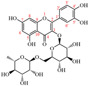
| Rutin Structure | Sample | Total Phenol Content (mg/g) |
|---|---|---|
 | OSP/Rutin-1 | 64.81 ± 1.51 c |
| OSP/Rutin-2 | 93.40 ± 0.42 b | |
| OSP/Rutin-3 | 123.92 ± 1.38 a |
| Sample | IC50 (μg/mL) |
|---|---|
| Rutin | 7.67 ± 0.15 e |
| OSP | 497.24 ± 5.30 a |
| OSP/Rutin-1 | 419.96 ± 5.99 b |
| OSP/Rutin-2 | 177.72 ± 5.40 c |
| OSP/Rutin-3 | 108.71 ± 0.81 d |
Disclaimer/Publisher’s Note: The statements, opinions and data contained in all publications are solely those of the individual author(s) and contributor(s) and not of MDPI and/or the editor(s). MDPI and/or the editor(s) disclaim responsibility for any injury to people or property resulting from any ideas, methods, instructions or products referred to in the content. |
© 2025 by the authors. Licensee MDPI, Basel, Switzerland. This article is an open access article distributed under the terms and conditions of the Creative Commons Attribution (CC BY) license (https://creativecommons.org/licenses/by/4.0/).
Share and Cite
He, C.; Bai, L.; Zhou, Y.; Liu, B.; Geng, S. Characterization of Okra Seed Protein/Rutin Covalent Complex and Its Application in Nanoemulsions. Foods 2025, 14, 1672. https://doi.org/10.3390/foods14101672
He C, Bai L, Zhou Y, Liu B, Geng S. Characterization of Okra Seed Protein/Rutin Covalent Complex and Its Application in Nanoemulsions. Foods. 2025; 14(10):1672. https://doi.org/10.3390/foods14101672
Chicago/Turabian StyleHe, Chengyun, Lu Bai, Yingxuan Zhou, Benguo Liu, and Sheng Geng. 2025. "Characterization of Okra Seed Protein/Rutin Covalent Complex and Its Application in Nanoemulsions" Foods 14, no. 10: 1672. https://doi.org/10.3390/foods14101672
APA StyleHe, C., Bai, L., Zhou, Y., Liu, B., & Geng, S. (2025). Characterization of Okra Seed Protein/Rutin Covalent Complex and Its Application in Nanoemulsions. Foods, 14(10), 1672. https://doi.org/10.3390/foods14101672