Article

Transcriptome Reveals the Key Genes Related to the Metabolism of Volatile Sulfur-Containing Compounds in Lentinula edodes Mycelium

1
College of Food Science & Technology, Huazhong Agricultural University, Wuhan 430070, China
2
State Key Laboratory of Resource Insects, Institute of Apicultural Research, Chinese Academy of Agricultural Sciences, Beijing 100193, China
3
Department of Nutrition, Food Science and Packaging, San Jose State University, San Jose, CA 95192, USA
4
Institute of Applied Mycology, Huazhong Agricultural University, Wuhan 430070, China
*
Author to whom correspondence should be addressed.
These authors contributed equally to this work.
Foods 2024, 13(14), 2179; https://doi.org/10.3390/foods13142179
Submission received: 5 June 2024 / Revised: 3 July 2024 / Accepted: 8 July 2024 / Published: 10 July 2024
(This article belongs to the Section Foodomics)

Abstract

:
Lentinula edodes (L. edodes) is a globally popular edible mushroom because of its characteristic sulfur-containing flavor compounds. However, the formation of the volatile sulfur-containing compounds in the mycelium of L. edodes has not been studied. We found that there were also sulfur-containing aroma compounds in the mycelium of L. edodes, and the content and composition varied at different stages of mycelial growth and development. The γ-glutamyl-transpeptidase (GGT) and cysteine sulfoxide lyase (C-S lyase) related to the generation of sulfur compounds showed the highest activities in the 15-day sample. Candidate genes for the metabolism of volatile sulfur compounds in mycelium were screened using transcriptome analysis, including encoding the GGT enzyme, C-S lyase, fatty acid oxidase, HSP20, and P450 genes. The expression patterns of Leggt3 and Leccsl3 genes were consistent with the measured activities of GGT and C-S lyase during the cultivation of mycelium and molecular dynamics simulations showed that they could stably bind to the substrate. Our findings provide insights into the formation of sulfur-containing flavor compounds in L. edodes. The mycelium of L. edodes is suggested for use as material for the production of sulfur-containing flavor compounds.

1. Introduction

Lentinula edodes (L. edodes) is one of the most popular mushrooms with rich nutrients [1] and characteristic flavor [2]. Sulfur-containing compounds, mainly including thioethers, thiols, and sulfur-containing heterocyclic compounds, contribute to their characteristic flavor [3]. Among sulfide compounds, dimethyl sulfide, dimethyl trisulfide, and methanethiol have a higher content and a sulfur flavor, making them characteristic odor substances of L. edodes [4]. Sulfur-containing compounds are the main flavor components in dried L. edodes, and their formation is mainly influenced by the heating temperature and time [5,6]. Currently, it is widely believed that the formation of sulfur-containing flavor substances mainly goes through two stages. Firstly, γ-glutamyl-transpeptidase (GGT) catalyzes lentinic acid to γ-glutamy lentinic acid, and cysteine sulfoxide lyase (C-S lyase) catalyzes γ-glutamy lentinic acid to products such as thiosulfate. These products then undergo a series of non-enzymatic reactions, followed by condensation and cleavage, ultimately forming a series of sulfur-containing flavor compounds [7,8]. Our previous studies showed that these two enzymes were not only the key enzymes for the formation of sulfur-containing volatile compounds in L. edodes but also involved in the formation of endogenous formaldehyde, which is associated with food safety and human health [9,10]. The GGT and C-S lyase families of L. edodes have seven (Leggt1–7) and five (Lecsl1–5) genes, which use γ-glutamyl diaminopimelic acid and S-methyl-L-cysteine sulfoxide as substrates, respectively. In a previous study, we clarified the role of Lecsl2 in the formation of L. edodes flavor and tested the enzymatic activity of GGT at different stages of L. edodes [10,11]. However, the functions of other genes in the C-S lyase family and the specific catalytic mechanisms of GGT are still under investigation.
Research on the flavor of L. edodes mainly focused on the fruiting body stage, to understand the formation mechanisms of sulfur-containing flavor compounds in L. edodes. Researchers analyzed the differences in volatile components at different development stages of L. edodes fruiting bodies. The growth and development of L. edodes fruiting bodies take a long time and undergo physiological metabolism during storage and processing [8], thus it is difficult to use L. edodes fruiting bodies to produce sulfur-containing flavor compounds. Compared with the fruiting body, L. edodes mycelium has a shorter growth cycle, easier culture and regeneration, and easier genome-editing-related operations; thus, it is a better research object for revealing the formation mechanism of aroma components in L. edodes and also could be processed as a food-flavoring agent or additive [12,13,14,15]. Studies indicated that L. edodes mycelium also contains amino acids, fatty acids, polysaccharides, and other components beneficial to human health, while studies on mycelium mainly focused on fermentation to produce polysaccharides and other active components. Regarding the flavor of the mycelium, studies have shown that L. edodes mycelium contains non-volatile taste components [16,17,18,19,20,21]. However, it is unclear whether characteristic volatile substances can be affected by the growth and development of mycelium [22,23]. In order to make better use of L. edodes mycelium, it is necessary to understand its flavor formation.
The objectives of this study were to explore the differences in flavor components in the prophase of L. edodes mycelial growth and screen candidate genes related to mycelium flavor formation and reveal the key gene action mechanism. Our findings will enable further studies of the formation mechanisms of L. edodes flavor and usage of L. edodes mycelium.

2. Materials and Methods

2.1. Strain and Culture

L. edodes, strain W1 (ACCC 50926) was provided by the Institute of Applied Mycology, Huazhong Agricultural University. The strain W1 was cultured on MYG medium 2% malt extract (Shuangxuan company, Beijing, China), 2% glucose (Sinopharm Chemical Reagent Co., Ltd., Shanghai, China), 0.1% yeast extract (Angelyeast, Yichang, China), 0.1% peptone (Shuangxuan company, Beijing, China), 2% agar (Biofroxx, Heidelberg, Germany) in 9 cm plates for 15, 35, and 55 days, respectively.

2.2. Microstructure Analysis by Scanning Electron Microscope (SEM)

The mycelium was transferred into 2.5% glutaraldehyde solution and fixed for 4 h. After the mixture was dried and gold sprayed, it was viewed under SEM to observe the microstructure. The magnification of the scanning electron microscope was 500.

2.3. Flavor Extraction and Analysis

2-butanone, 2-pentanone, 2-hexanone, 2-heptanone, 2-octanone, 2-nonone mixed standard (1 mg/L) used for analysis was purchased from German G.A.S (Hong Kong, China). One gram of dried mycelium was used to determine the volatile components by gas chromatography–ion mobility spectroscopy (GC-IMS). The headspace sampling conditions were as follows: incubation 10 min at 60 °C and sampling probe temperature 85 °C. GC-IMS conditions were as follows: MXT-5 chromatographic column (15 m × 0.53 mm, 1.0 μm), column temperature 60 °C, analysis time 30 min, IMS temperature 45 °C, and drift gas flow rate 150 mL/min. The carrier gas flow rate was 2 mL/min for 2 min and increased to 10 mL/min across the next 8 min, then increased to 100 mL/min across the next 15 min, and maintained there for an additional 5 min. A GC-IMS instrument supporting analysis software Vocal (version 0.1.3) was used for information collection and analysis. The application software had built-in Reporter and Gallery Plot plug-ins to plot the two-dimensional and fingerprint chromatograms of volatile components in the samples. C4~C9 ketones were used as the external standard standards to calculate the retention index of volatile components, which were used to match with the built-in National Institute of Standards and Technology (NIST) and IMS databases to conduct qualitative analysis of volatile components.

2.4. Enzyme Activities

The activities of GGT and C-S lyase were determined using our previous methods [10]. γ-glutamyl p-nitroaniline was used as a substrate to determine the GGT activity. S-methyl-L-cysteinyl was used as a substrate to measure the C-S lyase activity. Specific enzyme activity (U/g) was defined as the production of 1μ Mol p-nitroaniline or pyruvate per gram of total mycelium per minute.

2.5. RNA-seq and Transcriptomes Analysis

The RNA-seq sequencing experiment process included sample detection, library construction, quality inspection, and computer sequencing. The Illumina NovaSeq6000 sequencing platform (San Diego, CA, USA) was used for sequencing in PE150 mode. The original data were filtered to obtain clean data, and the sequence was aligned with the previously published reference genome of L. edodes strain W1-26 [24]. We used three different methods, edgeR, DESeq2, and log2FC, to calculate the number of differentially expressed genes. The intersection of the results from these three methods was taken. For edgeR and DESeq2, the threshold was p ≤ 0.001. For log2FC, the threshold was set to an absolute value greater than or equal to 2, which means a fold change of at least 4.

2.6. Transcriptome Validation

Due to individual differences among samples, in order to verify the reliability of transcriptome results, quantitative real-time PCR was used to measure the expression of related genes in samples with the same treatment but without transcriptome. The total RNA was extracted and reverse-transcribed to cDNA by HiScript II Q RT SuperMix for Qpcr (+gDNA wiper). Next, the AceQ qPCR SYBR Green Master Mix was used to detect the relative expression levels. Post-qRT-PCR calculations of relative gene expression levels were performed according to the method described in [25]. The primers for qRT-PCR were designed by Primer 3 (https://www.bioinformatics.nl/cgi-bin/primer3plus/primer3plus.cgi, accessed on 7 July 2024).

2.7. Molecular Docking and Molecular Dynamic Simulation

The 3D structures of GGT and CSL proteins were constructed using AlphaFold2 [26], with Global plDDT values of 91.699 and 92.671, respectively, indicating the reliability of the generated structures. The structures of S-methyl-L-cysteine sulfoxide and p-nitroaniline were built using the Avogadro program [27], and energy minimization was performed using the MMFF94s force field to optimize bond angles and lengths. Subsequently, semi-flexible molecular docking was carried out using the Autodock Vina [28] program. For the docking process, the docking coordinates for the Leggt3 protein were set as x = −1.8, y = 10.6, and z = 1.4, while for the Lecsl3 protein, the coordinates were x = −11.8, y = 2.8, and z = −9.6. The exhaustiveness parameter was set to 50, which determines the thoroughness of the docking search. During docking, the protein structure remained rigid while allowing the ligand’s rotatable bonds to move. After the docking process, the optimal complex construction was selected for molecular dynamics (MD) simulation study based on the binding energy and binding pose.
Based on molecular docking, molecular dynamics simulations were performed on GGT-γ-glutamyl p-nitroaniline and CSL-S-methyl-L cysteine sulfoxide. The geometric optimization of p-nitroaniline and methyl-L-cysteine sulfoxide was carried out using the ORCA [29] program with the r2SCAN-3c functional, and their single point energies were calculated using B3LYP/G D3 def2-TZVP def2/J RIJCOSX. The topological information of nitroaniline and S-methyl-L cysteine sulfoxide was generated using Ambertools [30] and their RESP charges were fitted using the Multiwfn program [31]. The systems were placed in a cubic TIP3P water tank and counter ions were added to neutralize the total charge of the system, maintaining a neutral system. Energy minimization was performed using the steepest descent method (1000.0 kJ/mol/nm) followed by conjugate gradient optimization (100.0 kJ/mol/nm) for the second energy minimization. Subsequently, the system was pre-equilibrated using NVT (1 ns, 298 K) and NPT (1 ns, 1 Bar) simulations, respectively.

2.8. Statistical Analysis

The experiments were conducted in triplicate and values were expressed as means ± SD (standard deviation). Data were analyzed using GraphPad Prism 5. The KEGG analysis was plotted using bioinformatics (https://www.bioinformatics.com.cn) [32]. The volcano map was drawn using Image GP (https://www.bic.ac.cn/ImageGP/, accessed on 7 July 2024) [33].

3. Results

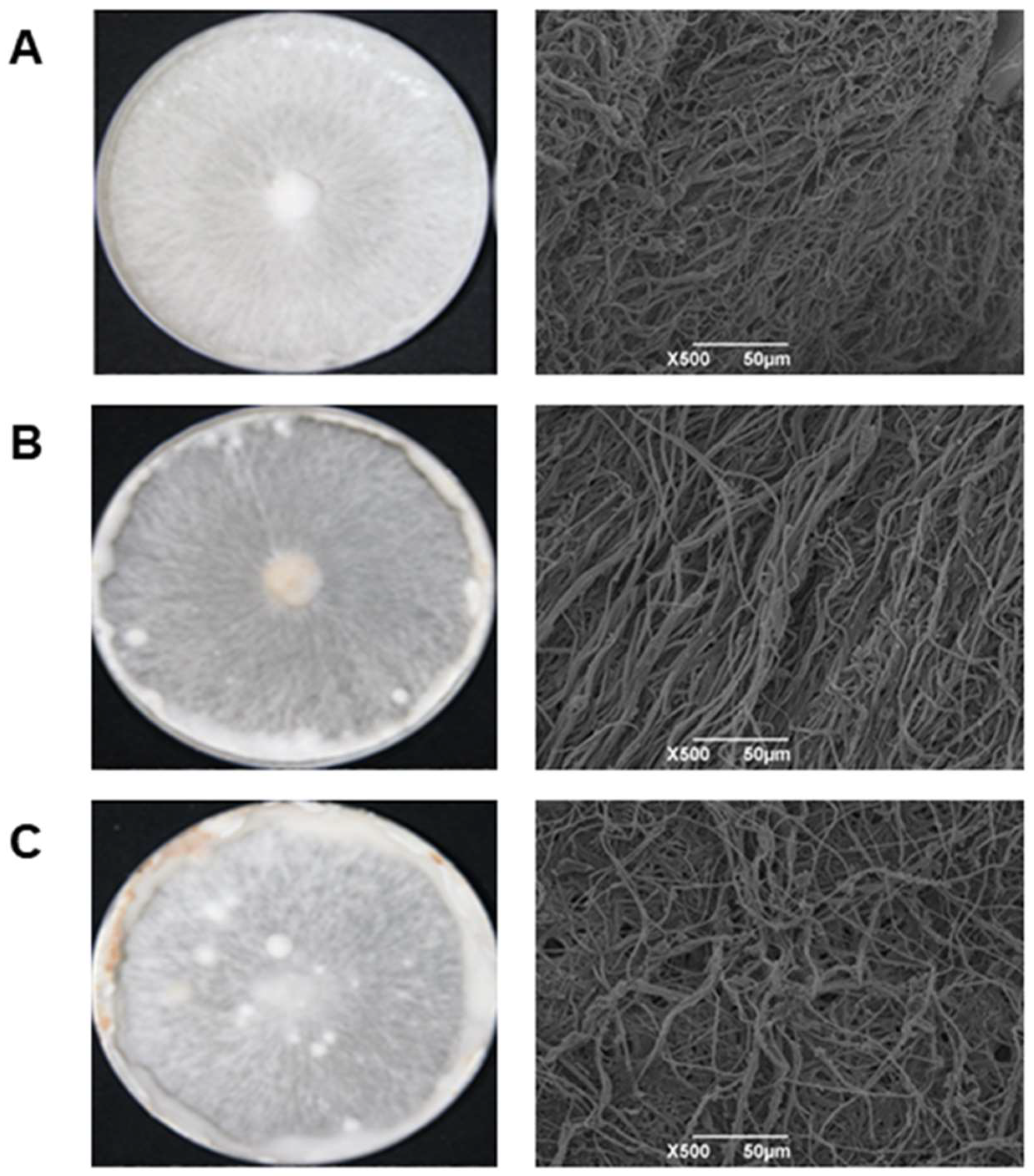
3.1. Morphology and Microstructure Analysis of Mycelium

The photographs of L. edodes mycelium collected at three different developmental stages are shown in Figure 1. As can be seen, the morphology of L. edodes mycelium changed significantly with the cultivated time. After 15 days of culturing, the mycelium extended to the edge of the culture dish. At this period, the mycelium was relatively dense, which was illustrated in the SEM results. As the space in the culture dish was limited, the mycelium began to differentiate [34]. When the culture time reached 35 days, the mycelium became denser. After 55 days of cultivation, the mycelium aggregated intensively and caused a significant increase in the number of kinks in the culture dish, and the mycelium at the edge turned brown [35,36]. During the early development stages of L. edodes mycelium, its biomass gradually increases and accumulates.

3.2. Analysis of Volatile Components in Mycelium

In this experiment, we detected volatile components in L. edodes at different mycelium stages through GC-IMS. As shown in Figure 2, the volatile components of the mycelium mainly varied with the culturing time. The main volatile components of L. edodes mycelium were acids, aldehydes, alcohols, and ketones, which were produced through amino acid, fatty acid, and carbohydrate metabolisms [37]. The levels of hexanal-M, 3-methylbutanal-M, 2-Methyl-2-propenal-M, and 3-methylbutanal-D did not change over time, indicating that they might be stable components of the L. edodes mycelium. L. edodes mycelium cultured for 15 and 55 days contained a higher level of n-Hexanol than that cultured for 35 days. The sulfur-containing volatile compounds in the mycelium cultured for 15 days were dimethyl trisulfide and 1-propanethiol. Dimethyl disulfide, diallyl sulfide, and 2-propanethiol were detected in the mycelium cultured for 35 and propylsulfide was detected in the mycelium cultured for 55 days [38,39]. Our results indicated that sulfur-containing volatile compounds in L. edodes were generated at the mycelium stage as well as hexanal-M, 3-methylbutanal-M, 2-methyl-2-propenal-M, and 3-methylbutanal-D. In summary, the L. edodes mycelium cultured for 15 days had the highest levels of total volatile components and sulfur-containing volatile compounds, possibly because of the vitality of L. edodes mycelium.

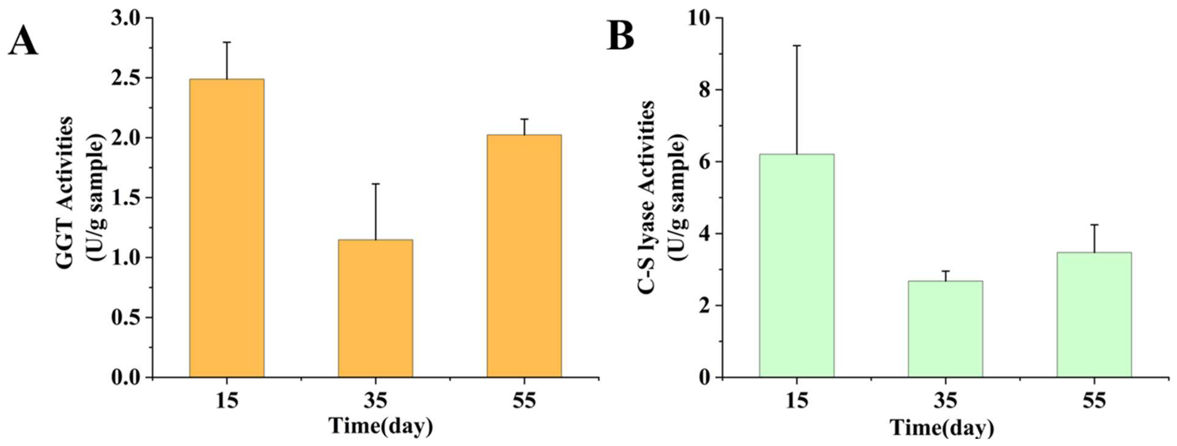
3.3. Enzyme Activities Analysis

The GGT and C-S lyase have been reported to be the key enzymes in the formation of volatile compounds in L. edodes; to reveal the mechanism of flavor formation in L. edodes mycelium, their activities in mycelium cultured for 15, 35, and 55 days were measured. The GGT and C-S lyase activities in different mycelium samples are shown in Figure 3. The GGT and C-S lyase activities of the 15-day sample were much higher than those of the 35- and 55-day samples, which could be why more volatile components were produced in the mycelium after 15 days. The GGT and C-S lyase activities of the 35-day sample were the lowest in the present study, and it was clear from the GC-IMS that this sample contained only a little n-Hexanol.

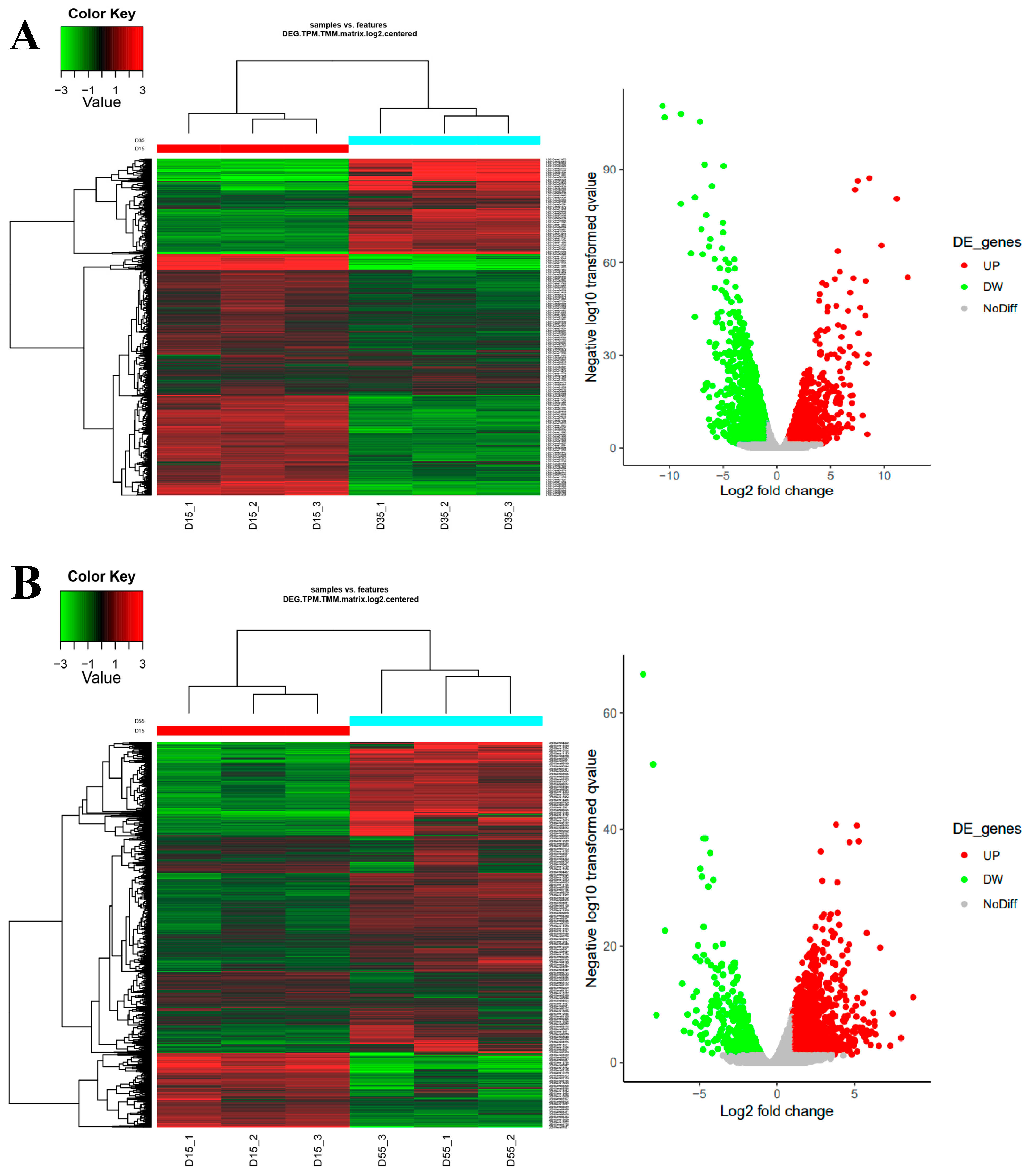
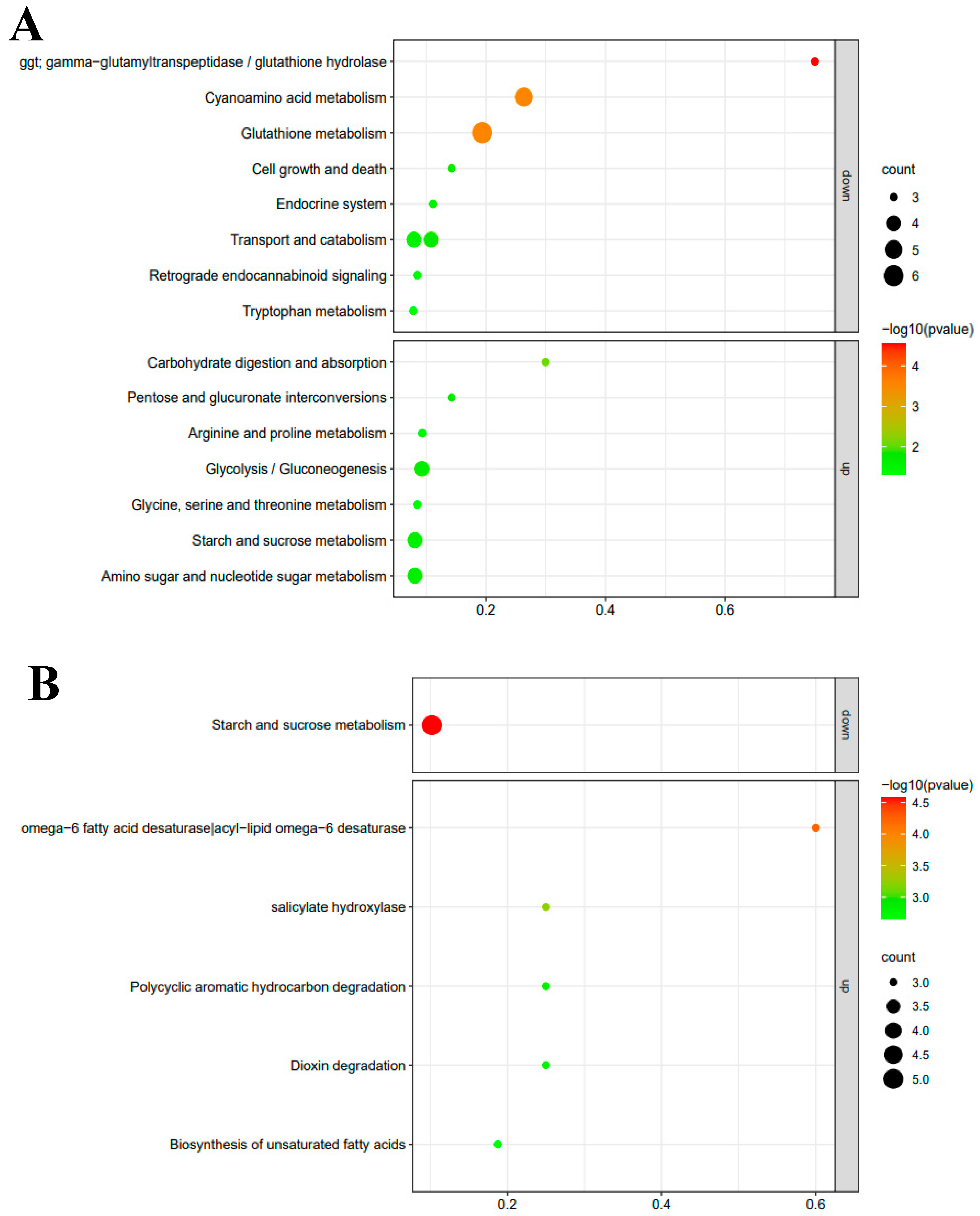
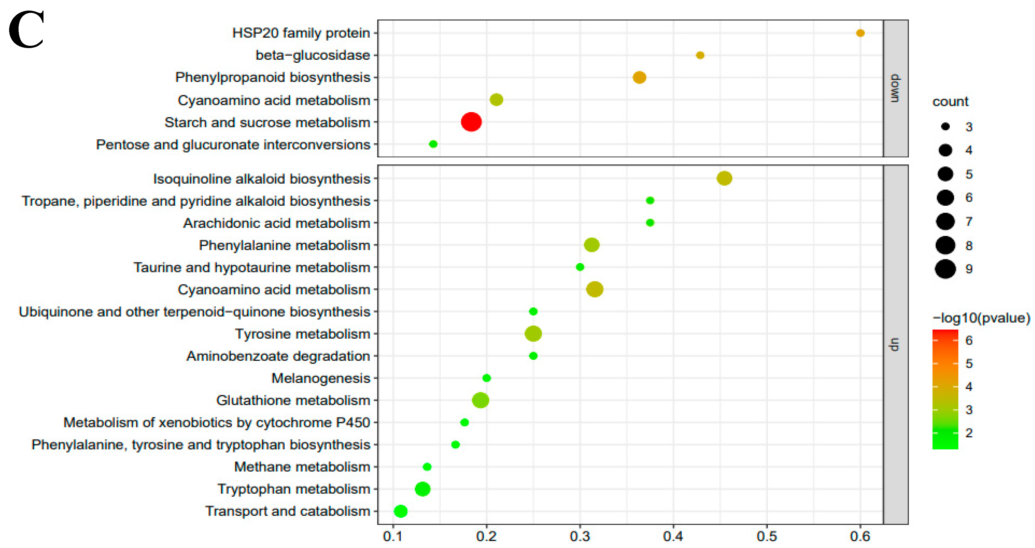
3.4. RNA-seq and Transcriptomes Analysis

In order to screen for differentially expressed genes with significant changes in expression levels, we used three different methods: edgeR, DESeq2, and log2FC (Figure 4). Comparing 35-day mycelium with 15-day mycelium, edgeR, DESeq2, and log2FC identified 659, 997, and 489 upregulated differentially expressed genes, respectively. Across all methods, there were 376 genes consistently identified as upregulated differentially expressed genes. For downregulated differentially expressed genes, edgeR, DESeq2, and log2FC identified 1378, 5650, and 735 genes, respectively. There were 525 genes consistently identified as downregulated differentially expressed genes across all methods. Comparing 55-day mycelium with 15-day mycelium, edgeR, DESeq2, and log2FC identified 1157, 1241, and 699 upregulated differentially expressed genes, respectively. Among these methods, there were 421 genes consistently identified as upregulated differentially expressed genes. For downregulated differentially expressed genes, edgeR, DESeq2, and log2FC identified 835, 4660, and 258 genes, respectively. Across all methods, there were 170 genes consistently identified as downregulated differentially expressed genes. Comparing 55-day mycelium with 35-day mycelium, edgeR, DESeq2, and log2FC identified 2097, 2065, and 1351 upregulated differentially expressed genes, respectively. Among these methods, there were 962 genes consistently identified as upregulated differentially expressed genes. For all differentially expressed genes, edgeR, DESeq2, and log2FC identified 1548, 1754, and 410 downregulated genes, respectively. Among these methods, there were 226 genes consistently identified as downregulated differentially expressed genes.
In order to screen candidate genes related to the metabolisms of sulfur-containing volatile compounds, KEGG enrichment analysis was conducted on the genes involved in the metabolic pathways. The metabolic pathways with significant changes are shown in Figure 5. Compared with the 15-day sample, three genes on the GGT-related metabolic pathway, Leggt2 (LE01Gene02907), Leggt3 (LE01Gene03045), and Leggt6 (LE01Gene06030), decreased significantly in the 35-day sample. The expressions of six genes in the glutathione metabolic pathway were also downregulated in the 35-day sample. Interestingly, four of the six genes belonged to the GGT gene family—the three mentioned above and Leggt4 (LE01Gene05244). However, the four GGT family genes related to the glutathione metabolism pathway were upregulated in the 55-day sample compared to the 35-day sample (Figure 5C). The results showed that the expression levels of the four GGT genes decreased first and then increased in mycelium during 55 days’ growth, which was consistent with the changes in GGT activity. Our results indicated that the changes in the expression of Leggt2–4 and Leggt6 genes directly affected the activity of GGT in the mycelium. However, there were few metabolic pathways and genes with significant changes in the 55-day sample compared with the 15-day sample. The differences were found in fatty acid and carbohydrate metabolisms (starch and sucrose metabolism). The genes enriched in two fatty acid metabolism pathways were LE01Gene02815, LE01Gene02816, and LE01Gene02817. The three genes encoded the fatty acid dehydrogenase (FAD) were reported to produce varied volatile profiles in L. edodes at the different fruiting body stages. In summary, the annotated metabolic pathways in mycelium were mainly amino acid, carbohydrate metabolic pathways, cyanoamino acid metabolism, glycolysis/gluconeogenesis, tryptophan metabolism, pentose and glucuronate interconversions, taurine and hypotaurine metabolism, arginine and proline metabolism and arachidonic acid metabolism. These results were similar to the metabolic pathways annotated in the fruiting bodies [40]. KEGG enrichment results showed that these candidate genes participate in flavor formation in L. edodes at the mycelium stage. Results suggested that the genes of the GGT family, FAD, HSP20, and P450 could be involved in the metabolism of sulfur-containing volatile compounds in L. edodes.

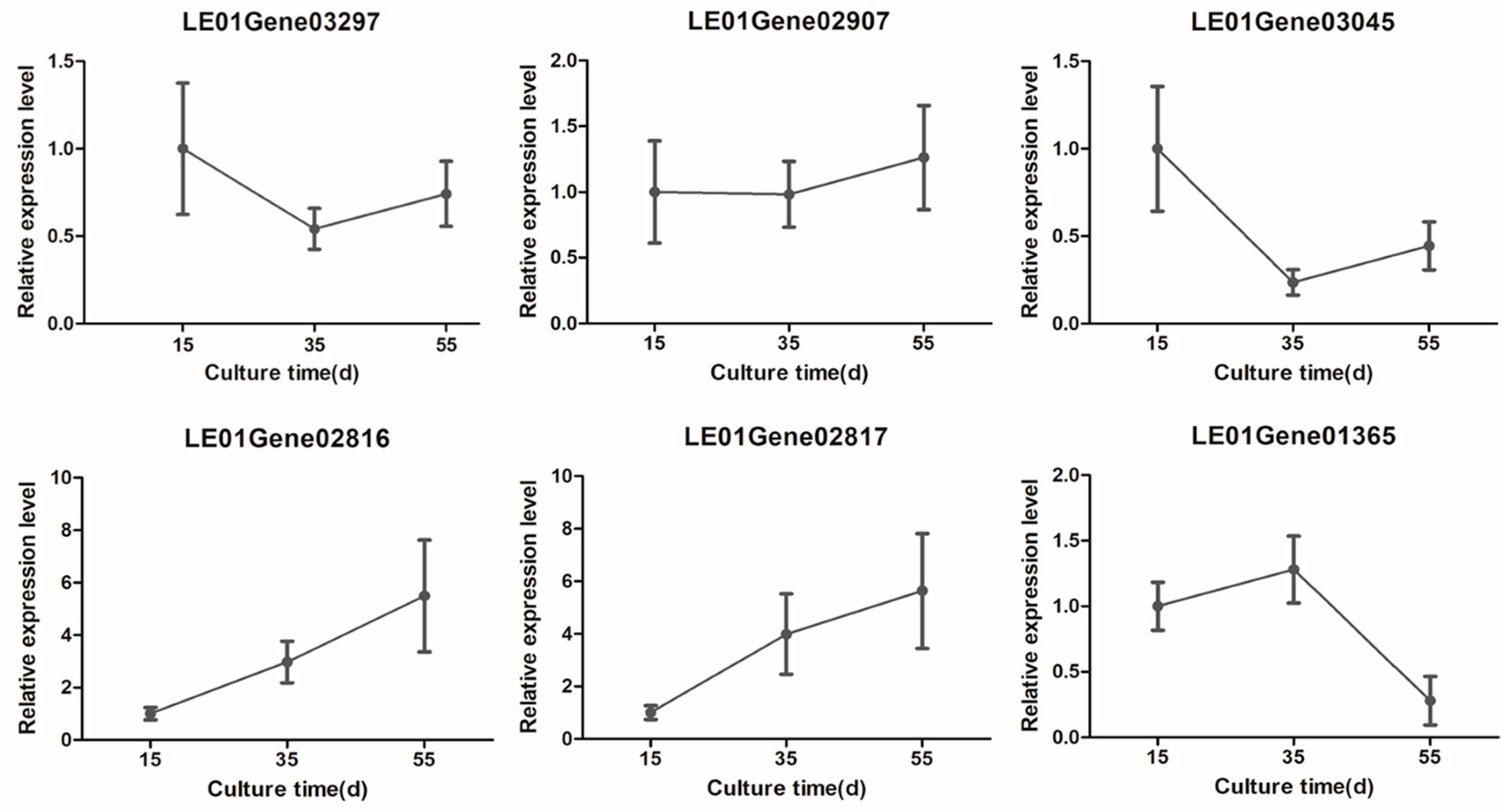
3.5. Transcriptome Validation

In order to verify the reliability of the transcriptome results, the expression of related genes was measured with the same treatment but without transcriptome sequencing [41]. As shown in Figure 6, the expression levels of the LE01Gene03297, LE01Gene02907, LE01Gene03045, LE01Gene02816, LE01Gene02817, and LE01Gene01365 genes were consistent with the results of the transcriptome analysis. The expression patterns of the Leggt3 (LE01Gene03045), and Leccsl3 (LE01Gene03297) genes were consistent with the measured activities of GGT and C-S lyase during the cultivation of the mycelium.

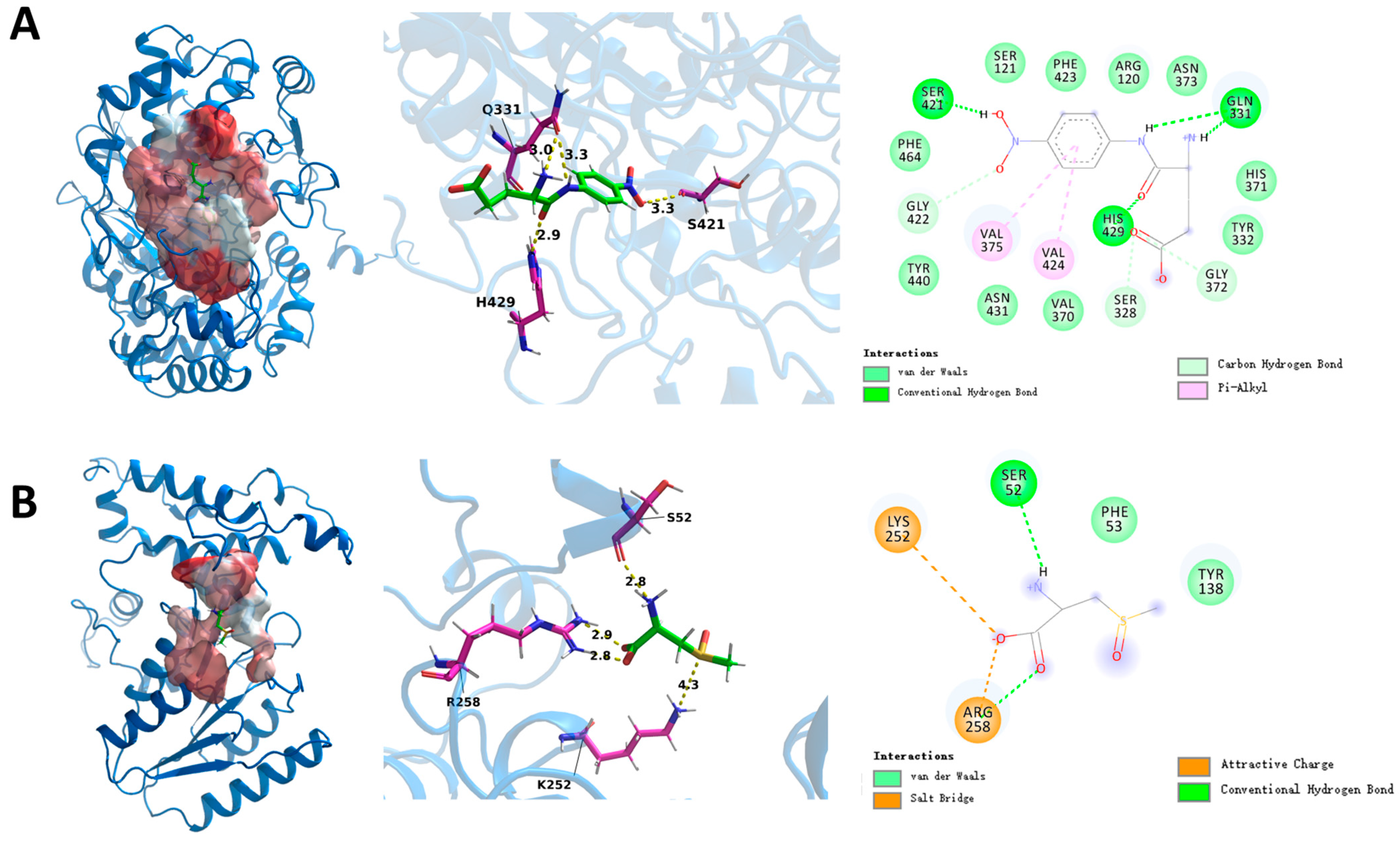
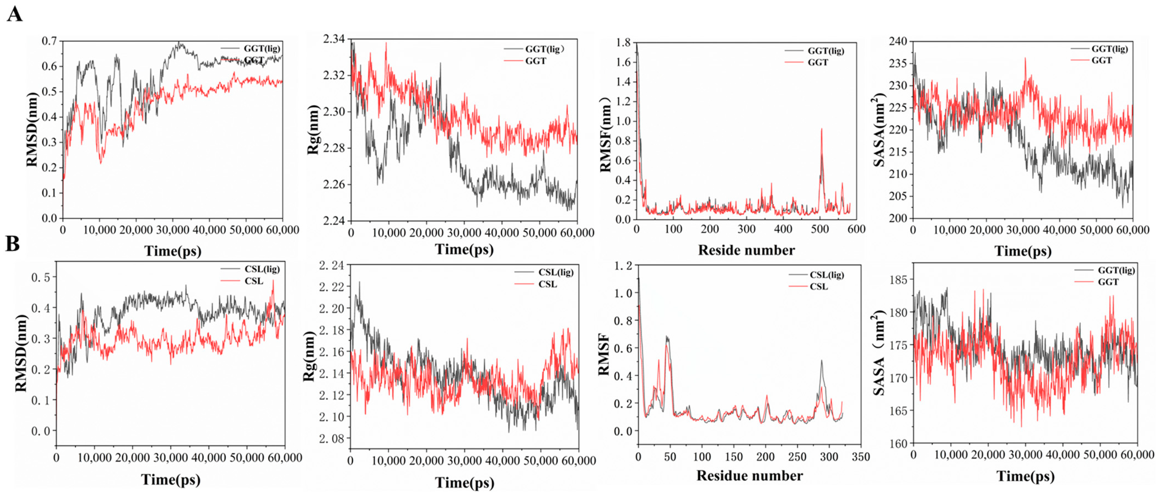
3.6. Mechanisms of Leggt3 and Lecsl3

The results of RNA-seq and qRT-PCR revealed that Leggt3 and Lecsl3 seemed to be the essential enzymes in the GGT and C-S lyase family, thus we chose to reveal the mechanisms of their interaction with substrates. When cloning the Lecsl3 gene, we discovered the actual length of the coding region was 966 bp instead of the 1497 bp shown in the database. The mutation of the 966th base from A to G made the 322nd amino acid code become the terminator TGA, which caused an early termination of protein translation. Therefore, we modeled the protein using the obtained sequence. Through molecular docking, as shown in Figure 7, we found that the substrates can bind to the hydrophobic pockets of Leggt3 and Lecsl3 and interact with key residues. Leggt3 mainly forms hydrogen bonds and Pi-Alkyl with the substrate, and the amino acid binding sites involved in substrate binding are VAL-375, VAL-424, GLN-331, HIS-429, and SER-421. In addition, there were some amino acids around the substrate that interact with the substrate through carbon-hydrogen bonds (Figure 7A). Lecsl3 forms hydrogen bonds and salt bridges with the substrate, LYS-252 forms hydrogen bonds with the substrate, and SER-52 forms salt bridges with the substrate, while AGR-258 forms salt bridges with the oxygen anion and hydrogen bonds with the oxygen atom (Figure 7B).
To investigate the stability of protein–substrate binding and the impact on the protein after binding, we performed a 60 ns molecular dynamics simulation based on the docking results, as shown in Figure 8, comparing the protein conformational changes between binding and non-binding of substrates. Calculation of the root-mean-square deviation (RMSD) of the protein backbone can assess the stability of protein conformation [42]. The radius of gyration (Rg) can reflect the potential plasticity of the protein structure [43]. As shown in Figure 8, according to the results of the RMSD analysis, the two systems gradually converged during the simulation process. Compared to the substrate-free Leggt3, the RMSD value of Leggt3 increases upon substrate binding, indicating that substrate binding induces conformational changes in Leggt3 and promotes the formation of an active site with catalytic functionality. Lecsl3 also exhibits a similar phenomenon upon substrate binding, but of smaller magnitude, reaching convergence within a very short time (~20 ns). The change in Rg value before and after protein–substrate binding better elucidates this phenomenon; this indicates that the protein structure becomes more compact after substrate binding. Root-mean-square fluctuation (RMSF) is commonly used to calculate the fluctuation in individual residues, thereby indicating the degrees of freedom of residue movements. We evaluated the flexibility of each amino acid residue in the protein before and after binding to the substrate by calculating the RMSF values. The results revealed that for both proteins, the RMSF values of most amino acids did not fluctuate before and after substrate binding, except for a few amino acids that displayed minor fluctuations.
Due to the molecular dynamics simulation being carried out in an aqueous environment, we also calculated the solvent-accessible surface area (SASA) (Figure 8) [44,45] of Leggt3 and Lecsl3 before and after binding with the substrate. From the graph (Figure 8), we can observe that the SASA value of Leggt3 decreased and its hydrophobicity was enhanced after binding with the substrate. In contrast, the SASA of Lecsl3 showed little change before and after substrate binding, and the SASA values were lower than those of Leggt3. This may be because the active pocket of Leggt3 was exposed and it interacts more strongly with water when the substrate binds. Conversely, the active pocket of Lecsl3 was smaller and partially exposed; therefore, even after substrate binding, there was no significant change in its contact with water.

4. Discussion

This study revealed the rule of flavor formation of L. edodes mycelium at different growth stages, enriching research on the flavor of L. edodes at various growth stages. By observing the growth of the mycelium, the biomass of mycelium was found to increase in the early stage of growth, and the hyphae gathered into clusters. Several sulfur-containing volatile compounds (dimethyl trisulfide, dimethyl disulfide, propylsulfide, and propanethiol) were detected in L. edodes mycelium. However, mycelium cultured for different numbers of days showed various levels of sulfur-containing volatile compounds. These results indicate that the sulfur-containing characteristic flavor components of L. edodes are already produced at the mycelium stage. In order to confirm the origins of sulfur-containing flavor components in mycelium, activities of GGT and C-S lyase were investigated, The results indicated that the enzyme activities of GGT and C-S lyase have a certain impact on the composition and content of aroma compounds.
By comparing transcriptome and KEGG analyses with the research results on the flavor components of L. edodes fruiting bodies at different stages, we also found that genes expect HSP20 and P450, which affect the growth and flavor differences of L. edodes mycelium and the fruiting bodies, are almost identical in the mycelium and fruiting bodies. This indicates that the influence of these genes on the flavor of L. edodes persists throughout. HSP20 is the ubiquitous heat shock protein in organisms and plays an important role in the heat tolerance of organisms, and sulfur-containing volatile compounds in L. edodes are generated by heat. It seemed that there might be a correlation between HSP20 and the generation of sulfur-containing volatile compounds, but there was not sufficient evidence to be certain. A study reported that the overexpression of HSP20 could promote the growth of L. edodes mycelium [46]. Therefore, this implies that HSP20 might affect the growth of L. edodes mycelium. Like HSP20, P450 is a ubiquitous enzyme system in organisms, and it has functional roles in growth, development, and secondary metabolisms [47,48]. Previous studies proved that P450 was vital during the production of sulfur-containing volatile compounds in plants [49,50]. In the present study, several P450 family genes were found to be related to the formation of sulfur-containing volatile compounds in the mycelium, including LE01Gene03374, LE01Gene04094, and LE01Gene10823. Elucidating the functions of HSP20 and P450 genes in the generation mechanism of volatile sulfur compounds in L. edodes is envisaged in the future. During the process of transcriptome verification, we found that the variation pattern of Leggt3 and Lecsl3’s gene expression was consistent with the trend of activity changes of GGT and C-S lyase. The potential binding sites and interactions with the substrate were determined through molecular docking calculations. Based on the molecular docking results, molecular dynamics simulations showed that they can stably bind to the substrate. However, as proteins in eukaryotic organisms, revealing whether they function as oligomers or require post-translational modifications to become active needs further experiments.
In this study, we systematically studied the formation mechanism of flavor in L. edodes mycelium and conducted a brief functional analysis of key genes. The growth of L. edodes from mycelium to fruiting bodies involves several stages: mycelium colonization in the cultivation bag, mycelium color transformation, primordium formation, and fruiting body emergence. It takes about two months from mycelium inoculation in the cultivation bag to complete mycelium color transformation. Therefore, we focused on the flavor formation mechanism in L. edodes mycelium before complete color transformation. Compared to fruiting bodies, mycelium is easier to obtain and cultivate in large quantities. These results supported the hypothesis that L. edodes mycelium can be studied to explore the mechanisms underlying flavor formation and it is possible to use the mycelium to produce characteristic L. edodes flavor. We identified some genes that may affect the flavor of L. edodes mycelium, and the formation of relevant flavor components of L. edodes can be regulated by controlling the expression of these genes.

5. Conclusions

The present study revealed the characteristics of mycelial growth and flavor formation in the mycelium stage of L. edodes, and a preliminary investigation of the mechanism of action of Leggt3 and Lecsl3 has also been conducted. In the first two months of growth, the amount of L. edodes mycelium continues to increase and the hyphae gather into clusters. During this process, characteristic L. edodes flavor compounds dimethyl trisulfide and dimethyl disulfide were also formed, and the genes and metabolic pathways related to flavor and the growth of L. edodes were the same in both the mycelium and fruiting body stages. The results not only provide novel insights for the study of sulfur-containing flavor compounds and formaldehyde biosynthesis in L. edodes but also lay a foundation for utilizing mycelium fermentation to produce sulfur-containing flavor compounds.

Author Contributions

Methodology, Z.L., S.G. and M.C.; Software, F.P. and L.C.; Validation, Z.L.; Investigation, Z.L., F.P., M.C. and Y.L.; Resources, W.H., Y.B., W.T. and Y.L.; Data curation, Z.L. and X.F.; Writing—original draft, Z.L., F.P., S.G., M.C., L.C. and Y.B.; Writing—review & editing, W.H., X.F., W.T. and Y.L.; Supervision, W.H. and Y.L.; Funding acquisition, Y.L. All authors have read and agreed to the published version of the manuscript.

Funding

The authors are grateful for the support provided by the Natural Science Foundation of China (No. 31601434), the Key Research and Development Projects in Agricultural and Rural Areas of Hubei Province (2022BBA0024), the Agricultural Science and Technology Innovation Center of Hubei Province (2021-620-000-001-031), and the Hubei Agriculture Research System (HBHZDZB-2021-023).

Institutional Review Board Statement

Not applicable.

Informed Consent Statement

Not applicable.

Data Availability Statement

The original contributions presented in the study are included in the article, further inquiries can be directed to the corresponding author.

Conflicts of Interest

The authors declare no conflict of interest.

References

  1. Sun, L.; Xin, G.; Hou, Z.; Zhao, X.; Xu, H.; Bao, X.; Xia, R.; Li, Y.; Li, L. Biosynthetic Mechanism of Key Volatile Biomarkers of Harvested Lentinula edodes Triggered by Spore Release. J. Agric. Food Chem. 2021, 69, 9350–9361. [Google Scholar] [CrossRef] [PubMed]
  2. Zhang, Y.; Wang, D.; Chen, Y.; Liu, T.; Zhang, S.; Fan, H.; Liu, H.; Li, Y. Healthy function and high valued utilization of edible fungi. Food Sci. Hum. Wellness 2021, 10, 408–420. [Google Scholar] [CrossRef]
  3. Lu, Y.; Zhang, M.; Huang, D. Dietary Organosulfur-Containing Compounds and Their Health-Promotion Mechanisms. Annu. Rev. Food Sci. Technol. 2022, 13, 287–313. [Google Scholar] [CrossRef]
  4. Schmidberger, P.C.; Schieberle, P. Changes in the Key Aroma Compounds of Raw Shiitake Mushrooms (Lentinula edodes) Induced by Pan-Frying as Well as by Rehydration of Dry Mushrooms. J. Agric. Food Chem. 2020, 68, 4493–4506. [Google Scholar] [CrossRef]
  5. Wen, X.; Li, W.; Li, W.; Chen, W.; Zhang, Z.; Wu, D.; Yang, Y. Quality characteristics and non-volatile taste formation mecha nism of Lentinula edodes during hot air drying. Food Chem. 2022, 393, 133378. [Google Scholar] [CrossRef]
  6. Xi, J.; Chen, X.; Du, J.; Zhong, L.; Hu, Q.; Zhao, L. Biosynthesis, behavior and fate of volatile organic sulfide in Lentinus edodes (Berk.) upon hot-air drying treatment. Food Chem. 2023, 412, 135528. [Google Scholar] [CrossRef] [PubMed]
  7. Sneeden, E.Y.; Harris, H.H.; Pickering, I.J.; Prince, R.C.; Johnson, S.; Li, X.; Block, E.; George, G.N. The sulfur chemistry of shiitake mushroom. J. Am. Chem. Soc. 2004, 126, 458–459. [Google Scholar] [CrossRef]
  8. Xu, L.; Fang, X.; Wu, W.; Chen, H.; Mu, H.; Gao, H. Effects of high-temperature pre-drying on the quality of air-dried shiitake mushrooms (Lentinula edodes). Food Chem. 2019, 285, 406–413. [Google Scholar] [CrossRef] [PubMed]
  9. Liu, Y.; Lei, X.-Y.; Chen, L.-F.; Bian, Y.-B.; Yang, H.; Ibrahim, S.A.; Huang, W. A novel cysteine desulfurase influencing or ganosulfur compounds in Lentinula edodes. Sci Rep. 2015, 5, 10047. [Google Scholar] [CrossRef]
  10. Lei, X.; Gao, S.; Feng, X.; Huang, Z.; Bian, Y.; Huang, W.; Liu, Y. Effects of GGT and C-S Lyase on the Generation of Endogenous Formaldehyde in Lentinula edodes at Different Growth Stages. Molecules 2019, 24, 4203. [Google Scholar] [CrossRef]
  11. Liu, Y.; Yuan, Y.; Lei, X.Y.; Yang, H.; Ibrahim, S.A.; Huang, W. Purification and characterisation of two enzymes related to endogenous formaldehyde in Lentinula edodes. Food Chem. 2013, 138, 2174–2179. [Google Scholar] [CrossRef] [PubMed]
  12. Gonzalez, A.; Cruz, M.; Losoya, C.; Nobre, C.; Loredo, A.; Rodriguez, R.; Contreras, J.; Belmares, R. Edible mushrooms as a novel protein source for functional foods. Food Funct. 2020, 11, 7400–7414. [Google Scholar] [CrossRef] [PubMed]
  13. Guillamon, E.; Garcia-Lafuente, A.; Lozano, M.; D’Arrigo, M.; Rostagno, M.A.; Villares, A.; Martinez, J.A. Edible mushrooms: Role in the prevention of cardiovascular diseases. Fitoterapia 2010, 81, 715–723. [Google Scholar] [CrossRef] [PubMed]
  14. Zou, G.; Nielsen, J.B.; Wei, Y. Harnessing synthetic biology for mushroom farming. Trends Biotechnol. 2023, 41, 480–483. [Google Scholar] [CrossRef] [PubMed]
  15. Rathore, H.; Prasad, S.; Kapri, M.; Tiwari, A.; Sharma, S. Medicinal importance of mushroom mycelium: Mechanisms and applications. J. Funct. Foods 2019, 56, 182–193. [Google Scholar] [CrossRef]
  16. Park, Y.-J.; Oh, T.-S.; Jang, M.-J. Effect of adding amino acids on the production of Gamma-Aminobutyric Acid (GABA) by mycelium of Lentinula edodes. Int. J. Food Eng. 2019, 15, 20180287. [Google Scholar] [CrossRef]
  17. Cao, X.; Liu, C.; Zhang, M.; Bi, R.; Fu, M.; Korik, E.; Chen, J.; Gao, J.; Semak, I.; Liu, J. Bovine lactoferrin and Lentinus edodes mycelia polysaccharide complex: The formation and the activity to protect islet β cells. Int. J. Biol. Macromol. 2021, 191, 811–820. [Google Scholar] [CrossRef] [PubMed]
  18. Li, W.; Chen, W.-C.; Wang, J.-B.; Feng, J.; Wu, D.; Zhang, Z.; Zhang, J.-S.; Yang, Y. Effects of enzymatic reaction on the generation of key aroma volatiles in shiitake mushroom at different cultivation substrates. Food Sci. Nutr. 2021, 9, 2247–2256. [Google Scholar] [CrossRef] [PubMed]
  19. Lu, X.; Zhao, Y.; Li, F.; Liu, P. Active polysaccharides from Lentinula edodes and Pleurotus ostreatus by addition of corn straw and xylosma sawdust through solid-state fermentation. Int. J. Biol. Macromol. 2023, 228, 647–658. [Google Scholar] [CrossRef]
  20. Yu, C.-X.; Zhang, Y.-R.; Ren, Y.-F.; Zhao, Y.; Song, X.-X.; Yang, H.-L.; Chen, M.-J. Composition and contents of fatty acids and amino acids in the mycelia of Lentinula edodes. Food Sci. Nutr. 2023, 11, 4038–4046. [Google Scholar] [CrossRef]
  21. Wang, W.-K.; Zhu, Y.; Tang, Y.; Lu, N.; Song, J.-L.; Yuan, W.-D.; Jia, Y. Non-Volatile Taste Components of Different Cultivated Mushrooms at Mycelia, Primordium, and Fruit Body Cultivation Stages. Int. J. Food Prop. 2016, 19, 1938–1948. [Google Scholar] [CrossRef]
  22. Chen, W.C.; Li, W.; Yang, Y.; Yu, H.L.; Zhou, S.; Feng, J.; Li, X.B.; Liu, Y.F. Analysis and Evaluation of Tasty Components in the Pileus and Stipe of Lentinula edodes at Different Growth Stages. J. Agric. Food Chem. 2015, 63, 795–801. [Google Scholar] [CrossRef] [PubMed]
  23. Wang, Y.; Yang, Z.; Chen, X.; Han, D.; Han, J.; Wang, L.; Ren, A.; Yu, H.; Zhao, M. Lenthionine, a Key Flavor Substance in Lentinula edodes, Is Regulated by Cysteine under Drought Stress. J. Agric. Food Chem. 2021, 69, 12645–12653. [Google Scholar] [CrossRef] [PubMed]
  24. Chen, L.F.; Gong, Y.H.; Cai, Y.L.; Liu, W.; Zhou, Y.; Xiao, Y.; Xu, Z.Y.; Liu, Y.; Lei, X.Y.; Wang, G.Z.; et al. Genome Sequence of the Edible Cultivated Mushroom Lentinula edodes (Shiitake) Reveals Insights into Lignocellulose Degradation. PLoS ONE 2016, 11, 20. [Google Scholar] [CrossRef] [PubMed]
  25. Liu, R.; Zhang, X.; Ren, A.; Shi, D.K.; Shi, L.; Zhu, J.; Yu, H.S.; Zhao, M.W. Heat stress-induced reactive oxygen species participate in the regulation of HSP expression, hyphal branching and ganoderic acid biosynthesis in Ganoderma lucidum. Microbiol Res. 2018, 209, 43–54. [Google Scholar] [CrossRef]
  26. Jumper, J.; Evans, R.; Pritzel, A.; Green, T.; Figurnov, M.; Ronneberger, O.; Tunyasuvunakool, K.; Bates, R.; Zidek, A.; Potapenko, A.; et al. Highly accurate protein structure prediction with AlphaFold. Nature 2021, 596, 583–589. [Google Scholar] [CrossRef] [PubMed]
  27. Hanwell, M.D.; Curtis, D.E.; Lonie, D.C.; Vandermeersch, T.; Zurek, E.; Hutchison, G.R. Avogadro: An advanced semantic chemical editor, visualization, and analysis platform. J. Cheminform. 2012, 4, 17. [Google Scholar] [CrossRef] [PubMed]
  28. Trott, O.; Olson, A.J. AutoDock Vina: Improving the speed and accuracy of docking with a new scoring function, efficient optimization, and multithreading. J. Comput. Chem. 2010, 31, 455–461. [Google Scholar] [CrossRef] [PubMed]
  29. Neese, F. The ORCA program system. Wiley Interdiscip. Rev. Comput. Mol. Sci. 2012, 2, 73–78. [Google Scholar] [CrossRef]
  30. Case, D.A.; Cheatham, T.E., III; Darden, T.; Gohlke, H.; Luo, R.; Merz, K.M., Jr.; Onufriev, A.; Simmerling, C.; Wang, B.; Woods, R.J. The amber biomolecular simulation programs. J. Comput. Chem. 2005, 26, 1668–1688. [Google Scholar] [CrossRef]
  31. Lu, T.; Chen, F. Multiwfn: A multifunctional wavefunction analyzer. J. Comput. Chem. 2012, 33, 580–592. [Google Scholar] [CrossRef] [PubMed]
  32. Kanehisa, M.; Sato, Y.; Kawashima, M. KEGG mapping tools for uncovering hidden features in biological data. Protein Sci. 2022, 31, 47–53. [Google Scholar] [CrossRef]
  33. Chen, T.; Liu, Y.-X.; Huang, L. ImageGP: An easy-to-use data visualization web server for scientific researchers. iMeta 2022, 1, e5. [Google Scholar] [CrossRef]
  34. Greenberg, E.P. Bacterial communication: Tiny teamwork. Nature 2003, 424, 134. [Google Scholar] [CrossRef]
  35. Boulianne, R.P.; Liu, Y.; Aebi, M.; Lu, B.C.; Kues, U. Fruiting body development in Coprinus cinereus: Regulated expression of two galectins secreted by a non-classical pathway. Microbiology 2000, 146, 1841–1853. [Google Scholar] [CrossRef] [PubMed]
  36. Kues, U.; Liu, Y. Fruiting body production in basidiomycetes. Appl. Microbiol. Biotechnol. 2000, 54, 141–152. [Google Scholar] [CrossRef] [PubMed]
  37. Sun, L.-B.; Zhang, Z.-Y.; Xin, G.; Sun, B.-X.; Bao, X.-J.; Wei, Y.-Y.; Zhao, X.-M.; Xu, H.-R. Advances in umami taste and aroma of edible mushrooms. Trends Food Sci. Technol. 2020, 96, 176–187. [Google Scholar] [CrossRef]
  38. Dermiki, M.; Phanphensophon, N.; Mottram, D.S.; Methven, L. Contributions of non-volatile and volatile compounds to the umami taste and overall flavour of shiitake mushroom extracts and their application as flavor enhancers in cooked minced meat. Food Chem. 2013, 141, 77–83. [Google Scholar] [CrossRef] [PubMed]
  39. Hiraide, M.; Miyazaki, Y.; Shibata, Y. The smell and odorous components of dried shiitake mushroom, Lentinula edodes I: Relationship between sensory evaluations and amounts of odorous components. J. Wood Sci. 2004, 50, 358–364. [Google Scholar] [CrossRef]
  40. Li, W.; Chen, W.-C.; Wang, J.-B.; Feng, J.; Wu, D.; Zhang, Z.; Zhang, J.-S.; Yang, Y. Screening candidate genes related to volatile synthesis in shiitake mushrooms and construction of regulatory networks to effectively improve mushroom aroma. J. Sci. Food Agric. 2021, 101, 5618–5626. [Google Scholar] [CrossRef]
  41. Luo, Y.; Wang, G.; Wang, C.; Gong, Y.; Bian, Y.; Zhou, Y. Selection and validation of reference genes for qRT-PCR in Lentinula edodes under different experimental conditions. Genes 2019, 10, 647. [Google Scholar] [CrossRef]
  42. Gholami, S.; Bordbar, A.-K. Exploring binding properties of naringenin with bovine β-lactoglobulin: A fluorescence, molecular docking and molecular dynamics simulation study. Biophys. Chem. 2014, 187–188, 33–42. [Google Scholar] [CrossRef]
  43. Mehranfar, F.; Bordbar, A.-K.; Parastar, H. A combined spectroscopic, molecular docking and molecular dynamic simulation study on the interaction of quercetin with β-casein nanoparticles. J. Photochem. Photobiol. B 2013, 127, 100–107. [Google Scholar] [CrossRef] [PubMed]
  44. Rehman, A.U.; Rafiq, H.; Rahman, M.U.; Li, J.; Liu, H.; Luo, S.; Arshad, T.; Wadood, A.; Chen, H.-F. Gain-of-Function SHP2 E76Q Mutant Recusing Autoinhibition Mechanism Associated with Juvenile Myelomonocytic Leukemia. J. Chem. Inf. Model 2019, 59, 3229–3239. [Google Scholar] [CrossRef] [PubMed]
  45. Wang, P.; Ji, R.; Ji, J.; Chen, F. Changes of metabolites of acrylamide and glycidamide in acrylamide-exposed rats pretreated with blueberry anthocyanins extract. Food Chem. 2019, 274, 611–619. [Google Scholar] [CrossRef]
  46. Ling, Y.-Y.; Ling, Z.-L.; Zhao, R.-L. Construction of a heat-resistant strain of Lentinus edodes by fungal Hsp20 protein over expression and genetic transformation. Front. Microbiol. 2022, 13, 1009885. [Google Scholar] [CrossRef]
  47. Shin, J.; Kim, J.-E.; Lee, Y.-W.; Son, H. Fungal cytochrome P450s and the P450 complement (cypome) of Fusarium graminearum. Toxins 2018, 10, 112. [Google Scholar] [CrossRef] [PubMed]
  48. Rudolf, J.D.; Chang, C.-Y.; Ma, M.; Shen, B. Cytochromes P450 for natural product biosynthesis in Streptomyces: Sequence, structure, and function. Nat. Prod. Rep. 2017, 34, 1141–1172. [Google Scholar] [CrossRef]
  49. Chen, S.; Glawischnig, E.; Jorgensen, K.; Naur, P.; Jorgensen, B.; Olsen, C.-E.; Hansen, C.H.; Rasmussen, H.; Pickett, J.A.; Halkier, B.A. CYP79F1 and CYP79F2 have distinct functions in the biosynthesis of aliphatic glucosinolates in Arabidopsis. Plant J. 2003, 33, 923–937. [Google Scholar] [CrossRef]
  50. Hull, A.K.; Vij, R.; Celenza, J.L. Arabidopsis cytochrome P450s that catalyze the first step of tryptophan-dependent indole-3-acetic acid biosynthesis. Proc. Natl. Acad. Sci. USA 2000, 97, 2379–2384. [Google Scholar] [CrossRef]
Figure 1. Macro morphology and microstructure of mycelium cultured for 15 days (A), 35 days (B), and 55 days (C), respectively.
Foods 13 02179 g001
Figure 2. Fingerprint of the main volatile components in mycelium cultured for different numbers of days.
Foods 13 02179 g002
Figure 3. Activities of GGT (A) and C-S lyase (B) in mycelium cultured for different numbers of days.
Foods 13 02179 g003
Figure 4. Differentially expressed genes (DEGs) in mycelium cultured for different days. D15, D35, and D55 represent L. edodes mycelium cultured for 15, 35, and 55 days, respectively.On the left is the heat map of the differential expression genes. This figure reflects the differential genes and gene expression changes in pairwise comparison of samples. For the color key, green means downregulation, and red represents upregulation of the transcripts. On the right is the volcano plot of DEGs in pairwise comparison of samples. This displays the analysis results of differentially expressed genes; in the figure, the horizontal axis represents the logarithmic value of the difference multiple, and the vertical axis represents the logarithmic value of the q value. Red is used to distinguish significantly upregulated genes, green is used to distinguish significantly downregulated genes (A) D15 vs. D35, (B) D15 vs. D55, (C) D35 vs. D55.
Foods 13 02179 g004aFoods 13 02179 g004b
Figure 5. Kyoto Encyclopaedia of Genes and Genomes (KEGG) enrichment analysis results. KEGG enrichment analysis of genes and metabolic pathways involved in significant downregulation and upregulation of expression levels between different samples. D15, D35, and D55 represent L. edodes mycelium cultured for 15, 35, and 55 days, respectively. (A) D15 vs. D35; (B) D15 vs. D55; (C) D35 vs. D55.
Foods 13 02179 g005aFoods 13 02179 g005b
Figure 6. Verification of the expression levels of selected transcripts by qRT-PCR.
Foods 13 02179 g006
Figure 7. From left to right are: the region and overall conformation of protein substrate binding; the local conformation, binding sites, and bond lengths of protein substrate binding region; the binding sites and binding modes of protein substrate binding. (A) Leggt3 with γ-glutamyl p-nitroaniline. (B) Lecsl3 with S-methyl-L cysteine sulfoxide.
Foods 13 02179 g007
Figure 8. Root-mean-square deviation (RMSD), radius of gyration (Rg), root-mean-square fluctuation (RMSF), and solvent-accessible surface area (SASA) before and after protein substrate binding. (A) Leggt3, (B) Lecsl3.
Foods 13 02179 g008
Disclaimer/Publisher’s Note: The statements, opinions and data contained in all publications are solely those of the individual author(s) and contributor(s) and not of MDPI and/or the editor(s). MDPI and/or the editor(s) disclaim responsibility for any injury to people or property resulting from any ideas, methods, instructions or products referred to in the content.

