Article

High Prevalence of Multidrug-Resistant, Biofilm-Forming Virulent Clostridium perfringens in Broiler Chicken Retail Points in Northeast India

by
Govindarajan Bhuvana Priya
1,
Kandhan Srinivas
2,
Heiborkie Shilla
2 and
Arockiasamy Arun Prince Milton
2,*
1
College of Agriculture, Central Agricultural University (Imphal), Kyrdemkulai 793105, Meghalaya, India
2
Division of Animal and Fisheries Sciences, ICAR Research Complex for NEH Region, Umiam 793103, Meghalaya, India
*
Author to whom correspondence should be addressed.
Foods 2023, 12(22), 4185; https://doi.org/10.3390/foods12224185
Submission received: 16 October 2023 / Revised: 16 November 2023 / Accepted: 17 November 2023 / Published: 20 November 2023
(This article belongs to the Section Food Microbiology)

Abstract

:
In light of the significant public health and food safety implications associated with Clostridium perfringens, this study aimed to isolate and characterize C. perfringens in samples obtained from broiler chicken retail points in Meghalaya, northeastern India. A total of 280 samples comprising meat, intestinal contents, water, and hand swabs were processed to detect contamination by C. perfringens. The isolates were subjected to toxinotyping, antimicrobial susceptibility testing, and biofilm-forming ability test. The overall occurrence of C. perfringens was 22.5% (17.74–27.85, 95% CI) with the highest recovery from intestine samples (31%; 22.13–41.03, 95% CI), followed by meat (23%, 15.17–32.49, 95% CI) and water samples (18%, 8.58–31.44, 95% CI). Type A was the predominant toxinotype (71.43%, 58.65–82.11, 95% CI), followed by Type A with beta2 toxin (17.46%, 9.05–29.10, 95% CI), Type C (7.94%, 2.63–17.56, 95% CI), and Type C with beta2 toxin (3.17%, 0.39–11.0, 95% CI). Nearly all (95.24%) isolates were multidrug resistant and 68.25% were biofilm formers. The predominance of multidrug-resistant and virulent Type A and Type C C. perfringens in retail broiler meat and intestines in the tribal-dominated northeastern region of India is of great concern from food safety and public health perspectives.

1. Introduction

Clostridium perfringens is a food-borne biological hazard of bacterial origin; they are Gram-positive rods thriving on anaerobic respiration and with spore-forming ability [1]. In addition to being an ubiquitous member of the environmental microflora, C. perfringens has the potential to cause diseases in various species, including poultry and humans [1]. The poultry industry is often marred by outbreaks of necrotic enteritis opportunistically caused by C. perfringens, resulting in severe economic losses. Colonization in poultry occurs early in life, even during the hatchery stage, with potential to disseminate in the broiler value chain [2]. The predominance of pathogenic strains of C. perfringens expressing various virulence factors over non-pathogenic strains triggers necrotic enteritis. In humans, the organism is commonly known for its food-borne importance amidst an array of other clinical manifestations [1]. In that regard, C. perfringens is the predominant bacterial and second-most common cause of food-borne illnesses in Canada, next to norovirus [3]. World Health Organization estimates in 2010 placed C. perfringens as the leading causal agent among food-borne intoxications, with 3,998,164 foodborne illnesses [4].
The virulence arsenal of C. perfringens comprises various toxin genes mediated by plasmids as well as chromosomes. Strains of C. perfringens are classified into seven toxinotypes (A, B, C, D, E, F and G) based on occurrence patterns of toxin genes (α, β, ε, ι, cpe and netB) [5]. The cpa gene, present on the variable sections of the chromosome, encodes the phospholipase C (α-toxin) and is identifiable in all toxinotypes [5,6]. The C. perfringens enterotoxin (CPE), often associated with food poisoning in humans, is either chromosome- or plasmid-mediated [1]. The CPE is usually present in marginal proportions of all C. perfringens strains, with higher occurrence in Type A strains, and is associated with bowel disturbances in humans and animals [7]. The beta toxin (cpb2) was first identified in the case of a Type C-infected piglet with haemorrhagic necrotic enteritis [8]. The beta2 toxin, capable of being produced by all toxinotypes, is considered a lethal and necrotizing factor that contributes to gastro-intestinal derangements in humans as well as animals [8]. Recently, a large clostridial toxin named TpeL was identified in Type C C. perfringens and was an important contributor to necrotic enteritis [9].
The presence of antimicrobial-resistant strains of bacteria in the food chain has been a constant public health concern. Antibiotic resistance in C. perfringens is chiefly mediated by plasmids, as well as transposons and insertion sequences [10]. Increasing reports of C. perfringens resistance to tetracycline, lincomycin, and erythromycin have been a growing concern over recent years, made worse by the ability of C. perfringens to produce biofilm and form spores [11]. Biofilms enhance resistance to antimicrobials and disinfectants and are associated with increased virulence and pathogenesis [12]. The molecular interplay of genes resulting in biofilm formation by C. perfringens was only recently elucidated [13]. The ability to produce spores, secrete toxins, form biofilms, and harbour antimicrobial resistance genes, coupled with a short (<10 min) doubling time, makes C. perfringens an important threat to humans and animals.
C. perfringens has been widely reported in various parts of India [1,14,15]. However, except for a case study of C. perfringens involving six birds on a farm, there are no other systematic reports of prevalence, virulence, and antimicrobial resistance of C. perfringens in the broiler chicken value chain in Meghalaya, a northeast Indian state with a dominant tribal population [16]. Interestingly, chicken intestines have culinary value regionally. Our objective was to determine the food-borne threat posed by C. perfringens.

2. Materials and Methods

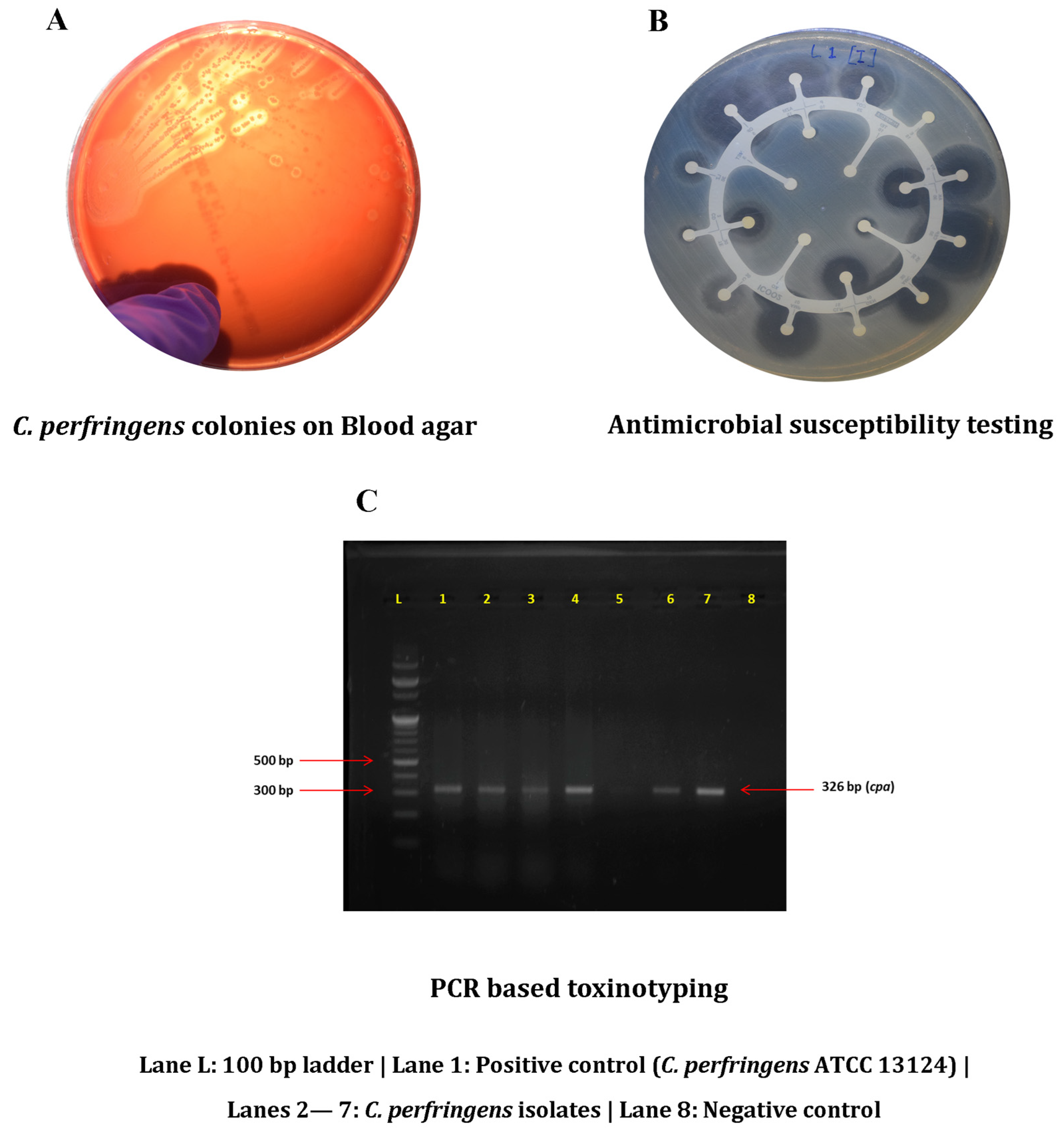
2.1. Sample Collection and Isolation

A total of 280 samples comprising broiler chicken meat (n = 100), chicken intestine (n = 100), slaughter water (n = 50), and butcher hand swabs (n = 30) were collected from retail chicken meat shops in 2 districts (East Khasi Hills and Ri-Bhoi) of Meghalaya, India. Hand swabs were collected during meat cutting, and slaughter water refers to the water used for slaughtering and meat cutting at the retail points. Within 2 h of collection, samples were inoculated in 10 mL of Robertson cooked meat (RCM) broth (HiMedia Laboratories, Mumbai, India). After incubation at 37 °C for 24 h, the enriched inoculum was plated onto 5% sheep blood agar plates (Figure 1) that were incubated at 37 °C for 24 h under an anaerobic environment induced with a gas pack system (Anaerogen™ 2.5 L; Thermo Scientific, Hampshire, UK). Presumptive identification of C. perfringens was attempted by exploiting the ability of the organism to produce a double zone of haemolysis on the sheep blood agar; suspected colonies were propagated in RCM broth. Further confirmation was achieved by PCR-based detection of cpa (α-toxin) using C. perfringens ATCC 13124 as a positive reference. Confirmed isolates were cryopreserved in brain heart infusion broth containing 15% glycerol and stocked at −80 °C.

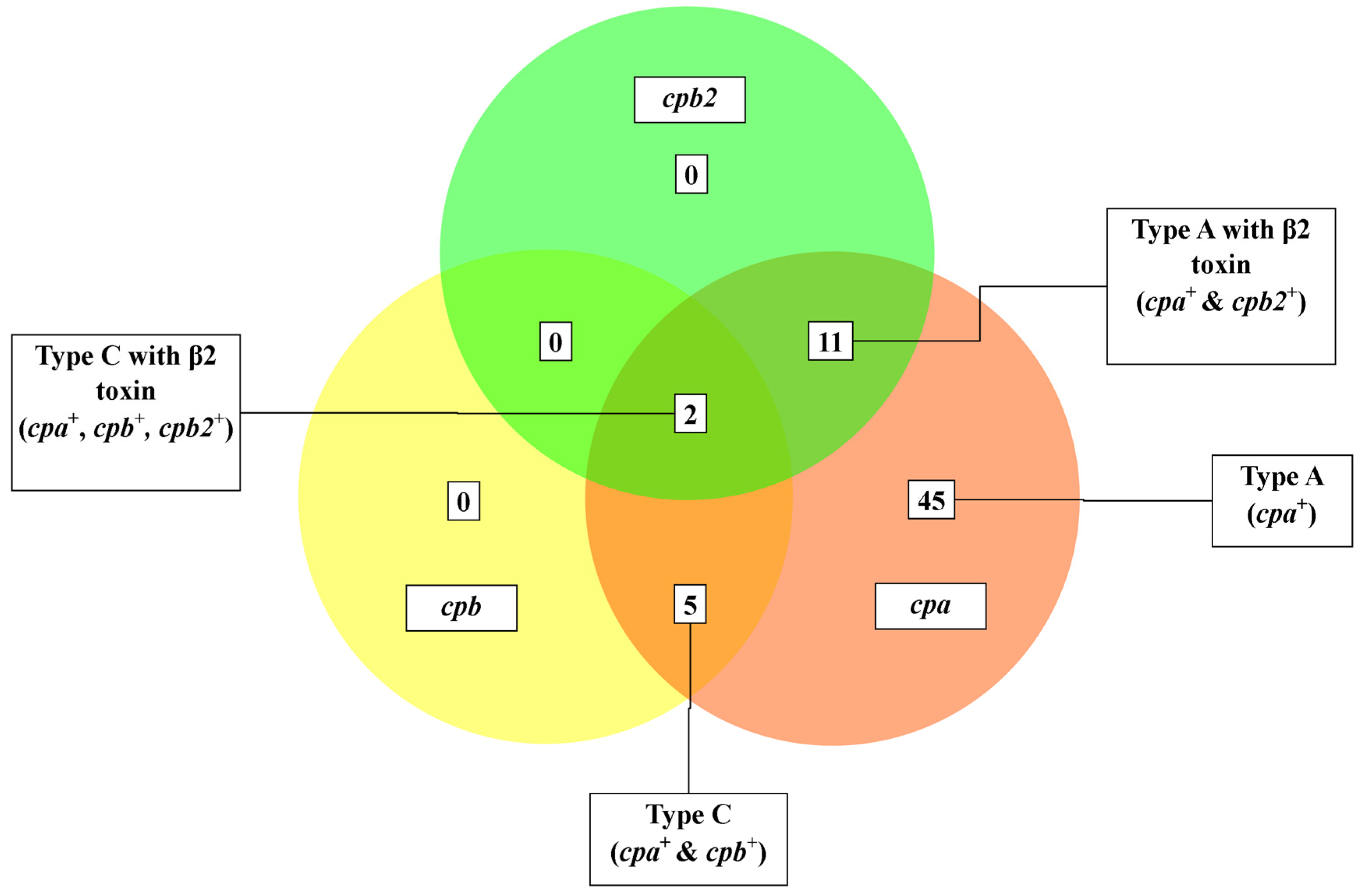
2.2. Toxinotyping of Confirmed Isolates

For all isolates, genomic DNA was extracted with a QIAmp DNA Mini Kit (Qiagen, Hilden, Germany) using the manufacturer’s protocol. Then, PCR-based toxinotyping was undertaken, as described, by screening for cpb, cpe, etx, iap, cpb2, and netB [17,18]. Additionally, isolates were screened for tpeL that codes for a large clostridial toxin [9]. Positive controls for various toxin genes were obtained from our previous studies [1,14]. Thermocycling was performed using an Eppendorf Master cycler® thermal cycler (Eppendorf, Hamburg, Germany), with PCR products separated on 1.5% agarose gels, stained with ethidium bromide, and amplicons visualized with UV illumination. All procedures were conducted twice to ensure consistency.

2.3. Antimicrobial Susceptibility Testing and MAR Indexing

The disk diffusion method (Kirby Bauer) was used to determine the antibiogram profile of C. perfringens isolates against antibiotics approved for clinical use. Erythromycin (ERY, 15 µg), clarithromycin (CLR, 15 µg), ampicillin (AMP, 10 µg), chloramphenicol (CHL, 30 µg), clindamycin (CLI, 2 µg), linezolid (LZD, 30 µg), ofloxacin (OFX, 5 µg), penicillin (PEN, 10 U), co-trimoxazole (STX, 25 µg), tetracycline (TET, 30 µg), and azithromycin (AZM, 15 µg) were placed on the lawn-inoculated plates of Mueller–Hinton agar, and incubated anaerobically at 37 °C for 24 h. Interpretative criteria for Staphylococcus aureus were adopted, as breakpoints for C. perfringens were unavailable [19]. All procedures were performed twice. Isolates resistant to 3 or more classes of antimicrobials were designated as multidrug resistant. Multiple antibiotic resistance (MAR) index of the isolates was calculated as the ratio of number of antibiotics towards which resistance is observed to the number of antibiotics exposed.

2.4. Evaluation of Biofilm-Forming Ability

A crystal violet-based assay was used to evaluate the biofilm-forming ability of C. perfringens isolates. First, pure isolates were propagated in tryptone soy broth (TSB) (Himedia Laboratories, Mumbai, India) anaerobically at 37 °C for 24 h. Further, the broth inoculum was diluted in fresh TSB and subsequently aliquoted to a 96-well polystyrene plate (Nunc™, Fisher Scientific, Waltham, MA, USA). After incubating the plate at 37 °C in an anaerobic environment for 24 h, the planktonic suspension was removed with the help of 1% phosphate-buffered saline (PBS) wash and wells were stained with crystal violet (1%) solution (SRL, Mumbai, India). After incubating at room temperature (30 min), wells were washed with 1% PBS. Ethanol was added to the stained wells and incubated for 15 min. Absorbance was read at 595 nm with a microplate reader (NanoQuant infinite M200PRO, Tecan, Männedorf, Switzerland) and biofilm-forming ability was interpreted as described [1].

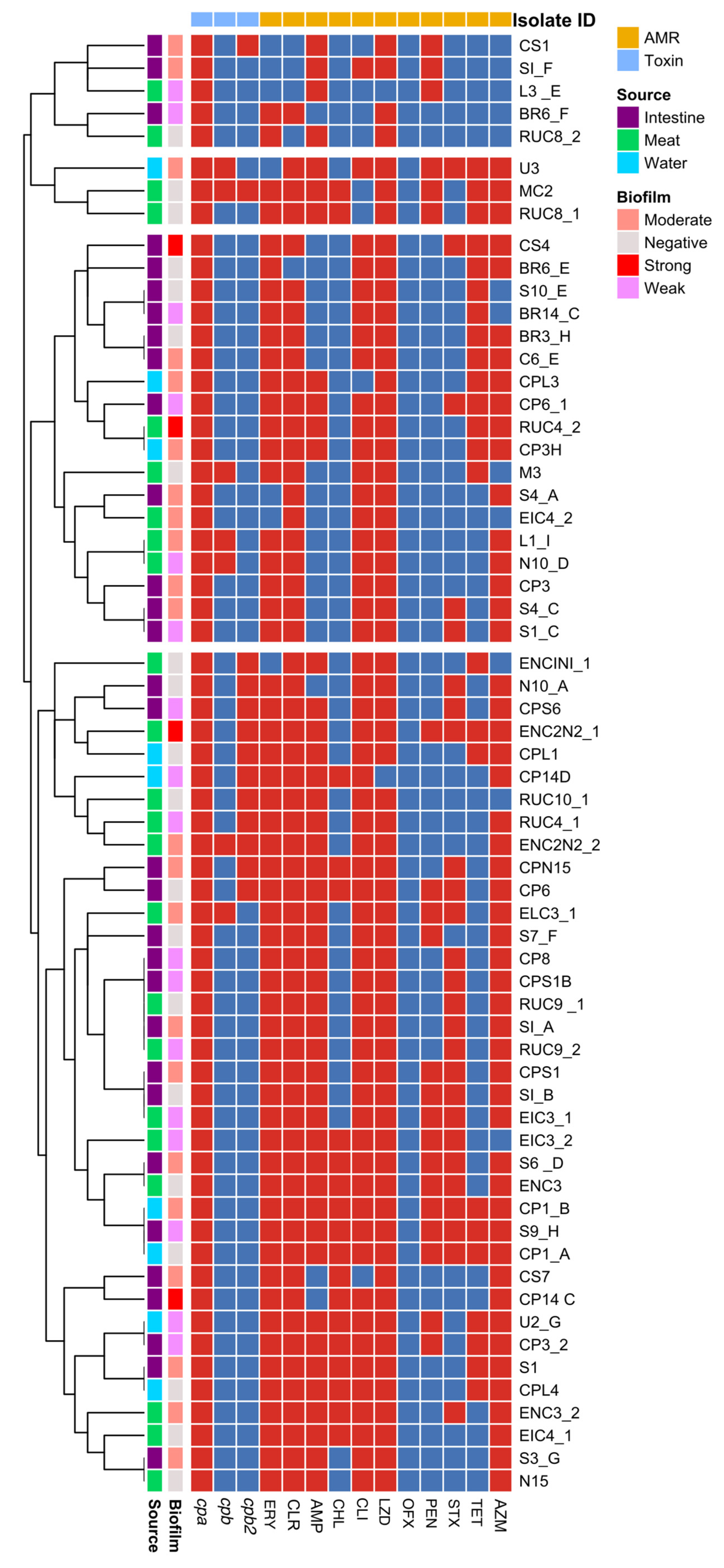
2.5. Hierarchical Clustering, Heatmap, and Correlation Plot Analysis

For all isolates, data were transformed into binary variables and used for construction of a heatmap and correlation plot analyses. Heatmaps with hierarchical clustering were constructed using “pheatmap” and “dendextend” (R software version 4.0.5) packages. Correlation plots with Spearman’s rank correlation method were made after converting data into a correlation matrix using the “corrplot” in R software version 4.0.5.

2.6. Statistical Analysis

Fisher’s exact test was performed with the help of MS-Excel to analyse the statistical association between various variables. For all the proportions, confidence intervals (CI) were calculated with 95% confidence level using binomial exact calculation (https://sample-size.net/confidence-interval-proportion/ accessed on 2 July 2023).

3. Results

3.1. Occurrence of C. perfringens

The overall occurrence of C. perfringens in retail chicken-associated samples was 22.5% (17.74–27.85 95% CI; 63/280) with the highest recovery from intestine samples (31%, 22.13–41.03 95% CI; 31/100), followed by meat samples (23%, 15.17–32.49 95% CI; 23/100) and water samples (18%, 8.58–31.44 95% CI; 9/50). However, none of the hand swab samples were positive for C. perfringens.

3.2. Molecular Toxinotyping

Molecular toxinotyping (Figure 2) of the 63 isolates by screening for toxin/virulence genes (cpa, cpb, etx, iap, cpb2, cpe, NetB, and tpeL) revealed the predominance of Type A toxinotype (45/63, 71.43%; 58.65–82.11 95% CI), followed by Type A with beta2 toxin (11/63, 17.46%; 9.05–29.10 95% CI), Type C (5/63, 7.94%; 2.63–17.56 95% CI) and Type C with beta2 toxin (2/63, 3.17%; 0.39–11.0 95% CI). The most common toxin/virulence gene was cpa (63/63, 100%; 94.31–100 95% CI), followed by cpb2 (13/63, 20.63%; 11.47–32.70 95% CI) and cpb (7/63, 11.11%; 4.59–21.56 95% CI). However, no signatures were detected among isolates for cpe, etx, iap, or netB; therefore, toxinotypes B, D, E, F and G were not present.

3.3. Antimicrobial Resistance Profiling and MAR Index

Regarding antimicrobial susceptibility testing (Table 1), the highest resistance was found in linezolid (61/63, 96.83%; 89–99.61 95% CI) followed by clarithromycin (58/63, 92.06%; 82.44–97.37 95% CI), erythromycin (56/63, 88.89%, 78.44–95.41 95% CI), clindamycin (55/63, 87.30%; 76.50–94.35 95% CI), azithromycin (51/63, 80.95%; 69.09–89.75 95% CI), ampicillin (45/63, 71.43; 58.65–82.11 95% CI), co-trimoxazole (26/63, 41.27%; 29.01–54.38 95% CI), tetracycline (24/63, 38.1%; 26.14–51.20 95% CI), penicillin (21/63, 33.33%; 21.95–46.34 95% CI), and chloramphenicol (18/63, 28.57; 17.89–41.35% 95% CI). All isolates were susceptible to ofloxacin (fluoroquinolone). Multidrug resistance was evaluated based on the criteria of resistance against three or more antimicrobial classes. In the present study, 60 of 63 isolates were multidrug resistant (95.24%; 86.71–99.01 95% CI). The most common resistance profiles were ERY-CLR-AMP-CLI-LZD-STX-AZM with the modal frequency (n = 6), followed by ERY-CLR-AMP-CLI-LZD-AZM and ERY-CLR-AMP-CLI-LZD-PEN-STX-AZM (n = 4). The MAR indices of the isolates ranged from 0.18 to 0.91 with a mean of 0.60. In terms of the isolation source, the mean MAR index values were 0.57, 0.59, and 0.72 for isolates from meat, intestine, and water, respectively (p > 0.5). Isolate S9_H from chicken intestine and isolates CP1_A and CP1_B recovered from water samples were resistant to 10 of 11 antimicrobials screened with a MAR index of 0.91. With the exception of isolate L3_E, the MAR indices of all other isolates were greater than 0.2.

3.4. Evaluation of Biofilm-Forming Ability

Regarding biofilm-forming ability, 43 of 63 isolates (68.25%, 55.31–79.42, 95% CI) were biofilm formers with a mean absorbance of 0.4010. Furthermore, 4, 22 and 17 isolates were deemed strong, medium, and weak biofilm formers, respectively. Mean absorbance of strong biofilm formers was 0.7217 (range: 0.6394 to 0.8746), whereas mean absorbance values of moderate and weak biofilm formers were 0.4388 (range: 0.1946 to 0.6229) and 0.2766 (range: 0.1874 to 0.3455), respectively. Isolate RUC4_2 recovered from chicken meat had the highest observed OD value. However, Fisher’s exact test could not identify any statistical association between biofilm-forming ability and other variables.

3.5. Heatmap-Based Hierarchical Clustering and Correlation Analyses

Heatmap construction with hierarchical clustering grouped isolates into four clusters (two large and two small clusters) (Figure 3). Cluster 1 comprised isolates belonging to Type A toxinotype, susceptible to co-trimoxazole, tetracycline, chloramphenicol, and ofloxacin. Source-wise clustering of isolates was observed (all members were isolated from the carcass). Cluster 2 was small and encompassed isolates resistant to azithromycin and tetracycline. However, all members of this small cluster were susceptible to ofloxacin. The large cluster (Cluster 3) represented isolates negative for cpb2 and that were phenotypically susceptible to chloramphenicol, ofloxacin, and penicillin. Source-based sub-clustering was appreciable within this cluster. The members of the other large cluster (4) were predominantly Type A toxinotype with increased susceptibility to ofloxacin. Correlation plot analysis with Spearman’s correlation coefficient (Figure 4) revealed high positive correlations between various variables. In the correlation plot, the colour and size of the circles represent the magnitude and direction of correlation. There were higher levels of positive correlation (ρ > 0.9) between resistance to macrolides such as erythromycin, clarithromycin, and azithromycin and resistance to ampicillin and linezolid. Biofilm-forming ability was highly associated with linezolid resistance.

4. Discussion

C. perfringens, a ubiquitous member of the gut microflora of normal poultry, is associated with consumption of under-processed protein-rich foods such as meat, which provides a suitable environment for this organism [20]. Contamination of chicken meat by C. perfringens is not uncommon [20].
In the present study, we identified multidrug-resistant, biofilm-producing C. perfringens isolates that harboured toxin genes such as cpa, cpb, and cpb2 in retail chicken-associated samples. In total, 22.5% of the 280 samples screened were positive for C. perfringens, with occurrence rates of 31, 23, and 18% in intestine, meat and water samples, respectively. Faecal prevalence of C. perfringens has been well established, with occurrence levels of C. perfringens in poultry intestinal contents ranging from 9.9 to 95%, with the wide range attributed to variable feeding and management conditions [2,21,22]. Furthermore, the use of antimicrobial growth promoters influences the level of C. perfringens in the gut microflora of poultry [2]. The presence of C. perfringens in intestinal samples in retail shops is of importance as chicken intestines have culinary value in various parts of the world including India and cross-contamination of other meat portions is another possibility.
Higher levels of C. perfringens in poultry meat, ranging from 18 to 88%, have been reported [20]. Differences in methodologies to isolate C. perfringens could explain the dispersion in the occurrence values in meat. Furthermore, the occurrence of C. perfringens in water samples (18%) from the slaughter area indicated that C. perfringens contaminated the slaughter environment, consistent with a study in a Chinese chicken production chain [23]. In that study, C. perfringens was also isolated from operators’ gloves, which contrasted with present results. The predominance of Type A toxinotype in this study has been validated in various parts of the world and in a variety of sources [11,14,24].
The presence of the cpb2 gene in 20.63% of isolates raises concerns of a potential threat to food safety, as this gene is implicated with aggravating gastro-intestinal symptoms in clinical cases linked to antibiotic-associated and sporadic diarrhoea [1]. Moreover, beta toxin is also considered the chief virulence factor in Type C cases [14]. The absence of other toxin genes such as etx, iap, cpe, netB, and tpeL in C. perfringens have been reported [1,24,25]. The presence of various toxin genes on the plasmids offers a wide diversity of pathovars of C. perfringens and allows for conversion of toxinotype by uptake or loss of plasmids [6].
The indiscriminate use of antibiotics in intensively reared animals such as poultry and pigs has resulted in the wide prevalence of multidrug-resistant strains. In the present study, there were very high resistance rates for linezolid (96.83%), clindamycin (87.30%), and macrolides such as erythromycin (92.06%), clarithromycin (88.89%), and azithromycin (80.95%). Reports of linezolid resistance in Gram-positive anaerobic bacteria have been steadily surfacing in recent years [26]. Similar levels of erythromycin resistance among C. perfringens were identified in an earlier report from Iran investigating raw beef [11]. Moreover, a study from Egypt reported broiler-origin C. perfringens with 100% resistance to macrolides and lincosamides [27]. Multidrug resistance has been a glaring trend among C. perfringens in recent years. Alarming levels of multidrug resistance were encountered in the present study (95.25%). Various researchers have hinted at the prevalence of multidrug-resistant strains of C. perfringens isolated from poultry origin [23,24,27]. The irrational and unregulated use of antibiotics at veterinary care and farm levels are regarded as probable sources of multidrug-resistant strains [28].
Biofilms are a food safety concern as they promote survival of C. perfringens within and outside hosts and enhance pathogenicity [29]. C. perfringens participate in both mono- and multi-species biofilms with enhanced survivability in the presence of oxygen [12]. In the present study, biofilm formation was confirmed in as many as 68.25% of C. perfringens isolates, a proportion that is slightly less than previously reported [1]. Disharmony in rates of multidrug resistance and biofilm is not uncommon and also occurs in Gram-negative bacteria [30].
Close clustering of isolates from meat, intestine, and water highlighted the ability of the organism to pass along the food chain, raising concerns of cross-contamination of surfaces, as suggested [23]. Close association of resistance to lincosamides and macrolides was attributed to macrolide-lincosamide-streptogramin B resistance, often encoded by erm(Q) or erm(B) genes [10]. Increased associations between linezolid resistance and biofilm-forming ability of the C. perfringens isolates are a concern, as linezolid is used for methicillin-resistant Staphylococcus aureus and vancomycin-resistant Enterococci [26].

5. Conclusions

This study sheds light on the concerning prevalence of multidrug-resistant, biofilm-forming virulent C. perfringens in broiler chicken retail points in Meghalaya, a northeastern state in India dominated by tribal populations with unique food preferences. The predominance of Type A and Type C toxinotypes in retail chicken meat and intestines merits increased attention from the perspectives of food safety and public health. The alarming rate of multidrug resistance among C. perfringens recovered from poultry advocates for the adequate cooking of meat before consumption. Further research is warranted to investigate the mechanisms underlying multidrug resistance in C. perfringens emanating from the poultry value chain.

Author Contributions

Conceptualization and methodology: G.B.P. and A.A.P.M.; investigation: G.B.P., K.S. and H.S.; formal analysis: G.B.P. and A.A.P.M.; writing—original draft and writing—review and editing: G.B.P. and A.A.P.M. All authors have read and agreed to the published version of the manuscript.

Funding

This research received no external funding.

Data Availability Statement

All data are available from the corresponding author and will be provided on request.

Acknowledgments

The authors are grateful to the Research Central Agricultural University (Imphal), the College of Agriculture, Central Agricultural University (Imphal), Kyrdemkulai, and the ICAR RC NEH, Umiam, Meghalaya, for providing the necessary facilities and supporting the study. We sincerely acknowledge the contribution of John P. Kastelic, University of Calgary, Canada, in improving the manuscript.

Conflicts of Interest

The authors declare no conflict of interest.

References

  1. Milton, A.A.P.; Momin, A.G.; Gandhale, P.N.; Das, S.; Ghatak, S.; Priya, G.B.; Firake, D.M.; Srinivas, K.; Momin, K.M.; Hussain, Z.; et al. Prevalence, toxinotyping, antimicrobial susceptibility and biofilm-forming ability of Clostridium perfringens isolated from free-living rodents and shrews. Anaerobe 2022, 77, 102618. [Google Scholar] [CrossRef] [PubMed]
  2. Van Immerseel, F.; De Buck, J.; Pasmans, F.; Huyghebaert, G.; Haesebrouck, F.; Ducatelle, R. Clostridium perfringens in poultry: An emerging threat for animal and public health. Avian Pathol. 2004, 33, 537–549. [Google Scholar] [CrossRef]
  3. Thomas, M.; Murray, R. Estimating the burden of food-borne illness in Canada. Can. Commun. Dis. Rep. 2014, 40, 299–302. [Google Scholar] [CrossRef] [PubMed]
  4. Kirk, M.D.; Pires, S.M.; Black, R.E.; Caipo, M.; Crump, J.A.; Devleesschauwer, B.; Döpfer, D.; Fazil, A.; Fischer-Walker, C.L.; Hald, T.; et al. World Health Organization Estimates of the Global and Regional Disease Burden of 22 Foodborne Bacterial, Protozoal, and Viral Diseases, 2010: A Data Synthesis. PLoS Med. 2015, 12, e1001940. [Google Scholar]
  5. Abdelrahim, A.M.; Radomski, N.; Delannoy, S.; Djellal, S.; Le Négrate, M.; Hadjab, K.; Fach, P.; Hennekinne, J.A.; Mistou, M.Y.; Firmesse, O. Large-Scale genomic analyses and toxinotyping of Clostridium perfringens implicated in foodborne outbreaks in France. Front. Microbiol. 2019, 10, 777. [Google Scholar] [CrossRef] [PubMed]
  6. Petit, L.; Gibert, M.; Popoff, M.R. Clostridium perfringens: Toxinotype and genotype. Trends Microbiol. 1999, 7, 104–110. [Google Scholar] [CrossRef] [PubMed]
  7. Freedman, J.C.; Shrestha, A.; McClane, B.A. Clostridium perfringens enterotoxin: Action, genetics, and translational applications. Toxins 2016, 8, 73. [Google Scholar] [CrossRef] [PubMed]
  8. Gao, X.; Yang, Q.; Huang, X.; Yan, Z.; Zhang, S.; Luo, R.; Wang, P.; Wang, W.; Xie, K.; Jiang, T.; et al. Effects of Clostridium perfringens beta2 toxin on apoptosis, inflammation, and barrier function of intestinal porcine epithelial cells. Microb. Pathog. 2020, 147, 104379. [Google Scholar] [CrossRef]
  9. Coursodon, C.F.; Glock, R.D.; Moore, K.L.; Cooper, K.K.; Songer, J.G. TpeL-producing strains of Clostridium perfringens type A are highly virulent for broiler chicks. Anaerobe 2012, 18, 117–121. [Google Scholar] [CrossRef]
  10. Adams, V.; Han, X.; Lyras, D.; Rood, J.I. Antibiotic resistance plasmids and mobile genetic elements of Clostridium perfringens. Plasmid 2018, 99, 32–39. [Google Scholar] [CrossRef]
  11. Hassani, S.; Pakbin, B.; Brück, W.M.; Mahmoudi, R.; Mousavi, S. Prevalence, Antibiotic Resistance, Toxin-Typing and Genotyping of Clostridium perfringens in Raw Beef Meats Obtained from Qazvin City, Iran. Antibiotics 2022, 11, 340. [Google Scholar] [CrossRef]
  12. Pantaléon, V.; Bouttier, S.; Soavelomandroso, A.P.; Janoir, C.; Candela, T. Biofilms of Clostridium species. Anaerobe 2014, 30, 193–198. [Google Scholar] [CrossRef] [PubMed]
  13. Charlebois, A.; Jacques, M.; Archambault, M. Comparative transcriptomic analysis of Clostridium perfringens biofilms and planktonic cells. Avian Pathol. 2016, 45, 593–601. [Google Scholar] [CrossRef] [PubMed]
  14. Milton, A.A.P.; Sanjukta, R.; Gogoi, A.P.; Momin, K.M.; Priya, G.B.; Das, S.; Ghatak, S.; Sen, A.; Kandpal, B.K. Prevalence, molecular typing and antibiotic resistance of Clostridium perfringens in free range ducks in Northeast India. Anaerobe 2020, 64, 102242. [Google Scholar] [CrossRef] [PubMed]
  15. Sarmah, H.; Hazarika, R.; Deka, P.; Sharma, R. Isolation and Characterization of Clostridium perfringens from Suspected Cases of Necrotic Enteritis (NE) in Broiler Chicken. Int. J. Livest. Res. 2019, 9, 221–232. [Google Scholar] [CrossRef]
  16. Das, A.; Mazumder, Y.; Dutta, B.K.; Shome, B.R.; Bujarbaruah, K.M.; Kumar, A. Clostridium perfringens Type A from Broiler Chicken with Necrotic Enteritis. Int. J. Poult. Sci. 2008, 7, 601–609. [Google Scholar] [CrossRef]
  17. Van Asten, A.J.A.M.; van der Wiel, C.W.; Nikolaou, G.; Houwers, D.J.; Gröne, A. A multiplex PCR for toxin typing of Clostridium perfringens isolates. Vet. Microbiol. 2009, 136, 411–412. [Google Scholar] [CrossRef]
  18. Tolooe, A.; Shojadoost, B.; Peighambari, S.M.; Tamaddon, Y. Prevalence of netB Gene among Clostridium perfringens Isolates Obtained from Healthy and Diseased Chickens. J. Anim. Vet. Adv. 2011, 10, 106–110. [Google Scholar] [CrossRef]
  19. M100 ED32; Performance Standards for Antimicrobial Susceptibility Testing. Clinical and Laboratory Standard Institute: Wayne, PA, USA, 2022.
  20. Guran, H.S.; Oksuztepe, G. Detection and typing of Clostridium perfringens from retail chicken meat parts. Lett. Appl. Microbiol. 2013, 57, 77–82. [Google Scholar] [CrossRef]
  21. Svobodová, I.; Steinhauserová, I.; Nebola, M. Incidence of Clostridium perfringens in broiler chickens in the Czech Republic. Acta Vet. Brno 2007, 76, S25–S30. [Google Scholar] [CrossRef]
  22. Fan, Y.C.; Wang, C.L.; Wang, C.; Chen, T.C.; Chou, C.H.; Tsai, H.J. Incidence and Antimicrobial Susceptibility to Clostridium perfringens in Premarket Broilers in Taiwan. Avian Dis. 2016, 60, 444–449. [Google Scholar] [CrossRef] [PubMed]
  23. Xu, W.; Wang, H.; Chen, S.; Chen, Y.; Liu, L.; Wu, W. Tracing Clostridium perfringens strains along the chicken production chain from farm to slaughter by multilocus sequence typing. Zoonoses Public Health 2021, 68, 431–442. [Google Scholar] [CrossRef] [PubMed]
  24. Jang, Y.S.; Kim, D.H.; Bae, D.; Kim, S.H.; Kim, H.; Moon, J.S.; Song, K.Y.; Chon, J.W.; Seo, K.H. Prevalence, toxin-typing, and antimicrobial susceptibility of Clostridium perfringens from retail meats in Seoul, Korea. Anaerobe 2020, 64, 102235. [Google Scholar] [CrossRef] [PubMed]
  25. Koo, B.S.; Hwang, E.H.; Kim, G.; Park, J.Y.; Oh, H.; Lim, K.S.; Kang, P.; Lee, H.Y.; Jeong, K.J.; Mo, I.; et al. Prevalence and characterization of Clostridium perfringens isolated from feces of captive cynomolgus monkeys (Macaca fascicularis). Anaerobe 2020, 64, 102236. [Google Scholar] [CrossRef] [PubMed]
  26. Zhang, S.; Liu, P.; Wang, Y.; Shen, Z.; Wang, S. Multiresistance gene cfr(C) in Clostridium perfringens of cattle origin from China. J. Antimicrob. Chemother. 2021, 76, 3310–3312. [Google Scholar] [CrossRef]
  27. Osman, K.M.; Elhariri, M. Antibiotic resistance of Clostridium perfringens isolates from broiler chickens in Egypt. OIE Rev. Sci. Tech. 2013, 32, 841–850. [Google Scholar] [CrossRef]
  28. Doyle, M.E. Multidrug-resistant pathogens in the food supply. Foodborne Pathog. Dis. 2015, 12, 261–279. [Google Scholar] [CrossRef]
  29. Hu, W.S.; Woo, D.U.; Kang, Y.J.; Koo, O.K. Biofilm and spore formation of Clostridium perfringens and its resistance to disinfectant and oxidative stress. Antibiotics 2021, 10, 396. [Google Scholar] [CrossRef]
  30. Cepas, V.; López, Y.; Muñoz, E.; Rolo, D.; Ardanuy, C.; Martí, S.; Xercavins, M.; Horcajada, J.P.; Bosch, J.; Soto, S.M. Relationship between Biofilm Formation and Antimicrobial Resistance in Gram-Negative Bacteria. Microb. Drug Resist. 2019, 25, 72–79. [Google Scholar] [CrossRef]
Figure 1. Isolation and characterization of C. perfringens recovered in the present study. (A) Isolation; (B) Antibiogram; (C) Toxinotyping.
Foods 12 04185 g001
Figure 2. Venn diagram depicting various toxinotypes of C. perfringens identified in the present study.
Foods 12 04185 g002
Figure 3. Heatmap analysis with hierarchical clustering depicting occurrence of toxin genes and resistance patterns of C. perfringens with respect to isolation source and biofilm-forming ability (ERY: Erythromycin; CLR: Clarithromycin; AMP: Ampicillin; CHL: Chloramphenicol; CLI: Clindamycin; LZD: Linezolid; OFX: Ofloxacin; PEN: Penicillin; STX: Co-trimoxazole; TET: Tetracycline; AZM: Azithromycin).
Foods 12 04185 g003
Figure 4. Correlation plot analysis depicting high levels of positive correlations between various attributes of C. perfringens (ERY: Erythromycin; CLR: Clarithromycin; AMP: Ampicillin; CHL: Chloramphenicol; CLI: Clindamycin; LZD: Linezolid; OFX: Ofloxacin; PEN: Penicillin; STX: Co-trimoxazole; TET: Tetracycline; AZM: Azithromycin).
Foods 12 04185 g004
Table 1. Virulence repertoire and antibiogram profile of C. perfringens isolated from broiler value chain.
Isolate IDSourceToxin GenesDesignated ToxinotypeAntimicrobial Susceptibility #MAR IndexBiofilm-Forming Ability ##
cpacpbcpb2ERYCLRAMPCHLCLILZDOFXPENSTXTETAZM
SI_FIntestine+Type AIIRIRRSRSIS0.36++
SI_AIntestine+Type ARRRIRRSSRSR0.64++
SI_BIntestine+Type ARRRSRRSRRIR0.73
S4_CIntestine+Type ARRSSRRSSRIR0.55++
S6 _DIntestine+Type ARRRRRRSRRSR0.82++
S9_HIntestine+Type ARRRRRRIRRRR0.91+
S4_AIntestine+Type AIRSSRRSSSSR0.36++
S3_GIntestine+Type ARRRSRRSSSIR0.55++
S1_CIntestine+Type ARRSSRRSSRIR0.55+
S10_EIntestine+Type ARRSSRRSSSRI0.45
S7_FIntestine+Type ARRRSRRSRSIR0.64
BR6_EIntestine+Type ARSSSRRSSSRR0.45
BR6_FIntestine+Type ARRSSIRSSSII0.27+
BR14_CIntestine+Type ARRSSRRSSSRI0.45+
CPN15Intestine++Type A with cpb2RRRRRRSSRSR0.72++
CP3Intestine+Type ARRSSRRSSSIR0.45++
BR3_HIntestine+Type ARRSSRRSSSRR0.55
S1Intestine+Type ARRRRRRSSSRR0.72++
CP6Intestine++Type A with cpb2RRRRRRSRRIR0.82
CS1Intestine++Type A with cpb2IIRIIRSRSSI0.27++
CPS1Intestine+Type ARRRSRRSRRIR0.73++
CS4Intestine+Type ARRSSRRSSRRR0.64+++
N10_AIntestine++Type A with cpb2RRSSRRSSRIR0.55
C6_EIntestine+Type ARRSSRRSSSRR0.55++
CS7Intestine+Type ARRSRSRSSSIR0.45++
CP14 CIntestine+Type ARRSRRRSSSIR0.55+++
CPS6Intestine++Type A with cpb2RRRSRRSSRIR0.64+
CPS1BIntestine+Type ARRRSRRSSRIR0.64+
CP8Intestine+Type ARRRSRRSSRIR0.64+
CP6_1Intestine+Type ARRRSRRSSRRR0.73+
CP3_2Intestine+Type ARRRRRRSRSRR0.82+
RUC9_2Meat+Type ARRRIRRSSRSR0.64+
ENC2N2_1Meat++Type A with cpb2RRRSRRSRRRR0.82+++
EIC4_1Meat+Type ARRRRRRSSSIR0.64
RUC4_1Meat++Type A with cpb2RRRSRRSSSSR0.55+
RUC9 _1Meat+Type ARRRIRRSSRSR0.64
L3 _EMeat+Type ASSRSSSSRSSS0.18+
L1_IMeat++Type CRRSSRRSSSSR0.45++
MC2Meat+++Type C with cpb2RRRRSRSRSRR0.73
N10_DMeat++Type CRRSSRRSSSSR0.45+
EIC4_2Meat+Type AIRSSRRSSSII0.27++
ELC3_1Meat++Type CRRRIRRSRRIR0.73++
RUC4_2Meat+Type ARRRIRRSSSRR0.64+++
ENCINI_1Meat++Type A with cpb2IRRSRRSSSRS0.45
EIC3_1Meat+Type ARRRSRRSRRIR0.72+
ENC2N2_2Meat+++Type C with cpb2RRRSRRSSSSR0.55++
EIC3_2Meat+Type ARRRRRRSRRIS0.72+
ENC3Meat+Type ARRRRRRSRRIR0.82
M3Meat++Type CRRSSRRSSSRS0.45
N15Meat+Type ARRRSRRSSSSR0.55
ENC3_2Meat+Type ARRRRRRSSRIR0.73++
RUC8_2Meat+Type ARSRSIRSSSIS0.27
RUC10_1Meat++Type A with cpb2RRRIRRSSSIS0.45
RUC8_1Meat+Type ARRRRIRSRIRR0.73
CPL4Water+Type ARRRRRRSSSRR0.73
U2_GWater+Type ARRRRRRSRSRR0.82+
CP1_AWater+Type ARRRRRRSRRRR0.91
CP1_BWater+Type ARRRRRRSRRRR0.91++
U3Water++Type CIRRSRRSRRRR0.73++
CP3HWater+Type ARRRSRRSSSRR0.64++
CP14DWater++Type A with cpb2RRRRRSSSSIR0.55+
CPL1Water++Type A with cpb2RRRSRRSSSRR0.64
CPL3Water+Type ARRRSIRSSSRR0.55++
# ERY: Erythromycin; CLR: Clarithromycin; AMP: Ampicillin; CHL: Chloramphenicol; CLI: Clindamycin; LZD: Linezolid; OFX: Ofloxacin; PEN: Penicillin; STX: Co-trimoxazole; TET: Tetracycline; AZM: Azithromycin; S: Sensitive; I: Intermediate; R: Resistance. ## +: weak positive; ++: moderate positive; +++: strong positive; −: negative.
Disclaimer/Publisher’s Note: The statements, opinions and data contained in all publications are solely those of the individual author(s) and contributor(s) and not of MDPI and/or the editor(s). MDPI and/or the editor(s) disclaim responsibility for any injury to people or property resulting from any ideas, methods, instructions or products referred to in the content.

