1. Introduction
All terrestrial and aquatic photoautotrophs, such as plants, microalgae, and macroalgae, universally produce carotenoids, the most prevalent class of tetraterpenoid pigments. Carotenoids can be categorized into two groups: (i) carotenes, which consist of a parent hydrocarbon chain without any functional group, such as β-carotene, and (ii) xanthophylls, which have oxygen as a functional group, exemplified by lutein and zeaxanthin. Carotenoids play a vital role in human health due to their antioxidant and provitamin A activities [
1,
2,
3].
Humans accumulate xanthophylls, namely lutein and zeaxanthin, as a macular pigment in the retina’s fovea and inner plexiform layer. These pigments play a crucial role in safeguarding the retinal membrane from the detrimental effects of short-wavelength high-intensity light and enhance visual acuity [
4,
5]. Moreover, clinical and epidemiological studies have observed the critical role of dietary lutein and zeaxanthin in cognitive improvement [
6,
7,
8], reducing the risk of age-related macular degeneration (AMD) [
5], age-related declines in muscle mass and strength [
9], cardiometabolic health [
10], and reducing the risk of cancer [
11,
12].
Colorful fruits and vegetables constitute the primary dietary source of carotenoids in the human diet [
13]. However, in recent years, microalgae, macroalgae, and food waste have also been explored to extract natural carotenoids [
14,
15,
16,
17,
18,
19]. Among the natural sources, marigold (
Tagetes patula L. and
T. erecta L.) flower petals are the most vital sources of carotenoids, especially lutein diesters [
20]. Considering the high content of lutein, the industry extensively uses marigold flowers for natural lutein production for functional food, feed, cosmetics, and pharmaceuticals [
19]. In addition to lutein extraction, marigold flowers are extensively used in poultry feed to improve egg yolk pigmentation, a symbol of high nutritional value and marketing quality eggs [
21,
22].
In marigold flower petals, lutein acylated with saturated fatty acids, such as stearic (C
18:0), palmitic (C
16:0), myristic (C
14:0), and lauric (C
12:0) acid moieties, are dominantly found [
23]. Interestingly, these fatty acids are also associated with other xanthophylls found in different plants [
24]. When the identical fatty acid is esterified on both sides of the lutein molecule, it results in a lutein homodiester (e.g., lutein-dimyristate). On the other hand, esterifying lutein with two different types of fatty acids forms a heterodiester (e.g., lutein-3-
O-myristate-3′-
O-palmitate) [
25]. Interestingly, as lutein is an asymmetric molecule due to the presence of a β-ionone ring at one side and an ε-ionone ring on another side, esterification with two different kinds of fatty acids gives rise to lutein ester regioisomers—for example, lutein-3
-O-palmitate-3′
-O-myristate and lutein-3-
O-myristate-3′-
O-palmitate.
Several studies have attempted to quantify the lutein contents in marigold flower petals. However, most of these studies quantified the lutein contents in saponified samples [
26,
27,
28]. In saponification, fatty acid moieties attached to the carotenoid molecules are removed, yielding free carotenoids. This process makes quantification easy; however, this approach does not provide information about naturally occurring carotenoid esters. In recent years, the availability of advanced liquid chromatography (LC)–mass spectrometry (MS) techniques means that it is possible to separate and identify the complex carotenoid esters regioisomers in the native samples [
29,
30].
Apart from carotenoids, phytosterols represent another class of beneficial compounds that help to regulate low-density lipoprotein (LDL) cholesterol levels and endothelial function [
31]. The US Food and Drug Administration (FDA) recommends a daily dietary intake of 2 g of non-esterified plant sterols to achieve the health advantages of reducing blood total and LDL cholesterol and lowering the risk of coronary heart disease (CHD) [
32].
Tocopherols (α-, β-, γ-, and δ; commonly known as vitamin E) are essential constituents of cellular lipids. They play a crucial role as a chain-breaking antioxidant, interrupting and mitigating the propagation of harmful oxidative reactions that can lead to cellular damage and degradation of lipids, proteins, and DNA [
33]. Consequently, this aids in reducing the susceptibility to diseases linked with oxidative stress, encompassing conditions such as cardiovascular diseases, neurodegenerative diseases, various types of cancer, and cognitive deterioration linked to aging [
34,
35,
36,
37]. Moreover, α-tocopherol is believed to improve skin health and immune function [
38].
Considering the commercial importance of lutein obtained from marigold flower petals, the objective of this work was to carry out an identification of prominent carotenoid esters from the flower petals of ten marigold cultivars by LC-diode-array detection (DAD)-MS, followed by their quantification by LC-DAD. In addition, tocopherols, phytosterols, and fatty acids were analyzed by gas chromatography (GC)–flame ionization detection (FID) and GC–MS methods. Furthermore, the 2,2′-azino-bis(3-ethylbenzothiazoline-6-sulfonic acid (ABTS•+) and 2,2-diphenyl-1-picrylhydrazyl (DPPH•) radical scavenging abilities of lipophilic extract were determined. The lutein contents of marigold cultivars commonly cultivated in Korea can help to recommend the lutein-rich and high flower-yielding cultivars for the industrial extraction of lutein and functional food preparations.
3. Results and Discussion
3.1. Identification of Carotenoids
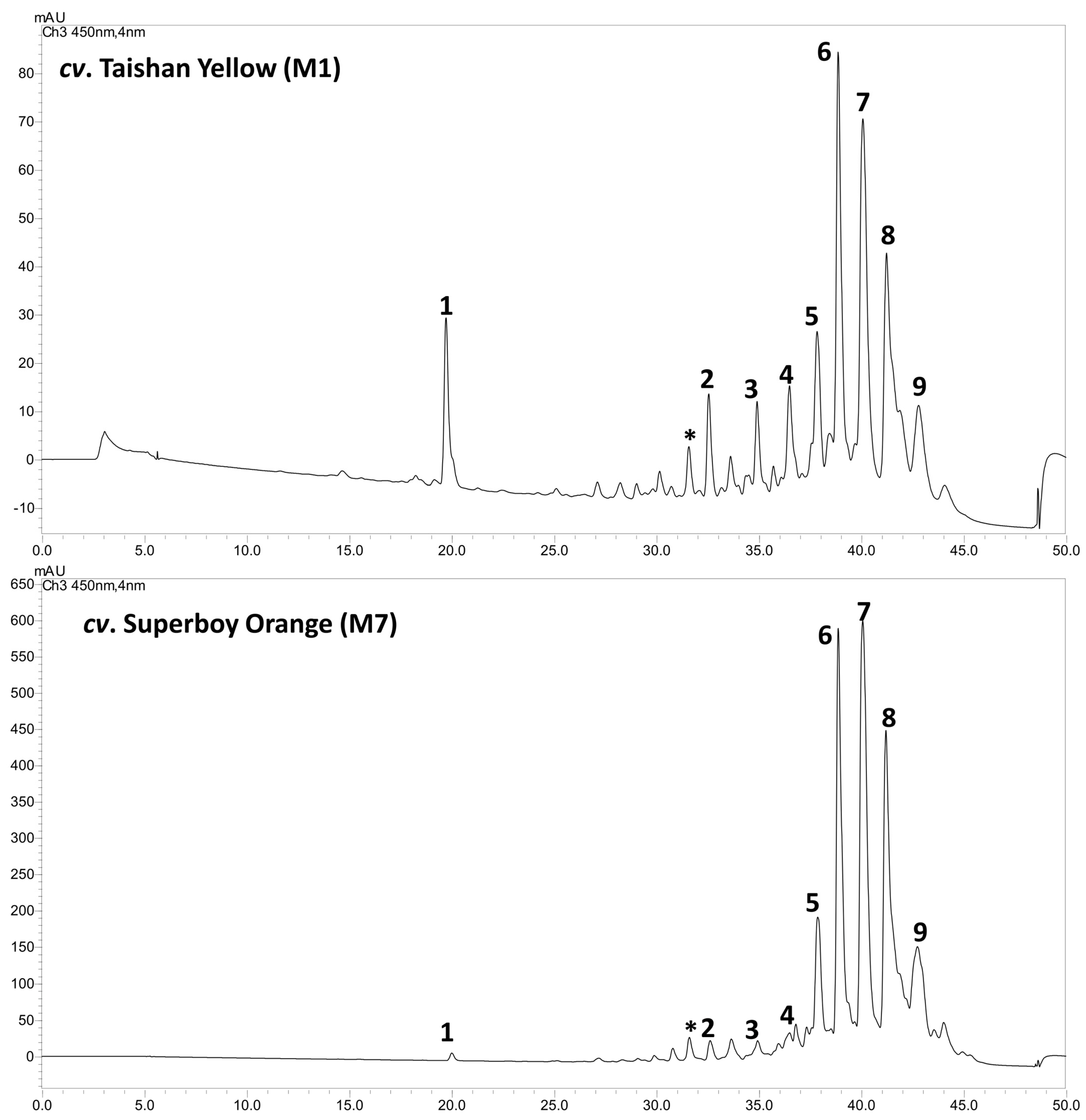
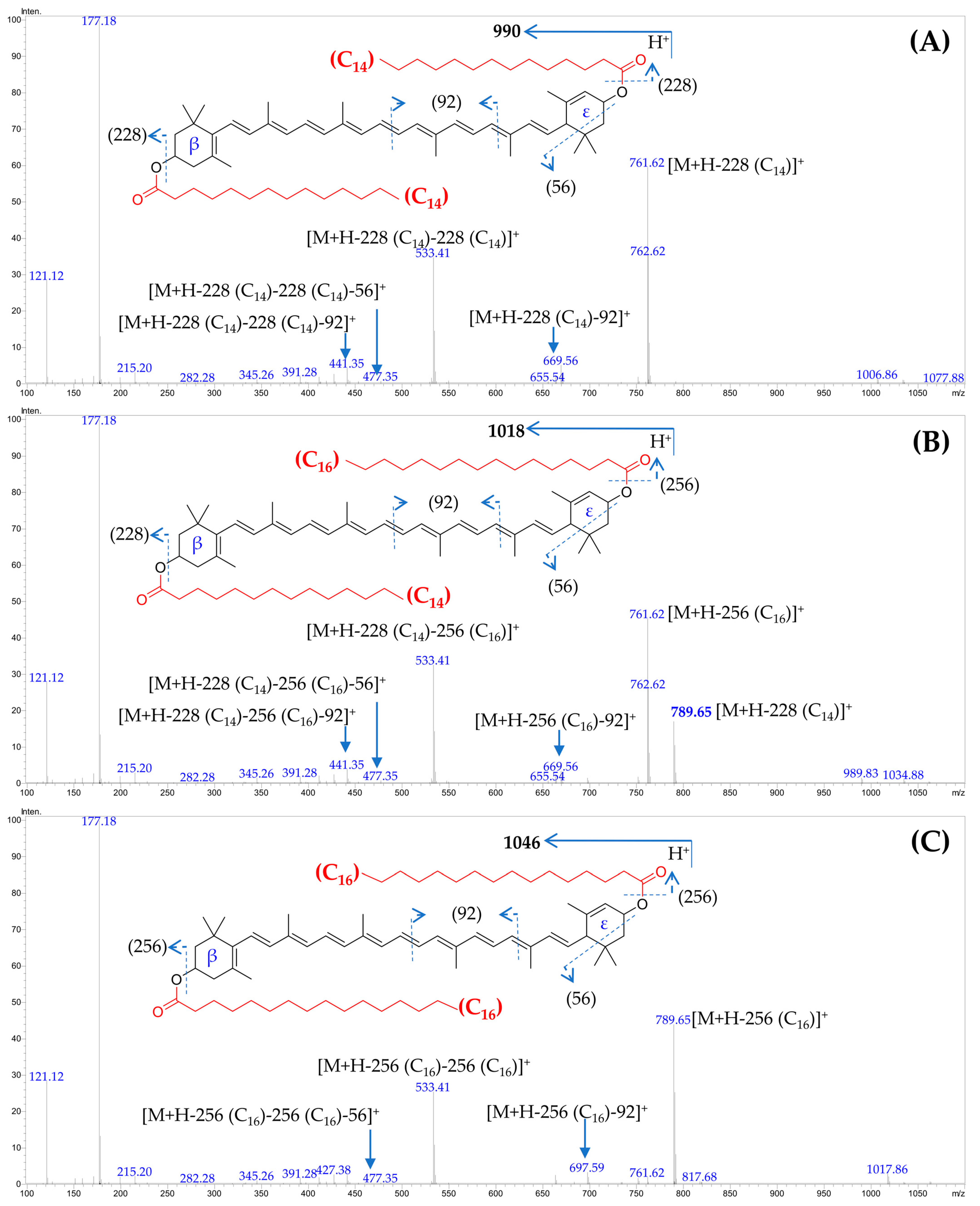
In the non-saponified lipophylic marigold extract, nine major carotenoids were identified (
Table 5,
Figure 1). The lutein homodiesters (attachment of the same fatty acid moieties at the β- and ε-rings of the lutein molecule) and heterodiesters (attachment of different fatty acid moieties at the β- and ε-rings of the lutein molecule) were found to dominate in the lipophylic marigold extract of all the studied cultivars. The neutral loss of either one [M+H-FA
1]
+ or the other [M+H-FA
2]
+, or both the fatty acid moieties [M+H-FA
1-FA
2]
+ were the characteristic fragment ions obtained from these lutein diesters (
Table 2,
Figure 2). The neutral loss of both fatty acid moieties gives rise to a fragment equivalent to the lutein carbon backbone at m/z 533. Additionally, we observed typical fragmentation patterns of ε-ring-containing structures, including the loss of 56 u due to retro-Diels-Alder fragmentation of the α-ionone moiety and of the carotenoid polyene chain, which involved the elimination of toluene (92 u) (
Figure 2) [
29,
47].
In the case of the lutein heterodiesters, such as peak 7 (
Figure 2B), the higher intensity of the fragment ion at m/z 761 [M+H-256 (palmitate)]
+, compared to m/z 789 [M+H-228 (myristate)]
+, indicated that palmitic acid is attached at the 3′-
O′ position of ε-ring, as loss of fatty acids from this position generates a more stable fragment ion than fatty acid acylated to the 3-
O-position of β-ring [
29]. Thus, this heterodiester was assigned as (all-
E)-lutein-3-
O-myristate -3′-
O-palmitate.
3.2. Quntititaive Analysis of Carotenoids and Tocopherols
In the present study, nine major carotenoids were identified and quantified from 10 marigold cultivars (
Table 6). Among the identified carotenoids, lutein diesters were the most dominating, accounting for 63.76–96.95% (
cv. Alaska and –
cv. Durango Orange, respectively) of total carotenoids. Other carotenoids, such as (all-
E)-lutein, (all-
E)-β-carotene, (all-
E)-violaxanthin-3,3′-dimyristate, and (all-
E)-violaxanthin-3-
O-myristate-3-
O′-palmitate were present in a small amount. Among the five major lutein-diesters identified and quantified, (all-
E)-lutein-3-
O-myristate-3′-
O-palmitate was the most dominating carotenoid amond the French (
T. patula L.)-type cultivars (M5–M10). Conversely, in
cv. Taishan Orange (M2) and
cv. Incall Orange (M3; Aztec type;
T. erecta L.), lutein dipalmitate was the dominant carotenoid.
In the present study, the total lutein contents varied significantly (
p < 0.05, Tukey HSD) among cultivars, ranging from 23.73–2613.47 µg/g fresh weight (FW) (
cv. Alaska and
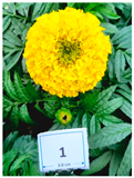
cv. Superboy Orange, respectively). The significantly highest contents of lutein were recorded in
cv. Superboy Orange, due to the presence of the highest amount of (all-
E)-lutein-3-
O-myristate-3-
O-laurate (212.13 µg/g FW), (all-
E)-lutein-dimyristate (602.86 µg/g FW), (all-
E)-lutein-3-
O-myristate-3′-
O-palmitate (753.86 µg/g FW), and (all-
E)-lutein-3-
O-stearate-3′-
O-palmitate (317.97 µg/g FW) (
Table 6).
Several studies have attempted to quantify the lutein contents in marigold flower petals. However, most of these studies quantified the lutein contents in saponified samples [
26,
27,
28]. In saponification, fatty acid moieties attached to the carotenoid molecules are removed, yielding free carotenoids. This process makes quantification easy; however, this approach does not provide information about naturally occurring carotenoid esters. In recent years, the availability of high-resolution mass spectrometry techniques has made it possible to separate and identify the complex carotenoid esters in native samples. In one such study, Piccaglia et al. [
48] recorded the predominance of lutein dimyristate, lutein myristate palmitate, and lutein dipalmitate in ten types of
T. patula and
T. erecta, with a lutein content of 167.7–5699.0 µg/g. Similarly, El-Sayed et al. [
20] also recorded the dominance of (all-
E)-lutein-dimyristate, (all-
E)-lutein-3-
O-myristate-3′-
O-palmitate, and (all-
E)-lutein-dipalmitate in the seven cultivars of
T. patula and
T. erecta, with a lutein content of 167–5752.0 µg/g. The total lutein contents recorded in the present investigation agree with these studies.
This study screened the lipophilic extract of marigold flower petals for tocopherol contents using LC-PDA-MS. Among the studied cultivars, 167.91–338.50 µg/g FW (
cv. Superboy Yellow and
cv. Taishan Orange, respectively) of α-tocopherol was recorded (
Table 6), while other forms (e.g., δ-, γ-, β-tocopherol) were not found in significant quantities.
To the best of our knowledge, previous studies on the tocopherol contents of marigold flower petals are not available. Gong et al. [
49] reported the dominance of α-tocopherol in T. erecta flower petals; however, quantitative analysis was not performed in their study. Among the 62 edible tropical plants screened for α-tocopherol contents, the highest content of 420 µg/g FW was recorded from Sauropus androgynus leaves, while Musa sapientum flower was found to contain 29.8 µg/g FW of α-tocopherol [
50]. Considering this, marigold flower petals contain a substantially higher amount of α-tocopherol than commonly consumed herbs. Moreover, α-tocopherol content recorded from marigold flower petals in the present study are substantially higher than the contents recorded in our recent studies from 18 Korean traditional green leafy vegetables (13.6–133.6 µg/g FW) [
51], leaf mustard (Brassica juncea (L.) czern.) cultivars (67.16–83.42 µg/g FW) [
43], and emerging green leafy vegetables, including Moringa oleifera (22.0–83.7 µg/g FW) [
44].
Attributable to the high contents of α-tocopherol in marigold flower petals, their enhanced intake can help to minimize the incidence of oxidative stress-related diseases [
36].
3.3. Ideal Cultivar for Food Fortification and Commercial Lutein Extraction
Marigold flower petals are the primary source of lutein for commercial extraction [
19]. Moreover, marigold flowers can be directly utilized as a food ingredient in fortifying foods with lutein [
52]. The present study suggests that among the studied cultivars, flower petals of
cv. Superboy Orange is the richest source of lutein, which can be utilized in food fortification.
In the present study, the yield of flower petals from the greenhouse experiments was used to calculate the yield on a per hectare (ha) basis. Results showed a flower petal yield of 19.35–47.37 Tons/ha (
cv. Durango Bee and
cv. Durango Red, respectively) (
Table 7). The previous studies also recorded a similar annual average production (30–60 tons/ha) of
T. erecta fresh flowers [
53].
In the present study, the highest (p < 0.05, Tukey HSD) contents of lutein and total carotenoids were obtained from cv. Superboy Orange. However, due to a substantially higher flower petal yield with high lutein contents, the cv. Durango Red can produce the highest lutein yield (94.45 kg/ha) and total carotenoid yield (97.13 kg/ha). These observations suggest that Durango Red is an ideal candidate for marigold cultivation and could be utilized for commercial lutein extraction.
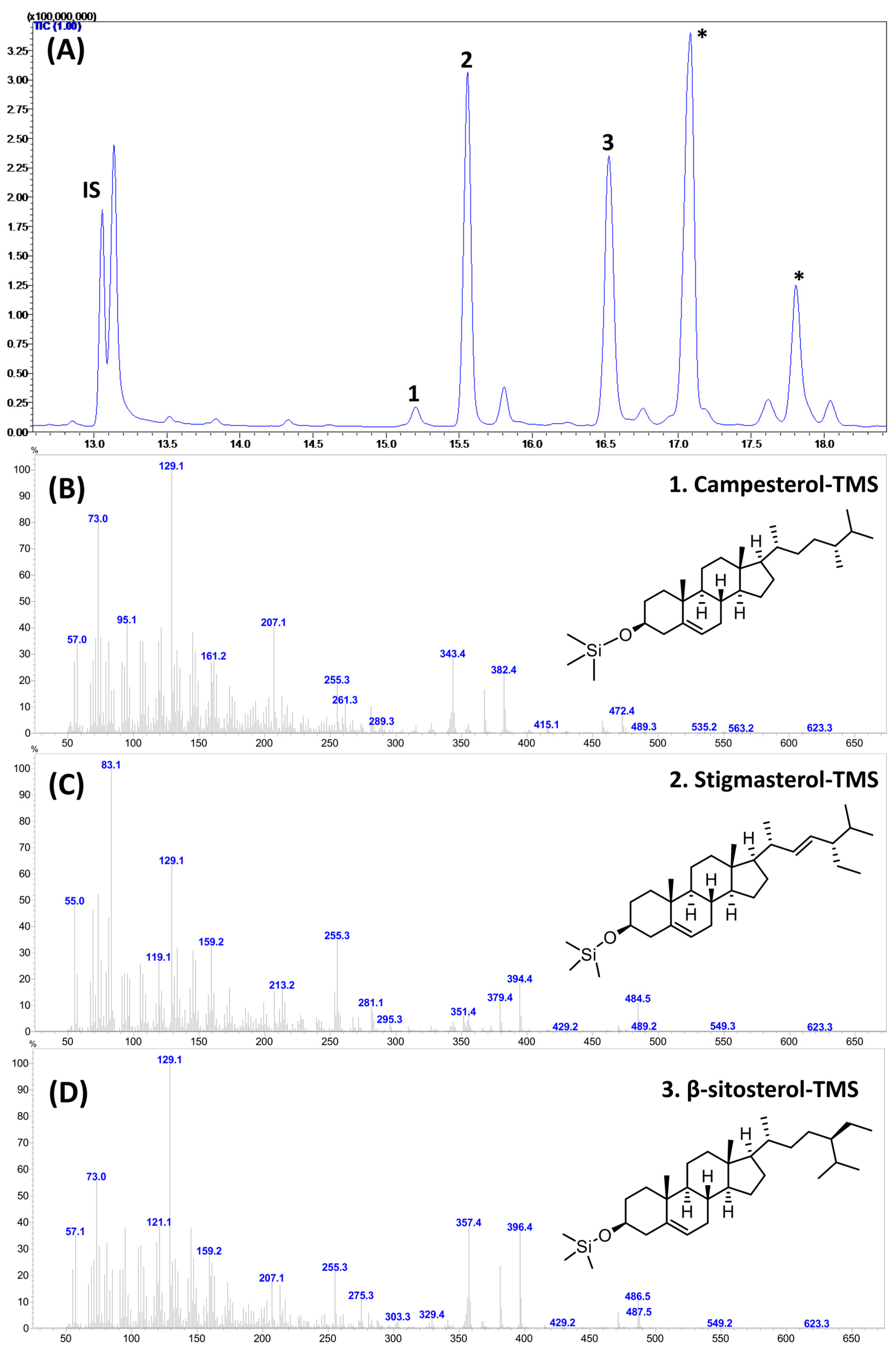
3.4. Phytosterols
In the present study, GC–MS analysis of trimethylsiloxy [−O-Si(CH
3)
3; TMS] derivatives revealed the presence of campesterol (Campest-5-en-3β-ol), stigmasterol (Stigmasta-5,22-dien-3β-ol), and β-sitosterol (Stigmast-5-en-3β-ol) in the studied samples (
Figure 3).
In this study, β-sitosterol was the most prevalent phytosterol among the studied cultivars, ranging between 127.08 (
cv. Superboy Yellow) and 191.99 µg/g FW (
cv. Taishan Yellow), which accounted for 46.57–62.43% (
cv. Taishan Orange and
cv. Superboy Yellow, respectively) of total phytosterol (
Table 8). The highest total sterol content of 333.42 µg/g FW was recorded from
cv. Taishan Yellow.
To the best of our knowledge, no detailed studies are available on the sterol composition of
Tagetes spp. However, we have previously recorded the predominance of β-sitosterol in herbs such as red lettuce (73.3 µg/g FW), fenugreek (
Trigonella foenum-graecum L.; 85.7 µg/g FW),
Moringa oleifera Lam. (175.9 µg/g FW),
Kaempferia parviflora Wall. Ex Baker (30.6–36.6 µg/g FW) [
54], and
Perilla frutescens Britt. (27.7–37.9 µg/g FW) [
55].
3.5. Fatty Acids
In this study, we identified seven prominent fatty acids from the flower petals of ten marigold cultivars, and their relative occurrence (expressed as percentages of the total fatty acids) was determined (
Table 9). Among the studied cultivars, palmitic acid (C
16:0; 33.36–47.43%) was the most dominant fatty acid, followed by linoleic (18:2n6c; 11.30–30.25%) and stearic acid (C
18:0; 15.99–24.57%). These three fatty acids together accounted for 72.13–86.44% (
cv. Superboy Orange and
cv. Taishan Yellow, respectively) of total fatty acids. Moreover, because of the dominance of palmitic and stearic acid, the total saturated fatty acids (SFAs) accounted for 58.17–75.90% (
cv. Durango Bee and
cv. Alaska, respectively) of total fatty acids. Interestingly, α-linoleic acid (C
18:3n3), which commonly occurs in photosynthetic tissues of the plant, was recorded as just 3.55–7.85% (
cv. Taishan Orange and
cv. Alaska, respectively) of total fatty acids.
Only a few reports are available on the fatty acid composition of
Tagetes spp. flowers. Gong et al. [
49] reported the dominance of linoleic acid (26.41%), palmitic acid (24.22%), and oleic acid (20.12%) in the
T. erecta flower lipids extracted utilizing supercritical CO
2 (SC-CO
2). Additionally, Zonta et al. [
56] reported the dominance of palmitic acid (31.35%), linoleic acid (22.89%), and oleic acid (15.44%) in
T. erecta flower petals. In agreement with these reports, in the present study, we also recorded the dominance of linoleic acid and palmitic acid; however, we recorded only 1.34–4.57% (
cv. Taishan Orange and
cv. Alaska, respectively) of oleic acid. In the present study, a substantial variation recorded for the fatty acid composition among the ten cultivars suggests the existence of significant genetic diversity among the marigold cultivars.
3.6. Antioxidant Activities
The lipophylic extract prepared from marigold flower petals showed ABTS
•+ and DPPH
• radical scavenging potential (
Table 10). In the present study, the extract prepared from
cv. Taishan Orange, Superboy Orange, and Durango Red presented the highest (
p < 0.05, Tukey HSD) ABTS
•+ scavenging activities of 402.33, 400.10, and 389.30 mg TE/100g FW, respectively. Conversely,
cv. Durango Orange, Taishan Orange, Superboy Orange, and Incall Orange exhibited the highest DPPH
• radical scavenging activities of 241.19, 250.82, 253.22, and 258.64 mg TE/100 g FW, respectively.
The ABTS
•+ and DPPH
• radical scavenging assays used in this study are based on the electron transfer (ET) mechanism. Consistent with earlier findings [
46,
57], a strong correlation (R = 0.787) was observed between these assays (
Table 8).
Carotenoids, tocopherols, ascorbic acid, and phenolic compounds (flavonoids and phenolic acids) are the principal antioxidants found in herbs [
58]. The present study reported a high correlation coefficient (R) of 0.803 and 0.686 between the total lutein contents and DPPH
• and ABTS
•+ radical scavenging activities, respectively (
Table 11). Gong et al. [
49] also reported a similar correlation coefficient between the total lutein contents of marigold flowers and DPPH
• and ABTS
•+ radical scavenging activities. Moreover, the present study reported a high correlation coefficient of 0.732 between the α-tocopherol contents and DPPH
• radical scavenging activities. These observations suggest that the radical scavenging activities of the marigold flower extracts are related to their lutein and α-tocopherol content.
3.7. Potential Impacts and Applications of the Obtained Results
Lutein is known for its potential health benefits, particularly in supporting eye health and acting as an antioxidant; the high lutein content with high antioxidant potential in marigold flower petals, especially cv. Superboy Orange, suggests an excellent candidate for incorporation into functional foods and nutraceuticals. Our findings regarding the cultivars with the highest lutein content can guide the development of lutein-enriched functional foods.
This study emphasizes the importance of maximizing lutein yield from marigold cultivation for cost-efficient extraction. Manufacturers can apply this insight to cultivate the high flower-yielding and lutein-rich cultivars, such as cv. Durango Red. This could lead to cost-effective production of lutein for various applications. Moreover, this study revealed that the contents of nutritionally vital constituents varied significantly among the marigold cultivars. These findings provide a strong foundation for further exploration of the genetic potential of marigolds to identify nutrient-rich cultivars for food fortification and commercial lutein extraction.
3.8. Limitations of the Study
In this study, ten cultivars of marigold commonly cultivated in Korea were grown under greenhouse conditions and utilized for the quantitative assessment of lutein-esters and other nutritionally vital constituents. Among the studied cultivars, cv. Durango Red produced the highest flower yield with high contents of lutein. These results suggest that cv. Durango Red is an ideal candidate for marigold cultivation and could be utilized for commercial lutein extraction. However, for commercial lutein extraction, marigolds are cultivated in the field. Under field conditions, the studied cultivars’ yield performance and lutein contents may change. Thus, field studies are warranted to establish the results obtained in the present study.
4. Conclusions
The commercial significance of lutein sourced from marigold flower petals is profound due to its versatile applications in functional food, feed, cosmetics, and pharmaceuticals. Significant differences in carotenoids (including lutein), α-tocopherol, phytosterols, fatty acid contents, and antioxidant activities were observed among the cultivars. The highest contents of total carotenoids (2723.11 µg/g FW) and lutein (2613.47 µg/g FW) were found in cv. Superboy Orange. The predominant fatty acids were palmitic acid (33.36–47.43%), followed by linoleic (11.30–30.25%) and stearic acid (15.99–24.57%). The highest level of total phytosterols (362.51 µg/g FW) was present in cv. Taishan Orange.
In addition to the high lutein contents, maximizing the lutein yield from the unit area of marigold cultivation holds paramount importance in the cost-efficient extraction of lutein. The present study suggests that due to the substantially higher flower petal yield with high lutein contents of 1995.90 µg/g FW, the cv. Durango Red can produce the highest lutein yield of 94.45 kg/ha. These observations suggest that cv. Durango Red is an ideal candidate for marigold cultivation and could be utilized for commercial lutein extraction. Moreover, cv. Superboy Orange is the richest source of lutein with the highest antioxidant potential, which can be used in functional food preparations.


 ,
, 
