Article

Molecular and Genetic Characterization of Colicinogenic Escherichia coli Strains Active against Shiga Toxin-Producing Escherichia coli O157:H7

1
Laboratorio de Inmunoquímica y Biotecnología, Centro de Investigación Veterinaria de Tandil (CIVETAN), CONICET, CICPBA, Facultad de Ciencias Veterinarias, UNICEN-Campus Universitario, Tandil B7000, Argentina
2
Food Science and Technology Department, Faculty of Veterinary Medicine, Universidad de Cordoba, 14071 Córdoba, Spain
3
Instituto de Ciencias biomédicas, Facultad de Medicina, Universidad de Chile, Santiago 8380453, Chile
*
Author to whom correspondence should be addressed.
Foods 2023, 12(14), 2676; https://doi.org/10.3390/foods12142676
Submission received: 30 May 2023 / Revised: 21 June 2023 / Accepted: 5 July 2023 / Published: 11 July 2023
(This article belongs to the Section Food Microbiology)

Abstract

:
The objective of this work was to molecularly and genotypically characterize and test the inhibitory activity of six colicinogenic Escherichia coli strains (ColEc) and their partially purified colicins against STEC O157:H7 isolated from clinical human cases. Inhibition tests demonstrated the activity of these strains and their colicins against STEC O157:H7. By PCR it was possible to detect colicins Ia, E7, and B and microcins M, H47, C7, and J25. By genome sequencing of two selected ColEc strains, it was possible to identify additional colicins such as E1 and Ib. No genes coding for stx1 and stx2 were detected after analyzing the genome sequence. The inhibitory activity of ColEc against STEC O157:H7 used as an indicator showed that colicins are potent growth inhibitors of E. coli O157:H7, being a potential alternative to reduce the presence of pathogens of public health relevance.

1. Introduction

Escherichia coli is a Gram-negative bacterium, part of the normal microbiota of the intestinal tract of warm-blooded animals, including humans. Most of them are nonpathogenic, but some can cause diseases such as watery diarrhea (WD), hemorrhagic colitis (HC), and hemolytic uremic syndrome (HUS). This disease, particularly severe in young children, is caused by Shiga toxin-producing Escherichia coli strains (STEC) that synthesize potent toxins and other virulence and adherence factors that contribute to their pathogenicity [1]. The main serotype associated with HUS worldwide is E. coli O157:H7 [2]. However, some non-O157 serotypes are frequently associated with HC and HUS in several countries. Argentina has the greatest worldwide prevalence, with an average incidence for HUS of 16/100,000 children younger than 5 years, without considering the sub-recording of the disease [3]. A report from Argentina in response to an FAO/WHO request confirmed that the etiologic agent most associated with HUS is STEC, with O157:H7 being the most frequently associated serotype, although there are more than 100 serotypes that have similar pathogenic potential [4]. STEC infection is associated with consumption and contact with contaminated foods and water, person-to-person transmission, and direct contact with infected animals, their feces, or their environment [5,6].
Bacterial communities are forced to compete with each other for nutrients and energy sources. They achieve this by producing proteins that inhibit the growth of phylogenetically related (reduced spectrum) or not-related (broad spectrum) strains. Bacteriocins are an important type of antimicrobial ribosomally synthesized peptides (in contrast with antibiotics, which are secondary metabolism products) and are present in all Eubacteria and Archaea genera [7,8].
In addition to their use as biopreservatives antagonistic to FBD (food-borne diseases) pathogens, bacteriocins have potential novel applications in animal and human health. Therefore, the search for producer strains and the functional characterization of these molecules is of paramount importance. Currently, the only bacteriocin approved worldwide for use as a biopreservative agent is nisin, synthesized by Lactococcus lactis, which has proven to be effective against multiple important pathogens, including Listeria monocytogenes, Clostridium botulinum, E. coli, and Salmonella spp. The use of nisin is not restricted only to the application in food but is marketed in disinfectant preparations for topical and intramammary application, applied to dairy cattle with mastitis due to Staphylococcus aureus, Streptococcus uberis, and Streptococcus agalactiae [9]. Nisin works by forming pores in the cell membrane, causing the leaching of important cellular compounds, including K+ ions, amino acids, and ATP, ultimately leading to cell death [10]. One of the main applications of bacteriocins in human health is the treatment of multi-resistant bacteria responsible for hospital-acquired infections [11].
Although the appearance of resistance or tolerance to bacteriocins is a factor to be considered in therapeutic applications, an alternative would be the use of combinations of bacteriocins from different origins or bacteriocins modified in their peptide sequence to evade the resistance acquired by target cells.
Colicins are the most studied bacteriocins produced by Gram-negative bacteria, with 25 different types being identified [12,13].
In E. coli, bacteriocins are classified based on their molecular weight as colicins (25–80 KDa) and microcins (smaller than 10 KDa). Colicins and microcins have a relatively narrow spectrum of activity, although, in general, microcins have wider antibacterial activity compared to colicins [14]. The colicin operons are encoded on the bacterial chromosome or on colicinogenic plasmids (pCol) that harbor, in addition to the colicin gene itself, all the genes necessary for their transport and immunity [13]. Among these classes of bacteriocin are colicins E1–E9, Ia, Ib, and S4. Microcins are frequently coded in the bacterial chromosome, and examples of these are microcins H47, M, or J25, among others. The action mechanism consists of the formation of pores in the target cell wall, the inhibition of peptidoglycan synthesis, and the inhibition of DNA replication or RNA transcription [15]. Colicin’s expression varies according to the bacterial growth phase, which in some cases occurs during the exponential phase [16,17], while in others, it occurs in the stationary phase [18,19].
Most of the bacteria considered probiotics belong to the Lactobacillus, Enterobacter, and Bifidobacterium genera [20,21]. However, the use of nonpathogenic strains of E. coli has been considered to achieve the reduction of infections caused by pathogenic bacteria in both animals and humans [22]. The use of E. coli as a probiotic is mainly accepted in Germany and other Eastern European countries. Wassenaar et al. (2016) [23] pointed out that E. coli is chosen as a probiotic in line with its presumed ubiquitous presence in the gut. An example is the E. coli Nissle 1917 strain, widely used in probiotic preparations. Colicins and colicin-like molecules derived from Gram-negative bacteria are easily and well-expressed in plants and fully functional, being up to 106 times more potent than antibiotics on a molar basis [24]. A promising alternative to circumvent the existing sanitary concerns regarding the use of E. coli under viable conditions is the use of colicins produced against pathogenic bacteria [25,26]. Numerous groups are constantly searching for new strains and new types of colicins [27,28], trying to express known colicins in different combinations [29] or new organisms, such as plants, to achieve higher yields and purity of the peptide extract [30].
The objective of this work was to molecularly and genotypically characterize and test the inhibitory activity of colicinogenic E. coli strains (ColEc) and their partially purified colicins against STEC O157:H7 isolated from clinical human cases.

2. Materials and Methods

2.1. Origin and Identification of E. coli and Indicator Strains

In this study, six strains isolated in 2004 from the bovine gastrointestinal tract were used and identified as E. coli 4.8, E. coli 8.2, E. coli 13.7, E. coli 24.7, E. coli 27.4, and E. coli 27.12. Colicinogenic E. coli strains were then identified as ColEc. These strains previously showed the absence of the different virulence factors: Shiga toxin (stx1 and stx2), intimin (eae), hemolysin (hlyA), thermo-stable toxin (stIa), and thermo-labile toxin (ltI) by polymerase chain reaction (PCR) genotypic analysis [31].
The absence of virulence genes was confirmed by a complementary study of the genetic profile using the PCR technique. This study included the associated genes, plus adhesion-associated genes such as intimin for E. coli O157 (eaeO157), hemolysin (ehxA), efa/iha, antigen 43 (AGN43), and intracellular domain associated protein-1 (AIDA1) factors.

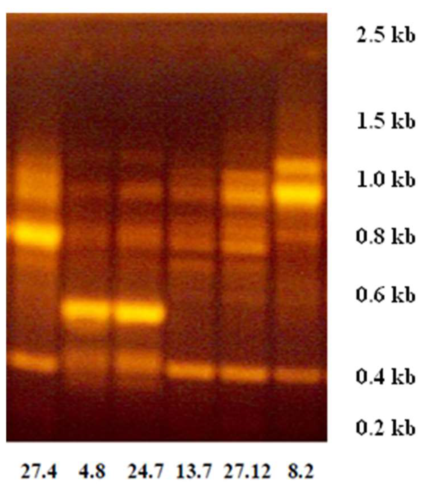
2.1.1. Genetic Profiles by Random Amplification of Polymorphic DNA (RAPD)

The determination of the genetic profiles of the selected bacteria was carried out using the random amplification of polymorphic DNA (RAPD) technique. The technique was performed based on the methodology developed by Birch et al. [32] with modifications. An aliquot of the tubes with the bacteria in stock (frozen at −80 °C) was seeded in 10 mL of Luria Bertani broth (LB, Britania, Argentina) and incubated at 37 °C under agitation for 18 h. Then, the OD600 of a 1/10 dilution of each culture was determined. For an OD value of 0.5, 500 μL of the culture was taken and centrifuged for 2 min at 12,000× g. The pellet obtained was resuspended in 500 μL of bidistilled water. The suspension obtained was boiled for 10 min, placed on ice, and centrifuged for 3 min at 13,000× g. The supernatant was extracted and frozen at −80 °C for later use.
The amplification was performed using the primer [32] M13: 5′GAGGGTGGCGGTTCT 3′. The PCR reaction cocktail was made in a final volume of 50 µL containing 20 mM (NH4)2SO4; 75 mM Tris-HCl pH 9.0; 0.1% (w/v) Tween 20; 2.5 mM MgCl2; 0.2 mM of each dNTP; 1 µM of the “primer”; 2.5 U of Taq DNA polymerase (Highway, Argentina); and 5 μL of the sample. The PCR reaction was performed in a thermocycler (Techne Genius, Thermo Fisher, Waltham, MA, USA) under the following conditions: initial temperature of 94 °C for 5 min, 40 cycles of 94 °C for 1 min, 50 °C for 1 min, and 72 °C for 1 min. The amplification products were analyzed by horizontal electrophoresis on a 1.8% agarose gel in the presence of ethidium bromide.

2.1.2. Serotyping

Serotyping O and H antigens were determined by means of a microagglutination technique in plates and tubes described by Guinée et al. [33] modified by Blanco et al. [34,35] using all available O (O1–O175) antisera plus six putative new O antigens (OX176 through OX181) [36] and H (H1–H56) antisera [37]. Non-specific agglutinins were removed by adsorption with the corresponding cross-reacting antigens. O antisera were produced in the Laboratory Reference of E. coli (LREC) (Lugo, Spain), whereas the H antisera were obtained from the Statens Serum Institut (Copenhagen, Denmark).

2.1.3. Origin of Indicator Strains

STEC O157:H7 was used as an indicator for the inhibitory activity of ColEc. STEC O157:H7 (n = 80) strains isolated between 1996 and 2013 from Argentinean and Chilean patients in clinical case studies of HUS, HC, and WD were used (unpublished data, Table 1). In addition, a STEC strain (identified as STEC 166), isolated from grazing cattle, was used [31].

2.2. Detection of Colicin Genes

ColEc were cultured on LB broth (Britania, Argentina) (overnight -ON- at 37 °C) with continuous shaking. DNA extraction was carried out by boiling 10 μL of this culture in 500 μL of sterile bidistilled water for 10 min.
Colicin genes were identified in colicinogenic strains by PCR monoplex using the primers listed in Table 2 [36,37]. In a final reaction volume of 25 μL, 2.5 μL DNA extract, 4 mM MgCl2, 0.5 units of Platinum Taq (Invitrogen), 1× reaction buffer (67 mM Tris/HCl -pH 8,8-, 16.6 mM (NH4)2SO4, 0.45% Triton X-100), 0.2 mg of gelatin, 0.2 mM dNTP, and 3 ng of each primer were used. The amplification cycle consisted of an initial step of 94 °C for 2 min, 30 cycles of 94 °C for 30 s, 55 °C for 30 s, 72 °C for 30 s, and a final step at 72 °C for 3 min. Samples were electrophoresed on 2% agarose gels stained with ethidium bromide and viewed under ultraviolet light.

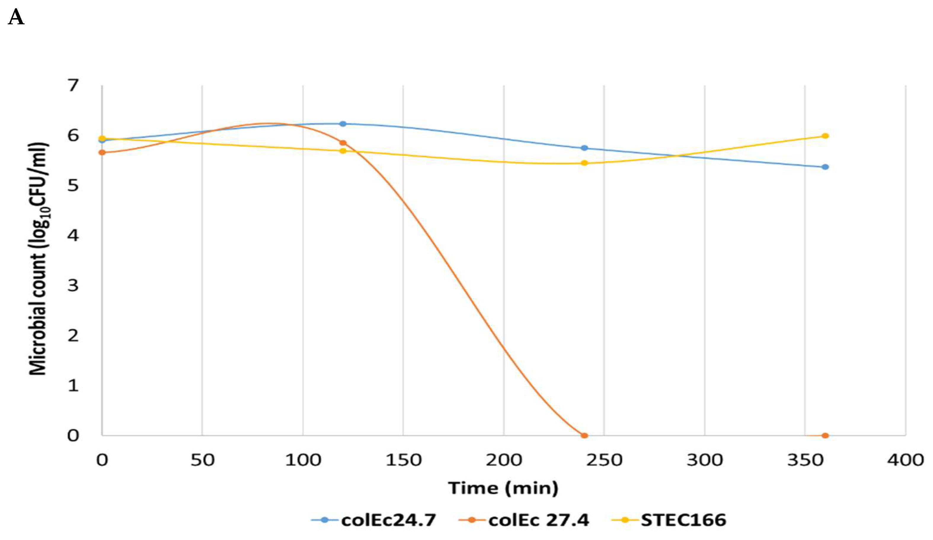
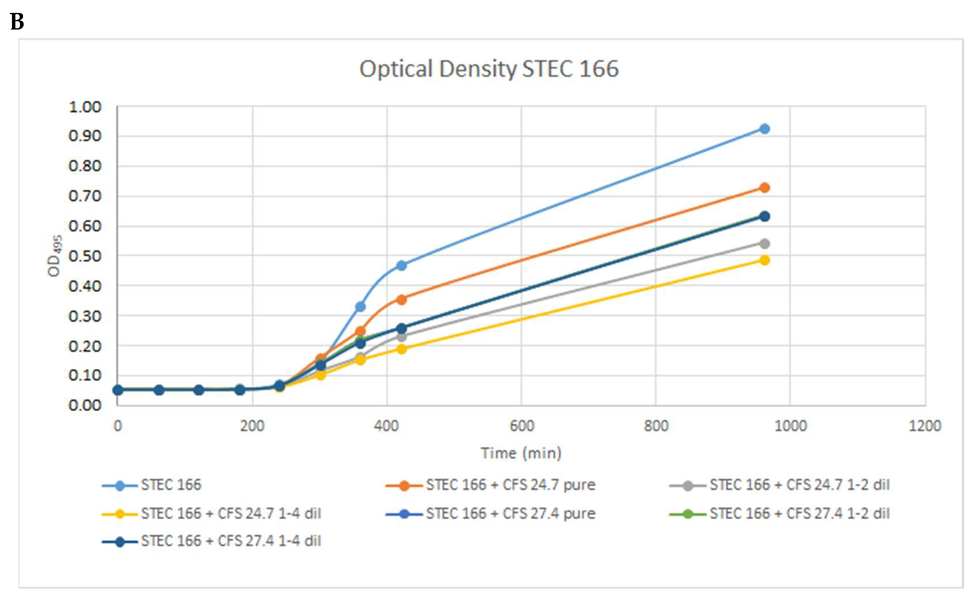
2.3. Determination of Inhibitory Activity of ColEc against STEC Strains

Assessment of the inhibitory activity of the ColEc strains was performed by puncture inhibition assay. First, the LB plates were punctured with each of the ColEc strains and incubated overnight at 37 °C. The colonies obtained were exposed to chloroform vapors for 1 h and then allowed to aerate for 30 min. Secondly, a soft agar containing 106 CFU/mL of each pathogenic strain supplied for this study was poured on them. The same procedure was carried out to rule out a possible inhibitory effect of the pathogenic strains over colicinogenic strains in the study. The presence of inhibition was considered when a translucent zone around the colony with the absence of growth of at least 1 mm was observed. The inhibitory tests were carried out in triplicate, and inhibition of the indicator strains was observed in all cases.

Determination of Inhibitory Activity of Colicins against STEC 166

Two ColEc strains (24.7 and 27.4) that inhibited the highest number of STEC strains from HUS and HC used were selected to characterize their colicin inhibitory activity. Strain 27.4, although it was not the one that obtained the highest percentage, was selected due to the optimal concentration and purity of the DNA obtained during the extraction process. The colicins were partially purified by obtaining a cell-free supernatant (CFS). The overnight cultures were centrifuged at 13,000× g and filtered with a 0.22 µm pore size syringe filter. The CFS was placed in a 96-well microplate at different concentrations, inoculated with 103 CFU/mL of STEC 166, and incubated at 37 °C. The average of the antimicrobial activity was determined by measuring optical density (OD495nm) each hour and by plate counting at 0, 2, 4, 6, and 8 h. These experiments were performed in triplicate, in three independent trials, and analyzed with INFOSTAT. They were expressed as the mean minus the standard deviation.

2.4. Differentiation Assay of Growth Inhibition by Phage or Colicin

With each ColEc strain, an inhibition assay was performed as described above. A small portion was then removed from each zone of growth inhibition and resuspended in 1 mL of LB broth containing two drops of chloroform. It was vortexed and left to stand for 5 min and 100 uL of this suspension was added to 3 mL of 0.4% LB agar containing 106 CFU/mL of the indicator strain STEC 166. The agar was poured onto a plate and incubated at 37 °C for 18 h. The presence of lysis plaques in each of the samples would show that the inhibition occurs due to the presence of bacteriophages. On the other hand, if confluent growth of the indicator strain is observed on the LB agar plates without the appearance of lysis plaques, it can be confirmed that the inhibition is produced by bacteriocins.

2.5. Stability of CFS to Physic-Chemical Treatments

CFS of the sequenced ColEC strains were subjected to several treatments to assess the stability of the potential antimicrobial substance contained in it. Those treatments consisted in:
I.
Temperature: The CFS extracts were heated in a thermal bath at 60 °C, 80 °C, and 100 °C; they were frozen at −20 and kept at 4 °C [38].
II.
pH changes: The pH of the CFS extracts were adjusted to approximately 4.5, 7.0, and 9.0 using HCl (Biopack 30%) and NaOH (Biopack 10N) (Larsen et al., 1993).
III.
Stability to protease: CFS extracts were incubated with Proteinase K (Sigma-Aldricht 20 ng/µL) at 37 °C for 15–60 min.
After each treatment, the CFS extracts’ activity was evaluated with the inhibition assay explained in point 2.4.

2.6. Virulence Genes Detection

The genome sequences previously obtained were analyzed with the Virulence Finder service provided by the Center for Genomic Epidemiology (www.cge.food.dtu.dk/services/VirulenceFinder, accessed on 27 February 2023), selecting a 90% threshold ID. After loading the assemblies, the graphical output of the program allowed us to detect the presence or absence of Shiga toxin genes [39].

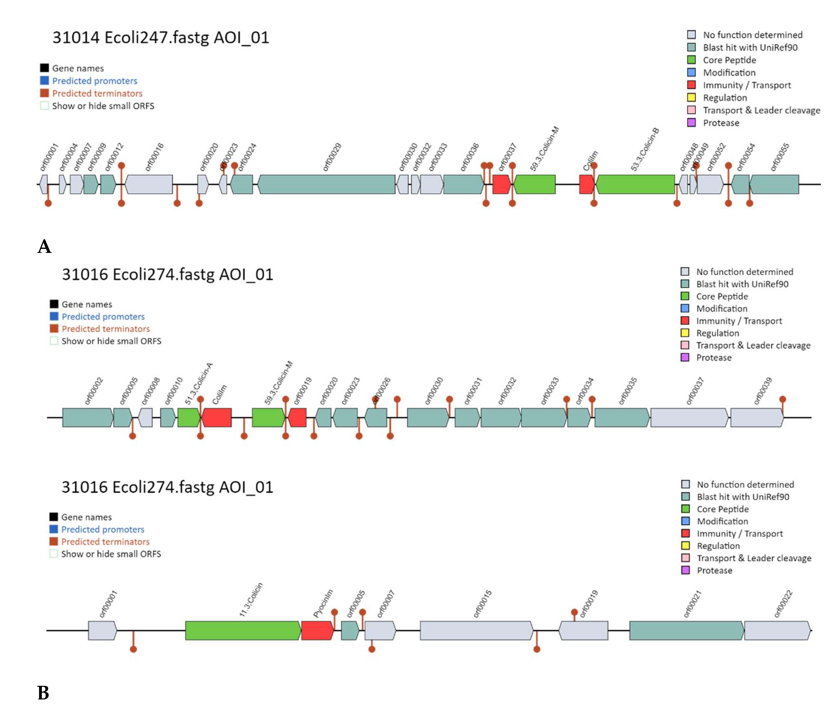
2.7. Strains Sequencing: Bacteriocin Mining

The DNA of the selected colicinogenic E. coli strains was extracted with the Wizard® Genomic DNA Purification Kit (Promega, Fitchburg, WI, USA), its quality and concentration were assessed with NanoDrop spectrophotometer (Thermo Fisher, USA) and sent to MicrobesNG (Birmingham, UK) using the standard whole genome sequencing with 30× coverage service.
The procedure run by the provider was as follows: genomic DNA was organized into libraries using the Nextera XT prep protocol. Then, they were sequenced through the Illumina Hi-Seq platform with 2 × 250 bp paired-end reads. Reads were trimmed using Trimmomatic (V 0.40), and the quality was assessed with the Samtools (V 1.4), Bedtools (V 2.29.2), and Bwa-mem (V 0.7.17) software. De novo assembly was carried out using SPAdes (V 3.15.4). Annotation was made by the RAST server. Assemblies’ metrics were calculated using QUAST (V 4.0). Next, the genera and family of each strain’s read map was calculated using the KRAKEN (V 2.0) software. Finally, an automated annotation was performed using PROKKA (V 1.14).
Genomes sequences assemblies were further analyzed for bacteriocin genes. BAGEL4 is a platform developed for the identification of bacteriocins and other ribosomally synthesized and post-translationally modified peptides (RiPPs) [40]. FASTA format sequences were uploaded onto the input page, followed by a graphical output showing the Areas of Interest (AOI) where each potential bacteriocin, as well as the immunity and modification genes related to its production, are identified on a color scheme. To re-confirm their identity, a Blast-p analysis was performed on the NCBI.

3. Results

3.1. Genetic Profile and Serotype Determination

The results of the PCR genotyping test for the different virulence and adherence factors showed that all the selected bacteria were negative.
Using the RAPD technique, it was possible to differentiate four genetic profiles among the six ColEc strains (Figure 1). Strain selection was made based on the best ability to produce growth inhibition zones of the indicator strain in terms of halo size and clarity, established in a previous study [31]. Strains 4.8 and 24.7 shared one genetic profile, strains 13.7 and 27.12 shared another profile, while strains 8.2 and 24.7 showed a profile that was different from each other and different from the profiles of the other strains.
With the antisera used, it was possible to identify the E. coli 4.8 strain as ONT, the E. coli 8.2 strain as O77, the E. coli 13.7 strain as O174: H8, the E. coli 24.7 ONT: H7 strain, the E. coli 27.4 as O26 H-, and E. coli strain 27.12 as O141.

3.2. PCR Screening

By PCR assays, it was confirmed that the colicinogenic strains under study encode the following colicins: E. coli 4.8: colicins B, microcin M, and microcin H47; E. coli 8.2: colicin Ia and microcin C7; E. coli 13.7 colicins Ia, E7, B, and microcin C7 and microcin J25; E. coli 24.7 colicin B, microcin M, and H47; E. coli 27.4; colicin Ia, E7, B, microcin C7, and J25 (Table 3). With the primers used, it was not possible to identify colicins in strain E. coli 27.12, despite having inhibitory activity, as could be demonstrated in previous studies [36]. The correspondence of the strain name of the cited study with the present study is as follows: Strain 1: 4.8, Strain 2: 8.2, Strain 3: 10.10, Strain 4: 13.7, Strain 5: 24.7, Strain 6: 27.4, and Strain 7: 27.12. Particularly in the present study, strain 10.10 was discarded.

3.3. Inhibition Assays

The results of the puncture inhibition test using ColEc strains against STEC from HC and HUS are shown in Table 3. All colicinogenic strains were found to inhibit more than 90% of the pathogenic strains. The Petri dish puncture inhibition assay showed a zone of inhibition against most pathogenic strains. The positive reading was considered when three replicates showed halos greater than 1 mm.
The ColEc strains selected because of these results to characterize colicin inhibitory activity were ColEc 24.7 and ColEc 27.4 (Colicin produced by ColEc 27.12 could not be identified using the primers of Table 2, and no further tested according to Section 2.5).
Results of the inhibitory activity of the colicins produced ColEc 24.7 and 27.4 strains against STEC 166 are shown in Figure 2. These results showed a difference in the antimicrobial activity of the CFS analyzed. The CFS of ColEc 27.4 showed potential bactericidal activity against pathogenic strains (Figure 2A) when determining the OD, observed by the absence of growth in the early stages and the absence of viable cells when plate assays were performed. These results are indicative of a bactericidal effect. On the other hand, we documented a very slight decrease in the viability of pathogenic strains exposed to SFC of ColEc 24.7, although this effect was not maintained over time. The optical density and viable cells were lower and proportional to the dilution of CFS used (Figure 2B). These results show an antibacterial activity of ColEc 27.4 CFS on STEC strains.

3.4. Differentiation Assay of Growth Inhibition by Phage or Colicin

In the six ColEc strains used, it was shown that the inhibition was due to the presence of bacteriocins and not of bacteriophages since confluent growth of the indicator strain was observed in all LB agar plates, with no lysis plaques indicating the presence of bacteriophages appearing in any of them.

3.5. Stability Assays

CFS from ColEc strains subjected to temperature, pH, and protease treatments showed a differential behavior. CFS from ColEc 24.7 was active after freezing and thawing at room temperature (25 °C), it also resisted a 10 min boiling at 100 °C but lost their inhibitory activity when incubated between 60 and 75 °C. When subjected to pH shifts, it only remains active at pH 7, while there was no activity at pH 4; 5; 9; 10. CFS from ColEc 27.4 showed inhibition subjected to freeze/thawing at room temperature, boiled for 10 min at 100 °C, and incubated between 60 and 75 °C. This strain of CFS also resisted pH variations maintaining its activity at pH 4; 5; 7; 9; 10. Both strains lost their inhibitory activity after Proteinase K incubation, allowing us to affirm the peptide nature of the antimicrobial substance in the CFS.

3.6. Virulence Finder

The sequenced ColEc strains (24.7 and 27.4) were negative for the Shiga toxin-associated genes (stx11 and stx2) both by PCR in previous studies [31] and in the search through the Virulence Finder service. These genes are the most important virulence factors associated with STEC.

3.7. Whole Genome Sequencing and Annotation: Bacteriocin Mining

The whole genome sequencing of ColEc 24.7 showed a guanine-cytosine (GC) percentage of 50.23%, with 717 contigs, while ColEc 27.4 presented a 50.56% GC in a total of 44 contigs. Annotation using PROKKA, after whole genome sequencing, confirmed the bacteriocins identified by PCR; additionally, we were able to detect the presence of Colicin, E1 in E. coli 24.7 and Colicin Ib in E. coli 27.4 that had not been identified by PCR.
BAGEL4 analysis showed in ColEc 24.7 the presence of 2 AOI from a total of 205 that coded for bacteriocins or other ribosomally synthesized and post-translationally modified peptides (RiPPs) (Figure 3A). In one of these areas, we were able to identify the presence of Colicin M and Colicin B structural peptide genes as well as their respective immunity proteins.
ColEc 27.4 presented 4 AOI from a total of 88 areas analyzed (Figure 3B). AOI 01 presented the Col Ib gene; AOI 02 presented the Col E7 gene, Col E2 immunity gene, and Col E8 lysis protein; and AOI 04 had Col A, Col B, and their respective immunity genes (Figure 3B).

4. Discussion

Numerous research groups have identified bacteriocin-producing strains capable of inhibiting pathogens such as enterotoxigenic Escherichia coli (ETEC) and STEC [25,41,42]. The use of non-pathogenic commensal strains of E. coli is promising since they present high specificity by inhibiting only phylogenetically related pathogenic strains. E. coli Nissle 1917 stands out, marketed in some European countries under the name Mutaflor® (©Pharma-Zentrale, Herdecke, Germany) whose inhibitory properties have been widely characterized, exhibiting microcin M/H47 production [43,44]. Administration of E. coli strains with antimicrobial activity available in commercial preparations has shown to outcompete and become dominant among the Enterobacteriaceae genus in the gut microbial communities of newborn and infant children [45]. The search for new strains and new molecules with comparable activity is a field of development [26,46,47]. Nevertheless, there has been criticism of the usage of Nissle 1917 as a probiotic due to several issues, such as the presence of a pathogenicity island called pks, which codifies and expresses a toxin called colibactin, a protein with a DNA alkylation activity that has been demonstrated to produce cross-linking damage on epithelial cell’s DNA [48]. This damage may lead to serious consequences such as colorectal cancer. A study led by Plequezuelos-Manzano in 2020 [49] was able to link the pks island to the mutational signature of colorectal cancer cells previously exposed to E. coli. In this regard, there have been studies focused on minimizing E. coli Nissle 1917 virulence factors using genome editing techniques such as CRISPR interference, this work made by Azam and Khan was able to knock down biofilm-related gene csgD and suppress virulence genes of csgA, csgB, fimA, fimH, ompR, luxS, and bolA related to fimbriae, two-component systems, quorum sensing and other functions on pathogenicity [50].
The ability to translocate through the intestinal epithelium to other tissues is a major concern on the safety of the probiotic strain. Pradhan and Weiss were able to prove, using human intestinal organoids, that E. coli Nissle was unable to cross the epithelial barrier and not affect the cells while other UPEC and EHEC bacteria did [51].
ColEc strains used were characterized as colicin producers [31] and grouped according to their genetic profiles (RAPD PCR). ColEc strains 4.8 and 24.7 share a genetic profile and harbor the same colicin and microcin genes. The profile of the other strains does not directly correlate to the presence of their colicin genes. Although they share a genetic profile, the serotypes observed differ between these strains. These differences may be due to a lack of discriminatory capacity of the RAPD technique.
The use of bioinformatic tools for sequence analysis is a robust and constantly growing field, the quality and quantity of sequence databases are an important factor to consider [52]. Sequence mining is a strategy to identify putative antimicrobial genes of interest in a specific bacterium. There are many platforms focused on these objectives, and we can highlight Bactibase [53], based on the Blast algorithm, which is a curated database for bacteriocins. AntiSMASH is another platform focused not only on bacteriocin but including other RiPPs. BAGEL4 is a database and mining suite with an advantageous user interface representing the AOI in a graphical layout displaying the genetic context of each putative bacteriocin. Some examples of the usage of these tools were the ones achieved by Cameron [54], who was able to identify multiple colicin and microcin genes (complete and truncated) in a sample obtained from soil, wastewater, and feces from feedlots in Alberta (Canada) combining sequence analysis on Bactibase and BAGEL4. Sabino [55] used BAGEL4 and AntiSMASH to detect lasso peptides produced by ruminal bacteria. In this work, we were able to detect previously unidentified bacteriocins (microcins and colicins) by combining PCR, genome sequencing, annotation, and mining for antimicrobial genes. This tool allowed us to confirm that the stx1 and stx2 genes were absent in the colicinogenic strain’s genome. This feature is of importance due to the severity of the HUS disease induced by the Shiga toxin.
Colicins can present three mechanisms of destruction of target cells: (i) creation of voltage-gated channels in the inner membrane of the target bacterium, (ii) action of a nuclease in the cytoplasm (DNase, 16S rRNase, and tRNase), or (iii) inhibition of peptidoglycan synthesis. This study identified and characterized the colicins produced by ColEc strains demonstrating that they have inhibitory activity against STEC O157:H7 strains, being able to be used as an alternative tool in the control of that pathogenic strains.
In this work, colicins and microcins have been detected with bacteriocin gene mining that was absent in PCR or genome annotation. These differences may be due to the quality of the assemblies obtained by the sequencing procedure. Genome annotation and mining tools are not without limitations due to the tedious and automated nature of these methods and the curation of genome databases compared to the mining tools. Several authors have approached this issue, such as Poptsova and Gogarten [56] and Kasaa [57].
This work showed that the ColEc strains under study could inhibit the bacterial growth of a high percentage of STEC O157:H7 strains obtained from clinical samples of patients with HC and HUS. Only a small percentage of the pathogenic strains showed resistance to the ColEc strains (Table 3). This percentage, although it is minor, is common to ColEc strains and deserves to be addressed in another study due to the dangerousness of the strains, and even subjected to sensitivity tests with other antimicrobial agents. These results indicate that colicinogenic strains and the inhibitory substances produced could be used to control STEC strains. The production of colicins in E. coli strains represents an important trait with respect to microbial survival and competition in the complex intestinal environment. A study by Micenková et al. [58] showed a new type of colicin, colicin Z, with a narrow inhibitory spectrum, being active only against enteroinvasive strains of E. coli (EIEC) and Shigella. ColE1 was produced from an E. coli K-12 strain containing the plasmid pColE1-K53 and effectively reduced L. monocytogenes populations in broth culture and on meat product surfaces [59]. Tahamtan et al. indicated that the use of colicin and biotherapy, rather than antibiotics, may be more effective for the control of E. coli K99 infection [60]. Therefore, the ability of colicinogenic strains to inhibit strains associated with important public health diseases such as HUS is remarkable. The antimicrobial substances present in the CFS of ColEc 27.4 maintained their activity against physical-chemical treatments of pH and temperature. This characteristic offers a potential application as antimicrobial agents on food to prevent or minimize the impact of bacterial contamination. The presence of virulence factors related to adherence and hemolysis is a serious concern about the potential application of a strain as a probiotic. However, this factor can be overcome by using antimicrobial substances such as colicins and microcins. These could be produced and purified en masse or by the cloning of the corresponding genes and factors necessary to achieve their expression and production in a safer microorganism.
Colicins and other bacteriocins are an alternative to antibiotics due to their comparatively narrow activity spectrum and relatively low frequency of resistance. Gillor et al. [61] reviewed potential applications of colicins and microcins to replace antibiotics in human or veterinary use or to apply them as food additives to prevent contamination with pathogens. However, to prevent the emergence of resistance, a combination of various colicins or microcins with distinct activities, different target receptors, or even molecular manipulation is a plausible strategy. Although combinations need to be evaluated, susceptible cells will have selective pressure on diverse points, and they will be, potentially, more constrained by tradeoffs and low fitness because of the metabolic ways involved in the resistance [62]. Our study shows that colicins are potent growth inhibitors of STEC O157:H7, being a potential alternative to reduce the presence of pathogens of public health relevance, and an alternative to use against multiresistant bacteria.
There are still pending issues arising from this study. It is interesting to study the resistance of STEC that did not display sensitivity against ColEc. A comparative study with other antimicrobial tools for the control of these pathogens could be carried out. The extraction and purification of colicins and microcins will be essential for their application in new pathogen control studies in biotechnological processes. And fundamentally, in vivo studies will be necessary to confirm its Generally Recognized As Safe character.
One of the most relevant aspects of colicins is the specificity and diversity of interactions that they undergo with various proteins during their action and during their production and release. The study of colicins, once the model for studies of bacterial toxins, has contributed significantly to progress in several fields. Colicins play some role in microbial communities. Researchers argue that they play a critical role in mediating microbial interactions [13]. The mystery not yet resolved involves knowing what that role is. Colicins can serve as anti-competitors, allowing a strain to invade an established microbial community. They can also play a defensive role and act to prohibit the invasion of other strains or species in an occupied niche or limit the advance of neighboring cells. It is likely that whatever roles bacteriocins play, these roles will change as components of the environment, both biotic and abiotic, change.
There is a need to further investigate the importance of colicins and microcins with respect to the impact of interactions with prokaryotic and eukaryotic cells, as well as their putative use in food biotechnology and medicine. Considering the search for new antimicrobial and anticancer agents, Enterobacteriaceae, and especially colicins and microcins, should be considered valid options.

Author Contributions

Funding acquisition, L.M.M.; Investigation, R.V.; Project administration, N.L.P.; Writing—original draft, M.D.G.; Writing—review & editing, M.J.R. and A.I.E. All authors have read and agreed to the published version of the manuscript.

Funding

This research was funded by PICT2015-2666, CONICET, Argentina, and University of Cordoba, grants “Ucoactiva”.

Data Availability Statement

Data is contained within the article.

Acknowledgments

The authors thank María Rosa Ortiz for her technical assistance.

Conflicts of Interest

The authors declare no conflict of interest.

References

  1. Rodríguez-Rubio, L.; Haarmann, N.; Schwidder, M.; Muniesa, M.; Schmidt, H. Bacteriophages of Shiga Toxin-Producing E. coli and Their Contribution to Pathogenicity. Pathogens 2021, 10, 404. [Google Scholar] [CrossRef]
  2. Bai, X.; Ylinen, E.; Zhang, J.; Salmenlinna, S.; Halkilahti, J.; Saxen, H.; Narayanan, A.; Jahnukainen, T.; Matussek, A. Comparative Genomics of Shiga Toxin-Producing E. coli Strains Isolated from Pediatric Patients with and without Hemolytic Uremic Syndrome from 2000 to 2016 in Finland. Microbiol. Spectr. 2022, 10, e00660-22. [Google Scholar] [CrossRef] [PubMed]
  3. Rivas, M.; Chinen, I.; Guth, B.E.C. Enterohemorrhagic (Shiga Toxin-Producing) Escherichia Coli. In E. coli in the Americas; Torres, A.G., Ed.; Springer International Publishing: Cham, Switzerland, 2016; pp. 97–123. ISBN 978-3-319-45092-6. [Google Scholar]
  4. FAO/OMS. Informe de la República Argentina en Respuesta a la Solicitud de Datos Sobre E. coli Productor de Verocitotoxina (VTEC)/E. coli Productor de Toxina de Shiga (STEC). 2016. Available online: https://rsa.conicet.gov.ar/wp-content/uploads/2016/07/Informe-FAO-OMS-Escherichia-coli-VTEC-STEC-Argentina.pdf (accessed on 1 April 2021).
  5. Bell, B.P.; Goldoft, M.; Griffin, P.M.; Davis, M.A.; Gordon, D.C.; Tarr, P.I.; Bartleson, C.A.; Lewis, J.H.; Barrett, T.J.; Wells, J.G.; et al. A Multistate Outbreak of E. coli O157:H7—Associated Bloody Diarrhea and Hemolytic Uremic Syndrome from Hamburgers: The Washington Experience. JAMA 1994, 272, 1349–1353. [Google Scholar] [CrossRef]
  6. Fernández, D.; Sanz, M.E.; Parma, A.E.; Padola, N.L. Short Communication: Characterization of Shiga Toxin-Producing E. coli Isolated from Newborn, Milk-Fed, and Growing Calves in Argentina. J. Dairy Sci. 2012, 95, 5340–5343. [Google Scholar] [CrossRef]
  7. Garcia-Gutierrez, E.; Mayer, M.J.; Cotter, P.D.; Narbad, A. Gut Microbiota as a Source of Novel Antimicrobials. Gut Microbes 2019, 10, 1–21. [Google Scholar] [CrossRef] [Green Version]
  8. Juturu, V.; Wu, J.C. Microbial Production of Bacteriocins: Latest Research Development and Applications. Biotechnol. Adv. 2018, 36, 2187–2200. [Google Scholar] [CrossRef] [PubMed]
  9. Cao, L.T.; Wu, J.Q.; Xie, F.; Hu, S.H.; Mo, Y. Efficacy of Nisin in Treatment of Clinical Mastitis in Lactating Dairy Cows. J. Dairy Sci. 2007, 90, 3980–3985. [Google Scholar] [CrossRef] [PubMed] [Green Version]
  10. Yap, P.G.; Lai, Z.W.; Tan, J.S. Bacteriocins from Lactic Acid Bacteria: Purification Strategies and Applications in Food and Medical Industries: A Review. Beni-Suef Univ. J. Basic Appl. Sci. 2022, 11, 51. [Google Scholar] [CrossRef]
  11. Piper, C.; Cotter, P.D.; Ross, R.P.; Hill, C. Discovery of Medically Significant Lantibiotics. Curr. Drug Discov. Technol. 2009, 6, 1–18. [Google Scholar] [CrossRef] [Green Version]
  12. Šmajs, D.; Weinstock, G.M. Genetic Organization of Plasmid ColJs, Encoding Colicin Js Activity, Immunity, and Release Genes. J. Bacteriol. 2001, 183, 3949–3957. [Google Scholar] [CrossRef] [PubMed] [Green Version]
  13. Cascales, E.; Buchanan, S.K.; Duché, D.; Kleanthous, C.; Lloubès, R.; Postle, K.; Riley, M.; Slatin, S.; Cavard, D. Colicin Biology. Microbiol. Mol. Biol. Rev. 2007, 71, 158–229. [Google Scholar] [CrossRef] [PubMed] [Green Version]
  14. Micenková, L.; Štaudová, B.; Bosák, J.; Mikalová, L.; Littnerová, S.; Vrba, M.; Ševčíková, A.; Woznicová, V.; Šmajs, D. Bacteriocin-Encoding Genes and ExPEC Virulence Determinants Are Associated in Human Fecal E. coli Strains. BMC Microbiol. 2014, 14, 109. [Google Scholar] [CrossRef] [PubMed] [Green Version]
  15. Riley, M.A.; Chavan, M.A. (Eds.) Bacteriocins: Ecology and Evolution; Springer: Berlin, Germany; New York, NY, USA, 2007; ISBN 978-3-540-36603-4. [Google Scholar]
  16. Corsini, G.; Baeza, M.; Monasterio, O.; Lagos, R. The Expression of Genes Involved in Microcin Maturation Regulates the Production of Active Microcin E492. Biochimie 2002, 84, 539–544. [Google Scholar] [CrossRef] [PubMed]
  17. Majeed, H.; Ghazaryan, L.; Herzberg, M.; Gillor, O. Bacteriocin Expression in Sessile and Planktonic Populations of Escherichia Coli. J. Antibiot. 2015, 68, 52–55. [Google Scholar] [CrossRef]
  18. Mader, A.; von Bronk, B.; Ewald, B.; Kesel, S.; Schnetz, K.; Frey, E.; Opitz, M. Amount of Colicin Release in E. coli Is Regulated by Lysis Gene Expression of the Colicin E2 Operon. PLoS ONE 2015, 10, e0119124. [Google Scholar] [CrossRef] [Green Version]
  19. Butala, M.; Sonjak, S.; Kamenšek, S.; Hodošček, M.; Browning, D.F.; Žgur-Bertok, D.; Busby, S.J.W. Double Locking of an E. coli Promoter by Two Repressors Prevents Premature Colicin Expression and Cell Lysis. Mol. Microbiol. 2012, 86, 129–139. [Google Scholar] [CrossRef]
  20. Monroy-Dasta, M.C.; Castro-Barrera, T.; Fernández-Perrino, F.J. Revisión Bibliográfica: Bacteriocinas Producidas Por Bacterias Probióticas. Available online: https://scholar.googleusercontent.com/scholar?q=cache:u-MgnYaDE8oJ:scholar.google.com/+18.%09Monroy-Dasta,+M.C.%3B+Castro-Barrera,+T.%3B+Fern%C3%A1ndez-Perrino,+F.J.+%E2%80%9CBacteriocinas+producidas+por+bacterias+probi%C3%B3ticas%E2%80%9D.+ContactoS.+2009,+73,+63%E2%80%9372.&hl=es&as_sdt=0,5 (accessed on 23 November 2022).
  21. Ruiz, M.J.; Colello, R.; Padola, N.L.; Etcheverría, A.I. Efecto inhibitorio de Lactobacillus spp. sobre bacterias implicadas en enfermedades transmitidas por alimentos. Rev. Argent. De Microbiol. 2017, 49, 174–177. [Google Scholar] [CrossRef]
  22. Kumar, P. Isolation and Characterization of Potential Probiotic E. coli Strains from Rat Faecal Samples. Am. J. Infect. Dis. 2009, 5, 112–117. [Google Scholar] [CrossRef]
  23. von Buenau, R.; Jaekel, L.; Schubotz, E.; Schwarz, S.; Stroff, T.; Krueger, M. E. coli Strain Nissle 1917: Significant Reduction of Neonatal Calf Diarrhea. J. Dairy Sci. 2005, 88, 317–323. [Google Scholar] [CrossRef] [Green Version]
  24. Schneider, T.; Hahn-Löbmann, S.; Stephan, A.; Schulz, S.; Giritch, A.; Naumann, M.; Kleinschmidt, M.; Tusé, D.; Gleba, Y. Plant-Made Salmonella Bacteriocins Salmocins for Control of Salmonella Pathovars. Sci. Rep. 2018, 8, 4078. [Google Scholar] [CrossRef]
  25. Schulz, S.; Stephan, A.; Hahn, S.; Bortesi, L.; Jarczowski, F.; Bettmann, U.; Paschke, A.-K.; Tusé, D.; Stahl, C.H.; Giritch, A.; et al. Broad and Efficient Control of Major Foodborne Pathogenic Strains of E. coli by Mixtures of Plant-Produced Colicins. Proc. Natl. Acad. Sci. USA 2015, 112, E5454–E5460. [Google Scholar] [CrossRef] [PubMed]
  26. Bosák, J.; Hrala, M.; Micenková, L.; Šmajs, D. Non-Antibiotic Antibacterial Peptides and Proteins of Escherichia Coli: Efficacy and Potency of Bacteriocins. Expert Rev. Anti-Infect. Ther. 2021, 19, 309–322. [Google Scholar] [CrossRef] [PubMed]
  27. Lozo, J.; Vukasinovic, M.; Strahinic, I.; Topisirovic, L. Characterization and Antimicrobial Activity of Bacteriocin 217 Produced by Natural Isolate Lactobacillus paracasei Subsp. paracasei BGBUK2-16. J. Food Prot. 2004, 67, 2727–2734. [Google Scholar] [CrossRef]
  28. Dolejšová, T.; Sokol, A.; Bosák, J.; Šmajs, D.; Konopásek, I.; Mikušová, G.; Fišer, R. Colicin U from Shigella Boydii Forms Voltage-Dependent Pores. J. Bacteriol. 2019, 201, e00493-19. [Google Scholar] [CrossRef]
  29. Fathizadeh, H.; Saffari, M.; Esmaeili, D.; Moniri, R.; Salimian, M. Evaluation of Antibacterial Activity of Enterocin A-Colicin E1 Fusion Peptide. Iran. J. Basic Med. Sci. 2020, 23, 1471–1479. [Google Scholar] [CrossRef]
  30. Stephan, A.; Hahn-Löbmann, S.; Rosche, F.; Buchholz, M.; Giritch, A.; Gleba, Y. Simple Purification of Nicotiana Benthamiana-Produced Recombinant Colicins: High-Yield Recovery of Purified Proteins with Minimum Alkaloid Content Supports the Suitability of the Host for Manufacturing Food Additives. Int. J. Mol. Sci. 2018, 19, 95. [Google Scholar] [CrossRef] [PubMed] [Green Version]
  31. Etcheverría, A.I.; Arroyo, G.H.; Perdigon, G.; Parma, A.E. E. coli with Anti-O157:H7 Activity Isolated from Bovine Colon. J. Appl. Microbiol. 2006, 100, 384–389. [Google Scholar] [CrossRef]
  32. Birch, M.; Denning, D.W.; Law, D. Rapid Genotyping of E. coli O157 Isolates by Random Amplification of Polymorphic DNA. Eur. J. Clin. Microbiol. Infect. Dis. 1996, 15, 297–302. [Google Scholar] [CrossRef]
  33. Guinée, P.A.M.; Jansen, W.H.; Wadström, T.; Sellwood, R. E. coli Associated with Neonatal Diarrhoea in Piglets and Calves. In Laboratory Diagnosis in Neonatal Calf and Pig Diarrhoea: Proceedings of a Workshop on Diagnostic Techniques for Enteropathogenic Agents Associated with Neonatal Diarrhoea in Calves and Pigs, Central Veterinary Institute, Department of Virology, Lelystad, The Netherlands, June 3–5, 1980; de Leeuw, P.W., Guinée, P.A.M., Eds.; Current Topics in Veterinary Medicine and Animal Science; Springer Netherlands: Dordrecht, The Netherlands, 1981; pp. 126–162. ISBN 978-94-009-8328-1. [Google Scholar]
  34. Blanco, J.E.; Blanco, M.; Blanco, J.; Mora, A.; Balaguer, L.; Mouriño, M.; Juarez, A.; Jansen, W.H. O Serogroups, Biotypes, and Eae Genes in E. coli Strains Isolated from Diarrheic and Healthy Rabbits. J. Clin. Microbiol. 1996, 34, 3101–3107. [Google Scholar] [CrossRef] [PubMed] [Green Version]
  35. Blanco, J.; Blanco, M.; Alonso, M.P.; Blanco, J.E.; Garabal, J.; González, E.A. Serogroups of E. coli Strains Producing Cytotoxic Necrotizing Factors CNF1 and CNF2. FEMS Microbiol. Lett. 1992, 96, 155–159. [Google Scholar] [CrossRef]
  36. Pradel, N.; Livrelli, V.; De Champs, C.; Palcoux, J.-B.; Reynaud, A.; Scheutz, F.; Sirot, J.; Joly, B.; Forestier, C. Prevalence and Characterization of Shiga Toxin-Producing E. coli Isolated from Cattle, Food, and Children during a One-Year Prospective Study in France. J. Clin. Microbiol. 2000, 38, 1023–1031. [Google Scholar] [CrossRef] [Green Version]
  37. Ørskov, F.; Ørskov, I. 2 Serotyping of Escherichia Coli**The Terminology Used to Describe the Different Classes of Bacterial Antigens Is Explained in the Preface. However, the Authors Would like to Mention That a Different Convention Is Used in Some Laboratories, for Example O:L, K:L, H:7 Is Equivalent to O1:K1:H7. In Methods in Microbiology; Bergan, T., Ed.; Academic Press: Cambridge, MA, USA, 1984; Volume 14, pp. 43–112. [Google Scholar]
  38. Larsen, A.G.; Vogensen, F.K.; Josephsen, J. Antimicrobial Activity of Lactic Acid Bacteria Isolated from Sour Doughs: Purification and Characterization of Bavaricin A, a Bacteriocin Produced by Lactobacillus Bavaricus MI401. J. Appl. Bacteriol. 1993, 75, 113–122. [Google Scholar] [CrossRef]
  39. Malberg Tetzschner, A.M.; Johnson, J.R.; Johnston, B.D.; Lund, O.; Scheutz, F. In Silico Genotyping of E. coli Isolates for Extraintestinal Virulence Genes by Use of Whole-Genome Sequencing Data. J. Clin. Microbiol. 2020, 58, e01269-20. [Google Scholar] [CrossRef]
  40. van Heel, A.J.; de Jong, A.; Song, C.; Viel, J.H.; Kok, J.; Kuipers, O.P. BAGEL4: A User-Friendly Web Server to Thoroughly Mine RiPPs and Bacteriocins. Nucleic Acids Res. 2018, 46, W278–W281. [Google Scholar] [CrossRef] [PubMed]
  41. Cotter, P.D.; Ross, R.P.; Hill, C. Bacteriocins—A Viable Alternative to Antibiotics? Nat. Rev. Microbiol. 2013, 11, 95–105. [Google Scholar] [CrossRef]
  42. Mushtaq, H.; Bakht, J.; Bacha, N. Screening of the Novel Colicinogenic Gram-Negative Rods against Pathogenic E. coli O157:H7. Indian J. Med. Microbiol. 2015, 33, S67–S72. [Google Scholar] [CrossRef]
  43. Pérez-Berezo, T.; Pujo, J.; Martin, P.; Le Faouder, P.; Galano, J.-M.; Guy, A.; Knauf, C.; Tabet, J.C.; Tronnet, S.; Barreau, F.; et al. Identification of an Analgesic Lipopeptide Produced by the Probiotic E. coli Strain Nissle 1917. Nat. Commun. 2017, 8, 1314. [Google Scholar] [CrossRef] [PubMed] [Green Version]
  44. Sonnenborn, U.; Schulze, J. The Non-Pathogenic E. coli Strain Nissle 1917—Features of a Versatile Probiotic. Microb. Ecol. Health Dis. 2009, 21, 122–158. [Google Scholar] [CrossRef]
  45. Micenková, L.; Bosák, J.; Smatana, S.; Novotný, A.; Budinská, E.; Šmajs, D. Administration of the Probiotic E. coli Strain A0 34/86 Resulted in a Stable Colonization of the Human Intestine During the First Year of Life. Probiotics Antimicrob. Proteins 2020, 12, 343–350. [Google Scholar] [CrossRef]
  46. Jordi, B.J.A.M.; Boutaga, K.; van Heeswijk, C.M.E.; van Knapen, F.; Lipman, L.J.A. Sensitivity of Shiga Toxin-Producing E. coli (STEC) Strains for Colicins under Different Experimental Conditions. FEMS Microbiol. Lett. 2001, 204, 329–334. [Google Scholar] [CrossRef] [PubMed]
  47. Chérier, D.; Patin, D.; Blanot, D.; Touzé, T.; Barreteau, H. The Biology of Colicin M and Its Orthologs. Antibiotics 2021, 10, 1109. [Google Scholar] [CrossRef]
  48. Nougayrède, J.P.; Chagneau, C.V.; Motta, J.P.; Bossuet-Greif, N.; Belloy, M.; Taieb, F.; Gratadoux, J.J.; Thomas, M.; Langella, P.; Oswald, E. A Toxic Friend: Genotoxic and Mutagenic Activity of the Probiotic Strain E. coli Nissle 1917|MSphere. Available online: https://journals.asm.org/doi/full/10.1128/mSphere.00624-21 (accessed on 22 December 2022).
  49. Pleguezuelos-Manzano, C.; Puschhof, J.; Rosendahl Huber, A.; van Hoeck, A.; Wood, H.M.; Nomburg, J.; Gurjao, C.; Manders, F.; Dalmasso, G.; Stege, P.B.; et al. Mutational Signature in Colorectal Cancer Caused by Genotoxic Pks+ E. coli. Nature 2020, 580, 269–273. [Google Scholar] [CrossRef]
  50. Azam, M.W.; Khan, A.U. CRISPRi-Mediated Suppression of E. coli Nissle 1917 Virulence Factors: A Strategy for Creating an Engineered Probiotic Using CsgD Gene Suppression. Front. Nutr. 2022, 9, 938989. [Google Scholar] [CrossRef] [PubMed]
  51. Pradhan, S.; Weiss, A.A. Probiotic Properties of E. coli Nissle in Human Intestinal Organoids. mBio 2020, 11, e01470-20. [Google Scholar] [CrossRef]
  52. Tu, M.; Cheng, S.; Lu, W.; Du, M. Advancement and Prospects of Bioinformatics Analysis for Studying Bioactive Peptides from Food-Derived Protein: Sequence, Structure, and Functions. TrAC Trends Anal. Chem. 2018, 105, 7–17. [Google Scholar] [CrossRef]
  53. Hammami, R.; Zouhir, A.; Le Lay, C.; Ben Hamida, J.; Fliss, I. BACTIBASE Second Release: A Database and Tool Platform for Bacteriocin Characterization. BMC Microbiol. 2010, 10, 22. [Google Scholar] [CrossRef] [PubMed] [Green Version]
  54. Cameron, A.; Zaheer, R.; Adator, E.H.; Barbieri, R.; Reuter, T.; McAllister, T.A. Bacteriocin Occurrence and Activity in E. coli Isolated from Bovines and Wastewater. Toxins 2019, 11, 475. [Google Scholar] [CrossRef] [PubMed] [Green Version]
  55. Sabino, Y.N.V.; de Araújo, K.C.; Assis, F.G.d.V.d.; Moreira, S.M.; Lopes, T.d.S.; Mendes, T.A.d.O.; Huws, S.A.; Mantovani, H.C. In Silico Screening Unveil the Great Potential of Ruminal Bacteria Synthesizing Lasso Peptides. Front. Microbiol. 2020, 11, 576738. [Google Scholar] [CrossRef]
  56. Poptsova, M.S.; Gogarten, J.P. Using Comparative Genome Analysis to Identify Problems in Annotated Microbial Genomes. Microbiology 2010, 156, 1909–1917. [Google Scholar] [CrossRef] [Green Version]
  57. Kassaa, I.A.; Rafei, R.; Moukhtar, M.; Zaylaa, M.; Gharsallaoui, A.; Asehraou, A.; Omari, K.E.; Shahin, A.; Hamze, M.; Chihib, N.-E. LABiocin Database: A New Database Designed Specifically for Lactic Acid Bacteria Bacteriocins. Int. J. Antimicrob. Agents 2019, 54, 771–779. [Google Scholar] [CrossRef] [PubMed]
  58. Micenková, L.; Bosák, J.; Kucera, J.; Hrala, M.; Dolejšová, T.; Šedo, O.; Linke, D.; Fišer, R.; Šmajs, D. Colicin Z, a Structurally and Functionally Novel Colicin Type That Selectively Kills Enteroinvasive E. coli and Shigella Strains. Sci. Rep. 2019, 9, 11127. [Google Scholar] [CrossRef] [PubMed] [Green Version]
  59. Marković, K.G.; Grujović, M.Ž.; Koraćević, M.G.; Nikodijević, D.D.; Milutinović, M.G.; Semedo-Lemsaddek, T.; Djilas, M.D. Colicins and Microcins Produced by Enterobacteriaceae: Characterization, Mode of Action, and Putative Applications. Int. J. Environ. Res. Public Health 2022, 19, 11825. [Google Scholar] [CrossRef] [PubMed]
  60. Tahamtan, Y.; Golestan, F.; Moazamian, E. Evaluation of Colicin Effect on the Induction of Treated Mice in Prevention of Infection Caused by E. coli K99. Int. J. Enteric Pathog. 2016, 4, 28–32. [Google Scholar] [CrossRef] [Green Version]
  61. Gillor, O.; Kirkup, B.C.; Riley, M.A. Advances in Applied Microbiology; Elsevier: Amsterdam, The Netherlands, 2004; ISBN 978-0-08-049020-5. [Google Scholar]
  62. Jin, X.; An, S.; Kightlinger, W.; Zhou, J.; Hong, S.H. Engineering E. coli to Produce and Secrete Colicins for Rapid and Selective Biofilm Cell Killing. AIChE J. 2021, 67, e17466. [Google Scholar] [CrossRef]
Figure 1. RAPD-PCR gel electrophoresis technique used to differentiate genetic profiles between selected ColEc strains. The numbers on the horizontal axis indicate the strain ID. The numbers on the vertical axis indicate the molecular size markers in kilobases (kb).
Foods 12 02676 g001
Figure 2. Results of the inhibitory activity of the colicins produced by the strains ColEc 24.7 and ColEc 27.4. (A) ColEc CFS inhibitory activity against STEC strain (log10 CFU/mL). (B) Optical density curves at 495nm and Log10 CFU/mL of STEC strain. Cultured in the presence of different SLC concentrations (pure, two-fold, and four-fold dilutions) of the antimicrobial substances of the selected strains.
Foods 12 02676 g002aFoods 12 02676 g002b
Figure 3. Bacteriocin Mining with BAGEL4. (A) Area of Interest (AOI) for ColEc 24.7 BAGEL4 showing Colicin M and Colicin B. (B) ColEc 27.4 1: AOI for Col Ib. 2: AOI for Col E7, Col E2 immunity gene, and Col E8 lysis gene AOI showing: Col B and Col M. Colicin core peptide gene (green) and immunity genes (red).
Foods 12 02676 g003
Table 1. STEC O157:H7 strains used as indicator strains for the inhibitory activity of ColEc.
nCodeSerotypeVirulence
Genes
OriginnCodeSerotypeVirulence
Genes
Origin
1 SHU-26O157:H7stx2 eaeHUS41BD 99O157:H7stx2, eae, hlyAHC
2E65-01O157:H7stx2 eaeHUS42BD 104O157:H7stx2, eae, hlyAHC
3E89-01O157:H7stx2 eaeHUS43BD 105O157:H7stx2, eae, hlyAHC
4E104-01O157:H7stx2 eaeHUS44BD 111O157:H7stx2, eae, hlyAHC
5E109-01O157:H7stx2 eaeHUS459O157:H7stx2, eae, hlyAHUS
6E110-01O157:H7stx2 eaeHUS46HUS 58O157:H7stx2, eae, hlyAHUS
7E111-01O157:H7stx2 eaeHUS4772O157:H7stx2, eae, hlyAHUS
8E112-01O157:H7stx2 eaeWD48HUS 73O157:H7stx2, eae, hlyAHUS
9E113-01O157:H7stx2 eaeWD4976O157:H7stx2, eae, hlyAHUS
10E114-01O157:H7stx2 eaeWD5077O157:H7stx2, eae, hlyAHUS
11E115-01O157:H7stx2 eaeWD51INE 84-3O157:H7stx2, eae, hlyAHUS
12E116-01O157:H7stx2 eaeWD52123O157:H7stx2, eae, hlyAHUS
13E109-02O157:H7stx2 eaeWD53HUS 125O157:H7stx2, eae, hlyAHUS
14E92-02O157:H7stx2 eaeHUS54HUS 126O157:H7stx2, eae, hlyAHUS
15E44-02O157:H7stx2 eaeHUS55HUS 127O157:H7stx2, eae, hlyAHUS
16E42-02O157:H7stx2 eaeWD56144O157:H7stx2, eae, hlyAHUS
17E123-01O157:H7stx2 eaeHC57147O157:H7stx2, eae, hlyAHUS
18E117-01O157:H7stx2 eaeWD58148O157:H7stx2, eae, hlyAHUS
19E118-01O157:H7stx2 eaeWD59HUS 156O157:H7stx2, eae, hlyAHUS
20E309-02O157:H7stx2 eaeHUS60HUS 161O157:H7stx2, eae, hlyAHUS
21E192-02O157:H7stx2 eaeWD61HUS 179O157:H7stx2, eae, hlyAHUS
22E165-02O157:H7stx2 eaeWD62HUS 196O157:H7vt1, stx2, eae, hlyAHUS
23 E161-02O157:H7stx2 eaeWD63INE 198-4O157:H7stx2, eae, hlyAHUS
24E121-02O157:H7stx2 eaeWD64226O157:H7stx2, eae, hlyAHUS
25E111-02O157:H7stx2 eaeWD65230O157:H7stx2, eae, hlyAHUS
26E110-02O157:H7stx2 eaeWD66238O157:H7stx2, eae, hlyAHUS
27E310-03O157:H7stx2 eaeWD67HUS 239O157:H7stx2, eae, hlyAHUS
28E211-03O157:H7stx2 eaeHC68272O157:H7stx2, eae, hlyAHUS
29E205-03O157:H7stx2 eaeHC69291O157:H7stx2, eae, hlyAHUS
30E121-03O157:H7stx2 eaeHC70HUS 303O157:H7stx2, eae, hlyAHUS
31E111-03O157:H7stx2 eaeHC71307O157:H7stx2, eae, hlyAHUS
32E92-03O157:H7stx2 eaeHUS72HUS 312O157:H7stx2, eae, hlyAHUS
33E16-03O157:H7stx2 eaeHC73HUS 316O157:H7stx2, eae, hlyAHUS
34E14-03O157:H7stx2 eaeHC74HUS 331O157:H7stx2, eae, hlyAHUS
35 GBO157:H7stx2 eaeHC75HUS 349O157:H7stx2, eae, hlyAHUS
36E30-00O157:H7stx2 eaeHUS76HUS 352O157:H7stx2, eae, hlyAHUS
37E3-99O157:H7stx2 eaeHUS77HUS 357O157:H7stx2, eae, hlyAHUS
38E93-03O157:H7stx2 eaeHC78358O157:H7stx2, eae, hlyAHUS
39BD 8O157:H7stx2, eae, hlyAHC79HUS 360O157:H7stx2, eae, hlyAHUS
40BD 14O157:H7stx2, eae, hlyAHC80HUS 361-4O157:H7stx2, eae, hlyAHUS
WD: Watery Diarrhea; HC: Hemorrhagic Colitis; HUS: Hemolytic Uremic Syndrome.
Table 2. Colicin primers used in PCR for colicin genes identification.
ColicinPrimerSequenceAmplicon
Size (Bp)
BcolicinB-FAAGAAAATGACGAGAAGACG493
colicinB-RGAAAGACCAAAGGCTATAAGG
E1colicinE1-FTGTGGCATCGGGCGAGAATA650
colicinE1-RCTGCTTCCTGAAAAGCCTTTT
E7ColE7-FGCATTCTGCCATCTGAAAT431
ColE7-RCTTCTGCCCACTTTCTTTCG
IaColIa-FGCATGCAAATGACGCTCTTA473
ColIa-RGAGGACGCCAGTTCTCTGTC
KColK-FCAGAGGTCGCTGAACATGAA469
ColK-RTCCGCTAAATCCTGAGCAAT
MColM-FGCTTACCACTTCGCAAAACC429
ColM-RGAGCGACTCTCCGATAATGC
S4ColS4-FTATATGGCCCAACTGCTGGT456
ColS4-RCGTAAGGACGGACACCTGTT
B17microcin B17-FTCACGCCAGTCTCCATTAGGTGTTGGCATT135
microcin B17-RTTCCGCCGCTGCCACCGTTTCCACCACTAC
H47microcin H47-FCACTTTCATCCCTTCGGATTG227
microcin H47-RAGCTGAAGTCGCTGGCGCACCTCC
J25microcin J25-FTCAGCCATAGAAAGATATAGGTGTACCAAT175
microcin J25-RTGATTAAGCATTTTCATTTTAATAAAGTGT
Vmicrocin V-FCACACACAAAACGGGAGCTGTT680
microcin V-RCTTCCCGCAGCATAGTTCCAT
Table 3. Inhibition test of ColEc strains on STEC strains isolated in clinical cases of HUS, HC, and WD and the STEC 166 strain isolated from grazing cattle (n = 81) and results de colicins and microcins production.
Colicigenic E. coli StrainsSTEC Strains InhibitedColicinMicrocin
ID StrainSerogroup% (Positive Reading)
4.8ONT90.1 (73)BM, H47
8.2O7790.1 (73)IaC7
13.7O17495.1 (77)Ia, E7, BC7, J25
24.7ONT95.1 (77)BM, H47
27.4O2692.6 (75)Ia, E7, BC7, J25
27.12O14192.6 (75)--
Disclaimer/Publisher’s Note: The statements, opinions and data contained in all publications are solely those of the individual author(s) and contributor(s) and not of MDPI and/or the editor(s). MDPI and/or the editor(s) disclaim responsibility for any injury to people or property resulting from any ideas, methods, instructions or products referred to in the content.

