Article

Antibiotic Resistance and Molecular Characterization of Cronobacter sakazakii Strains Isolated from Powdered Infant Formula Milk

by
Babak Pakbin
1,2,*,
Wolfram Manuel Brück
1,
Samaneh Allahyari
2,
John W. A. Rossen
3,4,5 and
Razzagh Mahmoudi
2
1
Institute for Life Technologies, University of Applied Sciences Western Switzerland Valais-Wallis, 1950 Sion, Switzerland
2
Medical Microbiology Research Center, Qazvin University of Medical Sciences, Qazvin 15315-3419, Iran
3
Department of Medical Microbiology and Infection Prevention, University Medical Center Groningen, University of Groningen, 9713 GZ Groningen, The Netherlands
4
Department of Pathology, University of Utah School of Medicine, 30 N 1900 E, Salt Lake City, UT 84132, USA
5
Laboratory of Clinical Microbiology and Infectious Diseases, Isala Hospital, 8025 AB Zwolle, The Netherlands
*
Author to whom correspondence should be addressed.
Foods 2022, 11(8), 1093; https://doi.org/10.3390/foods11081093
Submission received: 23 March 2022 / Revised: 28 March 2022 / Accepted: 4 April 2022 / Published: 11 April 2022
(This article belongs to the Section Food Microbiology)

Abstract

:
Background: Cronobacter sakazakii is a new emerging foodborne bacterial pathogen associated with severe lethal diseases such as meningitis, necrotizing enterocolitis, and septicemia in infants and neonates. Powdered infant formula milk (PIFM) has been recognized as one of the main transmission vehicles and contaminated sources of this pathogen. This study aimed to investigate the prevalence rate, genotypic and phenotypic antibiotic resistance profile, and clonal relatedness of C. sakazakii strains isolated from 364 PIFM samples collected from Tehran city, Iran. Methods: Culture-based methods, Kirby–Bauer disk diffusion antibiotic resistance testing, conventional Polymerase Chain Reaction (PCR), and Enterobacterial Repetitive Intergenic Consensus PCR (ERIC-PCR) assays were used in this study to detect and characterize the C. sakazakii isolates. Results: We isolated 25 C. sakazakii strains from PIFM samples (6.86%). The isolates were highly resistant to amoxicillin-clavulanic acid, amoxicillin, ampicillin, cefoxitin, cefepime, erythromycin, ceftriaxone, ciprofloxacin, and chloramphenicol and susceptible to gentamicin, tetracycline, norfloxacin, and azithromycin antibiotics. The blaCTX-M-1 gene was detected in 96% of the isolates. The isolates were categorized into eight distinct clonal types using the ERIC-PCR method, showing a high genetic diversity among the isolates. However, there was a significant correlation between the genotypic and phenotypic antibiotic resistance properties of the isolates. Conclusions: Novel microbial surveillance systems for detecting multi-drug-resistant C. sakazakii are required to control the contamination of this foodborne pathogen in infant foods.

1. Introduction

Cronobacter sakazakii, formerly known as Enterobacter sakazakii, is a Gram-negative, non-spore-forming, motile, peritrichous rod emerging bacterial foodborne pathogen belonging to the Enterobacteriaceae family [1]. C. sakazakii is an opportunistic pathogen causing several outbreaks worldwide and may cause severe infant meningitis, necrotizing enterocolitis, and septicemia with lethality rates between 40 and 80%. Additionally, this pathogen has been associated with several cases of aspiration pneumonia, urinary tract infections, abscess, wounds, diarrhea, and conjunctivitis in adults [2]. Elderly and newborns are the population groups most affected by this pathogen. However, C. sakazakii mainly infects neonates under one year of age [3]. C. sakazakii, as a foodborne pathogen, has been isolated from different food categories, including ready-to-eat foods, retail foods, dry powdered rice, and powdered infant formula milk (PIFM). Consumption of PIFM contaminated with C. sakazakii is the main reason for the incidence of intestinal and extra-intestinal infections caused by this pathogen in neonates [4].
During the first months of life, human and infant formula milk play a crucial role in the nutrition of neonate and infant. Human milk has been strongly recommended by international health and nutritional organizations and is recognized as the infant feeding gold standard. Additionally, PIFM (recognized as the best alternative) is widely used in the case of human milk being unavailable [5]. PIFM, the primary contamination source of C. sakazakii, is mainly due to contaminated manufacturing plants or ingredients used during production [6]. The incidence rate of C. sakazakii in PIFM products ranges from 3 to 30% worldwide. It is worth noting that due to the reconstitution of PIFM with slightly warm water, the viability of this pathogen may not be affected [7]. Consequently, consuming contaminated PIFM poses a serious risk of lethal meningitis and enteric infections, especially for immunodeficient and immature neonates.
Antibiotic therapy is the preferred way to prevent and treat C. sakazakii infections in neonates [8]. However, antibiotic resistance in bacterial foodborne pathogens is a significant challenge associated with high mortality and morbidity of foodborne infectious diseases resulting in high socioeconomic costs [9]. C. sakazakii is usually susceptible to the most generally clinically used antibiotics. However, resistance to one or more old-generation antimicrobial agents such as streptomycin, tetracycline, cephalothin, and gentamicin has been reported [10]. Notably, infections caused by this pathogen, especially those resulting in septicaemia and meningitis, require effective antibiotic treatments [11]. Therefore, resistant C. sakazakii strains in infant products are a serious problem, also as the consumers are generally immunologically vulnerable. Careless use of different antibiotics in animal farms and agriculture has encouraged multiple antibiotic resistance in C. sakazakii isolates [12].
Genotyping methods including Polymerase Chain Reaction (PCR) (DNA fingerprinting) and sequencing-based assays such as multi-locus sequence typing, random amplification of polymorphic DNA, repetitive element sequence-based PCR, ribotyping, and enterobacterial repetitive intergenic consensus (ERIC) PCR methods have been considered as practical tools to perform epidemiological surveillance to establish the similarity of bacterial pathogens isolated from food, environmental, and clinical samples [13,14]. Enterobacterial Repetitive Intergenic Consensus PCR (ERIC-PCR) is a rapid, cost-effective, and relatively efficient DNA fingerprinting PCR (ERIC sequences) method for genotyping Enterobacteriaceae family strains [15]. Several researchers used this method to analyze the genetic relatedness of C. sakazakii strains, and they found it efficient and practical in microbiological and epidemiological studies [16]. As mentioned, C. sakazakii is an emerging multi-drug-resistant genetically diverse foodborne pathogen commonly isolated from infant formula, consumed by neonates [12]. Thus, a better understanding of antibiotic resistance properties and genetic relatedness of C. sakazakii isolates from PIFM samples could help us to prevent the infections caused by this pathogen in neonates more efficiently. Although some previous studies regarding the prevalence, ecology, and antimicrobial susceptibility of Cronobacter spp. were conducted in Iran and several other countries around the world [8,9,10,11,12], data regarding the molecular characterization and antibiotic resistance of this foodborne pathogen are still limited in Iran. The present study characterized antibiotic resistance phenotypes, genes, and genetic relatedness of C. sakazakii strains isolated from industrial PIFM samples collected from local grocery stores in Tehran, Iran.

2. Materials and Methods

2.1. Collection of PIFM Samples

A total of 364 PIFM samples (according to the sample size formula [12], including different market brands commonly consumed in Iran) were purchased and collected from 46 local grocery stores located in different areas throughout Tehran city, Iran, between July 2016 to September 2019. All samples were transported to the food microbiology laboratory and stored in their original package at a cool and dry place before any microbiological analysis for C. sakazakii isolation.

2.2. Isolation and Identification of C. sakazakii

C. sakazakii was isolated from the collected PIFM samples and identified using the previously described method by Fei et al. (2017) [17]. Under aseptic conditions, twenty-five grams of each homogenized PMIF sample was dissolved and pre-enriched selectively with 225 mL of prewarmed sterilized buffered peptone water (BPW, HiMedia Laboratories, Mumbai, India) following incubation for 24 h at 37 °C. A measure of 10 mL of pre-enriched sample was mixed well with 90 mL of Enterobacteriaceae Enrichment broth (EE-broth, HiMedia Laboratories, Mumbai, India) at 44 °C for 18 h. Presumptive C. sakazakii strains were isolated from the inoculated enrichment media through surface plating on Violet Red Bile Glucose Agar (VRBGA, HiMedia Laboratories, Mumbai, India) at 44 °C for 24 h. The purple-red colonies, surrounded by colorless zone formation, were selected and subjected to an API 20E identification system (BioMerieux, Marcy-l’Étoile, France) and biochemically confirmed. C. sakazakii type strain ATCC 29544 was used as the positive control and reference strain [17].

2.3. Phenotypic and Genotypic Antimicrobial Susceptibility Profile of C. sakazakii Isolates

Antibiotic resistance testing of C. sakazakii isolates was carried out using the Kirby–Bauer disk diffusion method [18]. Twenty commercial antibiotic disks (Oxoid, UK) including nalidixic acid (NAL), 30 µg; cefoxitin (FOX), 30 µg; azithromycin (AZM), 15 µg; streptomycin (SPT), 10 µg; amoxicillin-clavulanic acid (AMC), 20/10 µg; imipenem (IPM), 10 µg; kanamycin (KAN), 30 µg; amoxicillin (AMX), 25 µg; norfloxacin (NOR), 10 µg; ampicillin (AMP), 10 µg; tetracycline (TET), 30 µg; levofloxacin (LVX), 5 µg; cefepime (FEP), 30 µg; gentamicin (GEN), 10 µg; colistin (CST) 10 µg; chloramphenicol (CHL), 30 µg; trimethoprim-sulfamethoxazole (SXT), 1.25/23.75 µg; erythromycin (ERY), 15 µg; ceftriaxone (CTR), 30 µg and ciprofloxacin (CIP), 5 µg were used in this study. The antibiotic resistance patterns were interpreted according to the CLSI guidelines [18]. The presence of beta-lactamase resistance genes including blaTEM, blaOXA, blaSHV, blaCTX-M-1, blaCTX-M-2, blaCTX-M-8, and blaCTX-M-9 in the isolates was researched by conventional PCR assays using the specific primers and thermal cycling programs previously described by Dallenne et al. (2010) [19] to reveal the genotypic antibiotic resistance profile of the C. sakazakii isolates. Escherichia coli ATCC 25922, Klebsiella pneumoniae ATCC 700603, and Staphylococcus aureus ATCC 25923 were used as positive and negative controls.

2.4. DNA Extraction

All C. sakazakii and control strains were inoculated and grown in bovine heart infusion (BHI, HiMedia Laboratories, Mumbai, India) broth overnight at 37 °C. Bacterial strains were subjected to total DNA extraction using the Sinaclon Gram-negative bacterial DNA extraction kit (Sinaclon Co., Tehran, Iran) according to the manufacturer’s instruction. The quality and quantity of the extracted genomes were evaluated using a NanoDrop-1000 spectrophotometer (Thermo Scientific, Waltham, MA, USA). The concentrations of all extracted genomes were adjusted to 50 μg/mL with phosphate-buffered saline (PBS, HiMedia Laboratories, Mumbai, India) prior to the PCR reactions.

2.5. ERIC-PRC Genotyping

An ERIC-PCR method was carried out for genotyping C. sakazakii isolates according to the method previously described by Ye et al. (2010) using the two primers ERIC1R 5′-ATG TAA GCT CCT GGG GAT TCA C-3′ and ERIC2 5′-AAG TAA GTG ACT GGG GTG AGC G-3′ [20]. For each strain, ERIC-PCR was performed in a 20 µL reaction volume containing 10 µL of 2X conventional PCR master mix (Ampliqon, Odense, Denmark), 1 µL of DNA template (50 ng/µL), 1 µL of each primer (10 pM), and nuclease-free water up to the final reaction volume. The thermal cycling program was: 5 min initial denaturation at 95 °C followed by 35 cycles of 1 min denaturation at 95 °C, 1 min annealing at 52 °C, and 8 min elongation at 65 °C. The amplification products were subjected to gel electrophoresis in 1.0% (w/v) agarose gel for 1.5 h at 100 V and then documented using UV transillumination and gel documentation systems (NovinPars Co., Tehran, Iran). ERIC-PCR profiles were analyzed using the PyElph software [21]. The dendrogram was constructed using the Dice coefficient and the Unweighted Pair Group Method with Arithmetic averages (UPGMA) method by NTSYS-pc software version 2.11 [22]. The ERIC-PCR pattern types of C. sakazakii isolates with a high similarity index (≥0.5) were regarded as closely related ERIC-PCR pattern types.

2.6. Statistical Analysis

The Chi-square test was employed for the evaluation of significant differences (p < 0.05) between the prevalence rates using the SPSS software version 22.0.1 (Chicago, IL, USA). All measurements were carried out in triplicates.

3. Results

3.1. Prevalence of C. sakazakii

In this study, we isolated C. sakazakii in 25 (6.86%) out of 364 PIFM samples, all confirmed by the API-20E biochemical evaluation.

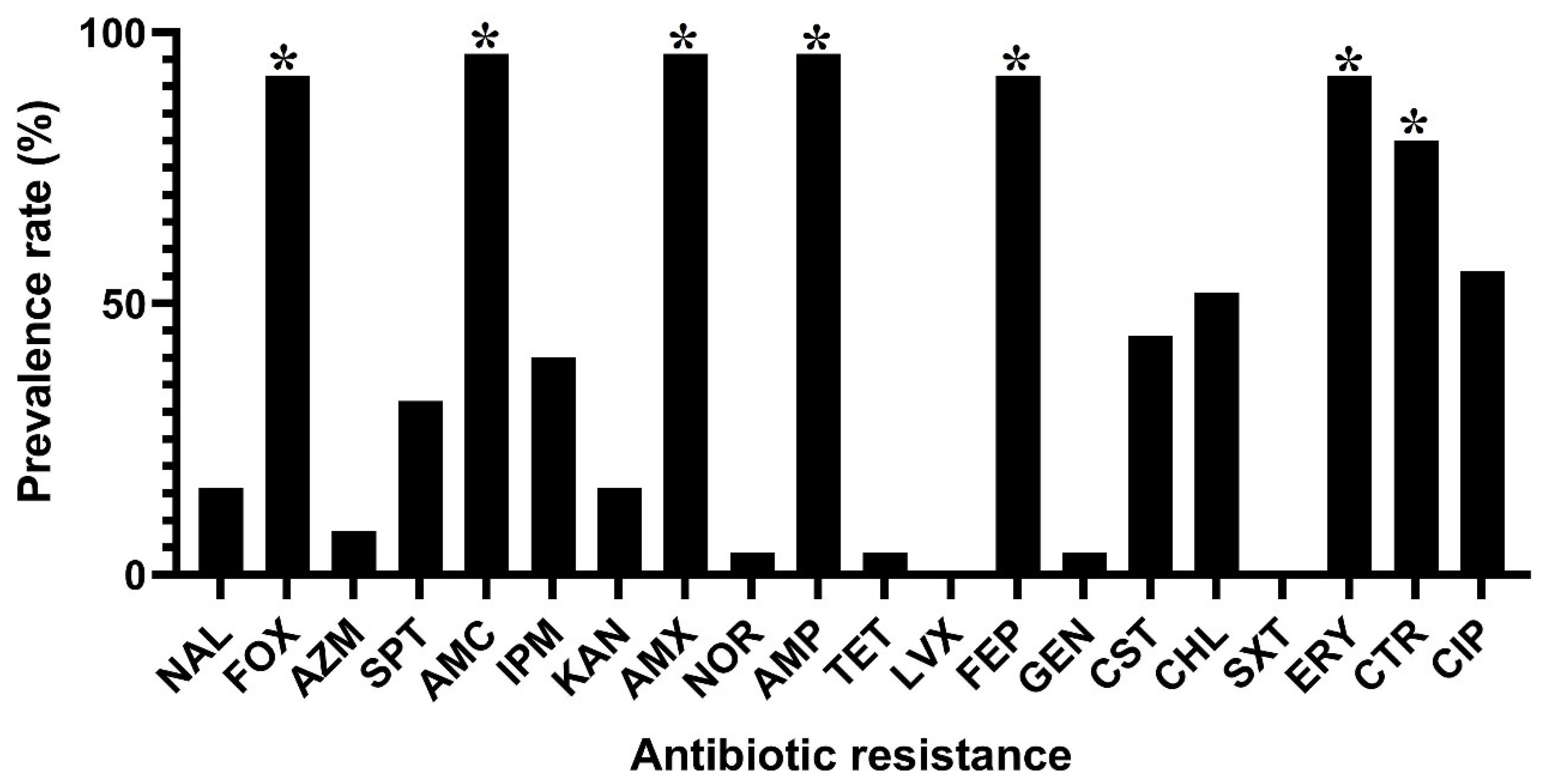
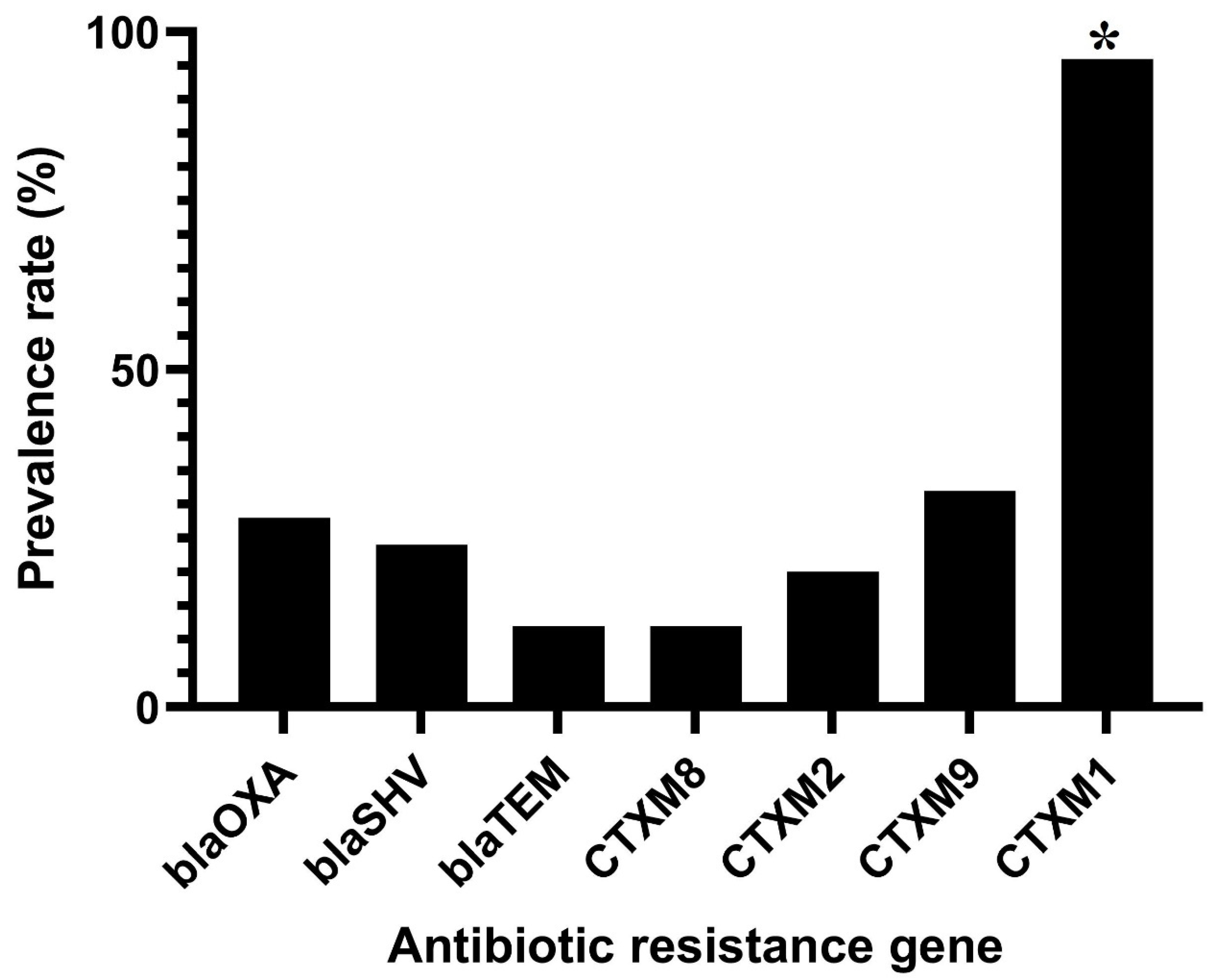
3.2. Antibiotic Resistance Phenotypes and Genotypes in C. sakazakii Isolates

Antibiotic resistance against nine different antibiotic categories and twenty commercial antibiotics was determined for all C. sakazakii isolates. Figure 1 shows the phenotypic resistance of C. sakazakii isolates against different antibiotics. All isolates were susceptible to trimethoprim/sulfamethoxazole and levofloxacin antibiotics. In contrast, C. sakazakii isolates were highly resistant to amoxicillin-clavulanic acid (24 isolates; 96%), amoxicillin (24 isolates; 96%), ampicillin (24 isolates; 96%), cefoxitin (23 isolates; 92%), cefepime (23 isolates; 92%), erythromycin (23 isolates; 92%), ceftriaxone (20 isolate; 80%), ciprofloxacin (14 isolates; 56%) and chloramphenicol (13 isolates; 52%) antibiotics. Also, these isolates were highly susceptible to gentamicin (1 isolate; 4%), tetracycline (1 isolate; 4%), norfloxacin (1 isolate; 4%) and azithromycin (2 isolates; 8%) antibiotics. As shown in Table 1, 24 (96%) out of 25 C. sakazakii isolates were resistant to at least three different antibiotic classes and considered multi-drug resistant (MDR). The multi-drug resistance pattern, including resistance to β-Lactam, Quinolone-fluoroquinolone, and Macrolide antibiotic classes, was the most prevalent MDR profile among the isolates (11 isolates; 44%). However, 8 (32%) out of 25 C. sakazakii isolates were resistant to an MDR profile consisting of six different classes of antibiotics: β-Lactam, Aminoglycoside, Lipopeptide, Phenicol, Quinolone-fluoroquinolone, and Macrolide. Several β-Lactamase encoding genes were detected in the isolates: blaCTX-M-1 (n = 24, 96%), blaTEM (n = 3, 12%), blaCTX-M-8 (n = 3, 12%), blaCTX-M-2 (n = 5, 20%), blaSHV (n = 6, 24%), blaOXA (n = 7, 28%) and blaCTX-M-9 (n = 8, 32%) (Figure 2).

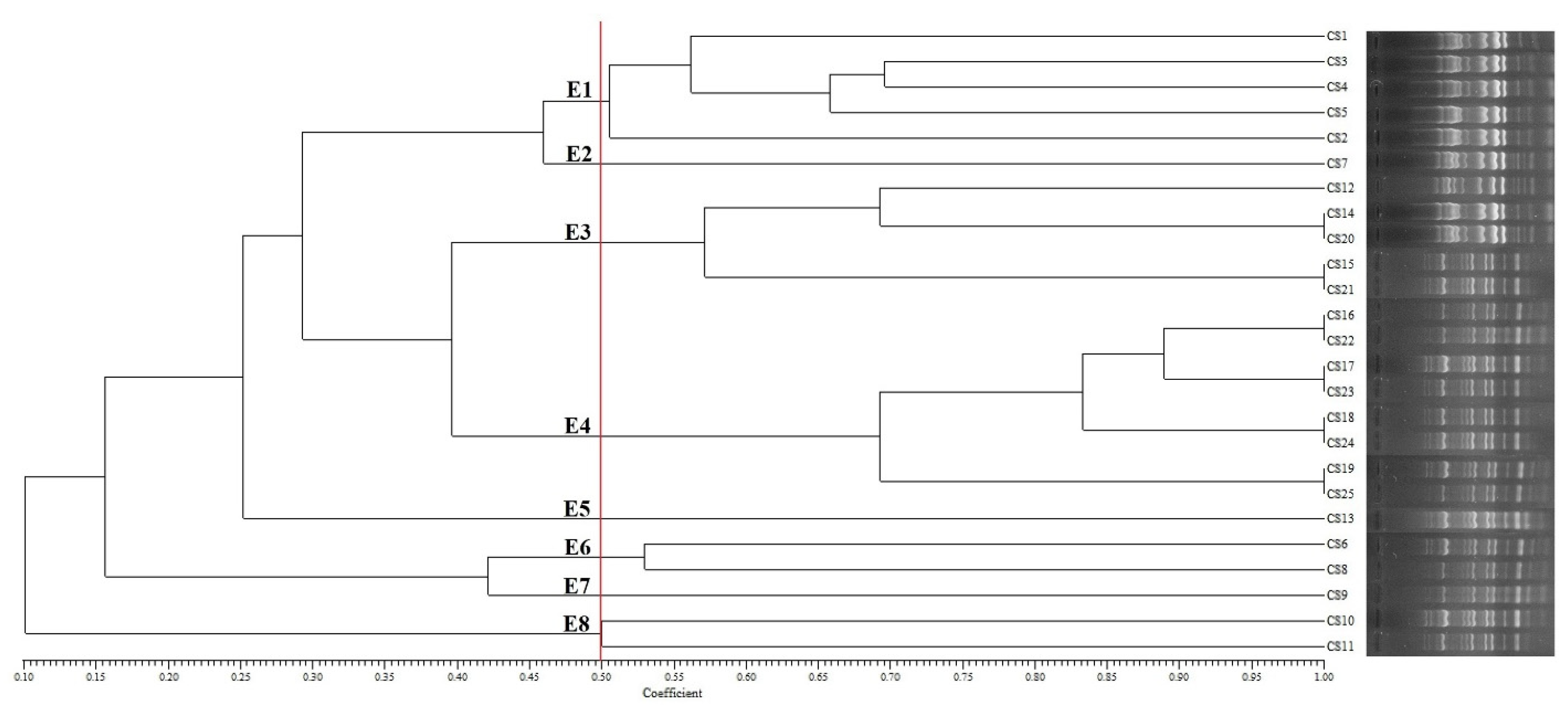
3.3. Genotyping of C. sakazakii Isolates

This study also evaluated genotypic polymorphism and genetic diversity among the C. sakazakii strains using ERIC-PCR fingerprint pattern analysis. Each C. sakazakii isolate produced ERIC-PCR banding patterns of 9 to 17 amplified PCR products, ranging from 1000 to 1800 bp (Figure 3). The C. sakazakii strains formed eight major clusters (E1–E8) with at least 50% ERIC-PCR profile similarity (cut-off value at 0.5). The Simpson’s Diversity Index was calculated at 0.83, demonstrating a high level of genetic diversity among the isolates. Table 2 shows the resistance phenotype, genotype, and ERIC-PCR types of the C. sakazakii isolates. Eight (32%) isolates were grouped in cluster E4. Clusters E1 and E3 included five isolates; however, other clusters contained only one (E2, E5, and E7) or two isolates (E6 and E8). As shown in Table 2, more antibiotic resistance genes were detected in C. sakazakii isolates with higher phenotypic antibiotic resistance. However, no significant relationship was observed between the ERIC-PCR clustering patterns and the phenotypic/genotypic antibiotic resistance profiles.

4. Discussion

C. sakazakii has recently been regarded as an emerging foodborne opportunistic pathogen isolated commonly from low-moisture foods, including powdered infant milk and formula [23]. C. sakazakii usually causes severe morbidity through some infectious diseases including meningitis, sepsis, necrotizing enterocolitis, and urinary tract infections in neonates and infants; however, this pathogen also significantly affects other age groups and leads to health complications in adults [24]. PIFM has recently been regarded the main vehicle of C. sakazakii transmission to infants implicating neonatal infections and has been considered as one of the most important concerns in children health around the world [6]. Other sources of this pathogen were also involved in cases of infectious diseases in children and other sensitive groups of people [25]. So far, limited studies are available on the prevalence rate of C. sakazakii PIFM.
In the present study, we determined the prevalence rate of this pathogen in PIFM samples collected from different grocery stores located in Tehran city, Iran. 6.86% of PIFM samples were contaminated with C. sakazakii. The prevalence rate of C. sakazakii in this study (Iran) was higher than that reported from Ireland (3.40%; 16 out of 470 PIF samples) [26] and China in 2016 (2.77%; 56 out of 2020 PIFM samples) [17], and lower than that reported from China in 2012 (23.0%; 84 out of 366 PIFM and baby food samples) [27], the USA (26.9%; 21 out of 78 households) [28], and Korea (83.0%; 113 out of 136 infant food samples) [29]. The prevalence rate in Tehran (6.86%) does not significantly differ from that in Qazvin city, Iran (5.08%; p < 0.05), reported in our previous study [12]. Contamination with C. sakazakii usually occurs during the post-pasteurization packaging or adding ingredients to the infant formula foods [23]. The thermal resistance of this pathogen is significantly higher than that of other Enterobacteriaceae species [30]. Stabilized membrane proteins and phospholipids and trehalose production and accumulation by C. sakazakii protect this pathogen from the dry conditions in low-moisture foods such as infant formula and improve its thermal resistance. Moreover, high solid sugar and fat content in infant formula foods also protect C. sakazakii from thermal stress [25,31,32]. Consequently, C. sakazakii can survive when exposed to hot and dry conditions. Our surveillance results in this and previous studies showed serious levels of C. sakazakii contamination in PIFM. Also, foodborne pathogens from contaminated foods may be introduced to other processed foods and food preparation areas as an important vehicle of cross-contamination. Depending on the initial solving water temperature, C. sakzakii may be removed during the infant food preparation [28].
The emergence of MDR foodborne bacterial pathogens has recently been regarded as one of the most important concerns in public health and a major challenge in food safety. Since a wide range of clinical and veterinary antibiotics are still being used worldwide for diseases prevention, growth promotion, and treatment of sick farm animals, the prevalence of MDR foodborne pathogens has been increasing [33]. It is worthwhile to note that antibiotic resistance genes can be transferred horizontally among different species of Enterobacteriaceae family isolates in humans, animals, environments, and foods [34]. In this study, we isolated 25 C. sakazakii strains from PIFM samples, collected from Tehran city (Iran), mostly resistant to amoxicillin-clavulanic acid, amoxicillin, ampicillin, cefoxitin, cefepime, erythromycin, and ceftriaxone, and utterly susceptible to trimethoprim/sulfamethoxazole and levofloxacin antibiotics. A previous study in Korea by Kim et al. (2008) showed a high level of antibiotic susceptibility among the C. sakazakii strains isolated from infant foods against ciprofloxacin, tetracycline, chloramphenicol, kanamycin, gentamicin, and nalidixic acid antibiotics; they reported 31.8 and 5.3% of the isolates resistant to ampicillin and cephalothin antibiotics, respectively [29]. A study carried out in the United States by Kilonzo-Nthenge et al. (2012) reported that 76.1, 66.6, 57.1, and 47.6% of C. sakazakii isolates were resistant to penicillin, tetracycline, ciprofloxacin, and nalidixic acid antibiotics, respectively, and all isolates were susceptible to gentamicin [28]. Another study conducted in China by Li et al. (2016) showed high resistance to amoxicillin-clavulanic acid, rifampicin, tetracycline, streptomycin, and ampicillin antibiotics among C. sakazakii isolated from retail milk-based infant foods [27]. Moreover, in China, Fei et al. (2017) isolated C. sakazakii from PIF samples with the highest resistance to cephalothin antibiotics [17]. Recently, we (in the year 2020) also performed a study in Iran, Qazvin city, and showed that C. sakazakii isolated from infant food samples were resistant to ampicillin, amoxicillin, ciprofloxacin, and tetracycline and susceptible to chloramphenicol, amikacin, and levofloxacin antibiotics [12]. Interestingly, the antibiotic resistance pattern of C. sakazakii isolated from infant food samples in this study is in accordance with other studies. However, unsupervised and reckless use of different clinical and veterinary antibiotics for improving animal health worldwide may contribute to some differences observed among the antibiotic resistance patterns of C. sakazakii strains isolated in different studies/geographic areas [17,35].
In this study, 96% of C. sakazakii isolates were MDR, which is higher than that previously reported in the USA and China [17,28]. The blaCTX-M-1 antibiotic resistance gene was mostly detected in our isolates. Farm animals exposed to different clinical and veterinary antibiotics for a long time can develop bacterial flora harboring different antibiotic resistance genes and resistance to various antibiotics [36]. blaCTX-M-1 is one of the most prevalent antibiotic resistance genes encoding extended-spectrum beta-lactamase (ESBL) in the Enterobacteriaceae family. Bacterial strains harboring blaCTX genes are commonly resistant to cephalosporin antibiotics such as cefoxitin, cefepime, ceftriaxone, etc. [37,38]. Notably, C. sakazakii strains isolated in this study harbored blaCTX genes and were resistant to cefoxitin, cefepime, and ceftriaxone antibiotics.
Different PCR-based genomic fingerprinting methods such as RAPD, ERIC, BOX, and rep-PCR assays have been used to evaluate the clonal relatedness and genetic diversity among foodborne pathogens isolated from food, environmental and clinical samples [13]. In this study, we used the ERIC-PCR patterns to determine the genetic relationship among the C. sakazakii strains isolated from the PIFM samples [20]. The results of ERIC-PCR indicated that eight (32%) isolates clustered together (E4). In addition, there were two clusters (E1 and E3), each containing five isolates and five clonal groups (E2, E5-E8), each containing just one or two isolates. No significant association between the antibiotic resistance and ERIC-PCR patterns of C. sakazakii isolates was found [20,39]. This contrasts with a study of Ye et al. (2008) in which they found a significant correlation between the antibiotic resistance profiles and genomic characterization of C. sakazakii isolates from instant formula milk [20]. However, significant positive correlations between the genotypic and phenotypic properties of antibiotic resistance were observed in most isolates. More investigations are needed to study the correlation between the genomic characterization and antimicrobial susceptibility profiles of C. sakazakii strains isolated from infant food samples using novel genomic analysis techniques.

5. Conclusions

In conclusion, we investigated the prevalence rate, antibiotic resistance, and genetic diversity of C. sakazakii strains isolated from PIFM samples collected from Tehran, Iran. C. sakazakii contamination in PIFM samples was frequently observed. This study showed that C. sakazakii isolates were resistant to amoxicillin-clavulanic acid, amoxicillin, ampicillin, cefoxitin, cefepime, erythromycin, and ceftriaxone antibiotics. The beta-lactamase encoding blaCTX-M-1 gene was detected in most isolates, and the isolates harboring this gene were resistant to cephalosporin antibiotics, including cefoxitin, cefepime, and ceftriaxone. Additionally, a high level of genetic diversity was observed among the isolates. We found a significant correlation between the phenotypic and genotypic antibiotic resistance properties of C. sakazakii isolates. Careless and irrational use of veterinary and clinical antimicrobial agents in farm and domestic animals may lead to the emergence of MDR C. sakazakii in infant foods.

Author Contributions

Conceptualization, B.P., W.M.B. and J.W.A.R.; methodology, B.P. and S.A.; software, B.P. and W.M.B.; validation, R.M.; formal analysis, B.P.; investigation, B.P.; resources, R.M.; data curation, B.P.; writing—original draft preparation, B.P.; writing—review and editing, J.W.A.R. and W.M.B.; visualization, S.A.; supervision, B.P.; project administration, R.M.; funding acquisition, B.P. All authors have read and agreed to the published version of the manuscript.

Funding

This research received no external funding.

Institutional Review Board Statement

Not applicable.

Informed Consent Statement

Not applicable.

Data Availability Statement

Not applicable.

Acknowledgments

We thank our colleagues and all technicians from the Medical Microbiology Research Center and the Central Research Laboratory of Qazvin University of Medical Sciences, who assisted us in this study.

Conflicts of Interest

The authors declare no conflict of interest.

References

  1. Healy, B.; Cooney, S.; O’Brien, S.; Iversen, C.; Whyte, P.; Nally, J.; Callanan, J.J.; Fanning, S. Cronobacter (Enterobacter sakazakii): An opportunistic foodborne pathogen. Foodborne Pathog. Dis. 2010, 7, 339–350. [Google Scholar] [CrossRef] [PubMed]
  2. Yong, W.; Guo, B.; Shi, X.; Cheng, T.; Chen, M.; Jiang, X.; Ye, Y.; Wang, J.; Xie, G.; Ding, J. An investigation of an acute gastroenteritis outbreak: Cronobacter sakazakii, a potential cause of food-borne illness. Front. Microbiol. 2018, 9, 2549. [Google Scholar] [CrossRef] [PubMed]
  3. Holý, O.; Forsythe, S. Cronobacter spp. as emerging causes of healthcare-associated infection. J. Hosp. Infect. 2014, 86, 169–177. [Google Scholar] [CrossRef] [PubMed] [Green Version]
  4. Bintsis, T. Foodborne pathogens. AIMS Microbiol. 2017, 3, 529. [Google Scholar] [CrossRef]
  5. McMullan, R.; Menon, V.; Beukers, A.G.; Jensen, S.O.; van Hal, S.J.; Davis, R. Cronobacter sakazakii infection from expressed breast milk, Australia. Emerg. Infect. Dis. 2018, 24, 393. [Google Scholar] [CrossRef] [Green Version]
  6. Henry, M.; Fouladkhah, A. Outbreak history, biofilm formation, and preventive measures for control of Cronobacter sakazakii in infant formula and infant care settings. Microorganisms 2019, 7, 77. [Google Scholar] [CrossRef] [Green Version]
  7. Chap, J.; Jackson, P.; Siqueira, R.; Gaspar, N.; Quintas, C.; Park, J.; Osaili, T.; Shaker, R.; Jaradat, Z.; Hartantyo, S. International survey of Cronobacter sakazakii and other Cronobacter spp. in follow up formulas and infant foods. Int. J. Food Microbiol. 2009, 136, 185–188. [Google Scholar] [CrossRef]
  8. Carvalho, G.G.; Calarga, A.P.; Teodoro, J.R.; Queiroz, M.M.; Astudillo-Trujillo, C.A.; Levy, C.E.; Brocchi, M.; Kabuki, D.Y. Isolation, comparison of identification methods and antibiotic resistance of Cronobacter spp. in infant foods. Food Res. Int. 2020, 137, 109643. [Google Scholar] [CrossRef]
  9. Franz, C.; den Besten, H.M.; Boehnlein, C.; Gareis, M.; Zwietering, M.H.; Fusco, V. Reprint of: Microbial food safety in the 21st century: Emerging challenges and foodborne pathogenic bacteria. Trends Food Sci. Technol. 2019, 84, 34–37. [Google Scholar] [CrossRef]
  10. Al-Nabulsi, A.A.; Osaili, T.M.; Elabedeen, N.A.Z.; Jaradat, Z.W.; Shaker, R.R.; Kheirallah, K.A.; Tarazi, Y.H.; Holley, R.A. Impact of environmental stress desiccation, acidity, alkalinity, heat or cold on antibiotic susceptibility of Cronobacter sakazakii. Int. J. Food Microbiol. 2011, 146, 137–143. [Google Scholar] [CrossRef]
  11. Lepuschitz, S.; Ruppitsch, W.; Pekard-Amenitsch, S.; Forsythe, S.J.; Cormican, M.; Mach, R.L.; Piérard, D.; Allerberger, F.; Group, E.S. Multicenter study of Cronobacter sakazakii infections in humans, Europe, 2017. Emerg. Infect. Dis. 2019, 25, 515. [Google Scholar] [CrossRef] [PubMed] [Green Version]
  12. Pakbin, B.; Mahmoudi, R.; Mousavi, S.; Allahyari, S.; Amani, Z.; Peymani, A.; Qajarbeygi, P.; Hoseinabadi, Z. Genotypic and antimicrobial resistance characterizations of Cronobacter sakazakii isolated from powdered milk infant formula: A comparison between domestic and imported products. Food Sci. Nutr. 2020, 8, 6708–6717. [Google Scholar] [CrossRef] [PubMed]
  13. Reade, M. Trends in DNA Fingerprinting Research; Nova Publishers: New York, NY, USA, 2005. [Google Scholar]
  14. Armstrong, G.L.; MacCannell, D.R.; Taylor, J.; Carleton, H.A.; Neuhaus, E.B.; Bradbury, R.S.; Posey, J.E.; Gwinn, M. Pathogen genomics in public health. N. Engl. J. Med. 2019, 381, 2569–2580. [Google Scholar] [CrossRef] [PubMed]
  15. Ku, Y.-H.; Lee, M.-F.; Chuang, Y.-C.; Yu, W.-L. Detection of plasmid-mediated β-lactamase genes and emergence of a novel AmpC (CMH-1) in Enterobacter cloacae at a medical center in Southern Taiwan. J. Clin. Med. 2018, 8, 8. [Google Scholar] [CrossRef] [Green Version]
  16. Gan, X.; Li, M.; Yan, S.; Wang, X.; Wang, W.; Li, F. Genomic Landscape and Phenotypic Assessment of Cronobacter sakazakii Isolated From Raw Material, Environment, and Production Facilities in Powdered Infant Formula Factories in China. Front. Microbiol. 2021, 12, 686189. [Google Scholar] [CrossRef]
  17. Fei, P.; Jiang, Y.; Feng, J.; Forsythe, S.J.; Li, R.; Zhou, Y.; Man, C. Antibiotic and desiccation resistance of Cronobacter sakazakii and C. malonaticus isolates from powdered infant formula and processing environments. Front. Microbiol. 2017, 8, 316. [Google Scholar] [CrossRef] [Green Version]
  18. Parra-Flores, J.; Aguirre, J.; Juneja, V.; Jackson, E.E.; Cruz-Córdova, A.; Silva-Sanchez, J.; Forsythe, S. Virulence and antibiotic resistance profiles of Cronobacter sakazakii and Enterobacter spp. involved in the diarrheic hemorrhagic outbreak in Mexico. Front. Microbiol. 2018, 9, 2206. [Google Scholar] [CrossRef] [Green Version]
  19. Dallenne, C.; Da Costa, A.; Decré, D.; Favier, C.; Arlet, G. Development of a set of multiplex PCR assays for the detection of genes encoding important β-lactamases in Enterobacteriaceae. J. Antimicrob. Chemother. 2010, 65, 490–495. [Google Scholar] [CrossRef] [Green Version]
  20. Ye, Y.; Wu, Q.; Xu, X.; Yang, X.; Dong, X.; Zhang, J. The phenotypic and genotypic characterization of Enterobacter sakazakii strains from infant formula milk. J. Dairy Sci. 2010, 93, 2315–2320. [Google Scholar] [CrossRef] [Green Version]
  21. Pavel, A.B.; Vasile, C.I. PyElph-a software tool for gel images analysis and phylogenetics. BMC Bioinform. 2012, 13, 9. [Google Scholar] [CrossRef] [Green Version]
  22. Rolf, J. NTSYS-PC, Numerical Taxonomy and Multivariate Analysis System, Version 2.11; T Exeter Software: Setauket, NY, USA, 2000. [Google Scholar]
  23. Chauhan, R.; Singh, N.; Pal, G.K.; Goel, G. Trending biocontrol strategies against Cronobacter sakazakii: A recent updated review. Food Res. Int. 2020, 137, 109385. [Google Scholar] [CrossRef] [PubMed]
  24. Elkhawaga, A.A.; Hetta, H.F.; Osman, N.S.; Hosni, A.; El-Mokhtar, M.A. Emergence of Cronobacter sakazakii in cases of neonatal sepsis in upper Egypt: First report in North Africa. Front. Microbiol. 2020, 11, 215. [Google Scholar] [CrossRef] [PubMed] [Green Version]
  25. Ling, N.; Forsythe, S.; Wu, Q.; Ding, Y.; Zhang, J.; Zeng, H. Insights into Cronobacter sakazakii biofilm formation and control strategies in the food industry. Engineering 2020, 6, 393–405. [Google Scholar] [CrossRef]
  26. O’Brien, S.; Healy, B.; Negredo, C.; Anderson, W.; Fanning, S.; Iversen, C. Prevalence of Cronobacter species (Enterobacter sakazakii) in follow-on infant formulae and infant drinks. Lett. Appl. Microbiol. 2009, 48, 536–541. [Google Scholar] [CrossRef] [PubMed]
  27. Li, Z.; Ge, W.; Li, K.; Gan, J.; Zhang, Y.; Zhang, Q.; Luo, R.; Chen, L.; Liang, Y.; Wang, Q. Prevalence and characterization of Cronobacter sakazakii in retail milk-based infant and baby foods in Shaanxi, China. Foodborne Pathog. Dis. 2016, 13, 221–227. [Google Scholar] [CrossRef] [PubMed]
  28. Kilonzo-Nthenge, A.; Rotich, E.; Godwin, S.; Nahashon, S.; Chen, F. Prevalence and antimicrobial resistance of Cronobacter sakazakii isolated from domestic kitchens in middle Tennessee, United States. J. Food Prot. 2012, 75, 1512–1517. [Google Scholar] [CrossRef] [PubMed]
  29. Kim, K.; Jang, S.S.; Kim, S.K.; Park, J.-H.; Heu, S.; Ryu, S. Prevalence and genetic diversity of Enterobacter sakazakii in ingredients of infant foods. Int. J. Food Microbiol. 2008, 122, 196–203. [Google Scholar] [CrossRef]
  30. Zhang, Y.; Xie, Y.; Tang, J.; Wang, S.; Wang, L.; Zhu, G.; Li, X.; Liu, Y. Thermal inactivation of Cronobacter sakazakii ATCC 29544 in powdered infant formula milk using thermostatic radio frequency. Food Control 2020, 114, 107270. [Google Scholar] [CrossRef]
  31. Dalkilic-Kaya, G.; Heperkan, D.; Juneja, V.K.; Heperkan, H.A. Thermal resistance of Cronobacter sakazakii isolated from baby food ingredients of dairy origin in liquid medium. J. Food Processing Preserv. 2018, 42, e13463. [Google Scholar] [CrossRef]
  32. Dancer, G.; Mah, J.H.; Rhee, M.S.; Hwang, I.G.; Kang, D.H. Resistance of Enterobacter sakazakii (Cronobacter spp.) to environmental stresses. J. Appl. Microbiol. 2009, 107, 1606–1614. [Google Scholar] [CrossRef]
  33. Mancilla-Becerra, L.M.; Lías-Macías, T.; Ramírez-Jiménez, C.L.; León, J.B. Multidrug-resistant bacterial foodborne pathogens: Impact on human health and economy. Pathog Bact 2019, 2019, 1–18. [Google Scholar]
  34. Sun, D.; Jeannot, K.; Xiao, Y.; Knapp, C.W. Horizontal gene transfer mediated bacterial antibiotic resistance. Front. Microbiol. 2019, 10, 1933. [Google Scholar] [CrossRef] [PubMed] [Green Version]
  35. Gao, J.; Han, Z.; Li, P.; Zhang, H.; Du, X.; Wang, S. Outer Membrane Protein F Is Involved in Biofilm Formation, Virulence and Antibiotic Resistance in Cronobacter sakazakii. Microorganisms 2021, 9, 2338. [Google Scholar] [CrossRef] [PubMed]
  36. Caniça, M.; Manageiro, V.; Abriouel, H.; Moran-Gilad, J.; Franz, C.M. Antibiotic resistance in foodborne bacteria. Trends Food Sci. Technol. 2019, 84, 41–44. [Google Scholar] [CrossRef]
  37. Zurfluh, K.; Nüesch-Inderbinen, M.; Morach, M.; Zihler Berner, A.; Hächler, H.; Stephan, R. Extended-spectrum-β-lactamase-producing Enterobacteriaceae isolated from vegetables imported from the Dominican Republic, India, Thailand, and Vietnam. Appl. Environ. Microbiol. 2015, 81, 3115–3120. [Google Scholar] [CrossRef] [Green Version]
  38. Holý, O.; Alsonosi, A.; Hochel, I.; Röderová, M.; Zatloukalova, S.; Mlynárčik, P.; Kolář, M.; Petrželová, J.; Alazraq, A.; Chmelař, D. Antibiotic susceptibility of Cronobacter spp. isolated from clinical samples. Pol. J. Microbiol. 2019, 68, 5. [Google Scholar] [CrossRef] [Green Version]
  39. Ye, Y.; Wu, Q.; Zhou, Y.; Dong, X.; Zhang, J. Analysis of major band of Enterobacter sakazakii by ERIC-PCR and development of a species-specific PCR for detection of Ent. sakazakii in dry food samples. J. Microbiol. Methods 2008, 75, 392–397. [Google Scholar] [CrossRef]
Figure 1. Prevalence rates of different antibiotic resistance phenotypes in C. sakazakii strains isolated from PIFM samples. * indicates significant differences (p > 0.05). NAL, nalidixic acid; FOX, cefoxitin; AZM, azithromycin; SPT, streptomycin; AMC, amoxicillin-clavulanic acid; IPM, imipenem; KAN, kanamycin; AMX, amoxicillin; NOR, norfloxacin; AMP, ampicillin; TET, tetracycline; FEP, cefepime; GEN, gentamicin; CST, colistin; CHL, chloramphenicol; ERY, erythromycin; CTR, ceftriaxone and CIP, ciprofloxacin.
Foods 11 01093 g001
Figure 2. Prevalence rates of ESBLs genes in C. sakazakii strains isolated from PIFM samples. * indicates significant differences (p > 0.05). blaSHV, blaSHV; blaOXA, blaOXA; blaTEM, blaTEM; CTXM1, blaCTX-M-1; CTXM2, blaCTX-M-2; CTXM8, blaCTX-M-8 and CTXM9, blaCTX-M-9.
Foods 11 01093 g002
Figure 3. UPGMA dendrogram of C. sakazakii isolates from PIFM samples indicating the Dice coefficient based on ERIC-PCR analysis.
Foods 11 01093 g003
Table 1. Multidrug resistance class patterns of C. sakazakii strains isolated from PIFM samples.
No. Classes of AntibioticsMultidrug Resistance Patterns a (No. Isolates in Each Pattern)No. Total Isolates (%) (n = 25)
TwoβLs-QNs (n = 1)1 (4)
ThreeβLs-QNs-MLs (n = 7)11 (44)
βLs-PNs-MLs (n = 4)
FourβLs-AGs-LPs-MLs (n = 1)3 (12)
βLs-PNs-MLs-QNs (n = 1)
βLs-AGs-LPs-QNs (n = 1)
FiveβLs-QNs-MLs-AGs-TCs (n = 1)2 (8)
βLs-QNs-MLs-AGs-LPs (n = 1)
SixβLs-QNs-AGs-LPs-MLs-PNs (n = 8)8 (32)
a βLs, β-lactams; AGs, aminoglycosides; LPs, lipopeptides; TCs, tetracyclines; PNs, phenicols; QNs, quinolones and fluoroquinolones; MLs, macrolides.
Table 2. Resistance phenotype, genotype and ERIC-PCR types in C. sakazakii strains isolated from PIFM samples.
No.IsolateResistance Phenotype aResistance Genotype bERIC-PCR Type
1CS1NAL, FOX, AZM, AMC, IPM, KAN, AMX, AMP, TET, FEP, GEN, ERYC1, S, O, TE1
2CS2NAL, FOX, AMC, IPM, KAN, AMX, AMP, FEP, ERY, CSTC1, C2, C9E1
3CS3FOX, AZM, AMC, IPM, KAN, AMX, AMP, FEP, ERY, CSTC1, S, OE1
4CS4FOX, AMC, IPM, KAN, NOR, AMP, FEP, CSTC1E1
5CS5NAL, FOX, AMC, AMX, AMP, FEP, ERY, CTRC1E1
6CS6FOX, AMC, AMX, AMP, FEP, CHL, ERY, CTRC1E6
7CS7FOX, SPT, AMC, AMX, AMP, FEP, CST, CHL, ERY, CTR, CIPC1, S, O, TE2
8CS8FOX, AMC, IPM, AMX, AMP, FEP, ERY, CTR, CIPC1E6
9CS9FOX, AMC, AMX, AMP, FEP, CHL, ERY, CTRC1E7
10CS10SPT, AMC, AMX, AMP, FEP, CST, CHL, ERY, CTR, CIPC1, C8, C9E8
11CS11FOX, AMC, IPM, AMX, AMP, FEP, ERY, CTR, CIPC1, C2, C9E8
12CS12FOX, SPT, AMC, AMX, AMP, CST, CHL, ERY, CTR, CIPC1, S, OE3
13CS13FOX, AMC, IPM, AMX, AMP, FEP, ERY, CIPC1E5
14CS14FOX, AMC, AMX, AMP, FEP, CHL, ERY, CIPC1E3
15CS15FOX, AMC, IPM, AMX, AMP, FEP, CST, CHL, ERY, CTR, CIPC1, C8, C9E3
16CS16NAL, FOX, AMC, AMX, AMP, FEP, ERY, CTRC1E4
17CS17FOX, AMC, AMX, FEP, CHL, ERY, CTR,C1E4
18CS18FOX, SPT, AMC, AMX, AMP, FEP, CST, CHL, ERY, CTR, CIPC1, C2, C9E4
19CS19IPM, AMX, AMP, FEP, CTR, CIPC1E4
20CS20FOX, AMC, AMX, AMP, FEP, CHL, ERY, CTRC1, S, OE3
21CS21FOX, SPT, AMC, AMX, AMP, CST, CHL, ERY, CTR, CIPC1, S, O, TE3
22CS22FOX, AMC, IPM, AMX, AMP, FEP, ERY, CTR, CIPC8E4
23CS23FOX, SPT, AMC, AMX, AMP, FEP, CST, CHL, ERY, CTR, CIPC1, C2, C9E4
24CS24FOX, AMC, IPM, AMX, AMP, FEP, ERY, CTR, CIPC1, C2, C9E4
25CS25FOX, SPT, AMC, AMX, AMP, FEP, CST, CHL, ERY, CTR, CIPC1, O, C9E4
a NAL, nalidixic acid; FOX, cefoxitin; AZM, azithromycin; SPT, streptomycin; AMC, amoxicillin-clavulanic acid; IPM, imipenem; KAN, kanamycin; AMX, amoxicillin; NOR, norfloxacin; AMP, ampicillin; TET, tetracycline; FEP, cefepime; GEN, gentamicin; CST, colistin; CHL, chloramphenicol; ERY, erythromycin; CTR, ceftriaxone; CIP, ciprofloxacin. b S, blaSHV; O, blaOXA; T, blaTEM; C1, blaCTX-M-1; C2, blaCTX-M-2; C8, blaCTX-M-8; C9, blaCTX-M-9.
Publisher’s Note: MDPI stays neutral with regard to jurisdictional claims in published maps and institutional affiliations.

