Review

Nature-Inspired Antimicrobial Surfaces and Their Potential Applications in Food Industries

1
Food Assurance, AgResearch, Palmerston North 4442, New Zealand
2
New Zealand Food Safety Science Research Centre, Palmerston North 4442, New Zealand
*
Author to whom correspondence should be addressed.
Foods 2022, 11(6), 844; https://doi.org/10.3390/foods11060844
Submission received: 14 February 2022 / Revised: 9 March 2022 / Accepted: 12 March 2022 / Published: 16 March 2022
(This article belongs to the Section Food Microbiology)

Abstract

:
Antimicrobial resistance (AMR) is a growing global concern and has called for the integration of different areas of expertise for designing robust solutions. One such approach is the development of antimicrobial surfaces to combat the emerging resistance in microbes against drugs and disinfectants. This review is a compressive summary of the work done in the field of material science, chemistry, and microbiology in the development of antimicrobial materials and surfaces that are inspired by examples in nature. The focus includes examples of natural antimicrobial surfaces, such as cicada wings or nanopillars, dragonfly wings, shrimp shells, taro leaves, lotus leaves, sharkskin, gecko skin, and butterfly wings, along with their mechanism of action. Techniques, compositions, and combinations that have been developed to synthetically mimic these surfaces against bacterial/viral and fungal growth in food-processing areas have also been discussed. The applications of synthetic mimics of natural antimicrobial surfaces in food-processing environments is still a naïve area of research. However, this review highlights the potential applications of natural antimicrobial surfaces in the food-processing environment as well as outlines the challenges that need mitigations.

Graphical Abstract

1. Introduction

Antimicrobial activity can be explained as a property of a compound or structure that enables either inhibition, reduction in growth rate, or inactivation (cell death) of microbes. Antibiotic drugs have been reported in the literature since the discovery by Alexander Fleming of the first antibiotic, penicillin, in 1928 [1,2]. However, the use of plant extracts to treat infections by some of the earliest civilisations indicates their long existence. Despite the discovery of several antibiotics, a challenge that has simultaneously emerged in the last two decades is antibiotic resistance. Antibiotic resistance is also an emerging issue for food industries, as there is the pressure exerted by the primary sector in the use of antibiotics and biocides (e.g., disinfectants, food and feed preservatives, or decontaminants), which has led to an increase in the emergence of antimicrobial resistance throughout the food chain [3]. Another related concern is the ability of the resistant populations to form biofilms in food-processing areas. This includes biofilms formed by the spore-forming bacteria, which are very resistant to disinfectants and mild to moderate thermal wash [4], and biofilms from non-spore-forming bacteria, which eventually develop resistance to the treatment and can lead to severe food poisoning and spoilage issues, beyond their role in transmitting antimicrobial resistance (AMR) in the community [5]. Nevertheless, a limited number of disinfectants can be used within food-processing areas, as they might not only leave residues in the final products but can also damage the surfaces of equipment, making it further challenging to reduce bacterial contamination in the final product [4]. The regular use of disinfectants has also increased the selection of resistant bacteria, and this along with the resistance against antibacterial agents is an emerging challenge for the food-processing industries [6,7] and a serious concern to public health.
AMR, resistance to disinfectants, and persistence of biofilms in food-processing industries have urged researchers to search for alternative options against microbial contaminants. One such promising area is the investigation of surfaces/structures found in nature that exhibits an antimicrobial contact killing mechanism. This review comprises a summary of work reported in the last two decades on the discovery of natural and nature-inspired antimicrobial surfaces, techniques used in their development, and their potential applications in food industries against foodborne pathogens. At present, the research on antimicrobial surfaces is limited to either lab-scale or pilot-scale studies. This review summarizes the reports on the efficacy of antimicrobial surfaces on foodborne pathogens and spoilage-related microorganisms, which emphasises the future potential in the food sector.

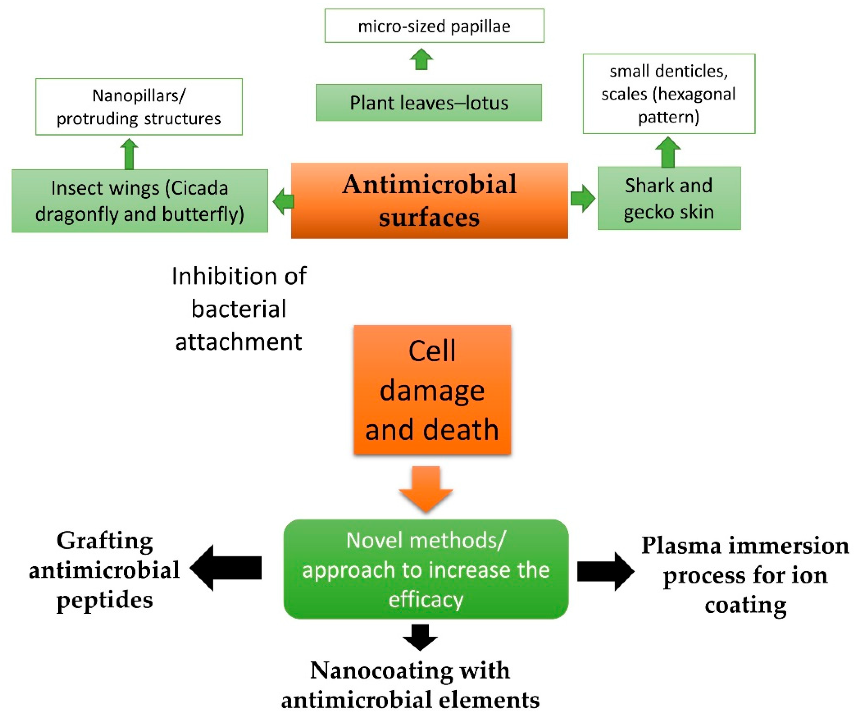
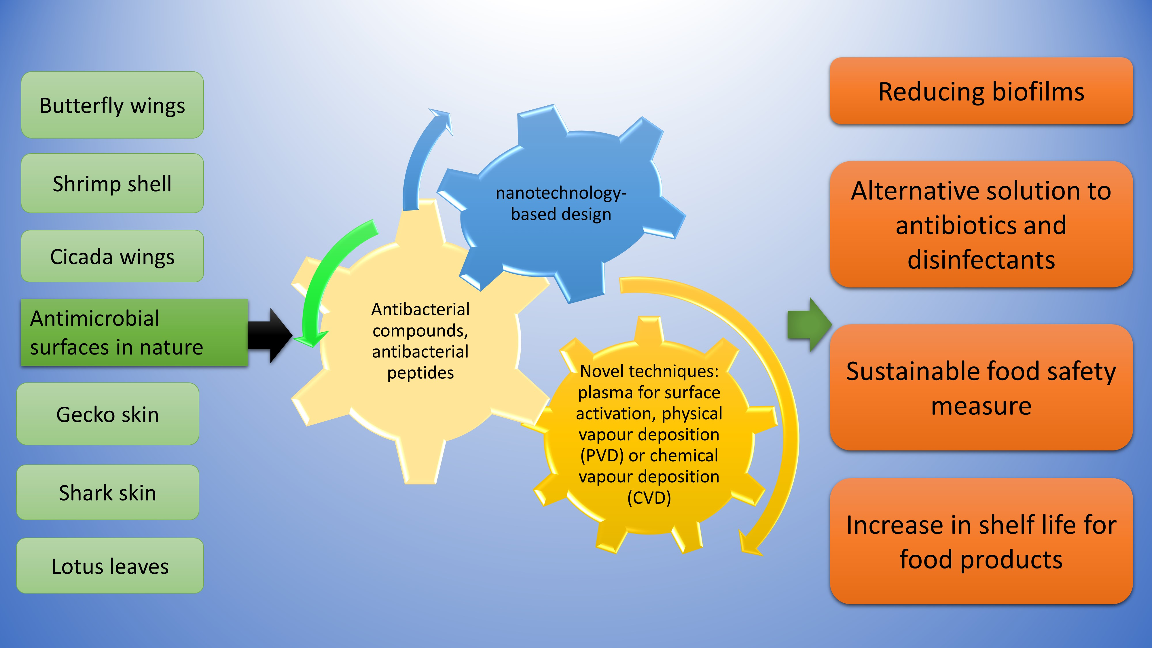
2. Antimicrobial Surfaces in Nature

Antimicrobial surfaces consist of specialized surface architectures or chemical compositions, which either prevent the attachment of microbial species by limiting their adhesion or impart a bactericidal action by disrupting and damaging the cell organelles [8]. Some examples of natural antimicrobial surfaces and their activities reported against bacterial strains are summarized in Table 1. It is to be noted that the table only includes the reported studies on specific strains, and there is still a wide research gap on the evaluation of the efficacy of these surfaces onto other strains or on actual food-grade premises.
Insect wings (cicada wings, dragonfly wings, and butterfly wings) consist of nano protrusions on the surface, which are capable of damaging the bacterial cell membranes, thereby acting as an antibacterial surface [8,9,10]. These structures also limit the colonization of any surviving bacterial cells. The structure and pattern of the dragonfly wing have been well investigated by researchers. The nanostructures on the dragonfly wings can also be explained as nanopillars that are postulated to follow more than one type of model for their killing mechanism [17]. For example, as per the biophysical model, when a bacterial cell comes in contact with the cicada wing, nanopillars can physically stretch the cell membranes, which in turn leads to bacterial rupture and death [17]. Another model indicated a thermodynamic relationship between the bactericidal mechanism of nanopatterned surfaces and the total free energy change of bacterial cells in a patterned surface (like cicada wings), thereby leading to a drastic increase in the contact adhesion area [18]. The contact angle is an important attribute that plays a key role in the inhibition of bacterial attachment as well as in cell disruption [18].
Plant leaves have also been investigated for antimicrobial properties. Lotus leaf surfaces have been reported to demonstrate superhydrophobic properties due to the presence of an air cushion that is entrapped at the liquid/solid interfaces, which limits the contact area and minimizes the biofouling [19,20]. Additionally, lotus leaves have recently been reported to demonstrate bactericidal activity due to the presence of micro-sized papillae and nano-sized wax tubes on the outermost layer with a similar aspect ratio to the bactericidal nanopillars [19]. Cellulose nanofibers (8–10 nm in diameter) in plants, which are made up of β 1,4 linked glucopyranose units, are known to exhibit antibacterial activities [21]. A study by Bixler et al. [16] studied and reported the liquid–solid–air interface between rice leaf and the environment, which indicated that surface roughness and chemistry influences wettability since the air pocket encourages a larger contact angle, thereby preventing a great deal of deposition. A similar mechanism could be postulated to prevent the colonization of microbes on these surfaces.
Another example of a different antimicrobial surface is sharkskin, which has also been extensively studied and mimicked using additional metal implants for increasing the antifouling effect. The structure of the skin comprises small denticles that are flexed against each other due to the elasticity of the skin underneath [22]. The overall structure of the scales on sharkskin represents a repetitive pattern of parallel ridges and grooves, which are found to be placed at a distance of a few millimetres [22]. This complicated, irregular structure plays an important role in preventing microbial settlement and colonization of the surface [23,24]. Gecko skin also has unique, dome-shaped, and closely layered scales (diameter of 100–190 µm and height of 50 µm) arranged in a hexagonal pattern with the regular presence of spinules (tiny hairs) [15]. The exposure of bacterial cells to gecko skin has been reported to result in immediate destruction and therefore cell death due to possible cell damage by the spinules [15].

3. Mimicking Antimicrobial Structures/Materials

The knowledge acquired from natural antimicrobial surfaces has been now combined with nanotechnology-based design and engineering solutions to generate surfaces that can further enhance the utility and life of antimicrobial surfaces. Specific elements and compounds of interest for fabrication to mimic the antibacterial structures are listed in Table 2.
Synthetic black silicon is known to have high aspect ratios with unique surface properties, such as high hydrophobicity and strong biological activity [25,35]. The study by Ivanova et al. [25] demonstrated a similarity between the bactericidal action of black silicon and dragonfly wings and demonstrated a killing rate of 450,000 bacterial cells min−1·cm−2, indicating potential biomedical and industrial applications. Reactive ion beam etching was used in this study to produce silicon nanofibers to mimic the dragonfly wings. Gold sputter-coated wings of dragonflies (common sand dragon) have also been reported to exhibit similar lethal effects on fungal cells, especially against Candida isolates [8,36]. Although the fungal cell structure is very different to that of bacterial cells [37], it can be concluded that the nanoscale topography effects can be extended to fungal de-contamination. Inspired by the lotus leaf, mesoporous silica micro-patterns with a layering of 1 μm silicon oxide have been tested in developing the nanopillars [38]. A study by Green et al. [39] tested the bactericidal efficacy of synthetically replicated spinule arrays consisting of small nano-tipped hairs (spinules) inspired by gecko skin. Eight strains of Lactobacillus spp. (probiotic human bacteria strains) were used to study their ability to colonize on synthetically replicated spinule arrays. Although bacterial inactivation was not quantified, the results indicated a 95% reduction in the population due to a combination of stretching between spinules and nanotip piercing as monitored using scanning electron microscopy [39].
Several synergistic combinations of the compounds/elements mentioned in Table 2 are available in the literature. However, only a few of them, such as graphene and black silicon, has been extensively used in the development of fabricated antibacterial surfaces against foodborne pathogens, indicating a potential for new combinations.

Advantages and Shortcomings of Nature-Inspired Antimicrobial Surfaces

Nanoengineered versions of the nature-inspired surfaces have their limitations as well as advantages as with any other technique/method that is currently in use. Some advantages include the physio-antimicrobial structure that does not rely on chemical components, which reduces the risk of developing chemical resistance or disinfectant-based chemical accumulation in the food chain [40]. Resistance to antibiotics and disinfectants is an emerging concern in the world, and the progress in the field of antimicrobial surfaces shows a potential step away from these chemical disinfectants. However, the use of certain chemicals in the formulation during nanoengineering leads to some limitations. For example, surfaces containing nano-silver, photocatalytic titanium oxides, and zinc oxide nanoparticles cannot be used for fresh produce disinfection or as an aerosol treatment for airborne disinfection, as they pose a risk of ingestion and inhalation [41]. Development of new antimicrobial surface and incorporation of antimicrobial polymers could be a lengthy and expensive process, and the financial gains could be long-standing benefits [42]. However, it can be postulated that with further research and progress on the development of nature-inspired, food-grade antimicrobial surfaces, the potential benefits, such as minimum biofilm formation, the longer shelf life of export quality products, and prevention of food recalls due to material-based cross-contamination, will eventually overcome the cost-associated challenges.

4. Effect of Antimicrobial Structures on Biofilms

Biofilms can be defined as communities of microorganisms that are attached to a surface with the help of polysaccharides and biomolecules secreted by them, and these groups act as a collective ecosystem consisting of one or many types of microbial contaminants that may be synergistic in nature [43,44]. Biofilm formation has been explained as a dynamic process that has five main steps: initial attachment, irreversible attachment, early development of biofilm architecture, maturation, and dispersion [4]. The freely suspended counterparts of the same organisms are known as planktonic cells, which differ significantly in their resistance, growth, and structure as compared to the cells/spores in biofilms [45]. Biofilms can be found on several types of surfaces, such as natural living tissues, natural aquatic systems, instruments of medical importance and functions, industrial machines, pipelines, drainage or potable water system piping, or even kitchen-based utensils. The factors influencing biofilm formation include secretion of extracellular matrix (ECM) by the individual planktonic cells. ECM consist of adhesins, polysaccharides, proteins, and DNA [46]. ECM has multiple functions in the sustainability of biofilms. This includes providing a glue-like medium for adhesion, protection against external stresses, and conferring a stable structure [47]. One of the key requisites in the formation of biofilms is the solid–liquid interface that needs to provide an optimum habitat for the cells to attach and grow. This can further be influenced by several factors, such as substratum, presence of other conditioning films on the substratum, hydrodynamics of the aqueous medium, characteristics of the medium, and the microbial strains involved [45,48]. Biofilms are a significant food safety concern for the food industries, as the strategies involved in cleaning and eradication are cost intensive, time-consuming, and in some cases might lead to huge disruptions in the processing lines and complete replacement of pipelines [49]. This can be further supported by reports demonstrating high acquired resistance by the cells in the biofilms when compared to their planktonic forms [50,51]. For example, a regular cleaning-in-place (CIP) regime used for planktonic cells that involve thermal treatment at 125 °C for 30 min could not lead to the complete inactivation of Bacillus spores in a 3-day-old biofilm [52]. A few examples of biofilm-forming bacteria and their relevance to food industries are indicated in Table 3.
The mechanism of action of natural antimicrobial surfaces against biofilms can be explained using two theories. As per the first one, natural antimicrobial surfaces can prevent biofilm formation by inhibiting the first step, i.e., the attachment of bacterial cells onto the target surface [8]. When the cells attach to the nanostructured surfaces of dragonfly wings, it is postulated to strongly adhere to the nanopillars using the extracellular polysaccharide (EPS) layer, which is then followed by shearing and membrane damage as the cell attempts to move across the nanotextured surface while it is immobilized [10].
Another theory based on the study by Linklater et al. (2017) proposed a different mechanism and indicated that the nanopillars might not be uniform in size in the Orthetrum villosovittatum dragonfly, which led to a different mechanism of cell damage unlike mechanistic models (using cicada wing nanopillars) [69]. The natural effect of nano topography of O. villosovittatum dragonfly wing on E. coli cells (involved in the formation of biofilm) indicated that although there is an initial attachment, the nanostructures lead to cell damage, which reduces proliferation and further sustainability of the biofilm [69]. The transmission electron micrographs indicated that the bacteria are attached to nanopillars via secreted EPS, with no direct contact of the membrane with the nanopillars. There is a large surface area attributed to the arrangement of nanopillars, which in turn leads to the initial adhesion through the EPS, followed by some movement of the bacterium. The attachment bends the taller nanopillars, which then penetrate the bacterial membrane, thereby rupturing the inner-cell membrane and the outer-cell membrane.
Carbon-infiltrated carbon nanotube (CICNT) surfaces have been reported to mimic a similar nanopillar structure to cicada wings, therefore conferring resistance to bacterial proliferation and biofilm formation by Staphylococcus aureus [70]. The mechanism of action is postulated to be the combined effect of hydrophobicity and size, as a result of which the bacterial cells do not get enough surface affinity for adhesion. A study by Cai et al. [71] investigated the effect of gecko cathelicidin on biofilm formation of Streptococcus mutans and reported interference in bacterial adhesion and the biofilm maturation stages. Cathelicidins are multifunctional antimicrobial peptides, which are effective against EPS synthesis, acid production, and bacterial acid tolerance. Although S. mutans are more relevant in the field of dentistry due to their carcinogenic potential and resistance to disinfection, the findings indicate a potential against foodborne pathogens especially, as this study emphasized low cytotoxicity and haemolysis of cathelicidin (Gj-CATH2) on mammalian cells [71]. A study by Chein et al. [72] investigated the role of the microscale structure of sharkskin on early bacterial attachment and biofilm formation by S. aureus and E. coli. This study evaluated the biofilm formation for 14 days, and it was evident that in comparison to any flat surface, the sharkskin (natural and synthetic) led to a significantly lesser number of viable cells in the biofilm. This also indicated that there was no significant difference in the attachment (measured using absorbance values); however, the growth, proliferation, and development of biofilm was reduced on sharkskin as compared to a flat surface that was used as a control [72]. Sharkskin consists of riblet ridges along the body axis, which act as a barrier to the flow of water close to the skin and potentially reduce the drag on the body. This, in turn, inhibits bacterial attachment and growth and hence acts as an antifouling agent. The epidermal mucus (consisting of antimicrobial peptides) of sharkskin inhibits the growth of a variety of microbes, and the structural micrography also reduces the settlement of bacterial contaminants; all of these factors are postulated to help against biofilm formation [72,73,74]. Synthetic nanofibrils of polypropylene (PP) that mimic the structure of the lotus leaf, consisting of low surface energy and hierarchical microstructure, were reported to be effective in reducing biofilm formation by E. coli [75]. The hydrophilic surface of PP was created using a technique known as oxygen and fluorine reactive ion etching (RIE), which led to changes in wettability and therefore reduced the bacterial adhesion by 68.7% as compared to untreated PP [75]. The superhydrophobic nature of taro (Colocasia esculenta) leaves has also been known to confer antifouling properties against bacteria [12]. The hydrophobic epicuticle layer on taro leaves consists of nanosized wax crystalloids that are convex, micro-sized surface structures, which create a high contact angle [12]. As a result, microbial cells fail to attach to the surface, and therefore, biofilm formation is inhibited. Taro leaves have been reported to show prevention of adhesion against Pseudomonas aeruginosa under both wet and dry conditions [12], indicating potential application in processing areas where the moisture content is high.
These findings indicate a significant potential of the antimicrobial surface topographies and surface designs that can be used in food-processing areas, which would increase the life of structures that are often compromised due to biofilms. The efficacy is also postulated to be increased by using synthetic bactericidal compounds/elements listed in Table 2.

4.1. Novel Techniques Used for the Development of Nanostructures

Antimicrobial coatings to enhance the antimicrobial efficacy of materials that are used to design and engineer surfaces that are based on learnings from nature have two main objectives: (1) controlled release of antibiotic agents and (2) eliminating free nanoparticles. Some examples of nanocoating are nanocomposite films containing metal nanoparticles, silver nanoparticles, copper nanoparticles, and zinc oxide nanoparticles. The traditional “wet” process of nanocoating involves the use of physical vapour deposition (PVD) or chemical vapour deposition (CVD). However, new techniques have been regularly sought and reported. Some novel techniques have been widely used to modify surfaces and have the advantage of being suitable for a wide range of polymers, ceramics, and metal surfaces. With this method, there are various models of protocols proposed. For example, the surfaces are first exposed to plasma for surface activation and then immersed into an aqueous antimicrobial component solution for absorption. Silver plasma immersion ion implantation process has been reported to be effective in grafting silver nanoparticles on titanium substrates, which were effective in inhibiting the growth of E. coli and S. aureus cells [76]. This technology has also been useful in improving the efficacy of bacterial inhibition on silver-treated polyurethane, which is the material used to manufacture catheters [77]. In a study by Gray et al. [77], the process involved silver deposition on the polymer films by a conventional electroless plating technique, followed by plasma treatments using a 2.45 GHz microwave generator. The growth inhibition of E. coli significantly improved polyurethane surfaces, and an inverse relationship between silver coverage and antibacterial efficacy was observed due to a reduced dissolution rate, with increasing particle size leading to less efficacy [77]. Another potential technique of ion deposition [78,79], especially using silver ion coating on stainless steel surfaces, has shown broad-spectrum antibacterial activities with potential applications in food industries [79].
Polymer-based nanocomposite coatings (PBNCs) consist of two phases; one is the matrix made up of polymers, and the other is nanoparticles. Antimicrobial polymeric nanomaterials have also gained a great deal of attention in this field due to their antibacterial/bactericidal action via different mechanism actions. For example, polymer-coated titania nanoparticles have demonstrated photocatalytic antibacterial properties as demonstrated by inactivation efficiency (95.7%) against S. aureus, which was further enhanced in the presence of UV (312 nm) light [80]. The mechanism of action was postulated as photogenerated reactive oxygen radicals that attack and disrupt the bacterial cell wall, resulting in cell death [80]. Silver nanoparticle fabricated into chitosan using stainless steel has been reported to be efficient against S. aureus as tested using disc diffusion-based assay for the zone of inhibition [81]. The mechanism of action is attributed to the slow release of silver ions from the polymer matrix, which is known to exhibit bactericidal properties due to either inhibition of the respiratory enzymes, disruption of metabolic activities, or the nucleic acid damage leading to consequent death of the microorganism [81,82]. Polymer (poly(N-isopropyl acrylamide))-coated silica nanoparticles have also demonstrated efficacy against Bacillus species (vegetative cells) as assessed using a preliminary growth inhibition assay via disc diffusion [83]. However, quantification of reduction in bacterial population was not conducted. Further developments and research on the biocompatibility of various materials used for antimicrobial grafting and coating along with the stability of these structures over time would increase applications in the processing industries.

4.2. Bioinspired Antimicrobial Peptides and Their Applications on Antimicrobial Surfaces

Antimicrobial peptides are naturally occurring antimicrobial peptides, which are cationic molecules that show resistance and inhibition against bacteria, some viruses, fungi, and parasites [84]. A few examples of antimicrobial peptides, which could be developed as potential alternatives to antibiotics, are summarised in Table 4. The mechanism of action by these antimicrobial peptides is through disruption of plasma cell membranes of the bacterial targets. The antimicrobial action is influenced by amphipathicity, hydrophobicity, structural folding, and polarity [85] depending on the structural specialities of these peptides. Researchers have been successful in generating synthetic antimicrobial peptides (SAMPs) that mimic the confirmation and activity up to various extents. Some examples are listed in Table 4.
Antimicrobial peptides derived from humans, plants, animals, and bacteria are under research, which aims to understand the opportunities of grafting them on the surface of polymers to thereby increase the antimicrobial activity [95]. This application would have two benefits; the natural physical topography of the surfaces will prevent biofilm formations, and the leaching ability of the antimicrobial peptides will add another layer of protection. However, the success largely depends upon the stability of the chemical bond established between the polymers and the AMPs. Grafting or immobilization of the AMPs on the polymer surfaces is accomplished using either physical or chemical methods. Examples of physical methods used in this regard are adsorption, layer-by-layer (LbL), and chemical methods (covalent bonding and Self-Assembled Monolayers (SAMs)) [96,97,98,99]. One of the challenges yet to be addressed is to develop methods to ensure the controlled release of grafted peptides to an extent that a minimum inhibitory concentration for commencement of antibacterial mechanism of action [100]. For the antibacterial action of these AMPs, the minimum concentration needs to be leached out, which can then disrupt the bacterial cell membranes by the formation of pores, followed by extended damage to the organelles, which lead to cell death [101,102]. Although further research in this field is required to facilitate the rational design of novel antimicrobial agents, it offers a great potential to be incorporated into surfaces for use in food-processing areas. Natural antimicrobial peptides (with no synthetic derivatives) that are extracted from microbes, plants, and animals have been well investigated and reported in literature and hence have not been included in this review.

5. Challenges and Research Gap

While antibiotic resistance has been widely reported in the literature, another challenge that has attracted food-safety experts is the emerging resistance to sanitization (antiseptics and disinfectants) [103,104]. The mechanism of resistance to sanitisers by bacterial cells could be (not limited to) due to reduced cellular permeability (gram-negative bacteria and spores), genetic mutations leading to acquired cellular mutations [105], and plasmid/transposon-mediated resistance [106]. Food-borne outbreaks due to consistent persistence of the pathogens in food-handling areas would continue with resistance to disinfectants, thereby posing a threat to human health. However, due to the limited research on specific disinfectants and target strains, information on mutational changes leading to resistance and the factors influencing these changes remain elusive. Nevertheless, the deposition of compounds/elements from disinfectants in the environment can attribute to the co-selection of multidrug-resistant bacteria [107,108,109]. Therefore, the development of alternative strategies, such as antimicrobial surfaces to reduce the use of disinfectants, is necessary. One of the most exciting future applications of nature-inspired novel antimicrobial structures is their use in inhibiting and inactivating AMR and sanitiser-resistant strains, which is a long-term concern across the globe and offers an alternative strategy for the management of foodborne illness.
A summary of the work presented in this review is shown in Figure 1.
The antimicrobial mechanisms of the naturally occurring surfaces, such as insect leaves, skins, and leaf surfaces, are known to be either physical or mechanical in origin. However, there is an unexplored plethora of information on how the cell membranes and structures of various microbial cells could be affected by these. For example, the theory behind the inhibition of biofilms covers two aspects; Bandara et al. [10] indicated inhibition of attachment, while Linklater et al. (2017) suggested that proliferation of cells in biofilms is prevented [69]. Similarly, less information exists on the antimicrobial effects of the nanofibrils or topographic morphologies on bacterial spore formers, such as Bacillus and Clostridium species. The spores form an extensively resistant external cortex that prevents them from external stresses, such as desiccation, pH, or even chemical disinfectants. The effect of nanostructures on bacterial spores remains elusive. Thermal treatment and high-pressure processing have been reported as effective ways to inactivate spores in food [110,111,112]. The removal of spores from the surfaces heavily relies on application-specific disinfectants [113]. Therefore, antimicrobial surfaces effective against spores would be beneficial for food industries.
Another research gap includes the specific effect on biofilms that comprise both vegetative cells and spores, such as B. cereus. While the efficiency against biofilms consisting of vegetative cells has been partially reported and highlighted in this review, the spores in biofilms are known to be more resistant to CIP regimes. The use of novel techniques, such as plasma activation and ion deposition, to improve the efficiency of antimicrobial surfaces indicates potential. However, the possibility to use them in food-grade premises is yet to be investigated, especially due to the long- and short-lived activated ions, which might affect the environment and food. The long-term efficacy of the antimicrobial surfaces, especially in the presence of regular wear and tear in a processing environment, is yet to be investigated, which would strengthen the prospects of applications. Additionally, many antibacterial surfaces are effective only in the presence of an aqueous solution and may prove less effective in killing airborne bacteria in the absence of a liquid medium [114]. Studies on the effect of these surfaces on the attachment of foodborne viruses, such as noroviruses, hepatitis A and E viruses, rotaviruses, and astroviruses [115], are not available in the literature. While viruses need a host cell to replicate and infect, the non-living surfaces can act as a carrier or transport between the host and viruses, and therefore, the inhibition of the initial adhesion of viruses would reduce the transmission.
AMR has now been reported to be associated with foodborne pathogens due to the use of antimicrobials in both animals and humans, which is postulated to select for resistant bacterial populations [116]. There is an evident lack of data on the efficacy of antibacterial surfaces and their mechanism of action against the major AMR strains, such as Klebsiella pneumoniae, non-typhoidal and typhoidal Salmonella and Mycobacterium tuberculosis, Campylobacter jejuni, L. monocytogenes, and Yersinia enterocolitica.

6. Conclusions and Future Perspective

This review summarizes the currently available studies that investigated the application of nature-inspired antimicrobial surfaces in the inhibition of foodborne pathogens and biofilm formation. Food-processing industries face a challenge of limited availability in the number of disinfectants, which could be used in food-grade environments. The increase in multidrug-resistant bacterial strains as well emerging resistance against disinfectants warrants alternative strategies that could use physical topologies to inactivate bacteria or reduce their proliferation. The provision of antimicrobial nature-inspired surfaces, in a combination of nano-particles, could be used for the fabrication of food-grade surfaces, especially in hard-to-reach areas, which might significantly reduce the requirement of chemical use in processing areas. This would not only be economical but also environmentally sustainable due to the minimum release of chemicals in the food chain. Further research is required to investigate the mechanisms of microbial inactivation, suitable materials and surface structure, as well as the hardiness and safety of these surfaces in food-processing environments.
Future research should consist of studies that evaluate the shelf life, efficacy, and economic perspectives of using nature-inspired nanomaterials in the food industries. The incorporation specifically should investigate the inhibition of biofilm formation by foodborne pathogens that are resistant to antibiotics and disinfectants. The studies (highlighted) in this review indicate the potential of using nanostructures in combination with techniques such as plasma immersion and ion etching, which can further enhance the activity of these structures. The incorporation of nanostructures in hard-to-reach kitchen areas in restaurants would be the stretch-goal using these structures; however, this would involve accomplishment of economic feasibility. Studies that validate the broad-spectrum efficacies of antimicrobial surfaces against bacteria, viruses, and fungi would support the applications of the next generation of antimicrobial surfaces in food industries.

Author Contributions

Conceptualization, G.B. and A.S. and writing—original draft preparation, A.S.; writing—review and editing, A.S. and G.B.; funding acquisition, G.B. All authors have read and agreed to the published version of the manuscript.

Funding

The research outlined in this study was supported by the Strategic Science investment Fund (SSIF), contract no-A25768 provided by AgResearch Ltd. Palmerston North, 4442, New Zealand.

Institutional Review Board Statement

Not applicable.

Informed Consent Statement

Not applicable.

Data Availability Statement

Data is contained within the article.

Acknowledgments

The authors wish to thank Delphine Rapp and Shuyan Wu for their technical inputs.

Conflicts of Interest

The authors declare no conflict of interest.

References

  1. Tan, S.Y.; Tatsumura, Y. Alexander Fleming (1881–1955): Discoverer of penicillin. Singap. Med. J. 2015, 56, 366–367. [Google Scholar] [CrossRef] [Green Version]
  2. da Cunha, B.R.; Fonseca, L.P.; Calado, C.R.C. Antibiotic Discovery: Where Have We Come from, Where Do We Go? Antibiotics 2019, 8, 45. [Google Scholar] [CrossRef] [Green Version]
  3. Capita, R.; Alonso-Calleja, C. Antibiotic-Resistant Bacteria: A Challenge for the Food Industry. Crit. Rev. Food Sci. Nutr. 2013, 53, 11–48. [Google Scholar] [CrossRef]
  4. Srey, S.; Jahid, I.K.; Ha, S.-D. Biofilm formation in food industries: A food safety concern. Food Control 2013, 31, 572–585. [Google Scholar] [CrossRef]
  5. Brauge, T.; Faille, C.; Leleu, G.; Denis, C.; Hanin, A.; Midelet, G. Treatment with disinfectants may induce an increase in viable but non culturable populations of Listeria monocytogenes in biofilms formed in smoked salmon processing environments. Food Microbiol. 2020, 92, 103548. [Google Scholar] [CrossRef]
  6. Baquero, F.; Negri, M.-C.; Morosini, M.-I.; Blázquez, J. Antibiotic-selective environments. Clin. Infect. Dis. 1998, 27, S5–S11. [Google Scholar] [CrossRef] [Green Version]
  7. Russell, A. Do biocides select for antibiotic resistance? J. Pharm. Pharmacol. 2000, 52, 227–233. [Google Scholar] [CrossRef] [PubMed]
  8. Elbourne, A.; Crawford, R.J.; Ivanova, E.P. Nano-structured antimicrobial surfaces: From nature to synthetic analogues. J. Colloid Interface Sci. 2017, 508, 603–616. [Google Scholar] [CrossRef]
  9. Jenkins, J.; Mantell, J.; Neal, C.; Gholinia, A.; Verkade, P.; Nobbs, A.H.; Su, B. Antibacterial effects of nanopillar surfaces are mediated by cell impedance, penetration and induction of oxidative stress. Nat. Commun. 2020, 11, 1626. [Google Scholar] [CrossRef] [PubMed]
  10. Bandara, C.D.; Singh, S.; Afara, I.O.; Wolff, A.; Tesfamichael, T.; Ostrikov, K.; Oloyede, A. Bactericidal Effects of Natural Nanotopography of Dragonfly Wing on Escherichia coli. ACS Appl. Mater. Interfaces 2017, 9, 6746–6760. [Google Scholar] [CrossRef] [Green Version]
  11. Liu, H.; Du, Y.; Wang, X.; Sun, L. Chitosan kills bacteria through cell membrane damage. Int. J. Food Microbiol. 2004, 95, 147–155. [Google Scholar] [CrossRef] [PubMed]
  12. Ma, J.; Sun, Y.; Gleichauf, K.; Lou, J.; Li, Q. Nanostructure on Taro Leaves Resists Fouling by Colloids and Bacteria under Submerged Conditions. Langmuir 2011, 27, 10035–10040. [Google Scholar] [CrossRef] [PubMed]
  13. Li, S.; Chen, A.; Chen, Y.; Yang, Y.; Zhang, Q.; Luo, S.; Ye, M.; Zhou, Y.; An, Y.; Huang, W.; et al. Lotus leaf inspired antiadhesive and antibacterial gauze for enhanced infected dermal wound regeneration. Chem. Eng. J. 2020, 402, 126202. [Google Scholar] [CrossRef]
  14. Dundar Arisoy, F.; Kolewe, K.W.; Homyak, B.; Kurtz, I.S.; Schiffman, J.D.; Watkins, J.J. Bioinspired Photocatalytic Shark-Skin Surfaces with Antibacterial and Antifouling Activity via Nanoimprint Lithography. ACS Appl. Mater. Interfaces 2018, 10, 20055–20063. [Google Scholar] [CrossRef] [PubMed]
  15. Watson, G.S.; Green, D.W.; Schwarzkopf, L.; Li, X.; Cribb, B.W.; Myhra, S.; Watson, J.A. A gecko skin micro/nano structure—A low adhesion, superhydrophobic, anti-wetting, self-cleaning, biocompatible, antibacterial surface. Acta Biomater. 2015, 21, 109–122. [Google Scholar] [CrossRef] [PubMed]
  16. Bixler, G.D.; Theiss, A.; Bhushan, B.; Lee, S.C. Anti-fouling properties of microstructured surfaces bio-inspired by rice leaves and butterfly wings. J. Colloid Interface Sci. 2014, 419, 114–133. [Google Scholar] [CrossRef]
  17. Pogodin, S.; Hasan, J.; Baulin, V.A.; Webb, H.K.; Truong, V.K.; Nguyen, T.H.P.; Boshkovikj, V.; Fluke, C.J.; Watson, G.S.; Watson, J.A. Biophysical model of bacterial cell interactions with nanopatterned cicada wing surfaces. Biophys. J. 2013, 104, 835–840. [Google Scholar] [CrossRef] [Green Version]
  18. Li, X. Bactericidal mechanism of nanopatterned surfaces. Phys. Chem. Chem. Phys. 2016, 18, 1311–1316. [Google Scholar] [CrossRef]
  19. Jiang, R.; Hao, L.; Song, L.; Tian, L.; Fan, Y.; Zhao, J.; Liu, C.; Ming, W.; Ren, L. Lotus-leaf-inspired hierarchical structured surface with non-fouling and mechanical bactericidal performances. Chem. Eng. J. 2020, 398, 125609. [Google Scholar] [CrossRef]
  20. Truong, V.K.; Webb, H.K.; Fadeeva, E.; Chichkov, B.N.; Wu, A.H.; Lamb, R.; Wang, J.Y.; Crawford, R.J.; Ivanova, E.P. Air-directed attachment of coccoid bacteria to the surface of superhydrophobic lotus-like titanium. Biofouling 2012, 28, 539–550. [Google Scholar] [CrossRef]
  21. LakshmiBalasubramaniam, S.; Patel, A.S.; Nayak, B.; Howell, C.; Skonberg, D. Antioxidant and antimicrobial modified cellulose nanofibers for food applications. Food BioSci. 2021, 44, 101421. [Google Scholar] [CrossRef]
  22. Kesel, A.; Liedert, R. Learning from nature: Non-toxic biofouling control by shark skin effect. Comp. Biochem. Physiol. A 2007, 4, S130. [Google Scholar] [CrossRef]
  23. Choi, W.; Lee, C.; Lee, D.; Won, Y.J.; Lee, G.W.; Shin, M.G.; Chun, B.; Kim, T.-S.; Park, H.-D.; Jung, H.W. Sharkskin-mimetic desalination membranes with ultralow biofouling. J. Mater. Chem. A 2018, 6, 23034–23045. [Google Scholar] [CrossRef]
  24. Jaggessar, A.; Shahali, H.; Mathew, A.; Yarlagadda, P.K. Bio-mimicking nano and micro-structured surface fabrication for antibacterial properties in medical implants. J. Nanobiotechnol. 2017, 15, 64. [Google Scholar] [CrossRef] [Green Version]
  25. Ivanova, E.P.; Hasan, J.; Webb, H.K.; Gervinskas, G.; Juodkazis, S.; Truong, V.K.; Wu, A.H.F.; Lamb, R.N.; Baulin, V.A.; Watson, G.S.; et al. Bactericidal activity of black silicon. Nat. Commun. 2013, 4, 2838. [Google Scholar] [CrossRef]
  26. Karahan, H.E.; Wiraja, C.; Xu, C.; Wei, J.; Wang, Y.; Wang, L.; Liu, F.; Chen, Y. Graphene materials in antimicrobial nanomedicine: Current status and future perspectives. Adv. Healthc. Mater. 2018, 7, 1701406. [Google Scholar] [CrossRef] [Green Version]
  27. Morka, K.D.; Wernecki, M.; Kędziora, A.; Książczyk, M.; Dudek, B.; Gerasymchuk, Y.; Lukowiak, A.; Bystroń, J.; Bugla-Płoskońska, G. The Impact of Graphite Oxide Nanocomposites on the Antibacterial Activity of Serum. Int. J. Mol. Sci. 2021, 22, 7386. [Google Scholar] [CrossRef]
  28. Alhadrami, H.; Al-Hazmi, F. Antibacterial activities of titanium oxide nanoparticles. J. Bioelectron. Nanotechnol. 2017, 2, 5. [Google Scholar]
  29. Zhou, J.; Wang, X. The osteogenic, anti-oncogenic and antibacterial activities of selenium-doped titanium dioxide coatings on titanium. Surf. Coat. Technol. 2020, 403, 126408. [Google Scholar] [CrossRef]
  30. Sagadevan, S.; Anita Lett, J.; Vennila, S.; Varun Prasath, P.; Saravanan Kaliaraj, G.; Fatimah, I.; Léonard, E.; Mohammad, F.; Al-Lohedan, H.A.; Alshahateet, S.F.; et al. Photocatalytic activity and antibacterial efficacy of titanium dioxide nanoparticles mediated by Myristica fragrans seed extract. Chem. Phys. Lett. 2021, 771, 138527. [Google Scholar] [CrossRef]
  31. Subhadarshini, S.; Singh, R.; Mandal, A.; Roy, S.; Mandal, S.; Mallik, S.; Goswami, D.K.; Das, A.K.; Das, N.C. Silver Nanodot Decorated Dendritic Copper Foam As a Hydrophobic and Mechano-Chemo Bactericidal Surface. Langmuir 2021, 37, 9356–9370. [Google Scholar] [CrossRef] [PubMed]
  32. Al-Dhabi, N.A.; Mohammed Ghilan, A.-K.; Arasu, M.V. Characterization of silver nanomaterials derived from marine Streptomyces sp. al-dhabi-87 and its in vitro application against multidrug resistant and extended-spectrum beta-lactamase clinical pathogens. Nanomaterials 2018, 8, 279. [Google Scholar] [CrossRef] [Green Version]
  33. Lee, B.; Lee, D.G. Synergistic antibacterial activity of gold nanoparticles caused by apoptosis-like death. J. Appl. Microbiol. 2019, 127, 701–712. [Google Scholar] [CrossRef] [PubMed]
  34. Almoudi, M.M.; Hussein, A.S.; Abu Hassan, M.I.; Mohamad Zain, N. A systematic review on antibacterial activity of zinc against Streptococcus mutans. Saudi Dent. J 2018, 30, 283–291. [Google Scholar] [CrossRef] [PubMed]
  35. Bhadra, C.M.; Werner, M.; Baulin, V.A.; Truong, V.K.; Al Kobaisi, M.; Nguyen, S.H.; Balcytis, A.; Juodkazis, S.; Wang, J.Y.; Mainwaring, D.E. Subtle variations in surface properties of black silicon surfaces influence the degree of bactericidal efficiency. Nano-Micro Lett. 2018, 10, 36. [Google Scholar] [CrossRef] [PubMed] [Green Version]
  36. Ahmad, T.; Wani, I.A.; Lone, I.H.; Ganguly, A.; Manzoor, N.; Ahmad, A.; Ahmed, J.; Al-Shihri, A.S. Antifungal activity of gold nanoparticles prepared by solvothermal method. Mater. Res. Bull. 2013, 48, 12–20. [Google Scholar] [CrossRef]
  37. Nowlin, K.; Boseman, A.; Covell, A.; LaJeunesse, D. Adhesion-dependent rupturing of Saccharomyces cerevisiae on biological antimicrobial nanostructured surfaces. J. R. Soc. Interface 2015, 12, 20140999. [Google Scholar] [CrossRef] [PubMed] [Green Version]
  38. Yang, H.; You, W.; Shen, Q.; Wang, X.; Sheng, J.; Cheng, D.; Cao, X.; Wu, C. Preparation of lotus-leaf-like antibacterial film based on mesoporous silica microcapsule-supported Ag nanoparticles. RSC Adv. 2014, 4, 2793–2796. [Google Scholar] [CrossRef]
  39. Green, D.W.; Lee, K.K.-H.; Watson, J.A.; Kim, H.-Y.; Yoon, K.-S.; Kim, E.-J.; Lee, J.-M.; Watson, G.S.; Jung, H.-S. High quality bioreplication of intricate nanostructures from a fragile gecko skin surface with bactericidal properties. Sci. Rep. 2017, 7, 41023. [Google Scholar] [CrossRef] [PubMed] [Green Version]
  40. Luo, L.; Zhou, Y.; Xu, X.; Shi, W.; Hu, J.; Li, G.; Qu, X.; Guo, Y.; Tian, X.; Zaman, A. Progress in construction of bio-inspired physico-antimicrobial surfaces. Nanotechnol. Rev. 2020, 9, 1562–1575. [Google Scholar] [CrossRef]
  41. Vaze, N.; Pyrgiotakis, G.; Mena, L.; Baumann, R.; Demokritou, A.; Ericsson, M.; Zhang, Y.; Bello, D.; Eleftheriadou, M.; Demokritou, P. A nano-carrier platform for the targeted delivery of nature-inspired antimicrobials using Engineered Water Nanostructures for food safety applications. Food Control 2019, 96, 365–374. [Google Scholar] [CrossRef] [PubMed]
  42. Kamaruzzaman, N.F.; Tan, L.P.; Hamdan, R.H.; Choong, S.S.; Wong, W.K.; Gibson, A.J.; Chivu, A.; Pina, M.d.F. Antimicrobial Polymers: The Potential Replacement of Existing Antibiotics? Int. J. Mol. Sci. 2019, 20, 2747. [Google Scholar] [CrossRef] [PubMed] [Green Version]
  43. Muhammad, M.H.; Idris, A.L.; Fan, X.; Guo, Y.; Yu, Y.; Jin, X.; Qiu, J.; Guan, X.; Huang, T. Beyond Risk: Bacterial Biofilms and Their Regulating Approaches. Front. Microbiol. 2020, 11, 928. [Google Scholar] [CrossRef] [PubMed]
  44. Zupančič, J.; Raghupathi, P.K.; Houf, K.; Burmølle, M.; Sørensen, S.J.; Gunde-Cimerman, N. Synergistic interactions in microbial biofilms facilitate the establishment of opportunistic pathogenic fungi in household dishwashers. Front. Microbiol. 2018, 9, 21. [Google Scholar] [CrossRef]
  45. Donlan, R.M. Biofilms: Microbial life on surfaces. Emerg. Infect. Dis. 2002, 8, 881–890. [Google Scholar] [CrossRef]
  46. Fazli, M.; Almblad, H.; Rybtke, M.L.; Givskov, M.; Eberl, L.; Tolker-Nielsen, T. Regulation of biofilm formation in Pseudomonas and Burkholderia species. Environ. Microbiol. 2014, 16, 1961–1981. [Google Scholar] [CrossRef] [PubMed]
  47. Limoli, D.H.; Jones, C.J.; Wozniak, D.J. Bacterial extracellular polysaccharides in biofilm formation and function. Microbiol. Spectr. 2015, 3, 29. [Google Scholar] [CrossRef] [Green Version]
  48. Lencova, S.; Svarcova, V.; Stiborova, H.; Demnerova, K.; Jencova, V.; Hozdova, K.; Zdenkova, K. Bacterial biofilms on polyamide nanofibers: Factors influencing biofilm formation and evaluation. ACS Appl. Mater. Interfaces 2020, 13, 2277–2288. [Google Scholar] [CrossRef]
  49. Shi, X.; Zhu, X. Biofilm formation and food safety in food industries. Trends Food Sci. Technol. 2009, 20, 407–413. [Google Scholar] [CrossRef]
  50. Chen, Z.; Jiang, X. Thermal resistance and gene expression of both desiccation-adapted and rehydrated Salmonella enterica serovar Typhimurium cells in aged broiler litter. Appl. Environ. Microbiol. 2017, 83, e00367-17. [Google Scholar] [CrossRef] [Green Version]
  51. Almatroudi, A.; Tahir, S.; Hu, H.; Chowdhury, D.; Gosbell, I.B.; Jensen, S.O.; Whiteley, G.S.; Deva, A.K.; Glasbey, T.; Vickery, K. Staphylococcus aureus dry-surface biofilms are more resistant to heat treatment than traditional hydrated biofilms. J. Hosp. Infect. 2018, 98, 161–167. [Google Scholar] [CrossRef] [PubMed]
  52. Gibson, H.; Taylor, J.; Hall, K.; Holah, J. Effectiveness of cleaning techniques used in the food industry in terms of the removal of bacterial biofilms. J. Appl. Microbiol. 1999, 87, 41–48. [Google Scholar] [CrossRef] [PubMed]
  53. Kumari, S.; Sarkar, P.K. Bacillus cereus hazard and control in industrial dairy processing environment. Food Control 2016, 69, 20–29. [Google Scholar] [CrossRef]
  54. Liu, X.-Y.; Hu, Q.; Xu, F.; Ding, S.-Y.; Zhu, K. Characterization of Bacillus cereus in Dairy Products in China. Toxins 2020, 12, 454. [Google Scholar] [CrossRef] [PubMed]
  55. Flint, S.; Palmer, J.; Bloemen, K.; Brooks, J.; Crawford, R. The growth of Bacillus stearothermophilus on stainless steel. J. Appl. Microbiol. 2001, 90, 151–157. [Google Scholar] [CrossRef] [PubMed] [Green Version]
  56. Meliani, A.; Bensoltane, A. Review of Pseudomonas attachment and biofilm formation in food industry. Poult. Fish. Wildl. Sci. 2015, 3, 2–7. [Google Scholar] [CrossRef]
  57. Allison, D.G.; Ruiz, B.; SanJose, C.; Jaspe, A.; Gilbert, P. Extracellular products as mediators of the formation and detachment of Pseudomonas fluorescens biofilms. FEMS Microbiol. Lett. 1998, 167, 179–184. [Google Scholar] [CrossRef] [PubMed]
  58. Elhariry, H.M. Biofilm formation by Aeromonas hydrophila on green-leafy vegetables: Cabbage and lettuce. Foodborne Pathog. Dis. 2011, 8, 125–131. [Google Scholar] [CrossRef]
  59. Moori Bakhtiari, N.; Tulabi, Z.; Alishahi, M. Biofilm-Producing Ability and Antibiotic Resistance Pattern of Pathogenic Strains of Aeromonas hydrophila. Jundishapur J. Microbiol. 2019, 12, e97640. [Google Scholar] [CrossRef] [Green Version]
  60. Daskalov, H. The importance of Aeromonas hydrophila in food safety. Food Control 2006, 17, 474–483. [Google Scholar] [CrossRef]
  61. Fagerlund, A.; Langsrud, S.; Møretrø, T. Microbial diversity and ecology of biofilms in food industry environments associated with Listeria monocytogenes persistence. Curr. Opin. Food Sci. 2021, 37, 171–178. [Google Scholar] [CrossRef]
  62. Papaioannou, E.; Giaouris, E.D.; Berillis, P.; Boziaris, I.S. Dynamics of biofilm formation by Listeria monocytogenes on stainless steel under mono-species and mixed-culture simulated fish processing conditions and chemical disinfection challenges. Int. J. Food Microbiol. 2018, 267, 9–19. [Google Scholar] [CrossRef] [PubMed]
  63. Nowak, J.; Visnovsky, S.B.; Pitman, A.R.; Cruz, C.D.; Palmer, J.; Fletcher, G.C.; Flint, S. Biofilm Formation by Listeria monocytogenes 15G01, a Persistent Isolate from a Seafood-Processing Plant, Is Influenced by Inactivation of Multiple Genes Belonging to Different Functional Groups. Appl. Environ. Microbiol. 2021, 87, e02349-20. [Google Scholar] [CrossRef] [PubMed]
  64. Mazaheri, T.; Cervantes-Huamán, B.R.H.; Bermúdez-Capdevila, M.; Ripolles-Avila, C.; Rodríguez-Jerez, J.J. Listeria monocytogenes Biofilms in the Food Industry: Is the Current Hygiene Program Sufficient to Combat the Persistence of the Pathogen? Microorganisms 2021, 9, 181. [Google Scholar] [CrossRef] [PubMed]
  65. Visvalingam, J.; Wang, H.; Youssef, M.K.; Devos, J.; Gill, C.O.; Yang, X. Spatial and temporal distribution of Escherichia coli on beef trimmings obtained from a beef packing plant. J. Food Prot. 2016, 79, 1325–1331. [Google Scholar] [CrossRef] [PubMed]
  66. Wang, R.; Kalchayanand, N.; King, D.A.; Luedtke, B.E.; Bosilevac, J.M.; Arthur, T.M. Biofilm formation and sanitizer resistance of Escherichia coli O157: H7 strains isolated from “high event period” meat contamination. J. Food Prot. 2014, 77, 1982–1987. [Google Scholar] [CrossRef]
  67. Wang, R.; Luedtke, B.E.; Bosilevac, J.M.; Schmidt, J.W.; Kalchayanand, N.; Arthur, T.M. Escherichia coli O157: H7 strains isolated from high-event period beef contamination have strong biofilm-forming ability and low sanitizer susceptibility, which are associated with high pO157 plasmid copy number. J. Food Prot. 2016, 79, 1875–1883. [Google Scholar] [CrossRef] [PubMed] [Green Version]
  68. Pagedar, A.; Singh, J.; Batish, V.K. Surface hydrophobicity, nutritional contents affect Staphylococcus aureus biofilms and temperature influences its survival in preformed biofilms. J. Basic Microbiol. 2010, 50, S98–S106. [Google Scholar] [CrossRef]
  69. Linklater, D.P.; Juodkazis, S.; Rubanov, S.; Ivanova, E.P. Comment on “bactericidal effects of natural nanotopography of dragonfly wing on Escherichia coli”. ACS Appl. Mater. Interfaces 2017, 9, 29387–29393. [Google Scholar] [CrossRef] [PubMed] [Green Version]
  70. Morco, S.R.; Williams, D.L.; Jensen, B.D.; Bowden, A.E. Structural biofilm resistance of carbon-infiltrated carbon nanotube coatings. J. Orthop. Res. 2021, in press. [Google Scholar] [CrossRef] [PubMed]
  71. Cai, S.; Meng, K.; Liu, P.; Cao, X.; Wang, G. Suppressive effects of gecko cathelicidin on biofilm formation and cariogenic virulence factors of Streptococcus mutans. Arch. Oral Biol. 2021, 129, 105205. [Google Scholar] [CrossRef] [PubMed]
  72. Chien, H.-W.; Chen, X.-Y.; Tsai, W.-P.; Lee, M. Inhibition of biofilm formation by rough shark skin-patterned surfaces. Colloids Surf. B Biointerfaces 2020, 186, 110738. [Google Scholar] [CrossRef] [PubMed]
  73. Wen, L.; Weaver, J.C.; Thornycroft, P.J.; Lauder, G.V. Hydrodynamic function of biomimetic shark skin: Effect of denticle pattern and spacing. Bioinspir. Biomim. 2015, 10, 066010. [Google Scholar] [CrossRef] [PubMed] [Green Version]
  74. Rakers, S.; Niklasson, L.; Steinhagen, D.; Kruse, C.; Schauber, J.; Sundell, K.; Paus, R. Antimicrobial Peptides (AMPs) from Fish Epidermis: Perspectives for Investigative Dermatology. J. Investig. Dermatol. 2013, 133, 1140–1149. [Google Scholar] [CrossRef] [Green Version]
  75. Kayes, M.I.; Galante, A.J.; Stella, N.A.; Haghanifar, S.; Shanks, R.M.Q.; Leu, P.W. Stable lotus leaf-inspired hierarchical, fluorinated polypropylene surfaces for reduced bacterial adhesion. React. Funct. Polym. 2018, 128, 40–46. [Google Scholar] [CrossRef]
  76. Cao, H.; Liu, X.; Meng, F.; Chu, P.K. Biological actions of silver nanoparticles embedded in titanium controlled by micro-galvanic effects. Biomaterials 2011, 32, 693–705. [Google Scholar] [CrossRef]
  77. Gray, J.E.; Norton, P.R.; Alnouno, R.; Marolda, C.L.; Valvano, M.A.; Griffiths, K. Biological efficacy of electroless-deposited silver on plasma activated polyurethane. Biomaterials 2003, 24, 2759–2765. [Google Scholar] [CrossRef]
  78. Echeverrigaray, F.; Echeverrigaray, S.; Delamare, A.; Wanke, C.; Figueroa, C.; Baumvol, I.; Aguzzoli, C. Antibacterial properties obtained by low-energy silver implantation in stainless steel surfaces. Surf. Coat. Technol. 2016, 307, 345–351. [Google Scholar] [CrossRef]
  79. Chen, R.; Ni, H.; Zhang, H.; Yue, G.; Zhan, W.; Xiong, P. A preliminary study on antibacterial mechanisms of silver ions implanted stainless steel. Vacuum 2013, 89, 249–253. [Google Scholar] [CrossRef]
  80. Kong, H.; Song, J.; Jang, J. Photocatalytic antibacterial capabilities of TiO2 biocidal polymer nanocomposites synthesized by a surface-initiated photopolymerization. Environ. Sci. Technol. 2010, 44, 5672–5676. [Google Scholar] [CrossRef]
  81. Pishbin, F.; Mouriño, V.; Gilchrist, J.B.; McComb, D.W.; Kreppel, S.; Salih, V.; Ryan, M.P.; Boccaccini, A.R. Single-step electrochemical deposition of antimicrobial orthopaedic coatings based on a bioactive glass/chitosan/nano-silver composite system. Acta Biomater. 2013, 9, 7469–7479. [Google Scholar] [CrossRef] [PubMed]
  82. Rai, M.; Yadav, A.; Gade, A. Silver nanoparticles as a new generation of antimicrobials. Biotechnol. Adv. 2009, 27, 76–83. [Google Scholar] [CrossRef] [PubMed]
  83. Chowdhury, P.; Saha, S.K.; Guha, A.; Saha, S.K. Chemical and biochemical activities of sonochemically synthesized poly(N-isopropyl acrylamide)/silica nanocomposite. Appl. Surf. Sci. 2012, 261, 598–604. [Google Scholar] [CrossRef]
  84. Pinto, I.B.; dos Santos Machado, L.; Meneguetti, B.T.; Nogueira, M.L.; Carvalho, C.M.E.; Roel, A.R.; Franco, O.L. Utilization of antimicrobial peptides, analogues and mimics in creating antimicrobial surfaces and bio-materials. Biochem. Eng. J. 2019, 150, 107237. [Google Scholar] [CrossRef]
  85. Palmieri, G.; Balestrieri, M.; Proroga, Y.T.R.; Falcigno, L.; Facchiano, A.; Riccio, A.; Capuano, F.; Marrone, R.; Neglia, G.; Anastasio, A. New antimicrobial peptides against foodborne pathogens: From in silico design to experimental evidence. Food Chem. 2016, 211, 546–554. [Google Scholar] [CrossRef] [PubMed]
  86. Dempsey, C.E.; Ueno, S.; Avison, M.B. Enhanced membrane permeabilization and antibacterial activity of a disulfide-dimerized magainin analogue. Biochemistry 2003, 42, 402–409. [Google Scholar] [CrossRef]
  87. Chung, P.Y.; Khanum, R. Antimicrobial peptides as potential anti-biofilm agents against multidrug-resistant bacteria. J. Microbiol. Immunol. Infect. 2017, 50, 405–410. [Google Scholar] [CrossRef] [PubMed]
  88. Skerlavaj, B.; Benincasa, M.; Risso, A.; Zanetti, M.; Gennaro, R. SMAP-29: A potent antibacterial and antifungal peptide from sheep leukocytes. FEBS Lett. 1999, 463, 58–62. [Google Scholar] [CrossRef] [Green Version]
  89. Nakajima, Y.; Ishibashi, J.; Yukuhiro, F.; Asaoka, A.; Taylor, D.; Yamakawa, M. Antibacterial activity and mechanism of action of tick defensin against Gram-positive bacteria. BBA Gen. Subj. 2003, 1624, 125–130. [Google Scholar] [CrossRef]
  90. Patterson-Delafield, J.; Martinez, R.J.; Lehrer, R.I. Microbicidal cationic proteins in rabbit alveolar macrophages: A potential host defense mechanism. Infect. Immun. 1980, 30, 180–192. [Google Scholar] [CrossRef] [Green Version]
  91. Lehrer, R.; Selsted, M.; Szklarek, D.; Fleischmann, J. Antibacterial activity of microbicidal cationic proteins 1 and 2, natural peptide antibiotics of rabbit lung macrophages. Infect. Immun. 1983, 42, 10–14. [Google Scholar] [CrossRef] [PubMed] [Green Version]
  92. Larrick, J.W.; Hirata, M.; Shimomoura, Y.; Yoshida, M.; Zheng, H.; Zhong, J.; Wright, S.C. Antimicrobial activity of rabbit CAP18-derived peptides. Antimicrob. Agents Chemother. 1993, 37, 2534–2539. [Google Scholar] [CrossRef] [PubMed] [Green Version]
  93. Palmieri, G.; Balestrieri, M.; Capuano, F.; Proroga, Y.T.R.; Pomilio, F.; Centorame, P.; Riccio, A.; Marrone, R.; Anastasio, A. Bactericidal and antibiofilm activity of bactenecin-derivative peptides against the food-pathogen Listeria monocytogenes: New perspectives for food processing industry. Int. J. Food Microbiol. 2018, 279, 33–42. [Google Scholar] [CrossRef] [PubMed]
  94. Fantner, G.E.; Barbero, R.J.; Gray, D.S.; Belcher, A.M. Kinetics of antimicrobial peptide activity measured on individual bacterial cells using high-speed atomic force microscopy. Nat. Nanotechnol. 2010, 5, 280–285. [Google Scholar] [CrossRef] [PubMed] [Green Version]
  95. Huan, Y.; Kong, Q.; Mou, H.; Yi, H. Antimicrobial peptides: Classification, design, application and research progress in multiple fields. Front. Microbiol. 2020, 11, 2559. [Google Scholar] [CrossRef] [PubMed]
  96. Nicolas, M.; Beito, B.; Oliveira, M.; Tudela Martins, M.; Gallas, B.; Salmain, M.; Boujday, S.; Humblot, V. Strategies for Antimicrobial Peptides Immobilization on Surfaces to Prevent Biofilm Growth on Biomedical Devices. Antibiotics 2022, 11, 13. [Google Scholar] [CrossRef] [PubMed]
  97. Escobar, A.; Muzzio, N.; Moya, S.E. Antibacterial Layer-by-Layer Coatings for Medical Implants. Pharmaceutics 2020, 13, 16. [Google Scholar] [CrossRef] [PubMed]
  98. Héquet, A.; Humblot, V.; Berjeaud, J.M.; Pradier, C.M. Optimized grafting of antimicrobial peptides on stainless steel surface and biofilm resistance tests. Colloids Surf. B Biointerfaces 2011, 84, 301–309. [Google Scholar] [CrossRef]
  99. Lombardi, L.; Falanga, A.; Del Genio, V.; Galdiero, S. A New Hope: Self-Assembling Peptides with Antimicrobial Activity. Pharmaceutics 2019, 11, 166. [Google Scholar] [CrossRef] [Green Version]
  100. Nguyen, L.T.; Haney, E.F.; Vogel, H.J. The expanding scope of antimicrobial peptide structures and their modes of action. Trends Biotechnol. 2011, 29, 464–472. [Google Scholar] [CrossRef]
  101. Hasan, J.; Crawford, R.J.; Ivanova, E.P. Antibacterial surfaces: The quest for a new generation of biomaterials. Trends Biotechnol. 2013, 31, 295–304. [Google Scholar] [CrossRef] [PubMed]
  102. Onaizi, S.A.; Leong, S.S.J. Tethering antimicrobial peptides: Current status and potential challenges. Biotechnol. Adv. 2011, 29, 67–74. [Google Scholar] [CrossRef] [PubMed]
  103. Russell, A.D. Bacterial resistance to disinfectants: Present knowledge and future problems. J. Hosp. Infect. 1999, 43, S57–S68. [Google Scholar] [CrossRef]
  104. Langsrud, S.; Sidhu, M.S.; Heir, E.; Holck, A.L. Bacterial disinfectant resistance—A challenge for the food industry. Int. Biodeterior. Biodegrad. 2003, 51, 283–290. [Google Scholar] [CrossRef]
  105. Russell, A. Bacterial resistance to disinfectants. Br. J. Infect. Control 2002, 3, 22–24. [Google Scholar] [CrossRef]
  106. Li, L.; Ye, L.; Kromann, S.; Meng, H. Occurrence of extended-spectrum β-lactamases, plasmid-mediated quinolone resistance, and disinfectant resistance genes in Escherichia coli isolated from ready-to-eat meat products. Foodborne Pathog. Dis. 2017, 14, 109–115. [Google Scholar] [CrossRef]
  107. Shrivastava, R.; Upreti, R.; Jain, S.; Prasad, K.; Seth, P.; Chaturvedi, U. Suboptimal chlorine treatment of drinking water leads to selection of multidrug-resistant Pseudomonas aeruginosa. Ecotoxicol. Environ. Saf. 2004, 58, 277–283. [Google Scholar] [CrossRef]
  108. Brown, E.E.; Cooper, A.; Carrillo, C.; Blais, B. Selection of multidrug-resistant bacteria in medicated animal feeds. Front. Microbiol. 2019, 10, 456. [Google Scholar] [CrossRef] [Green Version]
  109. M’ikanatha, N.M.; Sandt, C.H.; Localio, A.R.; Tewari, D.; Rankin, S.C.; Whichard, J.M.; Altekruse, S.F.; Lautenbach, E.; Folster, J.P.; Russo, A. Multidrug-resistant Salmonella isolates from retail chicken meat compared with human clinical isolates. Foodborne Pathog. Dis. 2010, 7, 929–934. [Google Scholar] [CrossRef]
  110. Zhu, S.; Naim, F.; Marcotte, M.; Ramaswamy, H.; Shao, Y. High-pressure destruction kinetics of Clostridium sporogenes spores in ground beef at elevated temperatures. Int. J. Food Microbiol. 2008, 126, 86–92. [Google Scholar] [CrossRef]
  111. Feeherry, F.E.; Munsey, D.T.; Rowley, D.B. Thermal inactivation and injury of Bacillus stearothermophilus spores. Appl. Environ. Microbiol. 1987, 53, 365–370. [Google Scholar] [CrossRef] [PubMed] [Green Version]
  112. Soni, A.; Oey, I.; Silcock, P.; Bremer, P. Bacillus spores in the food industry: A review on resistance and response to novel inactivation technologies. Compr. Rev. Food Sci. Food Saf. 2016, 15, 1139–1148. [Google Scholar] [CrossRef] [PubMed] [Green Version]
  113. Sagripanti, J.-L.; Bonifacino, A. Bacterial Spores Survive Treatment with Commercial Sterilants and Disinfectants. Appl. Environ. Microbiol. 1999, 65, 4255–4260. [Google Scholar] [CrossRef] [PubMed] [Green Version]
  114. Kohnen, W.; Jansen, B. Polymer materials for the prevention of catheter-related infections. Zent. Fur Bakteriol. 1995, 283, 175–186. [Google Scholar] [CrossRef]
  115. Atreya, C.D. Major foodborne illness causing viruses and current status of vaccines against the diseases. Foodborne Pathog. Dis. 2004, 1, 89–96. [Google Scholar] [CrossRef] [PubMed]
  116. White, D.G.; Zhao, S.; Simjee, S.; Wagner, D.D.; McDermott, P.F. Antimicrobial resistance of foodborne pathogens. Microbes Infect. 2002, 4, 405–412. [Google Scholar] [CrossRef]
Figure 1. A summary of the progress on antimicrobial surfaces.
Foods 11 00844 g001
Table 1. List of natural antimicrobial structures.
Antimicrobial SurfacesMicrobial Strains TestedReferences
Cicada wings—NanopillarsStaphylococcus aureus, Escherichia coli, and Klebsiella pneumoniae[9]
Dragonfly wingsEscherichia coli[10]
Chitosan from shrimp shellEscherichia coli and Staphylococcus aureus[11]
Taro leavesPseudomonas aeruginosa[12]
Lotus leavesE. coli and S. aureus[13]
SharkskinE. coli or S. aureus[14]
Gecko skinPorphyromonas gingivalis[15]
Butterfly wingEscherichia coli[16]
Table 2. Synthetic bactericidal compounds/elements used in developing antimicrobial surfaces.
Bactericidal
Compounds/Elements
Unique PropertiesEfficacy StudyReferences
Black siliconHigh aspect ratios like that of a dragonfly wing.Antibacterial against gram-negative and gram-positive bacteria and endospores[25]
Graphene, graphene oxide (GO), reduced GO (rGO), and graphene quantum dots (GQDs)Photo activator properties of graphite oxideEfficiency against S. aureus, E. coli, Pseudomonas aeruginosa, and Bacillus subtilis[26,27]
TitaniumOptical transparency and
refractive index and wide bandgap energy, photocatalytic activity
Efficient against E. coli and S. aureus[28,29,30]
SilverHydrophobic surface to inhibit the growth of bacterial floraEfficient against E. coli and B. subtilis[31,32]
GoldLarge surface-to-volume ratioEfficient against Salmonella typhimurium, Salmonella enteritidis, Salmonella typhi[33]
ZincSulfhydryl reactivity of the ionic compoundEfficient Streptococcus mutans[34]
Table 3. Biofilms in food industries—microbial strains of concern.
Biofilm-Forming StrainsIndustrial ConcernReferences
Bacillus cereusNegatively affects product quality and safety in dairy products. Produces emetic (cereulide) and enterotoxins (non-haemolytic enterotoxin, haemolysin BL, cytolysin K).[53,54]
Geobacillus stearothermophilusA common contaminant in powdered dairy products causing spoilage after reconstitution.[55]
Pseudomonas spp.Has been reported in dairy, meat, fresh produce, as well as ready-to-eat meal industries. Is capable of producing a high concentration of extracellular matrix (ECM) for strong attachment and is psychrophilic.[56,57]
Aeromonas hydrophilaHas been reported in produce and seafood industries, leading to product contamination, food poisoning, and zoonotic diseases.[58,59,60]
Listeria monocytogenesDairy, meat, fish, chilled vegetables, and ready-to-eat products have been known to be affected. Contamination leads to listeriosis outbreaks and therefore recalls.[61,62,63,64]
Escherichia coliDairy, meat, seafood industries are commonly affected. Reduction of shelf life, a food poisoning outbreak, and recalls have been reported, especially with Shiga toxin-producing E. coli (STEC).[65,66,67]
Staphylococcus aureusShelf-life reduction and food-safety concerns in meat-, poultry-, and dairy-processing industries.[68]
Table 4. Bioinspired peptides against foodborne pathogens.
Natural PeptidesBioinspired Derivatives PeptidesBactericidal Effect against StrainsReferences
Magainins from the skin of the African frog Xenopus laevisDisulphide-Dimerized Magainin AnalogueStenotrophomonas maltophilia and Escherichia coli.[86]
Cathelicidins in humansSMAP-29, a cathelicidin-derived peptide from sheep myeloid mRNAPotent antimicrobial activity against antibiotic-resistant clinical isolates of S. aureus (MRSA), vancomycin-resistant Enterococcus faecium (VREF), and mucoid P. aeruginosa[87,88]
Defensins in humansOrnithodoros defensin ABactericidal activity against Bacillus cereus, Enterococcus faecalis, and methicillin-resistant Staphylococcus aureus[89]
Cationic peptides (1 and 2) derived from rabbit lung macrophagesSynthetic CAP18 (106–142)Antibacterial effect on Bacillus subtilis, Listeria monocytogenes, and Streptococcus faecalis[90,91,92]
Bactenecin-Innate defence regulator peptide-1018 (IDR-1018)1018-derivative peptide named 1018-K6Bactericidal efficiency specifically against Listeria monocytogenes[93]
Cecropin A, the naturally occurring peptide in mothsCM15 synthetic peptideBactericidal effect on Escherichia coli[94]
Publisher’s Note: MDPI stays neutral with regard to jurisdictional claims in published maps and institutional affiliations.

