Article

Effects of NaCl Concentration on the Behavior of Vibrio brasiliensis and Transcriptome Analysis

College of Food Science and Nutritional Engineering, China Agricultural University, Beijing 100083, China
*
Author to whom correspondence should be addressed.
Foods 2022, 11(6), 840; https://doi.org/10.3390/foods11060840
Submission received: 19 February 2022 / Revised: 8 March 2022 / Accepted: 10 March 2022 / Published: 15 March 2022
(This article belongs to the Section Foodomics)

Abstract

:
The growth of Vibrio bacteria is affected by environmental conditions, and unfavorable conditions will produce different degrees of stress on Vibrio. The cells respond to the stress on the bacteria through changes in biological characteristics and transcriptomes. To study the effect of NaCl concentration on Vibrio brasiliensis, we have determined the biological characteristics of the 0%, 1%, 2%, 3%, 5%, and 7% NaCl concentrations cultured V. brasiliensis to research the salt stress to bacteria. We found that the biological properties of V. brasiliensis cultured with different NaCl concentrations were different, and the expression of outer membrane proteins of V. brasiliensis changed when it was grown under different NaCl concentrations. When bacteria cultured in higher NaCl concentrations (3%, 5% and 7% NaCl), the sodium-type flagellar protein MotY was found. Finally, the transcriptome analysis of V. brasiliensis cultured with 0% NaCl and 7% NaCl was carried out to find out the differentially expressed genes. We found that the same gene have opposite up-regulated and down-regulated expression in two treatments, indicating that these types of genes are regulated different in low and high osmotic stress.

Graphical Abstract

1. Introduction

Increasing seafood consumption around the world has attracted more attention to the control of bacterial infections. Vibrio species are Gram-negative bacteria which are facultative anaerobic and widely distributed in seawater and ocean animals [1]. In recent years, V. parahaemolyticus, V. vulnificus, and V. cholerae, have been reported to cause human intestinal infections, acute diarrhea and even shock death [2]. In recent years, the case of foodborne illnesses and wound infections caused by pathogenic Vibrio species have occurred. In the summer of 2015, there was an outbreak of V. parahaemolyticus infection caused by the consumption of raw oysters in British Columbia, Canada [3]. In August 2016, a cholera outbreak in South Korea resulted in acute watery diarrhea in three patients [4]. In 2018, an outbreak of cholera in the northern part of Algeria resulted in more than 200 patients with watery diarrhea symptoms and two deaths [5]. In many countries, pathogenic Vibrio species are a primary cause of bacterial diarrhea, possibly due to consumption of seafood or water contaminated with Vibrio pathogens. Among the pathogenic Vibrio species, the more harmful and pathogenic bacteria are V. parahaemolyticus, V. vulnificus, and V. cholerae. Clinical studies have found that there are three main diseases that occur after human infection with V. parahaemolyticus, that is acute gastroenteritis. V. vulnificus is an important opportunistic pathogen that can cause human wound infections and gastrointestinal disease, through exposure skin to seawater or consumption of seafood [6]. V. cholerae is the bacterial pathogen that causes cholera disease, devilling many developing countries and areas of poor environmental health, and causing approximately 95,000 deaths each year [7]. The growth and propagation of Vibrio need suitable environmental conditions. Although Vibrio most commonly occurred in large numbers in seafood, such as shellfish and fish [6], it has also been found in freshwater environment [8,9]. Their strong adaptability makes it difficult to control the Vibrio in many aquatic products, since treatments used in food processing and preservation often utilize stress factors for which Vibrio is resistant [10]. For instance, increasing salinity is one of the most commonly employed factors for food conservation and aquatic product processing due to the fact that NaCl does not only satisfy people’s tastes in aquatic products processing, but the high content of NaCl in food can potentially extend storage time by reducing water activity. Nonetheless, V. parahaemolyticus was isolated from pickled food containing high quantities of salt [11]. The ability to survive and breed in severe conditions is a prerequisite for food-borne bacteria.
Needless to say, microorganisms must be able to adapt to environmental changes or stresses. Outer membrane proteins (OMPs) located at the outermost area of the bacterial cells are likely to play a critical role in osmoregulation to adapt to environmental stress. The outer membrane structure and composition of Gram-negative bacteria are complex, so when extracting bacterial outer membrane proteins, multi-step processing is required to obtain relatively pure outer membrane proteins. At present, there are three main methods for the extraction of bacterial outer membrane proteins, namely Sodium dodecyl sulfate (SDS), Sarkosyl and PMSF methods [12]. Among them, Sarkosyl method is popular due to the variety of outer membrane proteins extracted. Sarkosyl (sodium lauryl sarcosinate) is a detergent that prepares the outer membrane by differential solubilization of the inner membrane [13]. In addition, liquid chromatography-tandem mass spectrometry (LC-MS/MS) provides a great deal of information about proteins and peptides in biological samples. Furthermore, LC-MS/MS can detect a relatively high number of peptides and amino acid sequences, so that the protein identification has a high confidence. For the past few years, LC-MS/MS methods were utilized in analysis and identification of bacterial OMPs. The possible roles of major OMPs of V. angullarum are shown to stimulate biofilm formation, which allows Vibrio to survive stressful environments [14]. The altered NaCl concentration of the culturing medium of V. alginolyticus [15] and V. parahaemolyticus [16] result in the change in the expression pattern of OMPs.
Bacteria may alter protein expression and metabolism to adaptation to the changing environment when they infect hosts or in a floating state. Additionally, rsb-encoding a signaling-hub stressosome has also been reported to respond to environmental stress through altered phosphorylated levels in Vibrio genera. Previous studies have shown that the stressosome phosphorylation level of the Gram-negative bacterium V. brasiliensis is regulated by environmental stress [17]. However, within Gram-negative bacteria, stressosome downstream proteins and activation of target genes have yet to know. Many studies have demonstrated that environmental stress has been manifested as the expression of virulence genes in some Vibrio that have implications on the competitiveness, stress tolerance, and the ability of pathogenic Vibrio to cause infection. For example, the high salt adaptability of V. parahaemolyticus strains significantly improves survival under environmental stresses [18]. Many studies have indicated that these conditions have a significant influence on the virulence and growth of pathogenic Vibrio species [19].
However, the survival strategies of Vibrio bacteria to withstand different kinds of environmental stress in the process of aquatic products processing are not well-understood. Most aquatic products need to be added with NaCl for further processing, but the Vibrio infected in aquatic products can survive by responses the NaCl stress. Although some studies on Vibrio bacteria such as V. parahaemolyticus and V. alginolyticus at different NaCl concentrations have been published [20,21], Vibrio how to respond to this environmental stress and related signal transduction pathways and which key genes expression changes are not yet clear.
Through the influence of different NaCl concentrations in the environment on the biological characteristics and gene expression of V. brasiliensis, we found that V. brasiliensis was affected differently under various NaCl concentrations. Transcriptome analysis further revealed that the bacteria are exposed to NaCl stress, eventually adapt to the new environment in response to stress by changing gene expression. In this study, V. brasiliensis has been used as a model strain to study the behavioral, morphometric and gene expression effect in response to NaCl stress. The morphology, viability, growth rate, swimming ability, outer membrane protein expression levels and transcriptomic analyses have been examined for V. brasiliensis cultivated in various NaCl conditions. We believe this study could provide mechanistic details as to how the Vibrio response to NaCl stress.

2. Materials and Methods

2.1. Bacterial Strains and Culture Conditions

Vibrio brasiliensis (LMG 20546) strain was purchased from Marine Culture Collection of China (MCCC). Cultures of V. brasiliensis were separately grown at 28 °C in 200 mL sterilized Marine Broth (Coolaber Science & Technology Co., Ltd., Beijing, China) medium supplemented with 0, 1%, 3%, 5% and 7% NaCl or on Marine Broth agar plates containing the same NaCl gradient. Liquid medium or solid plate containing 2% NaCl was use as a positive control of the salt stress experiment.

2.2. Growth Conditions of V. brasiliensis in Salt Stress

To study the tolerance of NaCl stress on the growth of V. brasiliensis, bacteria strain was cultured overnight in Marine Broth medium. The overnight cultures were diluted at 1:100 and then cultivated at 28 °C at 150 r/min in triplicate in Marine Broth medium containing various NaCl concentrations (0, 1%, 2%, 3%, 5%, and 7%) for 24 h. The bacterial growth rate was measured spectrophometrically (OD600) once every two hours. The bacterial liquids cultivated with 0%, 1%, 2%, 3%, 5%, and 7% NaCl were adjusted to an OD600 value of 0.8, and 100 μL of bacterial liquids were drawn to the liquid medium of the corresponding NaCl concentration and cultured for 18 h at 28 °C, and then the number of viable bacteria was calculated by the dilution coating method as the bacterial survival ability. Triplicate experiments and data were expressed as the mean ± standard deviation (SD).

2.3. Morphological Analysis of V. brasiliensis in Salt Stress

Morphological changes in liquid medium with different NaCl cultivated cells were monitored using a scanning electronic microscope (SEM) as previously described [22]. In brief, V. brasiliensis was cultured in 500 mL of Marine Broth 2216 at 28 °C for 150 r/min until OD600 reach 0.8. The V. brasiliensis were collected by centrifugation at 7645× g for 15 min at 4 °C. Cultural pellets were washed with phosphate buffer solution (PBS). Then, discarded the supernatant and added 2.5% glutaraldehyde (v/v) fixative solution, the mixture was stored at 4 °C overnight. Took appropriate number of cultural pellets were suspended and rinsed in 0.1 M (pH 7.0) PBS. Following, 1% osmic acid solution was used to immobilized samples with 1–2 h to fix cells on glass slides. The fixed cells were rinsed with 0.1 M PBS, dehydrated by ethanol treatment, and then, the samples so treated were mixed with a mixture of ethanol and isoamyl acetate (v/v = 1:1) for 30 min and dried. Bacterial culture samples were then coated and examined in a scanning electron microscope (SU8010, HITACHI, Tokyo, Japan) at an accelerating voltage of 3.0 kV.

2.4. Swimming Motility of V. brasiliensis in Salt Stress

The determination of the swimming ability of the bacteria was based on a previously published method with minor modification on OD600 value and the inoculation amount of bacterial solution [23,24]. Briefly, V. brasiliensis grown in MB medium with 0, 1%, 3%, 5%, and 7% NaCl concentration was cultivated to an OD600 of 0.6, and then 1.5 μL of the bacterial suspension was spread onto the Marine Broth plates containing 0.3% agar for the swimming test. The diameter of the halo resulting from bacteria swimming motion was measured after 18 h inoculation. Swimming motility were calculated from triplicate measurements and data were expressed as the mean ± standard deviation (SD).

2.5. Extraction and Analysis of Outer Membrane Proteins (OMPs)

2.5.1. OMPs Extraction

The outer membrane protein extraction according to previous method [25,26]. The V. brasiliensis cells were harvested by centrifugation at 7645× g for 15 min at 4 °C. Cell pellets were washed three times with 20 mmol/L Tris-HCl buffer (pH 8.0), before re-suspension in 20 mL Tris-HCl (pH 8.0). Cells were lysed by sonication (JY92-IIN, Ningbo Xinyi ultrasonic equipment Co., Ltd., Ningbo, China) at an output power of 200 W (pulse on 3 s, pulse off 3 s) until the bacteria liquid was clear and transparent. Cell debris was removed by centrifugation at 7645× g for 10 min at 4 °C. The supernatant containing crude membrane fraction was centrifuged at 57,500× g for 60 min at 4 °C. The resulting pellet was suspended in 3 mL 20 mmol/L Tris-HCl (pH 8.0) containing 2 mmol/L EDTA and 0.5% Sarkosyl solution to solubilize OMPs, and the mixture incubated at room temperature for 30 min. OMPs were recovered by centrifugation at 71,000× g for 30 min at 4 °C; the resulting pellet was re-suspended in 10 mmol/L PBS (pH 7.4). Protein concentration was determined based on Bradford method with bovine serum albumin as standard.

2.5.2. Sodium Dodecyl Sulfate—Polyacrylamide Gel Electrophoresis (SDS-PAGE)

The isolated OMPs as a result of salt stress was analyzed using a DYY-6C PowerStation (Beijing Liuyi Biological Technology Co., Ltd., Beijng, China) on 8–20% acrylamide precast gels (15 × 10 cm). Protein extraction were mixed in a 3:1 ratio with 4× loading buffer (10 μL) containing β-Mercaptoethanol. The mixture was denatured at 100 °C for 5 min before centrifugation at 10,000× g for 2 min using a benchtop centrifugation (Changsha Pingfan Instrument Co., Ltd., Changsha, China). Samples of 30 μL were loaded on the gel; the gels were stained with Coomassie brilliant blue R-250 dissolved in distaining solution. The resulting gels were scanned on a Clinx Geno Sens Capture system (Clinx Science Instruments Co., Ltd., Shanghai, China).

2.5.3. In Gel Tryptic Digest for Mass Spectrometry Analysis

SDS-PAGE gel containing protein-of-interest was diced into 1 mm3 pieces, the individual gel pieces were placed into 0.65 mL siliconized tubes (Thermo Scientific, Waltham, MA, USA). Following, disulfide bond within proteins were reduced with 25 mM dithiothreitol (DTT) and alkylated with 55 mM iodoacetamide. In-gel digestion was performed using sequencing grade-modified trypsin in 50 mM ammonium bicarbonate at 37 °C overnight. The peptides were extracted twice with 1% trifluoroacetic acid in 50% acetonitrile aqueous solution for 30 min. The peptide extracts were then centrifuged in a SpeedVac to reduce the volume.
For LC-MS/MS analysis, digested peptides were separated by a 60 min gradient elution at a flow rate 0.300 μL/min on a Thermo-Dionex Ultimate 3000 HPLC system, which was directly interfaced with the Thermo Orbitrap Fusion mass spectrometer. The analytical column was on an analytical reverse phase fused silica capillary column (75 μm ID, 150 mm length; Upchurch, Oak Harbor, WA, USA) packed with C-18 resin (300 A, 5 μm; Varian, Lexington, MA, USA). Mobile phase A consisted of 0.1% formic acid, and mobile phase B consisted of 100% acetonitrile and 0.1% formic acid. The Orbitrap Fusion mass spectrometer was operated in the data-dependent acquisition mode using Xcalibur 3.0 software, with a single full-scan mass spectrum in the Orbitrap (350–1550 m/z, 120,000 resolution) followed by 3 s data-dependent MS/MS scans in an Ion Routing Multipole at 30% normalized collision energy (HCD). The MS/MS spectra from each LC-MS/MS run were searched against the selected database using Proteome Discovery searching algorithm (version 1.4).

2.6. Transcriptome Analysis

Transcriptome analysis was performed on V. brasiliensis cultured in 0, 2%, and 7% NaCl. Three biological replicates were prepared for the bacteria cultured in each group. Total RNA was extracted from the bacteria using TRIzol® Reagent (Invitrogen, Carlsbad, CA, USA) according to the manufacturer’s instructions, and then the quality of RNA was determined by 2100 Bioanalyser (Agilent, Palo Alto, CA, USA) and Nanodrop 2000 (NanoDrop, Waltham, MA, USA) was used to quantify the RNA. Only high-quality RNA sample (OD260/280 = 1.8~2.0, RIN 6.5) was used to construct the library. RNA-seq transcriptome library was constructed using TruSeqTM RNA sample preparation Kit from Illumina (San Diego, CA, USA). Libraries were size selected for cDNA target fragments of 200 bp on 2% Low Range Ultra Agarose followed by PCR amplified using Phusion DNA polymerase (NEB, Ipswich, MA, USA) for 15 PCR cycles. After quantified by TBS380, paired-end RNA-seq sequencing library was sequenced with the Illumina HiSeq × TEN (2 × 150 bp read length). The processing of original images to sequences, base-calling, and quality value calculations were performed using the Illumina GA Pipeline (version 1.6), in which 150 bp paired-end reads were obtained. A Perl program was written to select clean reads by removing low-quality sequences, reads with more than 5% of N bases (unknown bases) and reads containing adaptor sequences. The data generated from Illumina platform were used for bioinformaticis analysis. All of the analyses were performed using the online platform of Majorbio Cloud Platform (www.majorbio.com, data has been deposited on 6 January 2022) from Shanghai Majorbio Bio-pharm Technology Co., Ltd. (Shanghai, China). High quality reads in each sample were mapped to the reference genome by using the Bowtie2 (http://bowtie-bio.sourceforge.net/bowtie2/index.shtml, data has been deposited on 6 January 2022) analysis tool. Then the rRNA contamination rate was assessed by randomly selecting 10,000 raw reads in each sample are aligned to Rfam database (http://rfam.xfam.org/, data has been deposited on 6 January 2022) with blast method. Based on the annotation results, percentage of rRNA in each sample is calculated, which is estimated as rRNA contamination. After that, gene expression analysis is performed by using analysis tools. Quantity gene and isoform abundances form single-end or paired-end RNA-Seq data using RSEM (http://deweylab.github.io/RSEM/, data has been deposited on 6 January 2022). FPKM and TPM method is used to calculated expression level, FPKM represents fragments per kilpbase of exon model per million mapped reads, TPM represents transcript per million mapped reads. The correlation analysis between samples and principal component analysis (PCA) were analysed using the RSEM software (http://deweylab.github.io/RSEM/, data has been deposited on 6 January 2022) on the online platform of Majorbio Cloud Platform (www.majorbio.com, data has been deposited on 6 January 2022), and TPM was used to an expression indicator. Furthermore, the Pearson Correlation algorithm, the Complete-linkage clustering method and the Euclidean Distance algorithm were used to analyse the correlation between samples. For each data set, and for each alignment and quantification protocol, we identified differentially expressed genes by using the DESeq2 (http://bioconductor.org/packages/release/bioc/html/DESeq2.html, data has been deposited on 6 January 2022) packages [27,28], with a p < 0.05 and abs(log2 Fold Change) ≥ 1 as the threshold of differential expression [29].

3. Results and Discussion

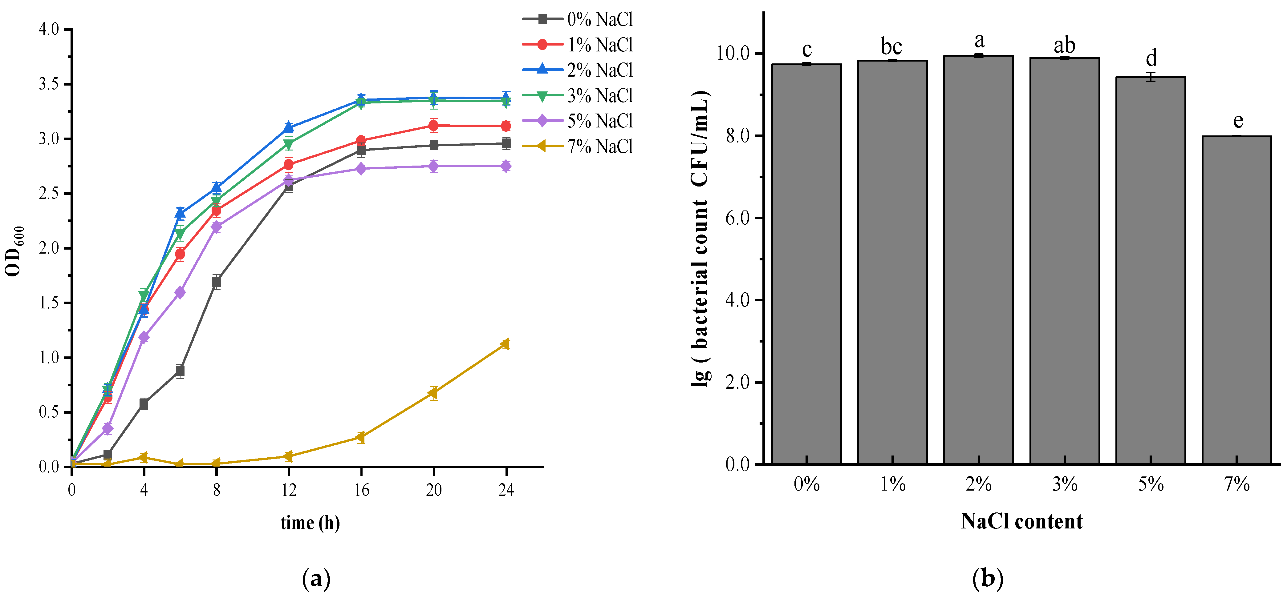
3.1. Effect of Salt Stress on Growth and Survival of V. brasiliensis

To study the effect of salt stress on the growth and survival of V. brasiliensis, growth rates of bacteria cultured in medium containing various NaCl concentrations are shown in Figure 1a. It was found that V. brasiliensis can survive without NaCl, as well as salinity as high as 7%. Previous experiments found that when salinity reached 9% or above, growth of V. brasiliensis was inhibited completely (Data not shown). Different strains of Vibrio spp. exhibited different salt tolerance. For example, pathogenic V. parahaemolyticus, a moderately halophilic and salt-requiring microorganism, cannot reproduce in a salt-free environment, but can grow in NaCl concentrations up to 8% [21].
Under salt conditions at 1% NaCl, 2% NaCl, and 3% NaCl, there was almost no lag phase after inoculation of V. brasiliensis, indicating that the strains quickly adapted to the new environment and began to grow exponentially. In addition, the log phase growth of V. brasiliensis was faster in 2% NaCl and 3% NaCl medium. However, for the cells grown in 0%, 5% and 7% NaCl, lag phase of the cells was longer, especially the cells cultured in 7% NaCl, it entered the logarithmic growth phase 12 h after inoculation, as seen in other Vibrio genus bacteria under salt stress [14]. This indicated that salinity had a certain effect on the adaptation of V. brasiliensis to the environment, making it stay longer in the adjustment period and grow slowly in the logarithmic phase. NaCl plays an important role in maintaining the osmotic pressure balance of cells. These findings suggested that lower salinities (2–3% NaCl) are most suitable or the growth and survival of V. brasiliensis. During salt stress when salt concentration is low, cells are in low osmotic pressure environment, causing the transportation of key substances exchanged between the cells and the environment cannot be reached since cell growth is affected. When salt concentration in the culture was 7%, cells were under high osmotic pressure conditions, material transportation were hindered, intracellular enzyme activity was reduced, metabolism was slow, and cell growth was restricted. The results showed that bacteria are able to adapt to survival in salt stress, suggesting that there should be a response regulation system to adjust different osmotic stresses [14]. The cell enumeration of V. brasiliensis cultivated in different NaCl content is shown in Figure 1b. Similar to the growth curve results, bacteria cultured in 2% and 3% NaCl medium showed the highest survival ability. The cell number of V. brasiliensis grew in culture medium with addition of 1%, 5% NaCl, and 7% NaCl were reduced significantly (p < 0.05). Additionally, the survival ability of cell cultured in 7% NaCl was reduced, mainly due to inhibition of cell growth in high salt condition. Previous studies using different concentrations of NaCl to culture V. parahaemolyticus showed that the survival ability of Vibrio gradually decreased with increasing (>6% NaCl) or decreasing (<1.0% NaCl) salinity [30]. And some studies shown that different strains of V. cholerae can tolerate the different maximum NaCl concentration. Growth curves shown that most of the strains can growth well in 0.5% to 5% NaCl. However, some strains barely grow or have a noticeable growth delay in 5% and 6% NaCl [31].

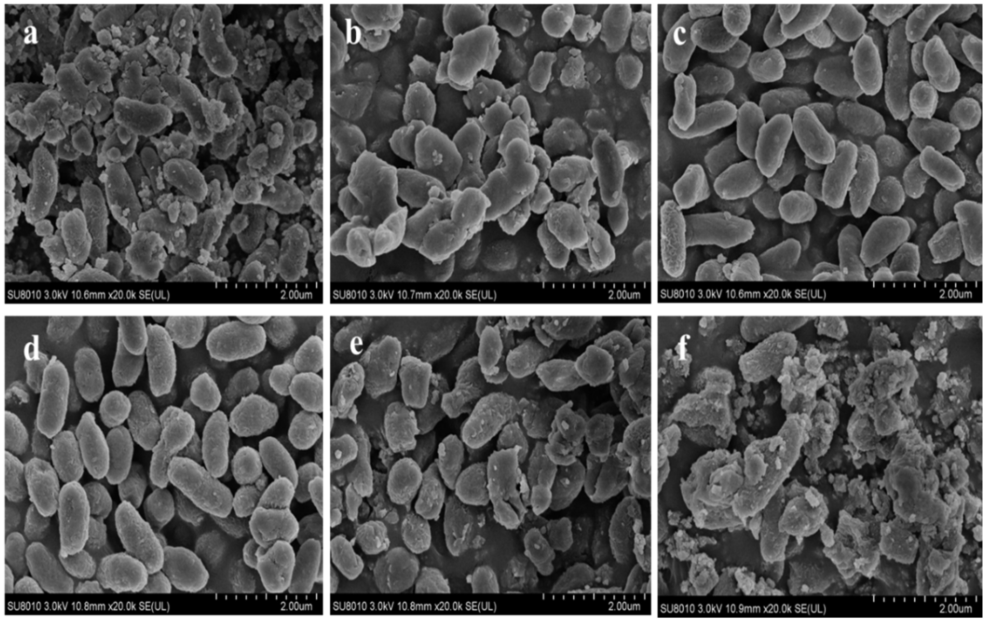
3.2. Morphological Analysis of V. brasiliensis in Response to Salt Stress

Scanning electron micrograph (SEM) revealed that the 2% NaCl and 3% NaCl content medium cultivated V. brasiliensis were mainly short rods, but 2% NaCl cultured cells surface was relatively smoother and exhibited better cell integrity than 3% NaCl (Figure 2c,d). Bacteria cultured in medium without NaCl, most cells had lost their integrity, resulting in cell debris as shown in Figure 2a. When the cells are in a low osmotic pressure environment, the cells tend to swell and break due to water absorption. However, significant structural changes, including rapture of cells and deformation of cells were observed after cultured in 1% NaCl (Figure 2b). When cultured in medium containing 5% and 7% NaCl, the cells lost their original shape and cells appeared to be lysed and ruptured to varying degrees, (Figure 2e). The medium containing 7% NaCl has led to complete cellular deformation (Figure 2f).

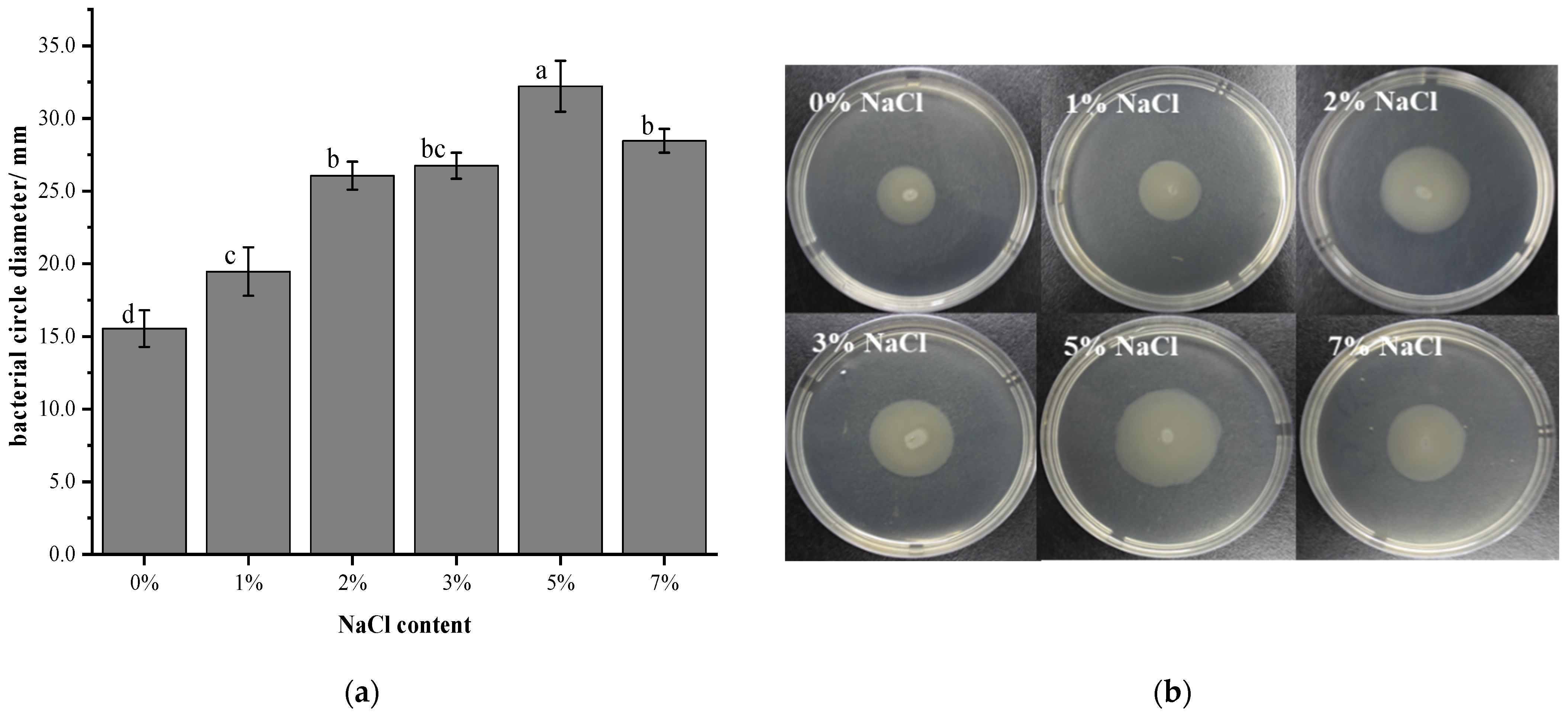
3.3. Effect of Salt Stress on the Swimming Ability of V. brasiliensis

Swimming motility of V. brasiliensis affected by salt stress were determined by measuring the diameter of the circle on the agar plate. Results in Figure 3 showed that bacteria cultured in 0% NaCl had the slowest motility among the test groups, and as the concentration of NaCl increased from 0% to 5%, the diameter of the bacterial circle also increased gradually. However, when the NaCl concentration was greater than 5%, the diameter was decreased. In addition, the diameter of the circle formed by V. brasiliensis grown in 7% NaCl medium was higher than 2%, 3% NaCl, and the bacterial circle diameter of the bacteria grown in 2% NaCl was less than that cultured in 3% NaCl, but there are no significant differences between them (p > 0.05). This result indicated that the concentration of NaCl has an effect on the swimming ability of V. brasiliensis. When grown in low NaCl concentration condition, the swimming ability of V. brasilienis was poor. Previous studies have shown that the V. salmonicida cells grown in 2.5% NaCl possess a higher motility than thsoe grown at 1% NaCl [32].
The effect of NaCl on the swimming ability of V. brasiliensis is mainly caused by the difference of Na+ concentration [33]. Studies indicating that Na+ drives the flagellum of V. cholerae, V. parahaemolyticus [34] and V. alginolyticus [35], mainly due to that the swimming ability of bacteria that use Na+ to drive their flagellar motors. Without NaCl, these is not enough sodium ion to drive their flagellar motors, which is the reason why V. brasiliensis has diminished swimming motility. However, the swimming ability of bacteria cultured with 3%, 5%, and 7% NaCl medium are higher than that of bacteria cultured with 2% NaCl. This may be due to more Na+ has led to swimming toward favorable environments in media through reversibly rotating the flagellar filaments [36].

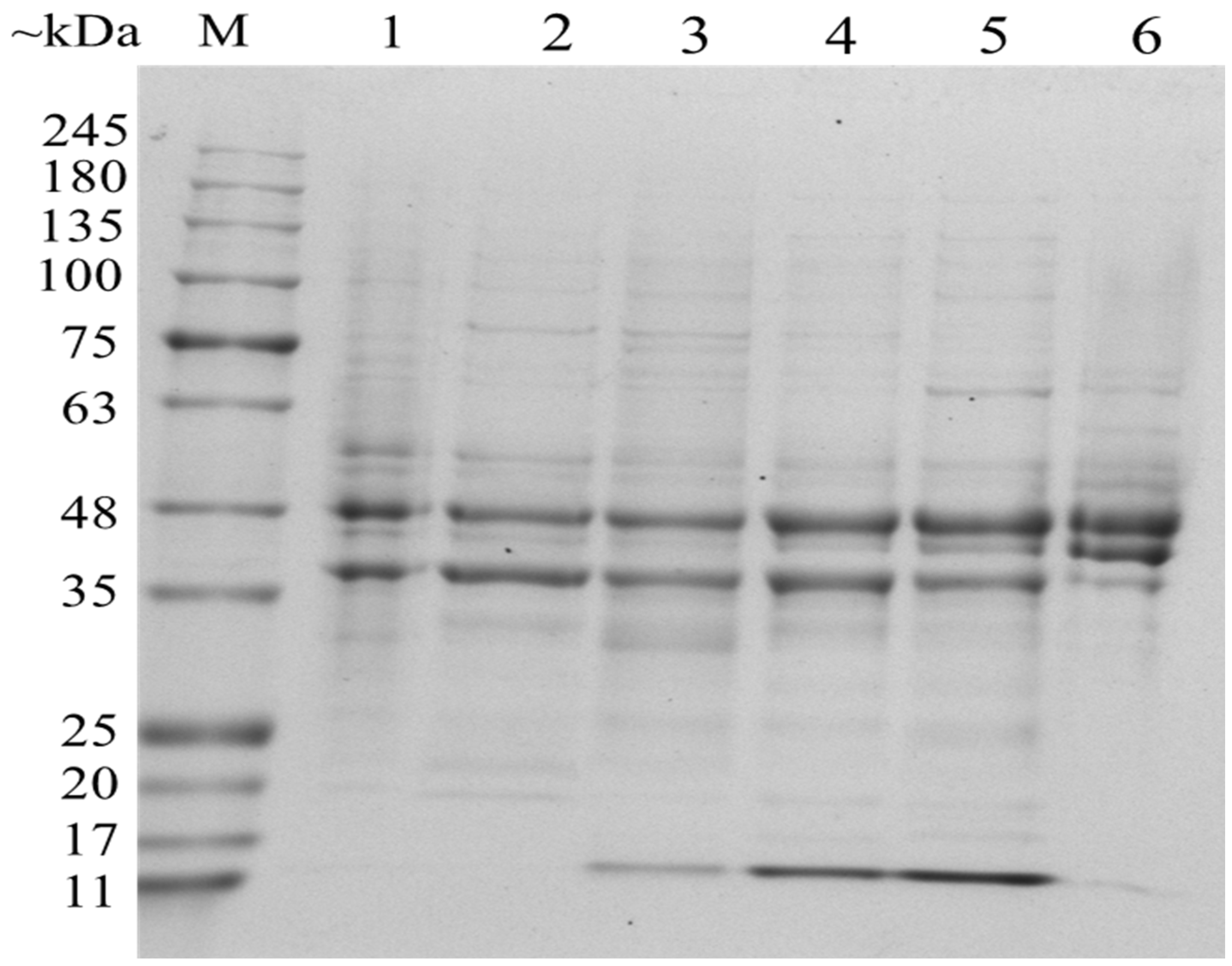
3.4. Analysis of Differentially Expressed OMPs in Response to Salt Stress

OMPs are special proteins that only exist in Gram-negative bacteria; they are composed of lipopolysaccharides (LPS), phospholipids (PLs), outer membrane proteins (OMPs) and lipoproteins (Lpp) [37]. OMPs play a vital role in material transportation of Gram-negative bacteria, ensuring the integrity of the outer membrane, sensing changes in the external environment, and signal transduction [38]. To examine the effect of NaCl on protein expression of the OMPs, SDS-PAGE electrophoresis was performed to analyze protein profiles of V. brasiliensis cultured in various NaCl concentration. As shown in Figure 4, two major protein bands with molecular weight in the range between 35 and 48 kDa (~48 kDa and ~37 kDa) with similar concentration and distribution have been expressed in most of the cultures, other than in cultures with 7% NaCl. The most obvious differences are expression of proteins around 15 kDa of V. brasiliensis cultured in 2%, 3%, and 5% NaCl, as well as the expression of protein with molecular weight around 55 kDa in cultures without NaCl. In addition, OMPs with molecular weight around 45 kDa cultured in 7% NaCl have higher protein expression than other NaCl concentration.
Identification of differentially expressed OMPs was performed by mass spectrometry. Some conserved OMPs were found (Table S1), the expression of these OMPs not affected by NaCl concentration, that is, these OMPs of V. brasiliensis are all expressed under various NaCl medium. Furthermore, there are OMPs that have porin activity in these conserved proteins, for example, outer membrane protein OmpU (VIBR0546_08029), outer membrane protein OmpA (VIBR0546_09484), Vitamin B12 transporter BtuB (btuB) and outer membrane protein (VIBR0546_02985). Among them, the OmpU is a major component of outer membrane, accounting for 60% of the outer membrane proteins [39]. Their conservation in Vibrio spp. suggesting it is importance to cell’s physiological processes. Previous studies suggesting that the OmpU seems to act as a sensor component in a signal transduction pathway in Vibrio [40]. But the synthesis of the OmpU protein is regulated by NaCl content of the cultured medium, OmpU accounts for 30% of the total outer membrane protein when V. cholerae cultured in 1.0% NaCl medium and 60% in without NaCl [39]. Besides, the outer membrane protein OmpK with nucleoside transmembrane transport protein activity is highly conserved in Vibrio, which has been recognized [41]. There have some proteins with efflux transmembrane transporter activity also conserved in various NaCl stress cultured, such as outer membrane channel protein (tolC), outer membrane protein (VIBR0546_13282), and outer membrane protein (VIBR0546_20323) that have not been reported.
However, the OMPs of V. brasiliensis changed when grown in different NaCl content medium, suggesting the bacterium can rapidly responding to salt stress through changes in outer membrane proteins. The results in Table 1 show that under NaCl stress conditions V. brasiliensis may need to regulate substances transport through special expression of its outer membrane proteins [42]. The expression of outer membrane proteins associated with bacterial pilus is affected by NaCl stress, and we found that Flp pilus assembly protein CpaB (VIBR0546_18016) and MSHA biogenesis protein MshL (VIBR0546_05478) only expressed when grown in 2% NaCl. The penicillin-binding protein activator LpoA was found in 2% NaCl and 3% NaCl medium cultured V. brasiliensis, respectively, which participated in the peptidoglycan biosynthetic process of bacterial and it was important for maintenance of cell shape and survival [43]. Such result explained that V. brasiliensis have typical Vibrio morphology when bacteria grown in the 2% NaCl and 3% NaCl medium. It should be pointed out that the outer membrane protein of sodium-type flagellar protein MotY (VIBR0546_10974), the component of the polar flagellar rotary motor is found only when the bacteria grown in 3%, 5%, and 7% NaCl. It is noteworthy that ferric aerobactin receptor protein was only found in 7% NaCl cultured bacteria’s outer membrane proteins, this may be due to that high salt stress affects iron absorption, and the expression of ferric aerobactin receptor protein is activated to improve iron utilization of the cells under high salt stress environment [44,45]. In addition, the putative long-chain fatty acid transport protein (VIBR0546_07642) has disappeared when bacteria cultivated in 5% and 7% NaCl stress medium, on the contrary, this protein has appeared when V. parahaemolyticus and V. alginolyticus were treated with Ultraviolet-C radiation stress [46]. Therefore, the OMPs that changes in salt stress can be considered as response salt stress proteins in V. brasiliensis and types of protein expressed in response to salt stress are different.

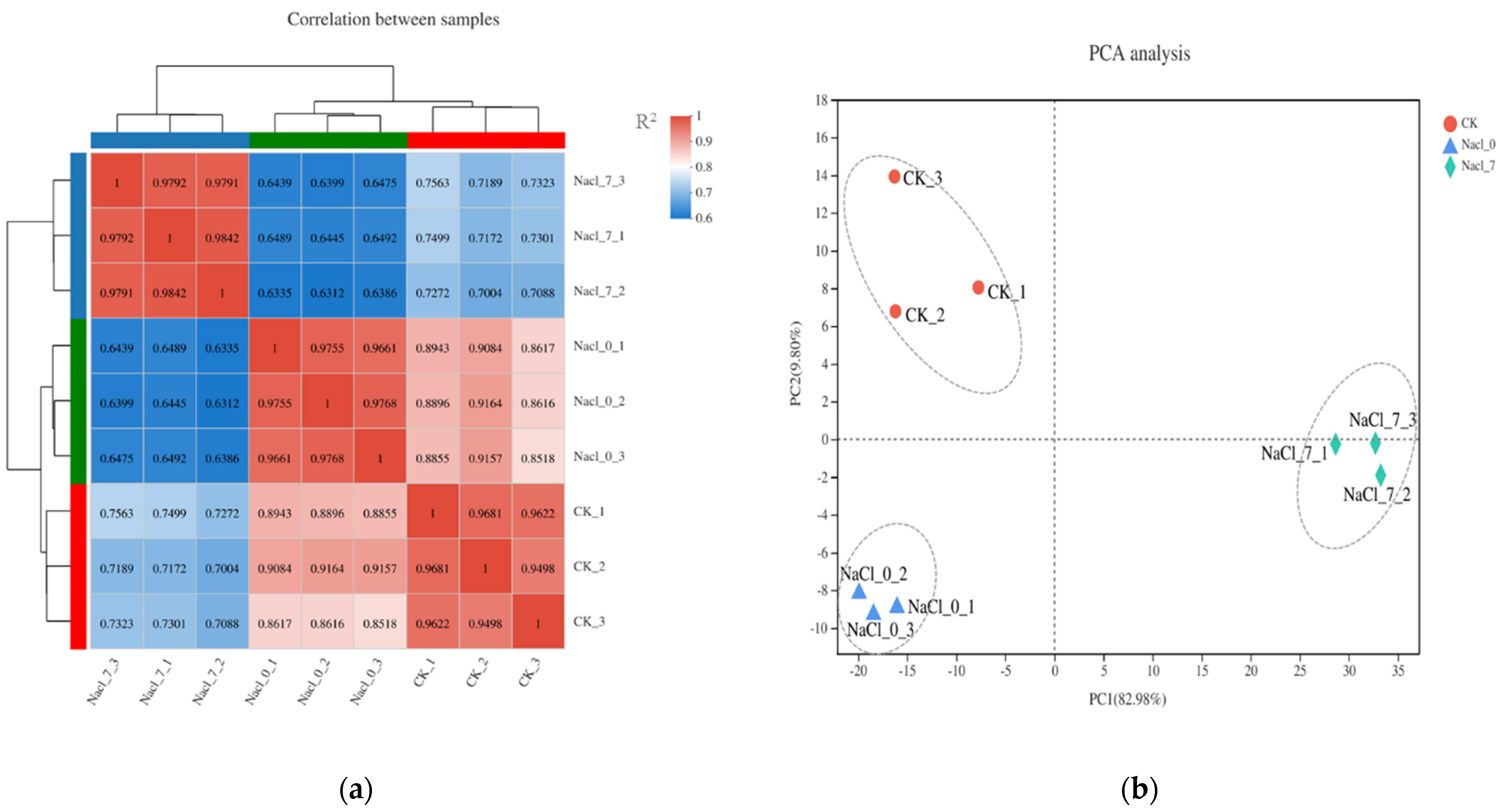
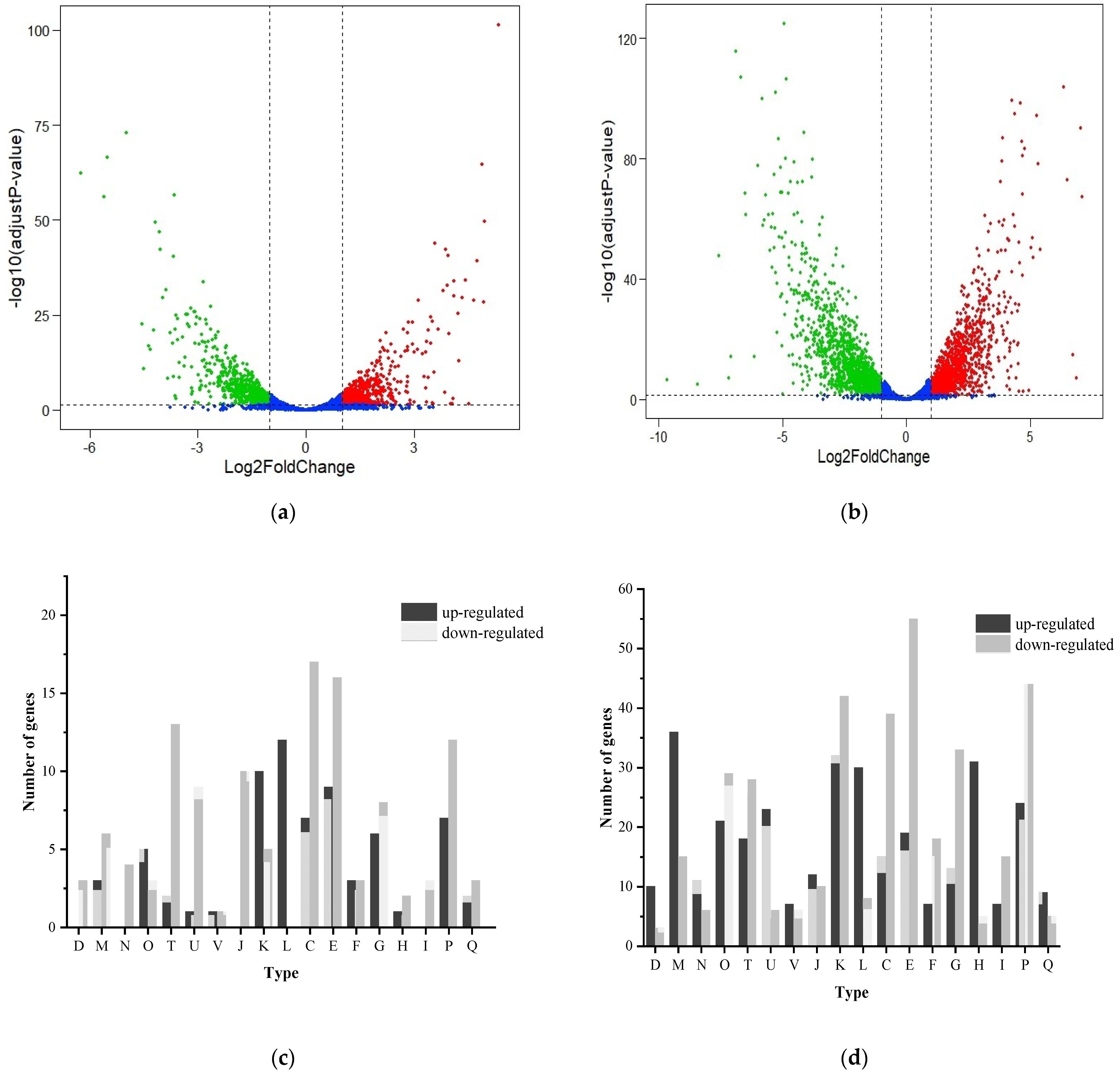
3.5. Transcriptome Analysis

It can be seen from Figure 5a that the correlation coefficient of CK (2% NaCl) of the control group samples were greater than 0.95, the correlation coefficient of the experimental group Nacl_0 (0% NaCl) samples were greater than 0.96, and the correlation coefficient of the experimental group Nacl_7 (7% NaCl) samples were greater than 0.98. The correlation coefficients of the repeated samples in the above groups were all above 0.95, indicated that the three biological replicate samples in each group had high similarity, therefore the transcription and sequencing results of the samples in each group should be reliable. In addition, the correlation coefficient of samples between different groups was relatively small, demonstrating that NaCl concentration had affected gene expression levels within V. brasiliensis. Similarly, PCA analysis in Figure 5b shows the repeated samples of each group have good correlation, which indicates the experimental data of V. brasiliensis transcriptome cultured in different NaCl are highly accurate.
The V. brasiliensis makes a series of responses to changes in the external environment by regulating its own gene expression. We performed transcriptome analysis of V. brasiliensis which cultured under 0% NaCl and 7% NaCl respectively, to isolate genes associated with salt stress. Compared to V. brasiliensis cultured in 2% NaCl, 1230 genes (577 up-regulated and 653 down-regulated) and 2504 genes (1215 up-regulated and 1289 down-regulated) were observed after 0% NaCl and 7% NaCl treatments, respectively (Figure 6a,b, Tables S2 and S3). It is interesting to note that the number of differentially expressed genes in 7% NaCl was significantly higher than in 0% NaCl, suggesting that when V. brasiliensis grown in 7% NaCl medium, this high concentration salt environment will generate greater stress to cell growth so that the bacteria by up-regulated and down-regulated self-genes in response to osmotic stress caused by high salt [31]. In order to describe the types and functions of genes with significant transcriptional changes more intuitively, COG function annotation analysis (Figure 6c,d) shows that the genes related to replication, recombination and repair of V. brasiliensis in 0% NaCl and 7% NaCl are notably up-regulated, indicating that under the stress of low or high osmotic stress, bacteria can activate their own repair mechanism to response the unfavorable environment. In addition, for genes involved in signal transduction mechanisms, cell motility, energy production and transformation, amino acid transport and metabolism, carbohydrate transport and metabolism, inorganic ion transport and metabolic functions, and the number of down-regulated genes was higher than those of up-regulated genes. This may be due to cells in unfavorable growth environment and most of the metabolism in the cell is blocked. To further obtain the genes that respond to salt stress, we integrated the genes with transcriptional changes and found that two sigma factors, including the RNA polymerase sigma factor RpoS (VIBR0546_05309) and putative anti-sigma F factor antagonist (VIBR0546_10019) were down-regulated in 0% NaCl and 7% NaCl stress. The anti-RNA polymerase sigma 70 factor down-regulated in 7% NaCl, showed in high osmotic stress the RNA polymerase sigma 70 factor can function normally (Table S4). In Gram-negative bacteria, the RNA polymerase sigma 70 factor plays a key role in responses to osmotic stress [47,48].
However, bacteria in low and high osmotic environment have distinct responses stress mechanism, through analysis the differentially expressed genes (selection criteria: Abs (log2FC) ≥ 1, p < 0.05) related to stress (Table 2 and Table 3). Changes in expression levels of salt stress-response genes in V. brasiliensis were concentration-specific, suggesting that the bacteria stress response system is complicated. We found that the same gene have opposite up-regulated and down-regulated expression in two treatments, indicate that this type of gene is regulated differently in low and high osmotic stress. The osmolarity response regulator (ompR), universal stress protein VspE, universal stress protein family 8, universal stress protein A, and NptA protein genes were all up-regulated in 0% NaCl medium but down-regulated in 7% NaCl, demonstrates that these genes can response to the low osmotic pressure environment outside the cell, which are conducive to the survival of bacteria in the low osmotic stress conditions. NptA protein gene (VIBR0546_00894) have phosphate ion transmembrane transporter activity and sodium: phosphate symporter activity function and involved in sodium-dependent phosphate transport process. Besides, previous studies of NptA in V. cholerae suggested that this protein may contribute to V. cholerae colonization in host [49], and further research has determined the stress response of NptA that contributes to the growth or stress resistance in V. vulnificus [50]. Up-regulation of such genes was in response to 0% NaCl stress. However, it was down-regulated in 7% NaCl conditions might show that this gene was inhibited under a higher osmotic pressure, and for that reason the growth and viability of V. brasiliensis are significantly reduced. In addition, there are some universal stress protein genes that have the same expressive characteristics as the genes mentioned above, which are up-regulated in 0% NaCl medium but are down-regulated under high osmotic pressure of 7% NaCl. The universal stress protein UspE gene, encode protein belongs to the universal stress proteins (USP) family. The UspE protein is important for cellular motility, and study demonstrates that uspE mutants are devoid of flagella in E. coli [51], and the UspE proteins enable cells to defense and escape environmental stress.
As shown previously, NaCl concentrations have influence on outer membrane protein gene transcription level, we found that expression of genes encoding outer membrane protein W (OmpW) (VIBR0546_07217), outer membrane protein OmpA (VIBR0546_09484), and outer membrane protein OmpK (VIBR0546_05598) were all down-regulated in 0% and 7% NaCl conditions. The ability in salt-tolerance was increased in E. coli overexpressed OmpW, the finding suggests that OmpW is necessary for environmental salt regulation in bacteria [52]. Previous studies have shown that bacteria in different salt stress can response to the cells osmotic imbalance through transporter system. The Na+/H+ antiporter, which is a sodium ion channel commonly used by cells and generate the electrochemical potential for the Na+ driven flagella motor by extruding Na+. The key gene, motY (VIBR0546_10974), for the rotation of the Na+ driven motor was found to be up-regulated in high NaCl (7% NaCl) medium, and the sodium-type flagella protein MotY was found in 3%, 5%, and 7% NaCl cultured bacteria’s outer membrane proteins, respectively. Furthermore, the Na+/H+ antiporter gene (VIBR0546_21150) was up-regulated as well, so when the bacteria cultured under higher NaCl concentrations, the higher Na+ concentration in the environment will give cell higher swimming ability. However, when the NaCl concentration is too high, it will cause fatal stress to growth and survival of V. brasiliensis, which diminishes its swimming ability. Although putative Na+/H+ antiporter gene (VIBR0546_01129) expression was also up-regulated in 0% NaCl cultured V. brasiliensis, the up-regulation of motY gene and the presence of sodium-type flagella protein MotY were not found, indicating that the Na+/H+ antiporter ultimately drives flagella rotation though the MotY protein [35].

4. Conclusions

Based on the different NaCl concentrations on the growth rate, survival, cell swimming ability, cell outer membrane proteins, and the transcriptome of V. brasiliensis were caried out to investigate the salt stress to the bacteria and cells responses to stress in this study. The results showed that 0–5% NaCl concentration had little effect on V. brasiliensis growth and survival, but 7% NaCl had a great influence on the bacteria. The NaCl concentration of the medium has an effect on the cellular morphology of V. brasiliensis, and the cells’ morphology were severely damaged under low-salt and high-salt stress cultures. The Penicillin-binding protein activator LopA OMP was only found in the cells cultured in 2% and 3% NaCl, and the protein contributed to maintaining the integrity of the cells. The OMP of Sodium-type flagellar protein MotY was found in the cells cultured with 3%, 5%, and 7% NaCl, indicating that V. brasiliensis produced this protein in response to external high-salt stress. Furthermore, some key genes in response to salt stress were discovered, such as NptA protein gene (VIBR0546_00894), osmolarity response regulator gene (ompR), putative threonine efflux protein gene (VIBR0546_17048) and Na+/H+ antiporter gene (VIBR0546_21150) and so on. For survival, V. brasiliensis responds to salt stress through differential gene expression. This study is conducive to understanding how V. brasiliensis responds to salt stress in the environment.

Supplementary Materials

The following supporting information can be downloaded at: https://www.mdpi.com/article/10.3390/foods11060840/s1, Table S1: OMPs of V. brasiliensis all expressed in different NaCl concentrations, Table S2: Stress-related differentially expressed genes screened by V. brasiliensis cultured in 0% NaCl. (Screening criteria: Abs (log2FC) ≥ 1, p < 0.05), Table S3: Stress-related differentially expressed genes screened by V. brasiliensis cultured in 7% NaCl. (Screening criteria: Abs (log2FC) ≥ 1, p < 0.05), Table S4: Differentially expressed sigma factors of V. brasiliensis cultured with different NaCl concentrations in transcriptome analysis.

Author Contributions

Conceptualization, X.J.; data curation, S.H.; funding acquisition, X.J.; investigation, S.H., Y.L., B.W. and X.J.; methodology, S.H. and X.J.; project administration, X.J.; resources, L.Y., Y.L., B.W. and X.J.; software, S.H.; supervision, L.Y. and X.J.; validation, S.H., Y.L. and B.W.; visualization, S.H. and X.J.; writing—original draft, S.H.; writing—review and editing, S.H., Y.L., B.W., L.Y. and X.J. All authors have read and agreed to the published version of the manuscript.

Funding

This work was funded by National Nature Science Foundation of China (No. 31901790).

Data Availability Statement

Data are contained within the article.

Conflicts of Interest

The authors declare no competing financial or other interest.

References

  1. Hernandez-Perez, A.; Soderhall, K.; Sirikharin, R.; Jiravanichpaisal, P.; Soderhall, I. Vibrio areninigrae as a pathogenic bacterium in a crustacean. J. Invertebr. Pathol. 2021, 178, 107517. [Google Scholar] [CrossRef] [PubMed]
  2. Osunla, C.A.; Okoh, A.I. Vibrio Pathogens: A public health concern in rural water resources in Sub-Saharan Africa. Int. J. Environ. Res. Public Health 2017, 14, 1188. [Google Scholar] [CrossRef] [Green Version]
  3. Taylor, M.; Cheng, J.; Sharma, D.; Bitzikos, O.; Gustafson, R.; Fyfe, M.; Greve, R.; Murti, M.; Stone, J.; Honish, L.; et al. Outbreak of Vibrio parahaemolyticus associated with consumption of raw Oysters in Canada, 2015. Foodborne Pathog. Dis. 2018, 15, 554–559. [Google Scholar] [CrossRef] [PubMed]
  4. Ha, S.M.; Chalita, M.; Yang, S.J.; Yoon, S.H.; Cho, K.; Seong, W.K.; Hong, S.; Kim, J.; Kwak, H.S.; Chun, J. Comparative genomic analysis of the 2016 Vibrio cholerae outbreak in South Korea. Front. Public Health 2019, 7, 228. [Google Scholar] [CrossRef] [Green Version]
  5. Parola, P.; Anani, H.; Eldin, C.; Dubourg, G.; Lagier, J.C.; Levasseur, A.; Casalta, J.P.; Raoult, D.; Fournier, P.E. Case Report: Vibrio cholerae biliary tract infections in two North Africans in France. Am. J. Trop. Med. Hyg. 2020, 102, 1306–1308. [Google Scholar] [CrossRef]
  6. Baker-Austin, C.; Oliver, J.D. Vibrio vulnificus. Trends Microbiol. 2020, 28, 81–82. [Google Scholar] [CrossRef]
  7. Yoon, S.H.; Waters, C.M. Vibrio cholerae. Trends Microbiol. 2019, 27, 806–807. [Google Scholar] [CrossRef] [PubMed]
  8. Cornejo-Granados, F.; Gallardo-Becerra, L.; Leonardo-Reza, M.; Ochoa-Romo, J.P.; Ochoa-Leyva, A. A meta-analysis reveals the environmental and host factors shaping the structure and function of the shrimp microbiota. PeerJ 2018, 6, e5382. [Google Scholar] [CrossRef] [PubMed]
  9. Mishra, P.; Samanta, M.; Mohanty, S.; Maiti, N.K. Characterization of Vibrio species isolated from freshwater fishes by ribotyping. Indian J. Microbiol. 2010, 50, 101–103. [Google Scholar] [CrossRef] [Green Version]
  10. Duche, O.; Tremoulet, F.; Glaser, P.; Labadie, J. Salt stress proteins induced in Listeria monocytogenes. Appl. Environ. Microbiol. 2002, 68, 1491–1498. [Google Scholar] [CrossRef] [PubMed] [Green Version]
  11. Lu, H.; Cao, W.; Hong, J.; Longwei, Z. Contamination status of Vibrio Parahaemolyticus in foods of wet markets in Chongming County of Shanghai. Occup. Health 2010, 26, 409–410. [Google Scholar] [CrossRef]
  12. Zhou, L.; Liu, H.; Zhan, W.; Song, W. Isolation and characteristics of major outer membrane proteins of aquatic pathogens Vibrio anguillarum and Vibrio alginolyticus. J. Fish. Sci. China 2003, 10, 31–35. [Google Scholar]
  13. Marchesi, V.T.; Andrews, E.P. Glycoproteins-isolation from cell membranes with lithium diiodosalicylate. Science 1971, 174, 1247–1248. [Google Scholar] [CrossRef] [PubMed]
  14. Kao, D.Y.; Cheng, Y.C.; Kuo, T.Y.; Lin, S.B.; Lin, C.C.; Chow, L.P.; Chen, W.J. Salt-responsive outer membrane proteins of Vibrio anguillarum serotype 01 as revealed by comparative proteome analysis. J. Appl. Microbiol. 2009, 106, 2079–2085. [Google Scholar] [CrossRef] [PubMed]
  15. Xu, C.G.; Wang, S.Y.; Ren, H.X.; Lin, X.M.; Wu, L.N.; Peng, X.X. Proteomic analysis on the expression of outer membrane proteins of Vibrio alginolyticus at different sodium concentrations. Proteomics 2005, 5, 3142–3152. [Google Scholar] [CrossRef] [PubMed]
  16. Xu, C.X.; Ren, H.X.; Wang, S.Y.; Peng, X.X. Proteomic analysis of salt-sensitive outer membrane proteins of Vibrio parahaemolyticus. Res. Microbiol. 2004, 155, 835–842. [Google Scholar] [CrossRef]
  17. Jia, X.; Wang, J.B.; Rivera, S.; Duong, D.; Weinert, E.E. An O-2-sensing stressosome from a Gram-negative bacterium. Nat. Commun. 2016, 7, 12381. [Google Scholar] [CrossRef] [PubMed]
  18. Kalburge, S.S.; Whitaker, W.B.; Boyd, E.F. High-salt preadaptation of Vibrio parahaemolyticus enhances survival in response to lethal environmental stresses. J. Food Prot. 2014, 77, 246–253. [Google Scholar] [CrossRef]
  19. Xiong, X.P.; Zhang, B.W.; Yang, M.J.; Ye, M.Z.; Peng, X.X.; Li, H. Identification of vaccine candidates from differentially expressed outer membrane proteins of Vibrio alginolyticus in response to NaCl and iron limitation. Fish Shellfish Immunol. 2010, 29, 810–816. [Google Scholar] [CrossRef]
  20. Xu, C.X.; Wang, S.Y.; Wu, L.N.; Ren, H.X.; Peng, X.X. Proteomic analysis on the expression of the outer membrane proteins of Vibrio alginolyticus at different Na+ concentrations. Mol. Cell. Proteom. 2004, 3, S161. [Google Scholar]
  21. Yang, L.; Zhan, L.; Han, H.; Gao, H.; Guo, Z.; Qin, C.; Yang, R.; Liu, X.; Zhou, D. The low-salt stimulon in Vibrio parahaemolyticus. Int. J. Food Microbiol. 2010, 137, 49–54. [Google Scholar] [CrossRef]
  22. Arias, C.R.; Lafrentz, S.; Cai, W.; Olivares-Fuster, O. Adaptive response to starvation in the fish pathogen Flavobacterium columnare: Cell viability and ultrastructural changes. BMC Microbiol. 2012, 12, 266. [Google Scholar] [CrossRef] [PubMed] [Green Version]
  23. Ringgaard, S.; Zepeda-Rivera, M.; Wu, X.; Schirner, K.; Davis, B.M.; Waldor, M.K. ParP prevents dissociation of CheA from chemotactic signaling arrays and tethers them to a polar anchor. Proc. Natl. Acad. Sci. USA 2014, 111, E255–E264. [Google Scholar] [CrossRef] [PubMed] [Green Version]
  24. Zhang, Y.B.; Liu, H.H.; Gu, D.; Lu, X.X.; Zhou, X.H.; Xia, X.D. Transcriptomic analysis of PhoR reveals its role in regulation of swarming motility and T3SS expression in Vibrio parahaemolyticus. Microbiol. Res. 2020, 235, 126448. [Google Scholar] [CrossRef] [PubMed]
  25. Gao, L.; Ouyang, M.; Zhang, H.; Rao, S.; Yin, Y. Biological characteristics of Vibrio parahaemolyticus during growth in adverse environment. Food Sci. 2018, 39, 177–182. [Google Scholar] [CrossRef]
  26. Lutwyche, P.; Exner, M.M.; Hancock, R.E.W.; Trust, T.J. A conserved Aeromonas-salmonicida porin provides protective immunity to rainbow-trout. Infect. Immun. 1995, 63, 3137–3142. [Google Scholar] [CrossRef] [Green Version]
  27. Levin, J.Z.; Yassour, M.; Adiconis, X.; Nusbaum, C.; Thompson, D.A.; Friedman, N.; Gnirke, A.; Regev, A. Comprehensive comparative analysis of strand-specific RNA sequencing methods. Nat. Methods 2010, 7, 709–715. [Google Scholar] [CrossRef] [Green Version]
  28. Parkhomchuk, D.; Borodina, T.; Amstislavskiy, V.; Banaru, M.; Hallen, L.; Krobitsch, S.; Lehrach, H.; Soldatov, A. Transcriptome analysis by strand-specific sequencing of complementary DNA. Nucleic Acids Res. 2009, 37, e123. [Google Scholar] [CrossRef] [PubMed] [Green Version]
  29. Gao, X.J.; Jiang, Z.Y.; Zhang, S.M.; Chen, Q.Y.; Tong, S.Q.; Liu, X.D.; Jiang, Q.; Yang, H.; Wei, W.H.; Zhang, X.J. Transcriptome analysis and immune-related genes expression reveals the immune responses of Macrobrachium rosenbergii infected by Enterobacter cloacae. Fish Shellfish Immun. 2020, 101, 66–77. [Google Scholar] [CrossRef] [PubMed]
  30. Shen, J.Y.; Ni, Y.; Gao, L. Biological characteristics of Vibrio parahaemolyticus growing in the medium of different salinity. Mod. Food 2018, 6, 91–94. [Google Scholar] [CrossRef]
  31. Fu, X.P.; Liang, W.L.; Du, P.C.; Yan, M.Y.; Kan, B. Transcript changes in Vibrio cholerae in response to salt stress. Gut Pathog. 2014, 6, 47. [Google Scholar] [CrossRef] [PubMed] [Green Version]
  32. Karlsen, C.; Paulsen, S.M.; Tunsjo, H.S.; Krinner, S.; Sorum, H.; Haugen, P.; Willassen, N.P. Motility and flagellin gene expression in the fish pathogen Vibrio salmonicida: Effects of salinity and temperature. Microb. Pathog. 2008, 45, 258–264. [Google Scholar] [CrossRef]
  33. Boehm, A.; Kaiser, M.; Li, H.; Spangler, C.; Kasper, C.A.; Ackermann, M.; Kaever, V.; Sourjik, V.; Roth, V.; Jenal, U. Second messenger-mediated adjustment of bacterial swimming velocity. Cell 2010, 141, 107–116. [Google Scholar] [CrossRef] [PubMed] [Green Version]
  34. Atsumi, T.; McCartert, L.; Imae, Y. Polar and lateral flagellar motors of marine Vibrio are driven by different ion-motive forces. Nature 1992, 355, 182–184. [Google Scholar] [CrossRef] [PubMed]
  35. Yorimitsu, T.; Homma, M. Na+-driven flagellar motor of Vibrio. Biochim. Biophys. Acta (BBA)-Bioenerg. 2001, 1505, 82–93. [Google Scholar] [CrossRef] [Green Version]
  36. Zhu, S.; Kojima, S.; Homma, M. Structure, gene regulation and environmental response of flagella in Vibrio. Front. Microbiol. 2013, 4, 410. [Google Scholar] [CrossRef] [Green Version]
  37. Konovalova, A.; Kahne, D.E.; Silhavy, T.J. Outer membrane biogenesis. Annu. Rev. Microbiol. 2017, 71, 539–556. [Google Scholar] [CrossRef] [PubMed]
  38. Jeeves, M.; Knowles, T.J. A novel pathway for outer membrane protein biogenesis in Gram-negative bacteria. Mol. Microbiol. 2015, 97, 607–611. [Google Scholar] [CrossRef] [Green Version]
  39. Chakrabarti, S.; Chaudhuri, K.; Sen, K.; Das, J. Porins of Vibrio cholerae: Purification and characterization of OmpU. J. Bacteriol. 1996, 178, 524–530. [Google Scholar] [CrossRef] [PubMed] [Green Version]
  40. Mathur, J.; Davis, B.M.; Waldor, M.K. Antimicrobial peptides activate the Vibrio cholerae sigma(E) regulon through an OmpU-dependent signalling pathway. Mol. Microbiol. 2007, 63, 848–858. [Google Scholar] [CrossRef]
  41. Wang, W.; Guo, S.; Gao, Y.; Liang, X.; Liu, L.; Pan, S. Comparative immunogenicity of outer membrane protein K and whole-cell antigens of Vibrio parahaemolyticus for diagnosis. Lett. Appl. Microbiol. 2021, 73, 460–470. [Google Scholar] [CrossRef] [PubMed]
  42. Simpson, L.M.; Engle, M.; Oliver, J.D. Effect of osmotic variation on the outer-membrane proteins of Vibrio vulnificus identification of a major heat-modifiable protein. Microbios 1994, 80, 209–214. [Google Scholar] [PubMed]
  43. Dorr, T.; Lam, H.; Alvarez, L.; Cava, F.; Davis, B.M.; Waldor, M.K. A novel peptidoglycan binding protein crucial for PBP1A-mediated cell wall biogenesis in Vibrio cholerae. PLoS Genet. 2014, 10, e1004433. [Google Scholar] [CrossRef] [PubMed] [Green Version]
  44. Tanabe, T.; Naka, A.; Aso, H.; Nakao, H.; Narimatsu, S.; Inoue, Y.; Ono, T.; Yamamoto, S. A novel aerobactin utilization cluster in Vibrio vulnificus with a gene involved in the transcription regulation of the iutA homologue. Microbiol. Immunol. 2005, 49, 823–834. [Google Scholar] [CrossRef] [PubMed]
  45. Bailey, D.C.; Drake, E.J.; Grant, T.D.; Gulick, A.M. Structural and functional characterization of aerobactin synthetase IucA from a hypervirulent pathotype of Klebsiella pneumoniae. Biochemistry 2016, 55, 3559–3570. [Google Scholar] [CrossRef]
  46. Ben Abdallah, F.; Lagha, R.; Ellafi, A.; Namane, A.; Rousselle, J.C.; Lenormand, P.; Kallel, H. Identification of outer membrane proteins altered in response to UVC-radiation in Vibrio parahaemolyticus and Vibrio alginolyticus. Indian J. Microbiol. 2012, 52, 660–665. [Google Scholar] [CrossRef] [PubMed] [Green Version]
  47. Abromaitis, S.; Koehler, J.E. The bartonella quintana extracytoplasmic function sigma factor RpoE has a role in bacterial adaptation to the arthropod vector environment. J. Bacteriol. 2013, 195, 2662–2674. [Google Scholar] [CrossRef] [Green Version]
  48. Tettmann, B.; Dotsch, A.; Armant, O.; Fjell, C.D.; Overhage, J. Knockout of extracytoplasmic function Sigma factor ECF-10 affects stress resistance and biofilm formation in Pseudomonas putida KT2440. Appl. Environ. Microbiol. 2014, 80, 4911–4919. [Google Scholar] [CrossRef] [PubMed] [Green Version]
  49. Lebens, M.; Lundquist, P.; Soderlund, L.; Todorovic, M.; Carlin, N.I.A. The nptA gene of Vibrio cholerae encodes a functional sodium-dependent phosphate cotransporter homologous to the type II cotransporters of eukaryotes. J. Bacteriol. 2002, 184, 4466–4474. [Google Scholar] [CrossRef] [PubMed] [Green Version]
  50. Staley, C.; Harwood, V.J. Differential expression of a sodium-phosphate cotransporter among Vibrio vulnificus strains. Microb. Ecol. 2014, 67, 24–33. [Google Scholar] [CrossRef]
  51. Nachin, L.; Nannmark, U.; Nystrom, T. Differential roles of the universal stress proteins of Escherichia coli in oxidative stress resistance, adhesion, and motility. J. Bacteriol. 2005, 187, 6265–6272. [Google Scholar] [CrossRef] [PubMed] [Green Version]
  52. Wu, L.N.; Lin, X.M.; Wang, F.P.; Ye, D.Z.; Xiao, X.; Wang, S.Y.; Peng, X.X. OmpW and OmpV are required for NaCl regulation in Photobacterium damsela. J. Proteome Res. 2006, 5, 2250–2257. [Google Scholar] [CrossRef] [PubMed]
Figure 1. Effect of salt concentration on growth of V. brasiliensis. (a) Growth rate of V. brasiliensis incubated in culture medium with different NaCl content. (b) Survival ability determination of the V. brasiliensis cultivated in different NaCl content medium. Values are means of three independent experiments. Different lowercase letters denote significant differences (p < 0.05).
Foods 11 00840 g001
Figure 2. Morphological of V. brasiliensis cells after cultivation in different NaCl concentrations. (a) Cells cultured in without NaCl medium; (b) cells cultured in 1% NaCl medium; (c) cells cultured in 2% NaCl medium (optimal growth NaCl content of V. brasiliensis); (d) cells cultured in 3% NaCl medium; (e) cells cultured in 5% NaCl medium; (f) cells cultured in 7% NaCl medium.
Foods 11 00840 g002
Figure 3. Swimming motility of V. brasiliensis under different NaCl concentrations. (a) 0.3% agar plates bacteria circle diameter (mm) were measured after cultivated in various NaCl medium, and the value is represented by three repetitions mean ± sd. Different lowercase letters denote significant differences (p < 0.05); (b) 0.3% agar medium obtained the swimming circle of V. brasiliensis cultured in various NaCl concentrations.
Foods 11 00840 g003
Figure 4. SDS-PAGE of OMPs of V. brasiliensis cultured in culture medium with different NaCl concentration. M: marker; lane 1, OMPs of V. brasiliensis cultured in 0% NaCl medium; lane 2, OMPs of V. brasiliensis cultured in 1% NaCl medium; lane 3, OMPs of V. brasiliensis cultured in 2% NaCl medium; lane 4, OMPs of V. brasiliensis cultured in 3% NaCl medium; lane 5, OMPs of V. brasiliensis cultured in 5% NaCl medium; lane 6, OMPs of V. brasiliensis cultured in 7% NaCl medium.
Foods 11 00840 g004
Figure 5. Correlation analysis between samples (a) and principal component analysis (PCA) of samples (b) of V. brasiliensis under different NaCl concentrations cultured. (a) Both the abscissa and the ordinate indicated the names of samples under different processing conditions, and each sample was set with three biological replicates. CK represent 2% NaCl concentration and as control; Nacl_0 represent 0% NaCl concentration; Nacl_7 represent 7% NaCl concentration. The shade of the square in the figure represents the correlation coefficient R2. Red indicates positive correlation, and blue indicates lower the correlation; (b) the three dots with the same color represent three biological replicates in each group.
Foods 11 00840 g005
Figure 6. The volcano plots of genes and COG functional annotations between different treatment. (a) Volcano plots displaying differential expressed genes between 0% NaCl and 2% NaCl cultured V. brasiliensis; (b) Volcano plots displaying differential expressed genes between 7% NaCl and 2% NaCl cultured V. brasiliensis. (Screening criteria for differentially expressed genes: adjust p value < 0.05, Abs (log2Fold Change) ≥ 1); (c,d) COG functional annotations of differentially expressed genes identified in 0% NaCl and 7% NaCl cultured bacteria, respectively. (Abs (log2Fold Change) ≥ 2). COG functional description represented by capital letters. D: Cell cycle control, cell division, chromosome partitioning; M: Cell wall/membrane/envelope biogenesis; N: Cell motility; O: Posttranslational modification, protein turnover, chaperones; T: Signal transduction mechanisms; U: Intracellular trafficking, secretion, and vesicular transport; V: Defense mechanisms; J: Translation, ribosomal structure and biogenesis; K: Transcription; L: Replication, recombination and repair; C: Energy production and conversion; E: Amino acid transport and metabolism; F: Nucleotide transport and metabolism; G: Carbohydrate transport and metabolism; H: Coenzyme transport and metabolism; I: Lipid transport and metabolism; P: Inorganic ion transport and metabolism; Q: Secondary metabolites biosynthesis, transport and catabolism; S: Function unknown (not shown in the picture).
Foods 11 00840 g006
Table 1. The special expression outer membrane proteins of V. brasiliensis in different NaCl concentrations cultured.
NaCl (%)AccessionGeneProteinsMw [KDa]Molecular Function
2E8LNG0lpoAPenicillin-binding protein activator LpoA 67.3enzyme regulator activity
2E8LPR8VIBR0546_18016Flp pilus assembly protein CpaB27.2NA
2E8LQ55VIBR0546_05478MSHA biogenesis protein MshL59.2NA
3E8LNG0lpoAPenicillin-binding protein activator LpoA 67.3enzyme regulator activity
3E8LS16VIBR0546_10974Sodium-type flagellar protein MotY33.5NA
5E8LS16VIBR0546_10974Sodium-type flagellar protein MotY33.5NA
5E8M0I9VIBR0546_09489Outer membrane protein OmpA35.1porin activity
7E8LS16VIBR0546_10974Sodium-type flagellar protein MotY33.5NA
7E8M0I9VIBR0546_09489Outer membrane protein OmpA35.1porin activity
7E8LX27VIBR0546_17898Ferric aerobactin receptor77.7siderophore uptake transmembrane transporter activity; signaling receptor activity
NA, it means there is no such content.
Table 2. The up and down-regulated genes of V. brasiliensis cultured in 0% NaCl medium.
Gene NameGene DescriptionFCLog2FCp ValueRegulate
VIBR0546_12042proton/glutamate symporter5.1652.3691.15 × 10−13up
nhaApH-dependent sodium/proton antiporter3.6721.8771.61 × 10−12up
VIBR0546_00894NptA protein11.8243.5645.86 × 10−12up
VIBR0546_21825putative outer membrane protein7.0402.8166.8 × 10−11up
VIBR0546_00270universal stress protein UspE2.9501.5616.62 × 10−8up
VIBR0546_01129putative Na+/H+ antiporter3.1821.6701.85 × 10−7up
VIBR0546_09929universal stress protein family 82.5111.3284.65 × 10−6up
VIBR0546_10589universal stress protein A2.8811.5272.87 × 10−5up
ompRosmolarity response regulator2.4711.3059.71 × 10−5up
VIBR0546_03992putative salt-induced outer membrane protein2.5671.3604.52 × 10−4up
VIBR0546_17778OmpR family two-component response regulator2.0391.0289.87 × 10−3up
VIBR0546_02830choline/carnitine/betaine transporter0.079−3.6603.18 × 10−60down
VIBR0546_00510outer membrane protein0.081−3.6317.45 × 10−24down
VIBR0546_06992sodium/proline symporter0.097−3.3668.10 × 10−14down
VIBR0546_03902putative sodium/sulfate symporter0.204−2.2952.04 × 10−13down
VIBR0546_13277Outer membrane protein0.263−1.9265.11 × 10−12down
VIBR0546_09484outer membrane protein OmpA0.250−2.0004.01 × 10−11down
VIBR0546_07217outer membrane protein W0.253−1.9844.35 × 10−11down
VIBR0546_05877OmpA family protein0.293−1.7721.04 × 10−10down
VIBR0546_18581sodium/alanine symporter0.383−1.3858.86 × 10−9down
VIBR0546_13282outer membrane protein0.396−1.3381.58 × 10−6down
VIBR0546_03420putative outer membrane protein0.370−1.4342.10 × 10−5down
VIBR0546_02985Outer membrane protein0.262−1.9339.81 × 10−5down
VIBR0546_16166putative arginine/ornithine antiporter0.478−1.0662.62 × 10−3down
Table 3. The up and down-regulated genes of V. brasiliensis cultured in 7% NaCl medium.
Gene NameGene DescriptionFCLog2FCp ValueRegulate
VIBR0546_17048putative threonine efflux protein26.1524.7091.9 × 10−43up
VIBR0546_21150Na+/H+ antiporter9.4963.2474.47 × 10−36up
VIBR0546_01064Ca2+/Na+ antiporter10.1003.3365.15 × 10−32up
VIBR0546_01411outer membrane protein4.1472.0521.00 × 10−30up
VIBR0546_01241proton/glutamate symport protein4.2432.0852.23 × 10−17up
VIBR0546_05214LPS assembly outer membrane complex protein LptD2.6831.4246.98 × 10−9up
VIBR0546_21825putative outer membrane protein3.5541.8301.01 × 10−7up
VIBR0546_10974sodium-type flagellar protein MotY precursor2.9061.5391.09 × 10−7up
VIBR0546_21600putative sodium-type flagellar protein MotY precursor6.9712.8015.54 × 10−6up
VIBR0546_03992putative salt-induced outer membrane protein2.4681.3043.57 × 10−5up
VIBR0546_06697sodium/glutamate symporter2.7051.4351.36 × 10−4up
VIBR0546_12742sodium/alanine symporter2.3261.2182.10 × 10−4up
VIBR0546_12482putative outer membrane protein2.2721.1847.17 × 10−4up
VIBR0546_07217outer membrane protein W0.008−6.8899.60 × 10−120down
VIBR0546_05309RNA polymerase sigma factor RpoS0.028−5.1637.74 × 10−90down
VIBR0546_00510outer membrane protein0.038−4.7267.75 × 10−47down
VIBR0546_20323outer membrane protein0.062−4.0073.32 × 10−37down
VIBR0546_00505putative anti-sigma regulatory factor0.030−5.0665.08 × 10−36down
VIBR0546_18581sodium/alanine symporter0.171−2.5521.12 × 10−19down
VIBR0546_09929universal stress protein family 80.160−2.6473.93 × 10−19down
VIBR0546_00270universal stress protein UspE0.221−2.1793.5 × 10−15down
VIBR0546_12042proton/glutamate symporter0.230−2.1238.13 × 10−15down
VIBR0546_09484outer membrane protein OmpA0.232−2.1101.90 × 10−13down
VIBR0546_10579universal stress protein UspB0.249−2.0051.88 × 10−11down
VIBR0546_06992sodium/proline symporter0.111−3.1716.54 × 10−10down
VIBR0546_10589universal stress protein A0.245−2.0263.43 × 10−7down
ompRosmolarity response regulator0.365−1.4525.85 × 10−7down
VIBR0546_00894NptA protein0.231−2.1161.03 × 10−4down
VIBR0546_05877OmpA family protein0.427−1.2291.20 × 10−4down
VIBR0546_05598outer membrane protein OmpK0.420−1.2521.06 × 10−3down
VIBR0546_01821Outer membrane protein0.217−2.2022.26 × 10−3down
Publisher’s Note: MDPI stays neutral with regard to jurisdictional claims in published maps and institutional affiliations.

