Review

Microorganisms—An Effective Tool to Intensify the Utilization of Sulforaphane

1
School of Food and Health, Beijing Technology and Business University, Beijing 100048, China
2
Key Laboratory of Functional Dairy, Ministry of Education, Department of Nutrition and Health, China Agricultural University, No. 17 Qinghua East Road, Haidian District, Beijing 100083, China
3
Institute of Agri-Food Processing and Nutrition, Beijing Academy of Agricultural and Forestry Sciences, Beijing 100097, China
*
Author to whom correspondence should be addressed.
Foods 2022, 11(23), 3775; https://doi.org/10.3390/foods11233775
Submission received: 20 October 2022 / Revised: 20 November 2022 / Accepted: 21 November 2022 / Published: 23 November 2022
(This article belongs to the Special Issue Food-Related Delivery Systems)

Abstract

:
Sulforaphane (SFN) was generated by the hydrolysis of glucoraphanin under the action of myrosinase. However, due to the instability of SFN, the bioavailability of SFN was limited. Meanwhile, the gut flora obtained the ability to synthesize myrosinase and glucoraphanin, which could be converted into SFN in the intestine. However, the ability of microorganisms to synthesize myrosinase in the gut was limited. Therefore, microorganisms with myrosinase synthesis ability need to be supplemented. With the development of research, microorganisms with high levels of myrosinase synthesis could be obtained by artificial selection and gene modification. Researchers found the SFN production rate of the transformed microorganisms could be significantly improved. However, despite applying transformation technology and regulating nutrients to microorganisms, it still could not provide the best efficiency during generating SFN and could not accomplish colonization in the intestine. Due to the great effect of microencapsulation on improving the colonization ability of microorganisms, microencapsulation is currently an important way to deliver microorganisms into the gut. This article mainly analyzed the possibility of obtaining SFN-producing microorganisms through gene modification and delivering them to the gut via microencapsulation to improve the utilization rate of SFN. It could provide a theoretical basis for expanding the application scope of SFN.

Graphical Abstract

1. Introduction

Sulforaphane (1-isothiocyanate-4-(methylsulfonyl) butane, SFN) is produced by the degradation of glucoraphanin under the action of myrosinase (β-D-thioglucosidase) [1,2,3]. SFN and its precursor (glucoraphanin) are important safeguard substances used by plants to defend themselves against insects, pathogens, and herbivores [4]. Recently, researchers have found that SFN possesses antioxidant, anti-inflammatory, and antibacterial functions [5,6]. In addition, SFN could prevent and cure cancer [7,8,9], improve the nervous system [10,11,12], and prevent coronavirus disease 2019 [13]. Therefore, SFN has attracted extensive attention from researchers around the world.
Although SFN has shown great positive effects on human health, it is an unstable compound. Therefore, it is difficult to obtain SFN directly from cruciferous plant tissues owing to its structural instability [14,15,16,17]. It has always been a research topic of interest to maintain the function of SFN stably in the human body. Exogenous hydrolysis and transformation research of glucoraphanin have suggested that SFN could be obtained outside plant tissues with high biological activity. Meanwhile, the key controlling factors for the exogenous transformation are glucoraphanin concentration and the activity of myrosinase [18,19]. Triska et al. found that SFN formation was controlled by a temperature-specific epithiospecifier protein (a myrosinase cofactor). The most suitable exogenous transformation condition is to ensure sufficient radish sprout content to continuously add myrosinase and to maintain a stable transformation temperature during the process [20].
However, external factors can also affect myrosinase synthesis during the cultivation of cruciferous plants. Polyethylene glycol can enhance the expression of the myrosinase gene, which can increase myrosinase synthesis [21]. Lower planting temperatures can also result in higher activity of the myrosinase obtained from cruciferous plants and increase the ability of myrosinase to convert glucoraphanin into SFN [22]. Although the production of plant-derived SFN is increased through exogenous environmental regulation, it is difficult for the human body to take up enough SFN from plant tissues due to the instability of the SFN structure [23]. Therefore, determining how to enhance the absorption rate of SFN in intestinal tissues has attracted the attention of researchers.
Studies show that the gut microbiome not only helps digest food ingested into the gastrointestinal tract, but also converts dietary pairs into more active products [24,25,26]. Lai et al. showed that after feeding F344 rats with cooked broccoli (without myrosinase), the microbes in the gut of the rats converted glucoraphanin into SFN, and SFN was detected in the blood [27]. However, studies on humans show that there are individual differences in the ability of intestinal flora to produce SFN [28,29].
Meanwhile, studies have reported that in the microbial conversion of glucoraphanin, probiotics, which are a class of active microorganisms that colonize the human intestinal environment, could improve the composition of human intestinal flora and enhance intestinal digestion [30,31]. Probiotics have also shown preventive and therapeutic effects on colorectal cancer, mental disorders, diabetes, and other diseases [32,33]. Therefore, probiotics are widely used in health products, dietary supplements, and prebiotics [34,35,36]. In contrast, studies have shown that lactic acid bacteria (LAB) can convert glucoraphanin from broccoli into SFN under suitable conditions, and high SFN concentrations were found in fermented broccoli puree [37,38]. The LAB fermentation results also indicated that, in addition to the mammalian gut flora, microorganisms from other sources could also degrade glucoraphanin and produce SFN.
Although microorganisms can convert glucoraphanin into SFN in the gut, the production of SFN is limited due to the insufficient number of microorganisms that can synthesize myrosinase in the intestine [4]. With the discovery of exogenous myrosinase, and the ability of LAB to ferment broccoli puree to produce high concentrations of SFN, researchers attempted to replicate this process in vivo [37,38].
Microorganisms that can synthesize myrosinase have been isolated from the gut and nature. However, the myrosinase-synthesizing efficiencies of the currently isolated microorganisms are weak, which results in low production rates of SFN. Therefore, it is necessary to modify microorganisms and deliver them into the intestine. This review discusses high myrosinase-synthesizing microorganisms, which were isolated and modified using genetic engineering and artificial selection, and the possibility of delivering these microorganisms into the intestine.

2. Intestinal Microorganisms Can Enhance the Utilization of SFN

2.1. Structure and Biochemical Characteristics of SFN, Glucoraphanin, and Myrosinase

SFN (1-isothiocyanate-4-(methylsulfonyl) is an isothiocyanates (a general formula R-N=C=S). Due to the presence of an active electrophilic carbon atom in the SFN group (-N=C=S), SFN is easily reversible with thiols under physiological conditions, resulting in pH-sensitive dithiocarbamates reacting with amines and forming thiourea [1,39,40]. This is the reason why SFN is sensitive to temperature and other conditions. SFN is converted by its precursor glucoraphanin under the enzymatic hydrolysis reaction of myrosinase. Meanwhile, glucoraphanin (4-methylsulfinyl butyl glucosinolate) is a methionine-derived aliphatic glucosinolate. Glucoraphanin is widely found in cruciferous plants, especially broccoli. Glucoraphanin is a water-soluble sulfur-containing anionic secondary metabolite consisting of a β-glucosinolate N-hydroxysulfate with a side chain center and a β-D-glucopyranose residue [41,42,43]. Glucoraphanin demonstrates no physiological activity. Therefore, the best form of utilization for SFN is to preserve.
Myrosinase, also known as β-glucosidase, is a ubiquitous enzyme in cruciferous plants that can efficiently degrade glucosinolates. The essence of myrosinase is a glycoprotein, and currently found in plants, aphids, and other myrosinase of the glycoside hydrolase family 1 [44,45]. The differences in myrosinase from different sources are mainly reflected in molecular weight, subunit number, and side chain sugar content, which leads to the different ability of myrosinase to degrade glucoraphanin.

2.2. Increasing the Intestinal SFN Production Rate Is a Scientific Approach to Enhance SFN Utilization

Although SFN has strong anticancer properties, it is very unstable and loses its biological activity under certain conditions, such as the presence of oxygen, which could reduce the utilization of SFN [46,47,48]. To improve SFN utilization, researchers considered the possibility of directly producing SFN in vivo and conducted in vitro simulation studies [49]. Xu et al. found that glucoraphanin could be converted into SFN under a simulated gastrointestinal environment in vitro, and the maximum conversion rate could reach 46.2%. Moreover, when glucoraphanin was directly fed to germ-free and human-microbiota-associated mice, SFN degradation products were found in the urine of the mice, which indicated that SFN could be produced in the intestinal environment and that SFN could be utilized [50].
Lai et al. used male F344 rats as an animal model and demonstrated that the cecum can also degrade glucoraphanin and produce SFN [27]. Once glucoraphanin was directly gavaged into male F344 rats, SFN was detected in the plasma of the rats after 120 min, and the level of SFN in the plasma remained constant for 1 h. In addition, a study indicated that the intestinal environment not only is a limiting factor for SFN utilization, but also has a promoting effect on SFN utilization [29]. However, humans can consume glucoraphanin-rich Brassica vegetables directly instead of consuming some glucoraphanin. Raw broccoli is used for studies on humans, and the results indicate that glucosinolates in broccoli are degraded in the human body and the degradation products of glucosinolates are detected in the blood and urine of volunteers [51]. Therefore, the studies on humans suggest that SFN can be produced in the human intestinal environment.
Meanwhile, the bioavailability of SFN in raw broccoli could reach 37%, which is significantly higher than that in cooked broccoli, and the consumption of cooked broccoli would delay the absorption of SFN [52]. Egner et al. found that the bioavailability of SFN was far superior to glucoraphanin in the human body [53]. Another study showed that the main reason for the higher bioavailability of SFN than sulforaphane is that glucoraphanin must be hydrolyzed to be absorbed [54].
However, because humans consume more cooked than raw vegetables, researchers have used cooked white cabbage to simulate the in vivo degradation of glucoraphanin in a rat duodenal model [55]. The results show that 82% of glucoraphanin is released from white cabbage seeds after 10 min, but no degradation of glucoraphanin is detected. However, the in vitro simulation results for the rat duodenum are different from the results from the study on male F344 rats. Therefore, Wu et al. explored the possibility of SFN production by the gut microbiota [28,56]. Research using a male C57BL/6 mouse shows that glucoraphanin could be degraded to SFN in the intestine, and the production of SFN is related to the intestinal flora. Hwang et al. show that 13 pmol/g fresh weight of SFN is produced in the gut after 120 min, and approximately 29% of the SFN is taken up and utilized by cells, indicating that the intestinal environment has a promoting effect on the production and utilization of SFN [57].
Meanwhile, Sangkret et al. found that the main elements affecting SFN production were myrosinase activity, temperature, pH, and reaction time [14]. Recently, researchers obtained a new myrosinase-producing bacterium from marine sediment (Marine Bacterium Shewanella baltica Myr-37) [58]. Once the reaction temperature is 40 °C and pH = 7.0, myrosinase can efficiently degrade sulforaphane to SFN in 25 min, the yield is 0.57 mg/mL, and the corresponding SFN conversion efficiency is 89%. However, intestinal myrosinase is also affected by epithiospecifier protein (EP) and sulfur–selenium interaction(S–Se) in the process of SFN formation [48,59,60]. EP interferes with the production of SFN, while S–Se induces the expression of myrosinase gene to produce more myrosinase.
The intestinal environment can degrade glucoraphanin to produce SFN without the action of plant-derived myrosinase, and the main factor for SFN production is the effect of the intestinal flora. However, the mechanisms by which the gut microbiota degrades glucoraphanin to produce SFN, and the gut microbes involved, are not clear. Therefore, researchers investigated the mechanisms of the microbial transformation of glucoraphanin.

2.3. Microorganisms Converted Glucoraphanin into SFN Using Myrosinase Synthesis

Myrosinase is a beta-thioglucosidase glucohydrolase that was originally discovered in cruciferous plants; it can resist in vitro damage and degrade glucoraphanin [61,62,63]. Studies have found that some microorganisms can also synthesize myrosinase (Table 1) [64,65,66]. Naoki Tani et al. first isolated a species of Enterobacter cloacae, which could synthesize myrosinase, but its molecular weight was smaller than the endogenous myrosinase of the plant. Meanwhile, Bacteroides thetaiotaomicron (another dominant species derived from the human colon) could convert glucosinolates into allyl isothiocya-nate [67]. With the further development of research on SFN production by intestinal flora, it was discovered that a variety of intestinal strains can degrade glucoraphanin to produce SFN [68].
With the development of research on human intestinal enzyme-producing flora, researchers successfully isolated Enterococcus gallinarum HG001 and Escherichia coli HG002, which could synthesize myrosinase from the intestines of C57BL/6 mice [69]. However, the mechanisms by which intestinal myrosinase produces SFN remain unclear. Watanabe et al., studying LAB as research objects, found that intestinal myrosinase may be involved in the metabolism of glucoraphanin through the β-glucoside-specific IIB, IIC, and IIA phosphotransferase system components (Figure 1) [72]. With the intestinal myrosinase synthesis mechanisms becoming clear, researchers further investigated the myrosinase synthesis capabilities of microorganisms to identify the types of microorganisms that synthesize myrosinase.
Researchers turned to fungal microbes that are ubiquitous. Rakariyatham et al. investigated the ability of Aspergillus sp. NR-4201 to synthesize myrosinase [77]. Aspergillus sp. NR-4201 converted all glucosinolates into allyl cyanide within 32 h, indicating it can synthesize myrosinase. Subsequently, Nuansri et al. also explored whether Aspergillus sp. NR46F13 could synthesize myrosinase [75], and their results show that 3.19 U mL–1 myrosinase was isolated from the medium of this strain after 48 h of culture. With the continuous exploration of microorganisms, researchers isolated Leclercia adecarboxylata and Citrobacter Wye1, which could synthesize myrosinase, from the soil [73,74].
Palop et al. investigated the potential of 42 Lactobacillus species to degrade glucosinolates, and the results indicate that strain R16 shows a strong ability to degrade glucosinolates [78]. In addition, Mullaney et al. explored the different abilities of L. plantarum KW30, Lactococcus lactis subsp. lactis KF147, and E. coli Nissle 1917, while E. cloacae was used to degrade glucoraphanin [70]. The comparative research of myrosinase obtained from different microbial sources shows that the myrosinase of the intestinal flora is more capable of degrading glucoraphanin than those of plant-derived microorganisms. Some researchers investigated the feasibility of using LAB fermentation as an alternative method to maintain myrosinase activity and enhance the bioconversion of glucosides into SFN [37]. The fermentation results show that the fermentation of LAB could achieve stable SFN production.
Some researchers further explored the effect of high-temperature sterilization or preheating broccoli puree on the fermentation of LAB to produce SFN [38,71]. The results show that high-temperature sterilization and preheating broccoli puree enhances the yields of SFN produced by LAB fermentation, and the SFN yield from broccoli puree that was preheated in advance is 16 times higher than that of non-preheated broccoli puree. Xu et al. show that using Pediococcus pentosaceus for fermentation in broccoli juice also produces more SFN [76]. Exogenous myrosinase could stably produce SFN under the condition of sufficient raw materials, which provides a new route for SFN production. In addition, the results indicate that microbial-derived myrosinase has the same efficacy as plant-derived myrosinase, but microbial-derived myrosinase is easier to obtain in vitro.
Microorganisms that could synthesize myrosinase have also been isolated from soil and broccoli, in addition to those isolated from the intestinal environment. However, the myrosinase-synthesizing abilities of the microorganisms were restricted by the culture conditions and their gene sequences; therefore, it was necessary to further investigate the factors that restrict the synthesis of high-yield myrosinase enzymes.

3. Improving the Myrosinase-Synthesizing Abilities of Microorganisms Is a Critical Approach to Increase SFN Production

3.1. Culture Conditions Influence the Efficiency of Myrosinase-Synthesizing Microorganisms

Microorganisms are organisms that can adapt to various complex growth environments and exhibit different physiological characteristics and life activities [79,80]. Enzymes synthesized by microorganisms have a long history of application in the fermentation industry, such as in the production of cheese, yogurt, milk powder, and the Chinese liquor, Daqu. Research shows that the efficiency of microbial myrosinase production is affected by the culture conditions (Figure 2) [79,81,82]. In these studies, nutrient content is an important factor in determining the synthesis of myrosinase by microorganisms, and the external environment also affects the myrosinase production efficiency of microorganisms. Therefore, it is very important to determine the factors that affect the myrosinase-synthesizing activities of microorganisms that stably express myrosinase genes.
Nutrients are important raw materials for the growth and metabolic activities of microorganisms, because nutrients can affect the ability of microorganisms to produce myrosinases, which can, in turn, affect the production of target substances [83]. A study reported that applying inulin as a carbon source can promote the growth of Lactobacillus and decrease enzymatic activity [79]. Lactose and galactose are mandatory carbon sources for promoting methionine gamma-lyase production by L. plantarum, and the results show that the level of methionine gamma-lyase production by L. plantarum with galactose as the carbon source increases by 16.7% [84]. Microorganisms have diverse preferences for their carbon sources; for example, L. brevis 145 prefers fructose [85]. However, LAB uses glucose to synthesize myrosinase [38].
Amino acids are another type of nutrient that can limit microbial myrosinase production, and L-methionine is an important limiting nitrogen source for methionine gamma-lyase synthesis; therefore, nitrogen sources can affect the microbial synthesis of myrosinase [84]. Studies on the synthesis of fibrinolytic protease in Bacillus flexus BF12 show that the best nitrogen source is beef extract [86]. West et al. demonstrate that uracil or cytosine is an important nitrogen source for the production of the polysaccharide curdlan by Agrobacterium species, and the polysaccharide concentration increases by 1.7-fold after adding pyrimidine [87]. In addition, a study on fermenting broccoli juice with Pediococcus pentosaceus shows that a nitrogen source containing large amounts of methionine is required as a supplement during the conversion of glucoraphanin into SFN by P. pentosaceus [76].
Although nutrients have important effects on the synthesis of myrosinase by microorganisms, environmental factors can also not be ignored [88,89,90]. A study on the abilities of microorganisms to produce pectinase at various pH and temperature ranges shows that the optimum pH for B. sphaericus MFW7 is 6.5 and the optimum temperature for Brevibacillus borstelensis (P35) is 60 °C [91]. However, environmental factors such as Tween-80, the initial moisture ratio, and magnesium sulphate all have effects on the production of Bacillus MSK-01 by laccase [92]. Moreover, a study reports that pH and temperature also affects the yield of myrosinase by Citrobacter Wye1, and the optimum pH and temperature are found to be 6.0 and 25 °C, respectively [73].
Carbon and nitrogen sources are the main nutrients that limit myrosinase synthesis by high-yielding microorganisms, and different microorganisms show diverse requirements for their carbon and nitrogen sources. Culture environment is another factor that limits high myrosinase synthesis, especially pH and temperature. Therefore, the optimal myrosinase-producing culture conditions for different high-myrosinase-synthesis microorganisms vary, which also provides a reference for the in vivo nutrient intake before the in vivo transport of high-myrosinase-synthesis microorganisms.

3.2. Modified Microorganisms with Higher Myrosinase-Synthesizing Abilities Can Promote SFN Production

Researchers have isolated microorganisms with myrosinase-synthesizing abilities from the gut flora and nature, but the myrosinase synthesis rates are not high, which results in low SFN utilization [69,93]. With the further development of gene research, targeted gene modification has become the most commonly used method for culturing microorganisms with high myrosinase yields. Genetically engineered myrosinase microorganisms with high-enzyme-producing capacities can be rapidly used in production [35].
Currently, some microorganisms that can produce exogenous myrosinase were discovered in the gastrointestinal tract and nature. However, the ability of these microorganisms to synthesize myrosinase is weak [65,69,70]. Research shows that transformation technology is the best approach to increase myrosinase production [94]. The principle of transformation is changing the permeability of the bacterial cell membrane, and the plasmid containing the target gene is taken into the bacterial cell [95]. Thus, two main factors restrict the success of bacterial transformation: the first factor is the generation of bacterial competent cells, and the second factor is that the plasmid carrying the target gene can correctly express the target gene in bacterial cells [96].
The technology for preparing competent cells is mainly divided into chemical, electrical, and physical methods [95]. In general, it is used to stimulate the microbial cell membrane by changing the external environment to obtain a microbial cell state that can easily take in external DNA [97]. In addition, the transformation effects of using arginine–glucose-functionalized hydroxyapatite nanoparticles to directly introduce the recombinant plasmid into Staphylococcus aureus MTCC 737 and E. coli DH5α are 109 CFU/µg plasmid DNA and 107 CFU/µg plasmid DNA [98]. The genes transformed by S. aureus MTCC 737 have extremely high stability, which is of great significance for the construction of recombinant microorganisms transformed with high SFN. Moreover, Pandey et al. used β-alanine/citric acid as a DNA carrier to deliver exogenous DNA up to 10 kb (kilobase, a common unit of DNA length, is equal to 1000 bp) to E. coli [99]. Pandey et al. show that the plant myrosinase gene can be introduced into microorganisms through nanoparticles, which will further improve the culture efficiency of microorganisms with high SFN transformation.
However, the stable expression of recombinant genes is another important factor that restricts the effect of bacterial transformation. Currently, the most commonly used recombinant gene vector is a plasmid, and the target protein could be successfully obtained after the target gene was transferred into E. coli [100]. Jain et al. found that RarA protein can stabilize the RecA-independent recombination of E. coli, especially on 200 bp homologous gene recombination [101]. The discovery of the RarA protein proves that some endogenous proteins of E. coli have the function of stabilizing recombinant genes. A two-plasmid-based pIT5 (IT = integrating, terminator protected) is being used for the transformation of E. coli [102]. The gene transformation process of pIT5 (containing a helper plasmid and a specialized recombinant plasmid) can be repeated multiple times to obtain more transformed strains.
The novel IncI1-type conjugative helper plasmid pSa42-91k could fuse with the plasmid pBackZeroT loaded with the MGEs ISEcp1 and IS15DI genes, resulting in gene transfer [103]. The discovery of plasmid pSa42-91k expanded the vector of gene transformation, reduced the secondary shear modification of the target gene, and reduced the risk of gene leakage. E. coli DH5α successfully expressed the transformed hematone gene and green fluorescent protein gene and obtained the target protein [104]. Therefore, Cabeci et al. transferred the myrosinase gene of Citrobacter Wye1 into E. coli [73]. The exogenous myrosinase gene could be expressed normally in E. coli, and the myrosinase synthesized by E. coli showed strong specificity for glucoraphanin. Wang et al. found that the myrosinase Rmyr gene from Rahnella inusitata could also be expressed heterologously in E. coli BL21 (DE3) [105]. The myrosinase synthesized by DE3 could degrade 97.84% of glucoraphanin. These studies suggest that genetically modified microorganisms synthesize more myrosinase to produce SFN.
However, the myrosinase gene in broccoli could also be expressed heterologously in microorganisms [106]. Both the myrosinase synthesized by genetically modified Saccharomyces cerevisiae and E. coli has higher catalytic efficiencies, but the activity of the myrosinase synthesized by S. cerevisiae is four times higher than that produced by E. coli. Recombinant Yarrowia lipolytica successfully expresses the myrosinase gene from Arabidopsis thaliana, and the yield of SFN increases by 92.53% after the recombinant Y. lipolytica is fermented 10 times [107]. Rosenbergova et al. achieve heterologous expression of the A. thaliana myrosinase gene in Pichia pastoris, and the myrosinase synthesized exhibits high activity [108]. Rosenbergova et al. found that microorganisms can be genetically engineered to have high levels of myrosinase synthesis.
At present, researchers found that there are three main sources of myrosinase genes, namely, plant myrosinase gene, microbial myrosinase gene, and animal myrosinase gene [44,45,73]. Among them, plant myrosinase genes and microbial myrosinase genes are the most commonly used heterologously expressed myrosinase genes, such as Arabidopsis genes THOGLUCOSIDASE1 (TGG1) and THIOGLUCOSIDASE2 (TGG2) [109,110].
Although targeted modifications of microorganisms by genetic engineering could achieve microbial metabolic factories with high levels of myrosinase synthesis, genetically engineered microorganisms may have the risk of gene leakage and contamination of the host genes in the human intestinal environment [43,111]. Therefore, it is safer and more reliable to obtain strains with high myrosinase synthesis through traditional artificial selection methods. Currently, researchers have isolated some bacterial samples with myrosinase genes that can be stably expressed, and bacterial myrosinase can stably convert glucoraphanin into SFN [69,72,74]. By configuring different types of deficient media, researchers conducted screening and optimization studies of microorganisms and obtained microorganisms that can stably express high-yield myrosinases through morphological identification, biochemical identification, and 16S rDNA sequence analysis [112].
Microorganisms with high myrosinase synthesis can be obtained through genetic-engineering-directed modifications and manual screening of defective media. The abilities of the screened microorganisms, which are high myrosinase-yielding, to convert glucoraphanin into SFN are significantly enhanced. However, the efficiency of these microorganisms in degrading glucoraphanin to produce SFN is still low. The main limiting factor is the low content of microorganisms, thereby necessitating the intake of microorganisms with a protective delivery system.

4. A Microencapsulated Delivery System Can Protect Microorganisms into Intestines

Although the human gut flora is confirmed to convert glucoraphanin into SFN, the ability of the intestinal flora to produce SFN is low due to limitations in the quantity and ability to synthesize myrosinase. As probiotic or intestinal strains that can produce SFN have been successfully isolated [69,72], transporting microorganisms with high myrosinase synthesis levels into the intestine has become an important approach to improve the ability of the intestinal flora to produce SFN. As the gut environment is complex, microorganisms delivered into the body that have high myrosinase synthesis are easily attacked by gastric acid and bile, which can reduce their survival rates [113]. Encapsulation, coating, and embedding technologies have been applied to the in vivo delivery of microorganisms to cope with the complex human environment and to improve the survival rates and stability of microorganisms (Table 2).
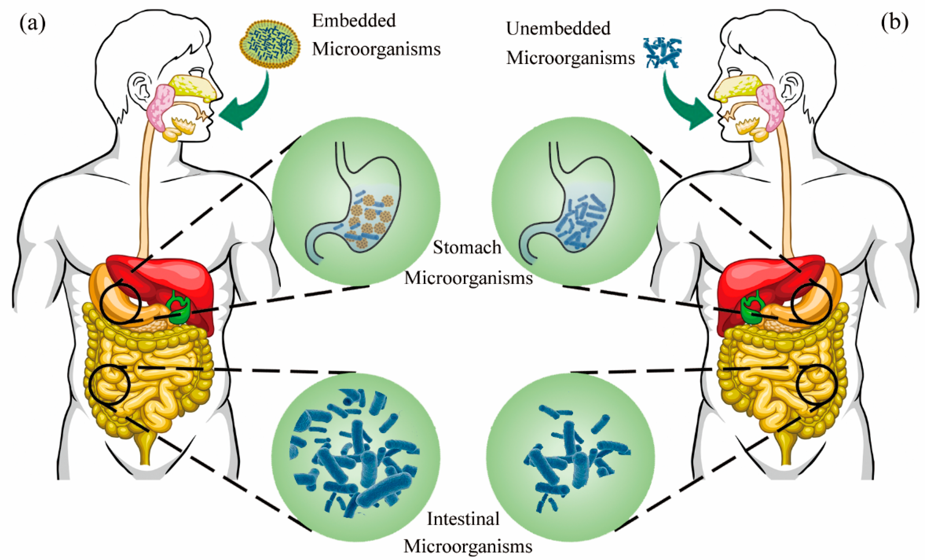
The current route for the in vivo delivery of microorganisms is by oral administration, and encapsulated probiotic supplements can stably exist in the body and colonize the intestinal tract and have certain abilities to clear intestinal bacteria (Figure 3) [116]. Microencapsulation or nanoencapsulation is the most commonly used technology to encapsulate microorganisms, and encapsulated microorganisms with different effects can be obtained by changing the encapsulation materials [36]. To resist the invasion of the acidic environment, microencapsulated technology was researched to improve the stability of encapsulated microorganisms and promote the ability of intestinal flora to metabolize glucoraphanin to produce SFN.
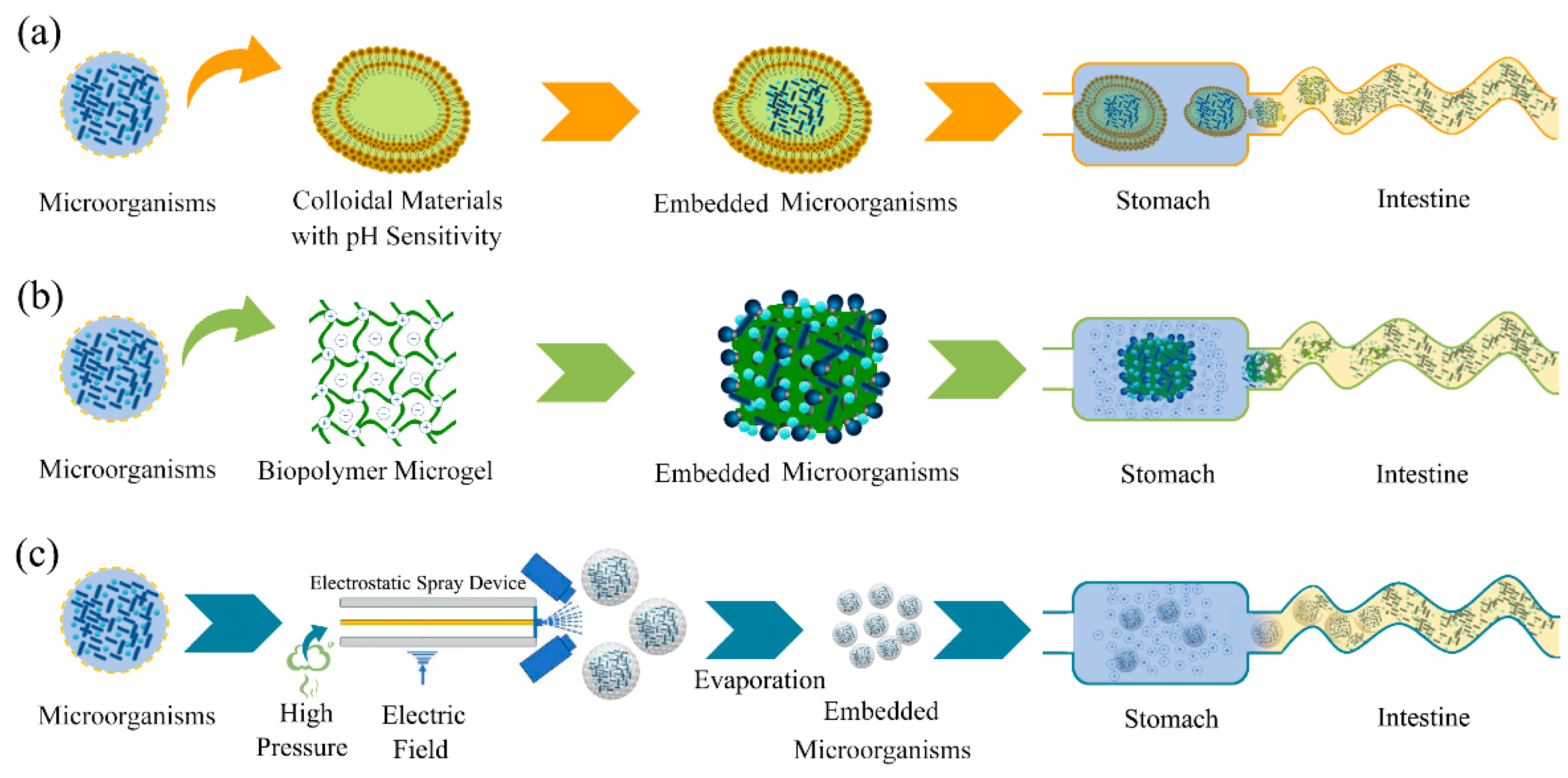
Furthermore, a modified alginate material was researched through material modification, and hydrogel beads were created from the modified alginate material [117]. The microorganisms protected by the hydrogel beads not only were protected from the gastric environment, but could also be released at different intestinal sites by controlling the content of the modified alginate. Cheng et al. used alginate and protamine as embedding materials and constructed an enzyme-triggered fuse-like microcapsule using combined electrostatic droplets [118]. The multilayered microcapsules have good protection against E. coli MG1655 during oral administration, and almost no E. coli is released in the gastric environment, but these bacteria are released 1 h after entering the intestinal environment (Figure 4a).
However, the oral core–shell microspheres constructed with sodium alginate and calcium ions as package materials exhibit colon-targeting properties. In addition, the residence times of local microorganisms are prolonged for the collapse release mechanism, and the numbers of microorganisms increases the bioavailability [119]. Kamguyan et al. developed a chitosan-coated microcontainer using L. rhamnosus as a model strain [120]. The chitosan-coated microcontainer was stable in the gastrointestinal environment and could specifically transport L. rhamnosus to the cecum or colon. The microorganisms were encapsulated in chitosan-coated alginate microparticles using emulsion technology, and feeding experiments show that the alginate microparticles can be used as a controlled release system to deliver live microorganisms to the gastrointestinal tract of abalone [121].
Biopolymer microgels are another form of encapsulation that can stabilize the entrapped microorganisms within the gastrointestinal environment and release them at the target intestinal site (Figure 4b) [122]. The stability of the microgels in the study was dependent on the different additives: acid-resistant microgels > control microgels > acid-resistant nanoemulsion microgels. Simulated in vitro colon experiments show that microorganisms are released due to fermentation of the microgels in the colon. Thiolated oxidized konjac glucomannan microspheres could achieve stable colonization of bifidobacteria within the intestinal environment [123]. Thiolated oxidized konjac glucomannan microspheres could also balance the intestinal flora and increase the abundance of bifidobacteria. Some researchers suggest that the multilayer electrospun structure is an important development direction for probiotic encapsulation and has important commercial value [124].
Electrospraying is a technique similar to electrospinning (Figure 4c) [125]. Small and uniform microcapsules can be prepared using electrospraying technology, and the activity and entrapment rate of electrospraying substances are higher than those of traditional spray drying methods. Electrospraying results of Premjit and Mitra show that 92.93% of Leuconostoc lactis is encapsulated, and the activity loss of encapsulated Leuconostoc lactis is 0.663 log CFU/g [126]. Huang et al. show that the encapsulation rate of electrospraying for L. plantarum is as high as 92.53%, and the encapsulation activity of L. plantarum by electrospraying is significantly higher than that by spray drying [127]. This also shows that electrospray technology has the characteristics of high encapsulation efficiency and high microbial activity, and also provides a reference for the encapsulation of SFN-transformed microorganisms.
Microencapsulation is another important form of microorganism encapsulation, and it has achieved long-term development [114,115,128]. Maillard conjugates and nanomaterials are currently one of the most commonly used microbial microencapsulation materials, which have the advantage of maintaining microbial stability and improving microbial intestinal colonization [129,130]. An in vitro simulation experiment shows that microcapsules can also improve the ability of microorganisms to produce myrosinases. Microencapsulation can deliver microorganisms that can synthesize myrosinase into the human intestinal environment, promote the stable existence and colonization of microorganisms in the intestinal environment, and can increase myrosinase synthesis.

5. Conclusions

Although intestinal microorganisms can synthesize myrosinase to degrade glucoraphanin and produce SFN, the myrosinase synthesis ability and colonization of the gut microbes are limited. Enhancing the myrosinase synthesis ability of gut microbes is the main effective means to promote the utilization rate of SFN. The application of transformation and microencapsulation technology to improve the myrosinase synthesis and colonization ability is feasible, and these technologies can enhance the absorption and utilization of SFN in the intestinal tract. Therefore, industries that produce high-yielding myrosinase microorganisms should consider transformation and microencapsulation technology, and it is important to provide technical support for the production and application of SFN. Meanwhile, attention should also be paid to the risk of leakage of transformed microbial genes and the uncontrollability of establishing a high-yield myrosinase microbial system in the intestine.

Author Contributions

Conceptualization and writing—original draft preparation, X.L.; methodology, Y.W.; formal analysis, P.W.; investigation, G.Z. and G.L.; writing—review and editing, J.L. All authors have read and agreed to the published version of the manuscript.

Funding

This research was funded by the Innovation and Capacity-building Project of Beijing Academy of Agriculture and Forestry Sciences (KJCX20220419) and the Beijing Technology and Business University Young Teachers Research Startup Fund Project (QNJJ2021-05).

Data Availability Statement

Not applicable.

Conflicts of Interest

The authors declare that there are no conflicts of interest.

References

  1. Yagishita, Y.; Fahey, J.W.; Dinkova-Kostova, A.T.; Kensler, T.W. Broccoli or Sulforaphane: Is It the Source or Dose That Matters? Molecules 2019, 24, 1029. [Google Scholar] [CrossRef] [Green Version]
  2. Abukhabta, S.; Ghawi, S.K.; Karatzas, K.A.; Charalampopoulos, D.; McDougall, G.J.; Allwood, J.W.; Verrall, S.R.; Lavery, S.; Latimer, C.; Pourshahidi, L.K.; et al. Sulforaphane-enriched extracts from glucoraphanin-rich broccoli exert antimicrobial activity against gut pathogens in vitro and innovative cooking methods increase in vivo intestinal delivery of sulforaphane. Eur. J. Nutr. 2021, 60, 1263–1276. [Google Scholar] [CrossRef] [PubMed]
  3. Hafezian, S.M.; Azizi, S.N.; Biparva, P.; Bekhradni, A. High-efficiency purification of sulforaphane from the broccoli extract by nanostructured SBA-15 silica using solid-phase extraction method. J. Chromatogr. B 2019, 1108, 1–10. [Google Scholar] [CrossRef]
  4. Janczewski, L. Sulforaphane and Its Bifunctional Analogs: Synthesis and Biological Activity. Molecules 2022, 27, 125. [Google Scholar] [CrossRef] [PubMed]
  5. Melrose, J. The Glucosinolates: A Sulphur Glucoside Family of Mustard Anti-Tumour and Antimicrobial Phytochemicals of Potential Therapeutic Application. Biomedicines 2019, 7, 821. [Google Scholar] [CrossRef] [PubMed] [Green Version]
  6. Zhang, Y.-J.; Wu, Q. Sulforaphane protects intestinal epithelial cells against lipopolysaccharide-induced injury by activating the AMPK/SIRT1/PGC-1alpha pathway. Bioengineered 2021, 12, 4349–4360. [Google Scholar] [CrossRef]
  7. Russo, M.; Spagnuolo, C.; Russo, G.L.; Skalicka-Wozniak, K.; Daglia, M.; Sobarzo-Sanchez, E.; Nabavi, S.F.; Nabavi, S.M. Nrf2 targeting by sulforaphane: A potential therapy for cancer treatment. Crit. Rev. Food Sci. Nutr. 2018, 58, 1391–1405. [Google Scholar] [CrossRef]
  8. Kaiser, A.E.; Baniasadi, M.; Giansiracusa, D.; Giansiracusa, M.; Garcia, M.; Fryda, Z.; Wong, T.L.; Bishayee, A. Sulforaphane: A Broccoli Bioactive Phytocompound with Cancer Preventive Potential. Cancers 2021, 13, 5618. [Google Scholar] [CrossRef]
  9. Daily, J.W.; Byoung-Seob, K.; Jina, R.; Meiling, L.; Weijun, Z.; Park, S. Isothiocyanate from broccoli, sulforaphane, and its properties. J. Med. Food 2019, 22, 121–126. [Google Scholar]
  10. Kim, J. Pre-Clinical Neuroprotective Evidences and Plausible Mechanisms of Sulforaphane in Alzheimer’s Disease. Int. J. Mol. Sci. 2021, 22, 201. [Google Scholar] [CrossRef]
  11. Ho-Sub, P.; Eun-Sang, H.; Ga-Young, C.; Hyun-Bum, K.; Kyun-Seob, P.; Jai-Yoon, S.; Yoonjin, H.; Geun Wook, C.; Byung Il, K.; Hyunwoo, P.; et al. Sulforaphane enhances long-term potentiation and ameliorate scopolamine-induced memory impairment. Physiol. Behav. 2021, 238, 113467. [Google Scholar]
  12. Bobermin, L.D.; Weber, F.B.; dos Santos, T.M.; Bello-Klein, A.; Wyse, A.T.S.; Goncalves, C.-A.; Quincozes-Santos, A. Sulforaphane Induces Glioprotection After LPS Challenge. Cell Mol. Neurobiol. 2022, 42, 829–846. [Google Scholar] [CrossRef]
  13. Ordonez, A.A.; Bullen, C.K.; Villabona-Rueda, A.F.; Thompson, E.A.; Turner, M.L.; Merino, V.F.; Yan, Y.; Kim, J.; Davis, S.L.; Komm, O.; et al. Sulforaphane exhibits antiviral activity against pandemic SARS-CoV-2 and seasonal HCoV-OC43 coronaviruses in vitro and in mice. Commun. Biol. 2022, 5, 1666. [Google Scholar] [CrossRef]
  14. Sangkret, S.; Pongmalai, P.; Devahastin, S.; Chiewchan, N. Enhanced production of sulforaphane by exogenous glucoraphanin hydrolysis catalyzed by myrosinase extracted from Chinese flowering cabbage (Brassica rapa var. parachinensis). Sci. Rep. 2019, 9, 101. [Google Scholar] [CrossRef] [PubMed] [Green Version]
  15. Zhang, S.; Ying, D.Y.; Cheng, L.J.; Bayrak, M.; Jegasothy, H.; Sanguansri, L.; Augustin, M.A. Sulforaphane in broccoli-based matrices: Effects of heat treatment and addition of oil. LWT Food Sci. Technol. 2020, 128, 1464. [Google Scholar] [CrossRef]
  16. Li, C.; Song, S.; He, Y.; Zhang, X.; Liu, H. CaCl2-HCl electrolyzed water affects glucosinolate metabolism and improves the quality of broccoli sprouts. Food Res. Int. 2021, 150, 1901. [Google Scholar] [CrossRef] [PubMed]
  17. Chen, W.; Wang, Y.; Xu, L.; Dong, J.; Zhu, X.; Ying, J.; Wang, Q.; Fan, L.; Li, C.; Liu, L. Methyl jasmonate, salicylic acid and abscisic acid enhance the accumulation of glucosinolates and sulforaphane in radish (Raphanus sativus L.) taproot. Sci. Hortic. 2019, 250, 159–167. [Google Scholar] [CrossRef]
  18. Triska, J.; Balik, J.; Houska, M.; Novotna, P.; Magner, M.; Vrchotova, N.; Hic, P.; Jilek, L.; Thorova, K.; Snurkovic, P.; et al. Factors Influencing Sulforaphane Content in Broccoli Sprouts and Subsequent Sulforaphane Extraction. Foods 2021, 10, 745. [Google Scholar] [CrossRef]
  19. Goharrizi, K.J.; Fatehi, F.; Nazari, M.; Salehi, F.; Maleki, M. Assessment of changes in the content of sulforaphane and expression levels of CYP79F1 and myrosinase genes and proteomic profile of Lepidium draba plant under water-deficit stress induced by polyethylene glycol. Acta Physiol. Plant 2020, 42, 991. [Google Scholar]
  20. Oloyede, O.O.; Wagstaff, C.; Methven, L. Influence of Cabbage (Brassica oleracea) Accession and Growing Conditions on Myrosinase Activity, Glucosinolates and Their Hydrolysis Products. Foods 2021, 10, 717. [Google Scholar] [CrossRef]
  21. Chen, X.; Li, Z.; Sun, X.; Ma, H.; Chen, X.; Ren, J.; Hu, K. New Method for the Synthesis of Sulforaphane and Related Isothiocyanates. Synthesis 2011, 1, 3991–3996. [Google Scholar] [CrossRef]
  22. Paone, P.; Cani, P.D. Mucus barrier, mucins and gut microbiota: The expected slimy partners? Gut 2020, 69, 2232–2243. [Google Scholar] [CrossRef]
  23. Luca, S.V.; Macovei, I.; Bujor, A.; Miron, A.; Skalicka-Wozniak, K.; Aprotosoaie, A.C.; Trifan, A. Bioactivity of dietary polyphenols: The role of metabolites. Crit. Rev. Food Sci. Nutr. 2020, 60, 626–659. [Google Scholar] [CrossRef] [PubMed]
  24. Magne, F.; Gotteland, M.; Gauthier, L.; Zazueta, A.; Pesoa, S.; Navarrete, P.; Balamurugan, R. The Firmicutes/Bacteroidetes Ratio: A Relevant Marker of Gut Dysbiosis in Obese Patients? Nutrients 2020, 12, 1789. [Google Scholar] [CrossRef] [PubMed]
  25. Ren-Hau, L.; Miller, M.J.; Jeffery, E. Glucoraphanin hydrolysis by microbiota in the rat cecum results in sulforaphane absorption. Food Funct. 2010, 1, 161–166. [Google Scholar]
  26. Li, F.; Hullar, M.A.J.; Beresford, S.A.A.; Lampe, J.W. Variation of glucoraphanin metabolism in vivo and ex vivo by human gut bacteria. Br. J. Nutr. 2011, 106, 408–416. [Google Scholar] [CrossRef] [Green Version]
  27. Angelino, D.; Jeffery, E. Glucosinolate hydrolysis and bioavailability of resulting isothiocyanates: Focus on glucoraphanin. J. Funct. Foods 2014, 7, 67–76. [Google Scholar] [CrossRef]
  28. Zhang, C.; Zhang, Y.; Li, H.; Liu, X. The potential of proteins, hydrolysates and peptides as growth factors for Lactobacillus and Bifidobacterium: Current research and future perspectives. Food Funct. 2020, 11, 1946–1957. [Google Scholar] [CrossRef]
  29. Xinzhou, W.; Peng, Z.; Xin, Z. Probiotics regulate gut microbiota: An effective method to improve immunity. Molecules 2021, 26, 6076. [Google Scholar]
  30. Monika, Y.; Mandeep; Pratyoosh, S. Probiotics of diverse origin and their therapeutic applications: A review. J. Am. Coll. Nutr. 2020, 39, 469–479. [Google Scholar]
  31. Gang, T.; Linyu, Z. Update on strategies of probiotics for the prevention and treatment of colorectal cancer. Nutr. Cancer 2022, 74, 27–38. [Google Scholar]
  32. How, Y.; Pui, L. Effect of prebiotics encapsulated with probiotics on encapsulation efficiency, microbead size, and survivability: A review. J. Food. Meas. Charac. 2021, 15, 4899–4916. [Google Scholar] [CrossRef]
  33. Monika, Y.; Pratyoosh, S. Efficient engineered probiotics using synthetic biology approaches: A review. Biotechnol. Appl. Biochem. 2020, 67, 22–29. [Google Scholar]
  34. Reque, P.M.; Brandelli, A. Encapsulation of probiotics and nutraceuticals: Applications in functional food industry. Trends Food Sci. Technol. 2021, 114, 1–10. [Google Scholar] [CrossRef]
  35. Yan Xue, C.; Ji Hui, W.; McAuley, C.; Augustin, M.A.; Terefe, N.S. Fermentation for enhancing the bioconversion of glucoraphanin into sulforaphane and improve the functional attributes of broccoli puree. J. Funct. Foods 2019, 61, 103461. [Google Scholar]
  36. Jian-Hui, Y.; Long-Yue, H.; Terefe, N.S.; Augustin, M.A. Fermentation-based biotransformation of glucosinolates, phenolics and sugars in retorted broccoli puree by lactic acid bacteria. Food Chem. 2019, 286, 616–623. [Google Scholar]
  37. Boehm, J.; Davis, R.; Murar, C.E.; Li, T.; McCleland, B.; Dong, S.; Yan, H.; Kerns, J.; Moody, C.J.; Wilson, A.J.; et al. Discovery of a crystalline sulforaphane analog with good solid-state stability and engagement of the Nrf2 pathway in vitro and in vivo. Bioorg. Med. Chem. 2019, 27, 579–588. [Google Scholar] [CrossRef]
  38. Zambrano, V.; Bustos, R.; Mahn, A. Insights about stabilization of sulforaphane through microencapsulation. Heliyon 2019, 5, 2899. [Google Scholar] [CrossRef]
  39. Jeon, M.; Lee, J.; Lee, H.K.; Cho, S.; Lim, J.H.; Choi, Y.; Pak, S.; Jeong, H.J. Sulforaphane mitigates mast cell-mediated allergic inflammatory reactions in in silico simulation and in vitro models. Immunopharmacol. Immunotoxicol. 2020, 42, 74–83. [Google Scholar] [CrossRef]
  40. Ge, M.; Zhang, L.; Cao, L.; Xie, C.; Li, X.; Li, Y.; Meng, Y.; Chen, Y.; Wang, X.; Chen, J.; et al. Sulforaphane inhibits gastric cancer stem cells via suppressing sonic hedgehog pathway. Int. J. Food Sci. Nutr. 2019, 70, 570–578. [Google Scholar] [CrossRef]
  41. Doheny-Adams, T.; Redeker, K.; Kittipol, V.; Bancroft, I.; Hartley, S.E. Development of an efficient glucosinolate extraction method. Plant Methods 2017, 13, 17. [Google Scholar] [CrossRef] [PubMed] [Green Version]
  42. Favela-Gonzalez, K.M.; Hernandez-Almanza, A.Y.; De la Fuente-Salcido, N.M. The value of bioactive compounds of cruciferous vegetables (Brassica) as antimicrobials and antioxidants: A review. J. Food Biochem. 2020, 3, e13414. [Google Scholar] [CrossRef]
  43. Yang, H.; Qin, J.; Wang, X.; Ei-Shora, H.M.; Yu, B. Production of plant-derived anticancer precursor glucoraphanin in chromosomally engineered Escherichia coli. Microbiol. Res. 2020, 238, 126484. [Google Scholar] [CrossRef] [PubMed]
  44. Francis, F.; Lognay, G.; Wathelet, J.P.; Haubruge, E. Characterisation of aphid myrosinase and degradation studies of glucosinolates. Arch. Insect Biochem. Physiol. 2002, 50, 173–182. [Google Scholar] [CrossRef]
  45. Roman, J.; Castillo, A.; Cottet, L.; Mahn, A. Kinetic and structural study of broccoli myrosinase and its interaction with different glucosinolates. Food Chem. 2018, 254, 87–94. [Google Scholar] [CrossRef]
  46. Lei, W.; Rose, D.; Pingfan, R.; Yue, Z. Development of prolamin-based composite nanoparticles for controlled release of sulforaphane. J. Agric. Food Chem. 2020, 68, 13083–13092. [Google Scholar]
  47. Wei, L.; Liu, C.; Zheng, H.; Zheng, L. Melatonin treatment affects the glucoraphanin-sulforaphane system in postharvest fresh-cut broccoli (Brassica oleracea L.). Food Chem. 2020, 307, 299. [Google Scholar] [CrossRef]
  48. Wu, Y.; Lv, C.; Zou, L.; Sun, J.; Song, X.; Zhang, Y.; Mao, J. Approaches for enhancing the stability and formation of sulforaphane. Food Chem. 2021, 345, 128771. [Google Scholar]
  49. Xinxing, X.; Mei, D.; Fei, L.; Fang, C.; Xiaosong, H.; Yuping, L.; Jihong, W. Effect of glucoraphanin from broccoli seeds on lipid levels and gut microbiota in high-fat diet-fed mice. J. Funct. Foods 2020, 68, 103858. [Google Scholar]
  50. Budnowski, J.; Hanske, L.; Schumacher, F.; Glatt, H.; Platz, S.; Rohn, S.; Blaut, M. Glucosinolates Are Mainly Absorbed Intact in Germfree and Human Microbiota-Associated Mice. J. Agric. Food Chem. 2015, 63, 8418–8428. [Google Scholar] [CrossRef]
  51. Sun, J.; Charron, C.S.; Novotny, J.A.; Peng, B.; Yu, L.; Chen, P. Profiling glucosinolate metabolites in human urine and plasma after broccoli consumption using non-targeted and targeted metabolomic analyses. Food Chem. 2020, 4, 309. [Google Scholar] [CrossRef]
  52. Martijn Vermeulen, I.W.M.; Armstrong, K.P.-K.; Van Den Berg, R.; Vaes, W.H.J. Bioavailability and Kinetics of Sulforaphane in Humans after Consumption of Cooked versus Raw Broccoli. J Agric Food Chem. 2008, 56, 10505–10509. [Google Scholar] [CrossRef] [PubMed]
  53. Egner, P.A.; Chen, J.G.; Wang, J.B.; Wu, Y.; Sun, Y.; Lu, J.H.; Zhu, J.; Zhang, Y.H.; Chen, Y.S.; Friesen, M.D.; et al. Bioavailability of Sulforaphane from two broccoli sprout beverages: Results of a short-term, cross-over clinical trial in Qidong, China. Cancer Prev. Res. 2011, 4, 384–395. [Google Scholar] [CrossRef] [PubMed]
  54. Shapiro, T.A.; Jhones, W.F.; Wade, K.L.; Stephenson, K.K.; Talalay, P. Chemoprotective Glucosinolates and Isothiocyanates of Broccoli Sprouts: Metabolism and Excretion in Humans1. Cancer Epidemiol. Biomark. Prev. 2001, 10, 501–508. [Google Scholar]
  55. Kuljarachanan, T.; Fu, N.; Chiewchan, N.; Devahastin, S.; Chen, X.D. In vitro digestion using dynamic rat stomach-duodenum model as an alternative means to assess bioaccessibility of glucosinolates in dietary fiber powder from cabbage. LWT Food Sci. Technol. 2021, 2, 151. [Google Scholar] [CrossRef]
  56. Yuanfeng, W.; Yuke, S.; Ye, Z.; Mupunga, J.; Ligen, Z.; Chao, L.; Shiwang, L.; Jianwei, M. Broccoli ingestion increases the glucosinolate hydrolysis activity of microbiota in the mouse gut. Int. J. Food Sci. Nutr. 2019, 70, 585–594. [Google Scholar]
  57. Hwang, E.-S.; Bornhorst, G.M.; Oteiza, P.I.; Mitchell, A.E. Assessing the Fate and Bioavailability of Glucosinolates in Kale (Brassica oleracea) Using Simulated Human Digestion and Caco-2 Cell Uptake Models. J. Agric. Food Chem. 2019, 67, 9492–9500. [Google Scholar] [CrossRef] [PubMed]
  58. Ye, Q.; Fang, Y.; Li, M.; Mi, H.; Liu, S.; Yang, G.; Lu, J.; Zhao, Y.; Liu, Q.; Zhang, W.; et al. Characterization of a Novel Myrosinase with High Activity from Marine Bacterium Shewanella baltica Myr-37. Int. J. Mol. Sci. 2022, 5, 23. [Google Scholar] [CrossRef]
  59. Mao, S.; Wang, J.; Wu, Q.; Liang, M.; Yuan, Y.; Wu, T.; Liu, M.; Wu, Q.; Huang, K. Effect of selenium-sulfur interaction on the anabolism of sulforaphane in broccoli. Phytochemistry 2020, 179, 112499. [Google Scholar] [CrossRef]
  60. Matusheski, N.V.; Ron, S.; Juvik, J.A.; Mithen, R.; Bennett, M.; Jeffery, E.H. Epithiospecifier Protein from Broccoli (Brassica oleracea L. ssp. italica) Inhibits Formation of the Anticancer Agent Sulforaphane. J. Agric. Food Chem. 2006, 54, 2069–2076. [Google Scholar] [CrossRef]
  61. Sporer, T.; Koernig, J.; Wielsch, N.; Gebauer-Jung, S.; Reichelt, M.; Hupfer, Y.; Beran, F. Hijacking the Mustard-Oil Bomb: How a Glucosinolate-Sequestering Flea Beetle Copes With Plant Myrosinases. Front. Plant Sci. 2021, 12, 1009. [Google Scholar] [CrossRef]
  62. Feng, Q.; Li, L.; Liu, Y.; Shao, X.; Li, X. Jasmonate regulates the FAMA/mediator complex subunit 8-THIOGLUCOSIDE GLUCOHYDROLASE 1 cascade and myrosinase activity. Plant Physiol. 2021, 187, 963–980. [Google Scholar] [CrossRef] [PubMed]
  63. Jafari, S.; Ryde, U.; Irani, M. QM/MM Study of the Catalytic Reaction of Myrosinase; Importance of Assigning Proper Protonation States of Active-Site Residues. J. Chem. Theory Comput. 2021, 17, 1822–1841. [Google Scholar] [CrossRef] [PubMed]
  64. Sicong, T.; Xiaodong, L.; Peng, L.; Xiaohong, Z.; Yujuan, S. Microbiota: A mediator to transform glucosinolate precursors in cruciferous vegetables to the active isothiocyanates. J. Sci. Food Agric. 2018, 98, 1255–1260. [Google Scholar]
  65. Sikorska-Zimny, K.; Beneduce, L. The Metabolism of Glucosinolates by Gut Microbiota. Nutrients 2021, 13, 2556. [Google Scholar] [CrossRef]
  66. Fahey, J.W.; Kensler, T.W. The Challenges of Designing and Implementing Clinical Trials With Broccoli Sprouts horizontal ellipsis and Turning Evidence Into Public Health Action. Front. Nutr. 2021, 8, 7290. [Google Scholar] [CrossRef]
  67. Elfoul, L.; Rabot, S.; Khelifa, N.; Quinsac, A.; Duguay, A.; Rimbault, A. Formation of allyl isothiocyanate from sinigrin in the digestive tract of rats monoassociated with a human colonic strain of Bacteroides thetaiotaomicron. FEMS Microbiol. Lett. 2001, 197, 99–103. [Google Scholar] [CrossRef]
  68. Luang-In, V.; Narbad, A.; Nueno-Palop, C.; Mithen, R.; Bennett, M.; Rossiter, J.T. The metabolism of methylsulfinylalkyl- and methylthioalkyl- glucosinolates by a selection of human gut bacteria. Mol. Nutr. Food Res. 2014, 58, 875–883. [Google Scholar] [CrossRef] [PubMed]
  69. Zhang, Y.; Huang, S.; Sun, J.; Song, X.; Jiang, C.; Wu, Y. Isolation and Characterization of Glucosinolate-Hydrolysis Enterococcus gallinarum HG001 and Escherichia coli HG002 from C57BL/6 Mouse Microbiota. Indian J. Microbiol. 2022, 62, 273–279. [Google Scholar] [CrossRef]
  70. Mullaney, J.A.; Kelly, W.J.; McGhie, T.K.; Ansell, J.; Heyes, J.A. Lactic Acid Bacteria Convert Glucosinolates to Nitriles Efficiently Yet Differently from Enterobacteriaceae. J. Agric. Food Chem. 2013, 61, 3039–3046. [Google Scholar] [CrossRef]
  71. Yan Xue, C.; Augustin, M.A.; Jegasothy, H.; Ji Hui, W.; Terefe, N.S. Mild heat combined with lactic acid fermentation: A novel approach for enhancing sulforaphane yield in broccoli puree. Food Funct. 2020, 11, 779–786. [Google Scholar]
  72. Watanabe, H.; Usami, R.; Kishino, S.; Osada, K.; Aoki, Y.; Morisaka, H.; Takahashi, M.; Izumi, Y.; Bamba, T.; Aoki, W.; et al. Enzyme systems involved in glucosinolate metabolism in Companilactobacillus farciminis KB1089. Sci. Rep. 2021, 11, 267. [Google Scholar] [CrossRef] [PubMed]
  73. Cebeci, F.; Mayer, M.J.; Rossiter, J.T.; Mithen, R.; Narbad, A. Molecular Cloning, Expression and Characterisation of a Bacterial Myrosinase from Citrobacter Wye1. Protein J. 2022, 41, 131–140. [Google Scholar] [CrossRef] [PubMed]
  74. Tie, Y.; Zhu, W.; Zhang, C.; Yin, L.; Zhang, Y.; Liu, L.; Yuan, H. Identification of Two Myrosinases from a Leclercia adecarboxylata Strain and Investigation of Its Tolerance Mechanism to Glucosinolate Hydrolysate. J. Agric. Food Chem. 2021, 69, 14151–14164. [Google Scholar] [CrossRef]
  75. Rakariyatham, N.; Butrindr, B.; Niamsup, H.; Shank, L. Screening of filamentous fungi for production of myrosinase. Braz. J. Microbiol. 2005, 36, 242–245. [Google Scholar] [CrossRef] [Green Version]
  76. Xu, X.; Bi, S.; Lao, F.; Chen, F.; Liao, X.; Wu, J. Induced changes in bioactive compounds of broccoli juices after fermented by animal- and plant-derived Pediococcus pentosaceus. Food Chem. 2021, 357, 144. [Google Scholar] [CrossRef]
  77. Rakariyatham, N.; Sakorn, P. Biodegradation of glucosinolates in brown mustard seed meal (Brassica juncea) by Aspergillus sp. NR-4201 in liquid and solid-state cultures. Biodegradation 2002, 13, 395–399. [Google Scholar] [CrossRef] [PubMed]
  78. Llanos Palop, M.; Smiths, J.P.; Brink, B.T. Degradation of sinigrin by Lactobacillus agilis strain R16. Int. J. Food Microbiol. 1995, 26, 219. [Google Scholar] [CrossRef] [PubMed]
  79. Cheng, F.; Chen, H.; Lei, N.; Zhang, M.; Wan, H.; Shu, G. Effect of Prebiotics, Inorganic Salts and Amino Acids for Cell Envelope Proteinase Production from Lactobacillus Plantarum Lp69. Acta Sci. Pol. Technol. Aliment. 2019, 18, 269–278. [Google Scholar] [PubMed]
  80. Min, Z.; Jia, Z.; Jun, X.; Dong, Z.; Zong-Wei, Q.; Dan, H.; Hui-Bo, L.; Hui-Bo, L. Effects of environmental factors on the microbial community changes during medium-high temperature Daqu manufacturing. Food Res. Int. 2022, 153, 110955. [Google Scholar]
  81. Rama, G.R.; Kuhn, D.; Beux, S.; Maciel, M.J.; Volken de Souza, C.F. Potential applications of dairy whey for the production of lactic acid bacteria cultures. Int. Dairy J. 2019, 98, 25–37. [Google Scholar] [CrossRef]
  82. Dahiya, D.; Manuel, J.V.; Nigam, P.S. An Overview of Bioprocesses Employing Specifically Selected Microbial Catalysts for gamma-Aminobutyric Acid Production. Microorganisms 2021, 9, 6628. [Google Scholar] [CrossRef]
  83. Nabot, M.; Guerin, M.; Sivakumar, D.; Remize, F.; Garcia, C. Variability of Bacterial Homopolysaccharide Production and Properties during Food Processing. Biology 2022, 11, 9762. [Google Scholar] [CrossRef] [PubMed]
  84. Alshehri, W.A. Bacterium Hafnia alvei secretes L-methioninase enzyme: Optimization of the enzyme secretion conditions. Saudi J. Biol. Sci. 2020, 27, 1222–1227. [Google Scholar] [CrossRef] [PubMed]
  85. Xiao, T.; Shah, N.P. Lactic acid produced by Streptococcus thermophilus activated glutamate decarboxylase (GadA) in Lactobacillus brevis NPS-QW 145 to improve gamma-amino butyric acid production during soymilk fermentation. LWT Food Sci. Technol. 2021, 137, 652. [Google Scholar] [CrossRef]
  86. Raj, T.S.; Athimoolam, S.; Vijayaraghavan, P. Biosynthesis and Characterization of a Novel Fibrinolytic Alkaline Serine Protease from Newly Isolated Bacillus flexus BF12 for Biomedical Applications. Curr. Pharm. Biotechnol. 2021, 22, 698–709. [Google Scholar] [CrossRef] [PubMed]
  87. West, T.P. Production of the Polysaccharide Curdlan by Agrobacterium species on Processing Coproducts and Plant Lignocellulosic Hydrolysates. Fermentation 2020, 6, 52. [Google Scholar] [CrossRef] [Green Version]
  88. Jing, Y.; Li, F.; Li, Y.; Jin, P.; Zhu, S.; He, C.; Zhao, J.; Zhang, Z.; Zhang, Q. Statistical optimization of simultaneous saccharification fermentative hydrogen production from corn stover. Bioengineered 2020, 11, 428–438. [Google Scholar] [CrossRef]
  89. Zhang, Q.; Li, Y.; Liu, Z.; Pu, Y.; Xia, L. Optimization of simultaneous saccharification and fermentation for ethanol production from steam-exploded cotton stalk. In Proceedings of the 2nd International Conference on Energy and Environmental Protection (ICEEP 2013), Guilin, China, 19–21 April 2013; Volume 724–725, p. 391. [Google Scholar]
  90. Guo, J.; Wei, J.; Huang, F.; Massey, I.Y.; Luo, J.; Yang, F. Optimization of microcystin biodegradation by bacterial community YFMCD4 using response surface method. Chemosphere 2021, 274, 7721. [Google Scholar] [CrossRef]
  91. Haile, S.; Ayele, A. Pectinase from Microorganisms and Its Industrial Applications. Sci. World J. 2022, 2022, 1881305. [Google Scholar] [CrossRef]
  92. Sondhi, S.; Saini, K. Response surface based optimization of laccase production from Bacillus sp. MSK-01 using fruit juice waste as an effective substrate. Heliyon 2019, 5, 811. [Google Scholar] [CrossRef] [Green Version]
  93. Ahmar, S.; Gill, R.A.; Jung, K.-H.; Faheem, A.; Qasim, M.U.; Mubeen, M.; Zhou, W. Conventional and Molecular Techniques from Simple Breeding to Speed Breeding in Crop Plants: Recent Advances and Future Outlook. Int. J. Mol. Sci. 2020, 21, 562. [Google Scholar] [CrossRef] [Green Version]
  94. Ren, J.; Na, D.; Yoo, S.M. Optimization of chemico-physical transformation methods for various bacterial species using diverse chemical compounds and nanomaterials. J. Biotechnol. 2018, 288, 55–60. [Google Scholar] [CrossRef]
  95. Ren, J.; Karna, S.; Lee, H.-M.; Yoo, S.M.; Na, D. Artificial transformation methodologies for improving the efficiency of plasmid DNA transformation and simplifying its use. Appl. Microbiol. Biotechnol. 2019, 103, 9205–9215. [Google Scholar] [CrossRef] [PubMed]
  96. Riley, L.A.; Guss, A.M. Approaches to genetic tool development for rapid domestication of non-model microorganisms. Biotechnol. Biofuels 2021, 14, 6144. [Google Scholar] [CrossRef] [PubMed]
  97. Wang, Y.; Wang, X.; Yu, L.; Tian, Y.; Li, S.; Leng, F.; Ma, J.; Chen, J. Effects of Sr2+ on the preparation of Escherchia coli DH5 alpha competent cells and plasmid transformation. Peerj 2020, 8, e8929. [Google Scholar]
  98. Deshmukh, K.; Ramanan, S.R.; Kowshik, M. Novel one step transformation method for Escherichia colt and Staphylococcus aureus using arginine-glucose functionalized hydroxyapatite nanoparticles. Mater. Sci. Eng. C 2019, 96, 58–65. [Google Scholar] [CrossRef] [PubMed]
  99. Pandey, A.; Devkota, A.; Sigdel, A.; Yadegari, Z.; Dumenyo, K.; Taheri, A. Citric acid/beta-alanine carbon dots as a novel tool for delivery of plasmid DNA into E. coli cells. Sci. Rep. 2021, 11, 1553. [Google Scholar] [CrossRef]
  100. Rosano, G.L.; Morales, E.S.; Ceccarelli, E.A. New tools for recombinant protein production in Escherichia coli: A 5-year update. Protein Sci. 2019, 28, 1412–1422. [Google Scholar] [CrossRef] [PubMed]
  101. Jain, K.; Wood, E.A.; Romero, Z.J.; Cox, M.M. RecA-independent recombination: Dependence on the Escherichia coli RarA protein. Mol. Microbiol. 2021, 115, 1122–1137. [Google Scholar] [CrossRef]
  102. Hao, N.; Chen, Q.; Dodd, I.B.; Shearwin, K.E. The pIT5 Plasmid Series, an Improved Toolkit for Repeated Genome Integration in E. coli. ACS Synth. Biol. 2021, 10, 1633–1639. [Google Scholar] [CrossRef]
  103. Chen, K.; Xie, M.; Chan, E.W.-C.; Chen, S. Delineation of ISEcp1 and IS26-Mediated Plasmid Fusion Processes by MinION Single-Molecule Long-Read Sequencing. Front. Microbiol. 2022, 12, 277. [Google Scholar] [CrossRef]
  104. Than, M.M.; Ruangsuriya, J.; Uthaipibull, C.; Srichairatanakool, S. Expression of fluorescent tagged recombinant erythroferrone protein. Asian Pac. J. Trop. Biomed. 2018, 8, 360–364. [Google Scholar]
  105. Wang, L.; Jiang, H.; Qiu, Y.; Dong, Y.; Hamouda, H.I.; Balah, M.A.; Mao, X. Biochemical Characterization of a Novel Myrosinase Rmyr from Rahnella inusitata for High-Level Preparation of Sulforaphene and Sulforaphane. J. Agric. Food Chem. 2022, 70, 2303–2311. [Google Scholar] [CrossRef]
  106. Curiqueo, C.; Mahn, A.; Castillo, A. Broccoli Myrosinase cDNA Expression in Escherichia coli and Saccharomyces cerevisiae. Biomolecules 2022, 12, 1872. [Google Scholar] [CrossRef] [PubMed]
  107. Wang, L.; Jiang, H.; Liang, X.; Zhou, W.; Qiu, Y.; Xue, C.; Sun, J.; Mao, X. Preparation of Sulforaphene from Radish Seed Extracts with Recombinant Food-Grade Yarrowia lipolytica Harboring High Myrosinase Activity. J. Agric. Food Chem. 2021, 69, 5363–5371. [Google Scholar] [CrossRef]
  108. Rosenbergova, Z.; Kantorova, K.; Simkovic, M.; Breier, A.; Rebros, M. Optimisation of Recombinant Myrosinase Production in Pichia pastoris. Int. J. Mol. Sci. 2021, 22, 627. [Google Scholar] [CrossRef]
  109. Ahuja, I.; Kissen, R.; Hoang, L.; Sporsheim, B.; Halle, K.K.; Wolff, S.A.; Ahmad, S.J.N.; Ahmad, J.N.; Bones, A.M. The Imaging of Guard Cells of thioglucosidase (tgg) Mutants of Arabidopsis Further Links Plant Chemical Defence Systems with Physical Defence Barriers. Cells 2021, 10, 924. [Google Scholar] [CrossRef] [PubMed]
  110. Cao, H.; Liu, R.; Zhang, J.; Liu, Z.; Fan, S.; Yang, G.; Jin, Z.; Pei, Y. Improving sulforaphane content in transgenic broccoli plants by overexpressing MAM1, FMOGS–OX2, and Myrosinase. Plant Cell Tissue Organ Cult. 2021, 146, 461–471. [Google Scholar] [CrossRef]
  111. Azizoglu, U.; Jouzani, G.S.; Yilmaz, N.; Baz, E.; Ozkok, D. Genetically modified entomopathogenic bacteria, recent developments, benefits and impacts: A review. Sci. Total Environ. 2020, 1, 734. [Google Scholar] [CrossRef] [PubMed]
  112. Yang, Q.; Luo, L.; Jiang, Y.; Zhang, J.; Tang, X.; Jiang, M.; Wu, Z.; Li, Y. A high amylase-producing bacterial strain exhibiting the phosphorus-dissolving ability, isolated from freshwater aquaculture pond. Aquacult. Res. 2020, 51, 4315–4326. [Google Scholar] [CrossRef]
  113. Mingfei, Y.; Jiaojiao, X.; Hengjun, D.; McClements, D.J.; Hang, X.; Lanjuan, L. Progress in microencapsulation of probiotics: A review. Compr. Rev. Food Sci. Food Saf. 2020, 19, 857–874. [Google Scholar]
  114. Lohrasbi, V.; Abdi, M.; Asadi, A.; Rohani, M.; Esghaei, M.; Talebi, M.; Amirmozafari, N. The effect of improved formulation of chitosan-alginate microcapsules of Bifidobacteria on serum lipid profiles in mice. Microb. Pathog. 2020, 149, 8821. [Google Scholar] [CrossRef]
  115. Zacchetti, B.; Andrianos, A.; van Dissel, D.; de Ruiter, E.; van Wezel, G.P.; Claessen, D. Microencapsulation extends mycelial viability of Streptomyces lividans 66 and increases enzyme production. BMC Biotechnol. 2018, 18, 7018. [Google Scholar] [CrossRef] [PubMed] [Green Version]
  116. Hung, Y.-P.; Lee, C.-C.; Lee, J.-C.; Tsai, P.-J.; Hsueh, P.-R.; Ko, W.-C. The Potential of Probiotics to Eradicate Gut Carriage of Pathogenic or Antimicrobial-Resistant Enterobacterales. Antibiotics 2021, 10, 1662. [Google Scholar] [CrossRef]
  117. Enck, K.; Banks, S.; Yadav, H.; Welker, M.E.; Opara, E.C. Development of a Novel Oral Delivery Vehicle for Probiotics. Curr. Pharm. Des. 2020, 26, 3134–3140. [Google Scholar] [CrossRef]
  118. Cheng, Q.; Liu, L.; Xie, M.; Li, H.; Ma, D.; Xue, W. A Colon-Targeted Oral Probiotics Delivery System Using an Enzyme-Triggered Fuse-Like Microcapsule. Adv. Healthc. Mater. 2021, 10, e19913. [Google Scholar] [CrossRef]
  119. Liu, H.; Cai, Z.; Wang, F.; Hong, L.; Deng, L.; Zhong, J.; Wang, Z.; Cui, W. Colon-Targeted Adhesive Hydrogel Microsphere for Regulation of Gut Immunity and Flora. Adv. Sci. 2021, 8, e17662. [Google Scholar] [CrossRef]
  120. Kamguyan, K.; Torp, A.M.; Christfort, J.F.; Guerra, P.R.; Licht, T.R.; Nielsen, L.H.; Zor, K.; Boisen, A. Colon-Specific Delivery of Bioactive Agents Using Genipin-Cross-Linked Chitosan Coated Microcontainers. ACS Appl. Bio Mater. 2021, 4, 752–762. [Google Scholar] [CrossRef]
  121. Masoomi Dezfooli, S.; Gutierrez-Maddox, N.; Alfaro, A.C.; Seyfoddin, A. Development of a microencapsulated probiotic delivery system for New Zealand black-footed abalone (Haliotis iris). Pharm. Dev. Technol. 2021, 26, 390–402. [Google Scholar] [CrossRef]
  122. Zhang, Z.; Gu, M.; You, X.; Sela, D.A.; Xiao, H.; McClements, D.J. Encapsulation of bifidobacterium in alginate microgels improves viability and targeted gut release. Food Hydrocoll. 2021, 116, 2672. [Google Scholar] [CrossRef]
  123. Liu, Y.; Liu, B.; Li, D.; Hu, Y.; Zhao, L.; Zhang, M.; Ge, S.; Pang, J.; Li, Y.; Wang, R.; et al. Improved Gastric Acid Resistance and Adhesive Colonization of Probiotics by Mucoadhesive and Intestinal Targeted Konjac Glucomannan Microspheres. Adv. Funct. Mater. 2020, 30, 214. [Google Scholar] [CrossRef]
  124. Ajalloueian, F.; Guerra, P.R.; Bahl, M.I.; Torp, A.M.; Te Hwu, E.; Licht, T.R.; Boisen, A. Multi-layer PLGA-pullulan-PLGA electrospun nanofibers for probiotic delivery. Food Hydrocoll. 2022, 123, 2886. [Google Scholar] [CrossRef]
  125. Coelho, S.C.; Estevinho, B.N.; Rocha, F. Encapsulation in food industry with emerging electrohydrodynamic techniques: Electrospinning and electrospraying—A review. Food Chem. 2021, 339, 2872. [Google Scholar]
  126. Premjit, Y.; Mitra, J. Optimization of Electrospray-Assisted Microencapsulation of Probiotics (Leuconostoc lactis) in Soy Protein Isolate-Oil Particles Using Box-Behnken Experimental Design. Food Bioprocess Technol. 2021, 14, 1712–1729. [Google Scholar] [CrossRef]
  127. Huang, R.-M.; Feng, K.; Li, S.-F.; Zong, M.-H.; Wu, H.; Han, S.-Y. Enhanced survival of probiotics in the electrosprayed microcapsule by addition of fish oil. J. Food Eng. 2021, 307, 2613. [Google Scholar] [CrossRef]
  128. Oberoi, K.; Tolun, A.; Sharma, K.; Sharma, S. Microencapsulation: An overview for the survival of probiotic bacteria. J. Microbiol. Biotechnol. Food Sci. 2019, 9, 280–287. [Google Scholar] [CrossRef]
  129. Nooshkam, M.; Varidi, M. Maillard conjugate-based delivery systems for the encapsulation, protection, and controlled release of nutraceuticals and food bioactive ingredients: A review. Food Hydrocoll. 2020, 100, 2670. [Google Scholar] [CrossRef]
  130. Razavi, S.; Janfaza, S.; Tasnim, N.; Gibson, D.L.; Hoorfar, M. Nanomaterial-based encapsulation for controlled gastrointestinal delivery of viable probiotic bacteria. Nanoscale Adv. 2021, 3, 2699–2709. [Google Scholar] [CrossRef]
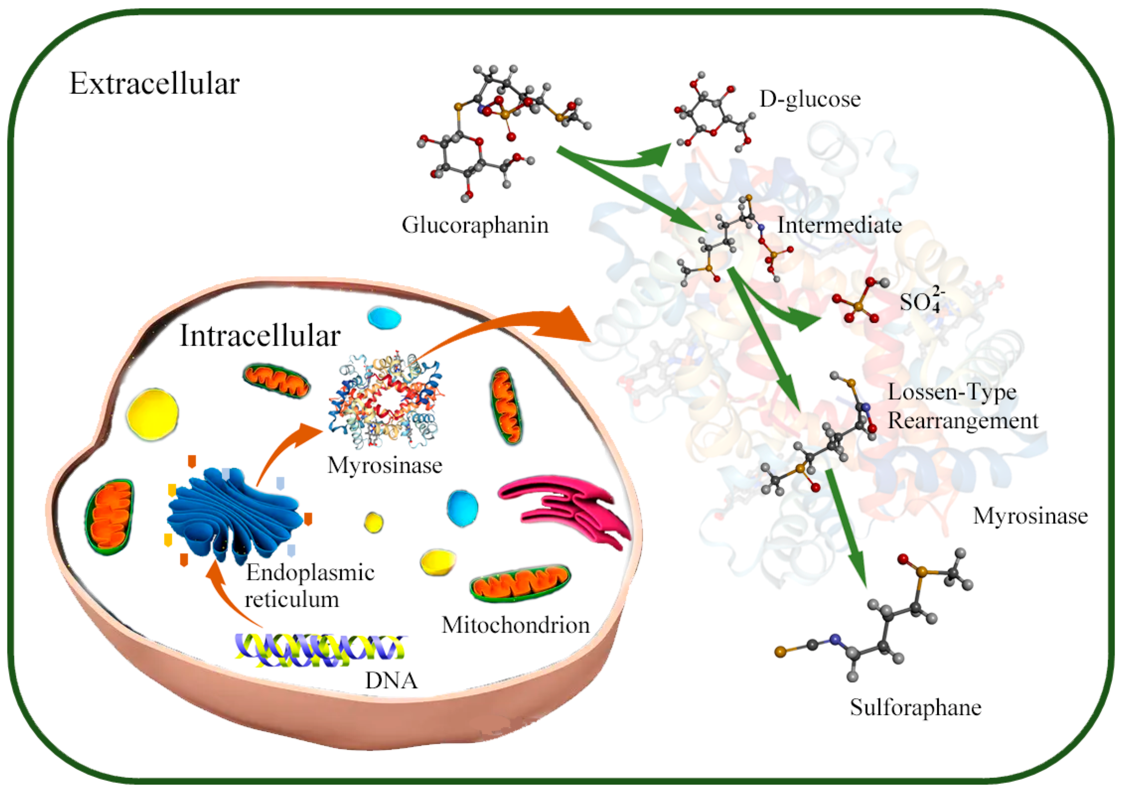
Figure 1. Mechanism of SFN production by microorganisms. In microorganisms, myrosinase is synthesized and secreted to environment through ribosome and endoplasmic reticulum under the myrosinase gene regulation; the glucoraphanin is converted into sulforaphane under the action of myrosinase in extracellular environment.
Foods 11 03775 g001
Figure 2. Factors affecting the synthesis of myrosinase by microorganisms.
Foods 11 03775 g002
Figure 3. Protect effect of micro-encapsulated delivery system on probiotics. (a) Microorganisms enter the intestine under the protection of microcapsules; (b) microorganisms enter the intestine without the protection of microcapsules.
Foods 11 03775 g003
Figure 4. The protection principle of different embedding methods for microorganisms. (a) Microspheres/microcapsules mainly used colloidal materials with pH sensitivity to achieve multilayer embedding of microorganisms, and microcapsules disintegrated layer by layer to help microorganisms targeting release at the specific site in intestine. (b) Biopolymer microgels mainly protected microorganisms by adding colloidal antacids. Colloidal antacids maintained a neutral internal pH of biopolymer microgels under stomach conditions due to the ability of the hydroxyl ions for colloidal antacids to neutralize hydrogen ions arising from the gastric fluids. (c) Electrospraying made the emulsion dispersed into uniform and stable micron or nanometer droplets by high pressure and applied electric field.
Foods 11 03775 g004
Table 1. Microorganisms with myrosinase synthesis function.
StrainSourceSubstrateProductsTransformation AbilityReferences
Lactobacillus agilis R16NSGlucoiberin/glucoraphaninNS10%[68]
Enterococcus casseliflavus CP1Human fecesGlucoiberin/glucoraphaninIberin/SFN40–50%
Escherichia coli VL8Human fecesGlucoiberin/glucoraphaninGlucoiberverin/glucoerucin80–90%
Enterococcus gallinarum HG001Mouse fecesGlucosinolateIsothiocyanate39.54%[69]
Escherichia coli HG002Mouse fecesGlucosinolateIsothiocyanate29.17%
L. plantarum KW30NSGlucoraphanin, etc.SFN, etc.30–33%[70]
Lactococcus lactis subsp. lactis KF147NSGlucoraphanin, etc.SFN, etc.30–33%
E. coli Nissle 1917NSGlucoraphanin, etc.Glucoerucin, etc.65–78%
Bacteroides thetaiotaomicronHuman fecesSinigrinAllyl isothiocyanateNS[71]
Companilactobacillus farciminis KB1089PicklesSinigrinAllyl isothiocyanateNS[72]
Citrobacter Wye1SoilSinigrinAllylcyanideNS[73]
Leclercia adecarboxylataSoilSinigrinAllylcyanideNS [74]
Aspergillus sp. NR46F13SoilSinigrin NS[75]
LABBroccoliGlucoraphaninSFNNS[37]
LABBroccoliGlucoraphaninSFNNS[38]
Pediococcus pentosaceusNatural fermented cherry juiceGlucoraphaninSFNNS[76]
Aspergillus sp. NR-4201NSGlucosinolateAllylcyanideNS[77]
Lactobacillus agilis R16NSSinigrinAllyl isothiocyanateNS[70]
Note: Not explicitly stated (NS).
Table 2. Delivery system of microorganisms.
Bacterial StrainsCompositionsDelivery SystemsFunctionsReferences
Lactobaccillus casei NCDC 298Modified alginateHydrogelProtects probiotics from enzymatic hydrolysis[106]
E. coli MG1655Alginate and protamineMicrocapsuleProtects probiotics from acidity and bile salts[107]
NSAlginateMicrosphereAntiacid and colon targeting[108]
L. rhamnosusChitosanMicrocontainersTargeted delivery of probiotics[109]
ExiguobacteriumChitosan and alginateMicroparticlesTargeted delivery of probiotics[110]
Bifidobacterium pseudocatenulatumCalcium alginateMicrogelsImprove the stability of probiotics[43]
BifidobacteriumThiolated oxidized konjac glucomannanMicrospheresImproves intestinal colonization of probiotics[111]
Lactobacillus rhamnosus GGAmylopectinNanofibersEnhanced probiotic delivery capabilities[112]
BifidobacteriumAlginateMicrocapsulesImprove the survival rate of probiotics[114]
Streptomyces lividans 66AlginateMicro-encapsulationImprove the enzyme production capacity of probiotics[115]
Note: Not explicitly stated (NS).
Publisher’s Note: MDPI stays neutral with regard to jurisdictional claims in published maps and institutional affiliations.