
Eco-Friendly Edible Packaging Systems Based on Live-Lactobacillus kefiri MM5 for the Control of Listeria monocytogenes in Fresh Vegetables

 , , ,
, , ,  and
and 
Abstract
:
1. Introduction
2. Materials and Methods
2.1. Materials for Films and Coatings Preparation
2.2. Film Characterization
2.2.1. Thickness
2.2.2. Barrier Properties
2.3. Active Films and Coatings Preparation
2.3.1. Bacterial Strains
2.3.2. Anti-Listeria Activity Determination
2.3.3. Preparation of Cultures for “On Food” Studies
2.4. Preparation of Polysaccharides-Based Active Films (PoAF) and Coatings (PoAC)
2.4.1. Polysaccharides-Based Active Films (PoAF)
2.4.2. Polysaccharides-Based Active Coatings (PoAC)
2.5. Preparation of Protein-Based Active Films (PrAF) and Coatings (PrAC)
2.5.1. Protein-Based ACTIVE Films (PrAF)
2.5.2. Protein-Based Active Coatings (PrAC)
2.6. Anti-Listeria Activity Determination in Polysaccharides and Protein-Based Films and Coatings: “In Vitro” Studies
2.7. Evaluation of the Antibacterial Activity of Active Films and Coatings in Inoculated Whole Vegetables: “On Food” Studies
2.8. Statistical Analysis
3. Results
3.1. Film Characterization
3.1.1. Thickness
3.1.2. Barrier Properties
3.2. Anti-Listeria Activity of L. kefiri MM5 Strain and of Active Films and Coatings: “In Vitro” Studies
3.3. Anti-Listeria Activity of Films (PoAF) and Coatings (PoAC) in Inoculated Whole Vegetables: “On Food” Studies
3.4. Anti-Listeria Activity of Films (PrAF) and Coatings (PrAC) in Inoculated Whole Vegetables: “On Food” Studies
4. Discussion
5. Conclusions
Author Contributions
Funding
Institutional Review Board Statement
Informed Consent Statement
Data Availability Statement
Conflicts of Interest
References
- Dhumal, C.V.; Sarkar, P. Composite edible films and coatings from food-grade biopolymers. J. Food Sci. Technol. 2018, 55, 4369–4383. [Google Scholar] [CrossRef] [PubMed]
- Han, J.H.; Gennadios, A. Edible films and coatings: A review. In Innovations in Food Packaging; Elsevier Academic Press: Oxford, UK, 2005; pp. 239–262. [Google Scholar]
- Dea, S.; Ghidelli, C.; Pérez-Gago, M.; Plotto, A. Coatings for minimally processed fruits and vegetables. In Edible Coatings and Films to Improve Food Quality; CRC Press: Boca Raton, FL, USA, 2011; pp. 243–289. [Google Scholar]
- Valdés, A.; Ramos, M.; Beltrán, A.; Jiménez, A.; Garrigós, C.M. State of the art of antimicrobial edible coatings for food packaging applications. Coatings 2017, 7, 56. [Google Scholar] [CrossRef]
- Zubair, M.; Ullah, A. Recent advances in protein derived bionanocomposites for food packaging applications. Crit. Rev. Food Sci. Nutr. 2020, 60, 406–434. [Google Scholar] [CrossRef] [PubMed]
- Morillon, V.; Debeaufort, F.; Blond, B.; Capelle, M.; Voilley, A. Factors affecting the moisture permeability of lipid-based edible films: A Review. Crit. Rev. Food Sci. Nutr. 2002, 42, 67–89. [Google Scholar] [CrossRef] [PubMed]
- Athmaselvi, K.A.; Sumitha, P.; Revathy, B. Development of Aloe vera based edible coating for tomato. Int. Agrophysics. 2013, 27, 369–375. [Google Scholar] [CrossRef]
- Jorge, E.D.A.; Villa-Rodríguez, J.A.; Villegas-Ochoa, M.A.; Tortoledo-Ortiz, O.; Olivas, G.I.; Ayala-Zavala, J.F.; González-Aguilar, G.A. Effect of edible coatings on bioactive compounds and antioxidant capacity of tomatoes at different maturity stages. J. Food Sci. Technol. 2014, 51, 2706–2712. [Google Scholar] [CrossRef]
- Fagundes, C.; Moraes, K.; Pérez-Gago, M.B.; Palou, L.; Maraschin, M.; Monteiro, A.R. Effect of active modified atmosphere and cold storage on the postharvest quality of cherry tomatoes. Postharvest Biol. Technol. 2015, 109, 73–81. [Google Scholar] [CrossRef]
- Villafañe, F. Edible coatings for carrots. Food Rev. Int. 2017, 33, 84–103. [Google Scholar] [CrossRef]
- Johnson, E.M.; Jung, D.Y.; Jin, D.Y.; Jayabalan, D.R.; Yang, D.S.H.; Suh, J.W. Bacteriocins as food preservatives: Challenges and emerging horizons. Crit. Rev. Food Sci. Nutr. 2018, 58, 2743–2767. [Google Scholar] [CrossRef]
- Yang, S.C.; Lin, C.H.; Sung, C.T.; Fang, J.Y. Antibacterial activities of bacteriocins: Application in foods and pharmaceuticals. Front. Microbiol. 2014, 5, 241, Erratum in: Front. Microbiol. 2014, 5, 683. [Google Scholar] [CrossRef] [Green Version]
- Cintas, L.M.; Casaus, M.P.; Herranz, C.; Nes, I.F.; Hernandez, P.E. Review: Bacteriocins of lactic acid bacteria. Food Sci. Technol. Int. 2001, 7, 281–305. [Google Scholar] [CrossRef]
- Riley, M.A.; Wertz, J.E. Bacteriocins: Evolution, ecology, and application. Ann. Rev. Microbiol. 2002, 56, 117–137. [Google Scholar] [CrossRef] [PubMed]
- Zehra, S.A.; Javed, S.; Nadeem, S.G.; Hakim, S.T. Lactic acid bacteria from fresh fruits and vegetables as biocontrol agent of foodborne bacterial pathogens. RADS J. Biol. Res. Appl. Sci. 2014, 5, 36–45. [Google Scholar]
- Cálix-Lara, T.F.; Rajendran, M.; Talcott, S.T.; Smith, S.B.; Mille, R.K.; Castillo, A.; Sturino, J.M.; Taylor, T.M. Inhibition of Escherichia coli O157: H7 and Salmonella enterica on spinach and identification of antimicrobial substances produced by a commercial lactic acid bacteria food safety intervention. Food Microbiol. 2014, 38, 192–200. [Google Scholar] [CrossRef]
- Agriopoulou, S.; Stamatelopoulou, E.; Sachadyn-Król, M.; Varzakas, T. Lactic acid bacteria as antibacterial agents to extend the shelf life of fresh and minimally processed fruits and vegetables: Quality and safety aspects. Microorganisms 2020, 8, 952. [Google Scholar] [CrossRef]
- Alakomi, H.L.; Skyttä, E.; Saarela, M.; Mattila-Sandholm, T.; Latva-Kala, K.; Helander, I.M. Lactic acid permeabilizes gram-negative bacteria by disrupting the outer membrane. Appl. Environ. Microbiol. 2000, 66, 2001–2005. [Google Scholar] [CrossRef]
- Martínez-Hernández, G.B.; Navarro-Rico, J.; Gómez, P.A.; Otón, M.; Artés, F.; Artés-Hernández, F. Combined sustainable sanitising treatments to reduce Escherichia coli and Salmonella enteritidis growth on fresh-cut kailan-hybrid broccoli. Food Control 2015, 47, 312–317. [Google Scholar] [CrossRef]
- Mishra, S.K.; Malik, R.K.; Panwar, H.; Barui, A.K. Microencapsulation of reuterin to enhance long-term efficacy against food-borne pathogen Listeria monocytogenes. 3 Biotech 2018, 8, 23. [Google Scholar] [CrossRef]
- Trias, R.; Badosa, E.; Montesinos, E.; Bañeras, L. Bioprotective Leuconostoc strains against Listeria monocytogenes in fresh fruits and vegetables. Int. J. Food Microbiol. 2008, 127, 91–98. [Google Scholar] [CrossRef]
- EFSA BIOHAZ Panel (EFSA Panel on Biological Hazards); Ricci, A.; Allende, A.; Bolton, D.; Chemaly, M.; Davies, R.; Fernandez Escamez, P.S.; Girones, R.; Herman, L.; Koutsoumanis, K.; et al. Scientific Opinion on the Listeria monocytogenes contamination of ready-to-eat foods and the risk for human health in the EU. EFSA J. 2018, 16, e05134. [Google Scholar] [CrossRef]
- Swaminathan, B.; Gerner-Smidt, P. The epidemiology of human listeriosis. Microbes Infect. 2007, 9, 1236–1243. [Google Scholar] [CrossRef] [PubMed] [Green Version]
- Beuchat, L.R. Listeria monocytogenes: Incidence on vegetables. Food Control 1996, 7, 223–228. [Google Scholar] [CrossRef]
- EFSA BIOHAZ Panel (EFSA Panel on Biological Hazards); Koutsoumanis, K.; Alvarez-Ordo~nez, A.; Bolton, D.; Bover-Cid, S.; Chemaly, M.; Davies, R.; De Cesare, A.; Herman, L.; Hilbert, F.; et al. Scientific Opinion on the public health risk posed by Listeria monocytogenes in frozen fruit and vegetables including herbs, blanched during processing. EFSA J. 2020, 18, 6092. [Google Scholar] [CrossRef]
- Marik, C.M.; Zuchel, J.; Schaffner, D.W.; Strawn, L.K. Growth and Survival of Listeria monocytogenes on Intact Fruit and Vegetable Surfaces during Postharvest Handling: A Systematic Literature Review. J. Food Prot. 2020, 83, 108–128. [Google Scholar] [CrossRef] [PubMed]
- Farber, J.M.; Peterkin, P.I. Listeria monocytogenes, a food-borne pathogen. Microbiol. Rev. 1991, 55, 476–511, Erratum in: Microbiol. Rev. 1991, 55, 752. [Google Scholar] [CrossRef] [PubMed]
- Gálvez, A.; Abriouel, H.; López, R.L.; Ben Omar, N. Bacteriocin-based strategies for food biopreservation. Int. J. Food Microbiol. 2007, 120, 51–70. [Google Scholar] [CrossRef]
- Ramos, B.; Brandão, T.R.S.; Teixeira, P.; Silva, C.L.M. Biopreservation approaches to reduce Listeria monocytogenes in fresh vegetables. Food Microbiol. 2020, 85, 103282. [Google Scholar] [CrossRef]
- Montanari, A.; Zurlini, C. Biopolimero da Scarti Dell’industria Alimentare. Italian Industrial Invention Patent # 1,399,500, 16 April 2011. [Google Scholar]
- Bradstreet, R.B. The Kjeldhal Method for Organic Nitrogen; Academic Press Incorporated: New York, NY, USA, 1965. [Google Scholar]
- Pomeranz, Y.; Meloan, E.C. Food Analysis: Theory & Practise; AVI Publishing Company: Westport, CT, USA, 1978. [Google Scholar]
- Benton, J.J. Kjeldhal Method for Nitrogen Determination; Micro-Macro Publishing: Athens, GA, USA, 1991. [Google Scholar]
- AOAC International. Official Methods of Analysis; AOAC International: Arlington, VA, USA, 1995. [Google Scholar]
- ASTM F1249-20; Standard Test Method for Water Vapor Transmission Rate through Plastic Film and Sheeting Using a Modulated Infrared Sensor. ASTM International: West Conshohocken, PA, USA, 2020.
- ASTM F1927-20; Standard Test Method for Determination of Oxygen Gas Transmission Rate, Permeability and Permeance at Controlled Relative Humidity through Barrier Materials Using a Coulometric Detector. ASTM International: West Conshohocken, PA, USA, 2020.
- Olivas, G.I.; Barbosa-Canovas, G.V. Alginate–calcium films: Water vapor permeability and mechanical properties as affected by plasticizer and relative humidity. LWT-Food Sci. Technol. 2008, 41, 359–366. [Google Scholar] [CrossRef]
- Bamdad, F.; Goli, A.H.; Kadivar, M. Preparation and characterization of proteinous film from lentil (Lens culinaris) Edible film from lentil (Lens culinaris). Food Res. Int. 2006, 39, 106–111. [Google Scholar] [CrossRef]
- Wagh, Y.R.; Pushpadass, H.A.; Emerald, F.M.; Nath, B.S. Preparation and characterization of milk protein films and their application for packaging of Cheddar cheese. J. Food Sci. Technol. 2014, 51, 3767–3775. [Google Scholar] [CrossRef]
- Chen, J.; Wu, A.; Yang, M.; Ge, Y.; Pristijono, P.; Li, J.; Xu, B.; Mi, H. Characterization of sodium alginate-based films incorporated with thymol for fresh-cut apple packaging. Food Control 2021, 126, 108063. [Google Scholar] [CrossRef]
- Lavrič, G.; Oberlintner, A.; Filipova, I.; Novak, U.; Likozar, B.; Vrabič-Brodnjak, U. Functional nanocellulose, alginate and chitosan nanocomposites designed as active film packaging materials. Polymers 2021, 13, 2523. [Google Scholar] [CrossRef] [PubMed]
- Kaewprachu, P.; Osako, K.; Benjakul, S.; Tongdeesoontorn, W.; Rawdkuen, S. Biodegradable protein-based films and their properties: A comparative study. Packag. Technol. Sci. 2016, 29, 77–90. [Google Scholar] [CrossRef]
- Bugnicourt, E.; Schmid, M.; Nerney, O.M.; Wildner, J.; Smykala, L.; Lazzeri, A.; Cinelli, P. Processing and validation of whey-protein-coated films and laminates at semi-industrial scale as novel recyclable food packaging materials with excellent barrier properties. Adv. Mater. Sci. Eng. 2013, 2013, 496207. [Google Scholar] [CrossRef]
- Faust, S.; Foerster, J.; Lindner, M.; Schmid, M. Effect of glycerol and sorbitol on the mechanical and barrier properties of films based on pea protein isolate produced by high-moisture extrusion processing. Polym. Eng. Sci. 2021, 62, 95–102. [Google Scholar] [CrossRef]
- Standard UNI 10534:1995; Food packaging. Guidelines for modified atmosphere food packaging. UNI Italian National Unification Body: Milan, Italy, 1995.
- Cazón, P.; Velazquez, G.; Ramírez, J.A.; Vázquez, M. Polysaccharide-based films and coatings for food packaging: A review. Food Hydrocoll. 2017, 68, 136–148. [Google Scholar] [CrossRef]
- Wua, F.; Misraa, M.; Mohantya, A.K. Challenges and new opportunities on barrier performance of biodegradable polymers for sustainable packaging. Prog. Polym. Sci. 2021, 117, 101395. [Google Scholar] [CrossRef]
- Rhim, J.W. Physical and mechanical properties of water resistant sodium alginate films. LWT-Food Sci. Technol. 2004, 37, 323–330. [Google Scholar] [CrossRef]
- Wang, L.; Auty, M.A.E.; Kerry, J.P. Physical assessment of composite biodegradable films manufactured using whey protein isolate, gelatin and sodium alginate. J. Food Eng. 2010, 96, 199–207. [Google Scholar] [CrossRef]
- Dobson, A.; Cotter, P.D.; Ross, R.P.; Hill, C. Bacteriocin production: A probiotic trait? Appl. Environ. Microbiol. 2012, 78, 1–6. [Google Scholar] [CrossRef]
- Gillor, O.; Etzion, A.; Riley, M.A. The dual role of bacteriocins as anti- and probiotics. Appl. Microbiol. Biotechnol. 2008, 81, 591–606. [Google Scholar] [CrossRef] [Green Version]
- Tripathi, M.K.; Giri, S.K. Probiotic functional foods: Survival of probiotics during processing and storage. J. Funct. Foods 2014, 9, 225–241. [Google Scholar] [CrossRef]
- Sáez-Orviz, S.; Marcet, I.; Rendueles, M.; Díaz, M. Preparation of edible films with Lactobacillus plantarum and lactobionic acid produced by sweet whey fermentation. Membranes 2022, 12, 115. [Google Scholar] [CrossRef] [PubMed]
- Melchior, S.; Marino, M.; Innocente, N.; Calligaris, S.; Nicoli, M.C. Effect of different biopolymer-based structured systems on the survival of probiotic strains during storage and in vitro digestion. J. Sci. Food Agric. 2020, 100, 3902–3909. [Google Scholar] [CrossRef] [PubMed]
- Nair, M.S.; Tomar, M.; Punia, S.; Kukula-Koch, W.; Kumar, M. Enhancing the functionality of chitosan- and alginate-based active edible coatings/films for the preservation of fruits and vegetables: A review. Int. J. Biol. Macromol. 2020, 164, 304–320. [Google Scholar] [CrossRef] [PubMed]
- Léonard, L.; Gharsallaoui, A.; Ouaali, F.; Degraeve, P.; Waché, Y.; Saurel, R.; Oulahal, N. Preferential localization of Lactococcus lactis cells entrapped in a caseinate/alginate phase separated system. Colloids Surf. B Biointerfaces 2013, 109, 266–272. [Google Scholar] [CrossRef]
- López-Rubio, A.; Sanchez, E.; Wilkanowicz, S.; Sanz, Y.; Lagaron, J.M. Electrospinning as a useful technique for the encapsulation of living bifidobacteria in food hydrocolloids. Food Hydrocoll. 2012, 28, 159–167. [Google Scholar] [CrossRef]
- Adilah, Z.A.M.; Jamilah, B.; Hanani, Z.A.N. Functional and antioxidant properties of protein-based films incorporated with mango kernel extract for active packaging. Food Hydrocoll. 2018, 74, 207–218. [Google Scholar] [CrossRef]
- Pop, O.L.; Pop, C.R.; Dufrechou, M.; Vodnar, D.C.; Socaci, S.A.; Dulf, F.V.; Minervini, F.; Suharoschi, R. Edible Films and Coatings Functionalization by Probiotic Incorporation: A Review. Polymers 2019, 12, 12. [Google Scholar] [CrossRef]
- Mihalca, V.; Kerezsi, A.D.; Weber, A.; Gruber-Traub, C.; Schmucker, J.; Vodnar, D.C.; Dulf, F.V.; Socaci, S.A.; Fărcaș, A.; Mureșan, C.I.; et al. Protein-Based Films and Coatings for Food Industry Applications. Polymers 2021, 13, 769. [Google Scholar] [CrossRef]
- Goossens, Y.; Schmidt, T.G.; Kuntscher, M. Evaluation of food waste prevention measures—The use of fish products in the food service sector. Sustainability 2020, 12, 6613. [Google Scholar] [CrossRef]
- Helkar, P.B.; Sahoo, A.K.; Patil, N.J. Review: Food industry by-products used as a functional food ingredients. Int. J. Waste Resour. 2016, 6, 248–253. [Google Scholar] [CrossRef]
- Nora, S.M.S.; Ashutosh, S.; Vijaya, R. Potential utilization of fruit and vegetable wastes for food through drying or extraction techniques. Nov. Technol. Nutr. Food Sci. 2017, 1, NTNF.000506. [Google Scholar]
- Luque, R.; Clark, J.H. Valorisation of food residues: Waste to wealth using green chemical technologies. Sustain. Chem. Process. 2013, 1, 10. [Google Scholar] [CrossRef]
- Voisin, A.S.; Guéguen, J.; Huyghe, C.; Jeuffroy, M.H.; Magrini, M.B.; Meynard, J.M.; Mougel, C.; Pellerin, S.; Pelzer, E. Legumes for feed, food, biomaterials and bioenergy in Europe: A review. Agron. Sustain. Dev. 2014, 34, 361–380. [Google Scholar] [CrossRef]
- Mateos-Aparicio, I.; Redondo-Cuenca, A.; Villanueva-Suarez, M.J.; Zapata-Revilla, M.A.; Tenorio-Sanz, M.D. Pea pod, broad bean pod and okara, potential sources of functional compounds. LWT-Food Sci. Technol. 2010, 43, 1467–1470. [Google Scholar] [CrossRef]
- LEGUVAL Project. Targets the Use and Valorisation of by-Products of Processed Grain Legumes by the Extraction of the Proteinaceous Fraction. Available online: http:www.leguval.eu (accessed on 13 May 2022).
- PROLIFIC Project. Valorization of Agro-Industrial Residues in a Cascading Approach. Available online: https:www.prolific-project.eu (accessed on 13 May 2022).
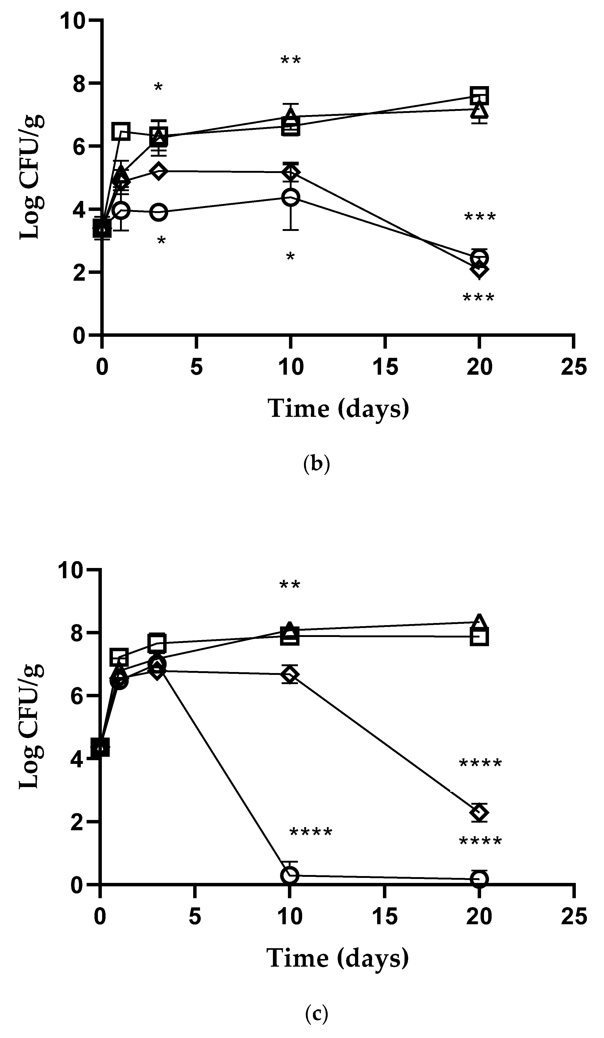
 ], PoAC [☐, ♢, ●]. Error bars represent standard deviations. p-values of < 0.05 (*), p < 0.01 (**), p < 0.001 (***) and p <0.0001 (****) were considered significant by t-test and ANOVA. ns stands for not statistically significant.
], PoAC [☐, ♢, ●]. Error bars represent standard deviations. p-values of < 0.05 (*), p < 0.01 (**), p < 0.001 (***) and p <0.0001 (****) were considered significant by t-test and ANOVA. ns stands for not statistically significant.
], PoAC [☐, ♢, ●]. Error bars represent standard deviations. p-values of < 0.05 (*), p < 0.01 (**), p < 0.001 (***) and p <0.0001 (****) were considered significant by t-test and ANOVA. ns stands for not statistically significant.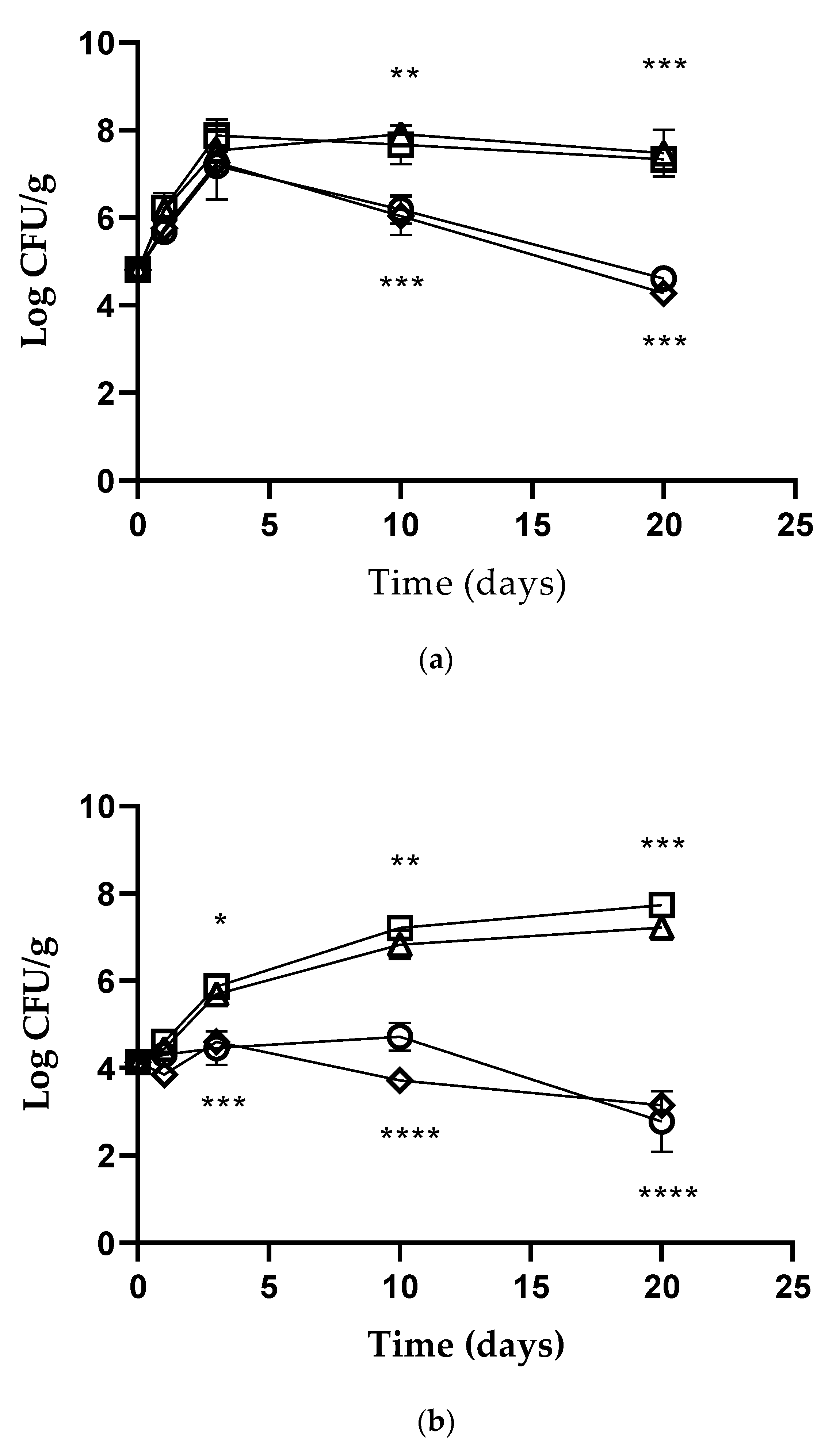
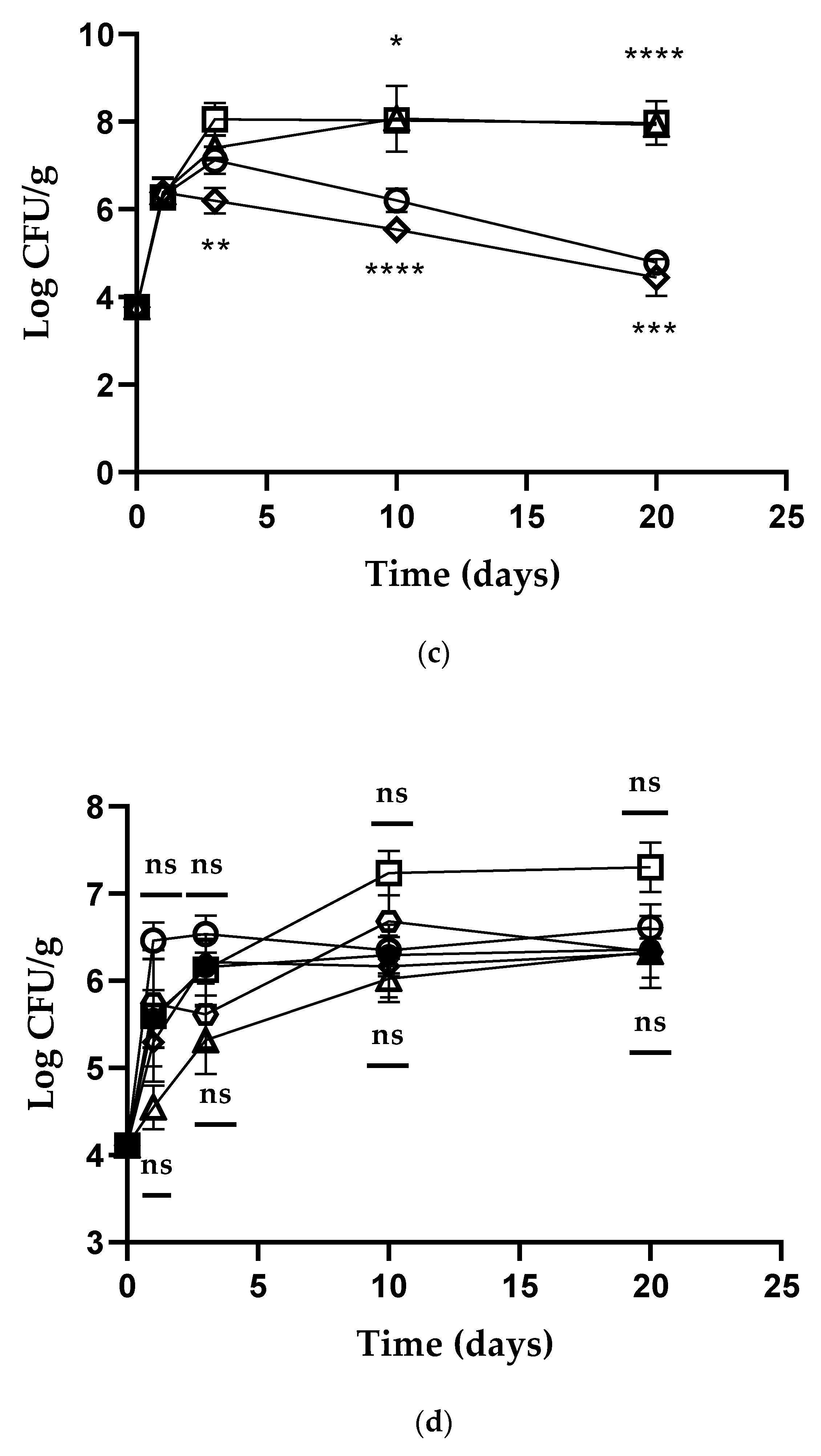 ], PrAC [☐, ♢, ●]. Error bars represent standard deviations. p-values of < 0.05 (*), p < 0.01 (**), p < 0.001 (***) and p < 0.0001 (****) were considered significant by t-test and ANOVA. ns stands for not statistically significant.
], PrAC [☐, ♢, ●]. Error bars represent standard deviations. p-values of < 0.05 (*), p < 0.01 (**), p < 0.001 (***) and p < 0.0001 (****) were considered significant by t-test and ANOVA. ns stands for not statistically significant.
], PrAC [☐, ♢, ●]. Error bars represent standard deviations. p-values of < 0.05 (*), p < 0.01 (**), p < 0.001 (***) and p < 0.0001 (****) were considered significant by t-test and ANOVA. ns stands for not statistically significant.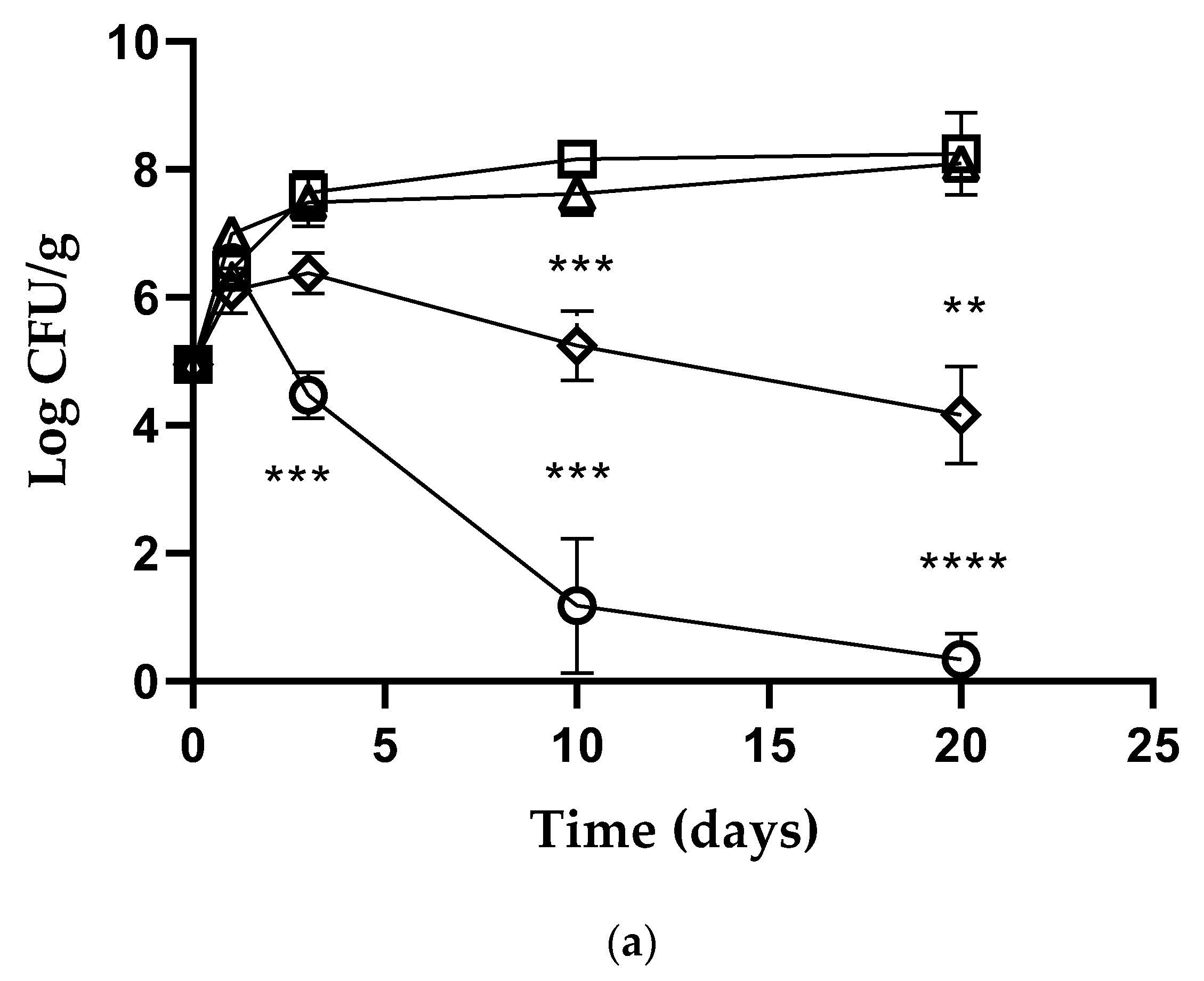


| Sample | Thickness (µm) |
|---|---|
| Polysaccharides film | 89.10 ± 11.10 |
| Protein film | 160.70 ± 14.10 |
| Sample | Test Conditions | WVTR [g/(m2 24 h)] | OTR [cm3/(m2 24 h)] |
|---|---|---|---|
| Polysaccharides film | T 23 °C RH 10% | 1.73 ± 0.07 | 0.10 ± 0.02 |
| Protein film | 16.46 ± 2.00 | 2.90 ± 0.13 |
Publisher’s Note: MDPI stays neutral with regard to jurisdictional claims in published maps and institutional affiliations. |
© 2022 by the authors. Licensee MDPI, Basel, Switzerland. This article is an open access article distributed under the terms and conditions of the Creative Commons Attribution (CC BY) license (https://creativecommons.org/licenses/by/4.0/).
Share and Cite
Iseppi, R.; Zurlini, C.; Cigognini, I.M.; Cannavacciuolo, M.; Sabia, C.; Messi, P. Eco-Friendly Edible Packaging Systems Based on Live-Lactobacillus kefiri MM5 for the Control of Listeria monocytogenes in Fresh Vegetables. Foods 2022, 11, 2632. https://doi.org/10.3390/foods11172632
Iseppi R, Zurlini C, Cigognini IM, Cannavacciuolo M, Sabia C, Messi P. Eco-Friendly Edible Packaging Systems Based on Live-Lactobacillus kefiri MM5 for the Control of Listeria monocytogenes in Fresh Vegetables. Foods. 2022; 11(17):2632. https://doi.org/10.3390/foods11172632
Chicago/Turabian StyleIseppi, Ramona, Chiara Zurlini, Ilaria Maria Cigognini, Mariarosaria Cannavacciuolo, Carla Sabia, and Patrizia Messi. 2022. "Eco-Friendly Edible Packaging Systems Based on Live-Lactobacillus kefiri MM5 for the Control of Listeria monocytogenes in Fresh Vegetables" Foods 11, no. 17: 2632. https://doi.org/10.3390/foods11172632
APA StyleIseppi, R., Zurlini, C., Cigognini, I. M., Cannavacciuolo, M., Sabia, C., & Messi, P. (2022). Eco-Friendly Edible Packaging Systems Based on Live-Lactobacillus kefiri MM5 for the Control of Listeria monocytogenes in Fresh Vegetables. Foods, 11(17), 2632. https://doi.org/10.3390/foods11172632