Article

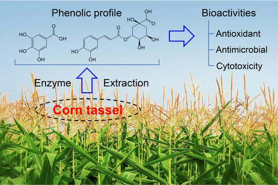
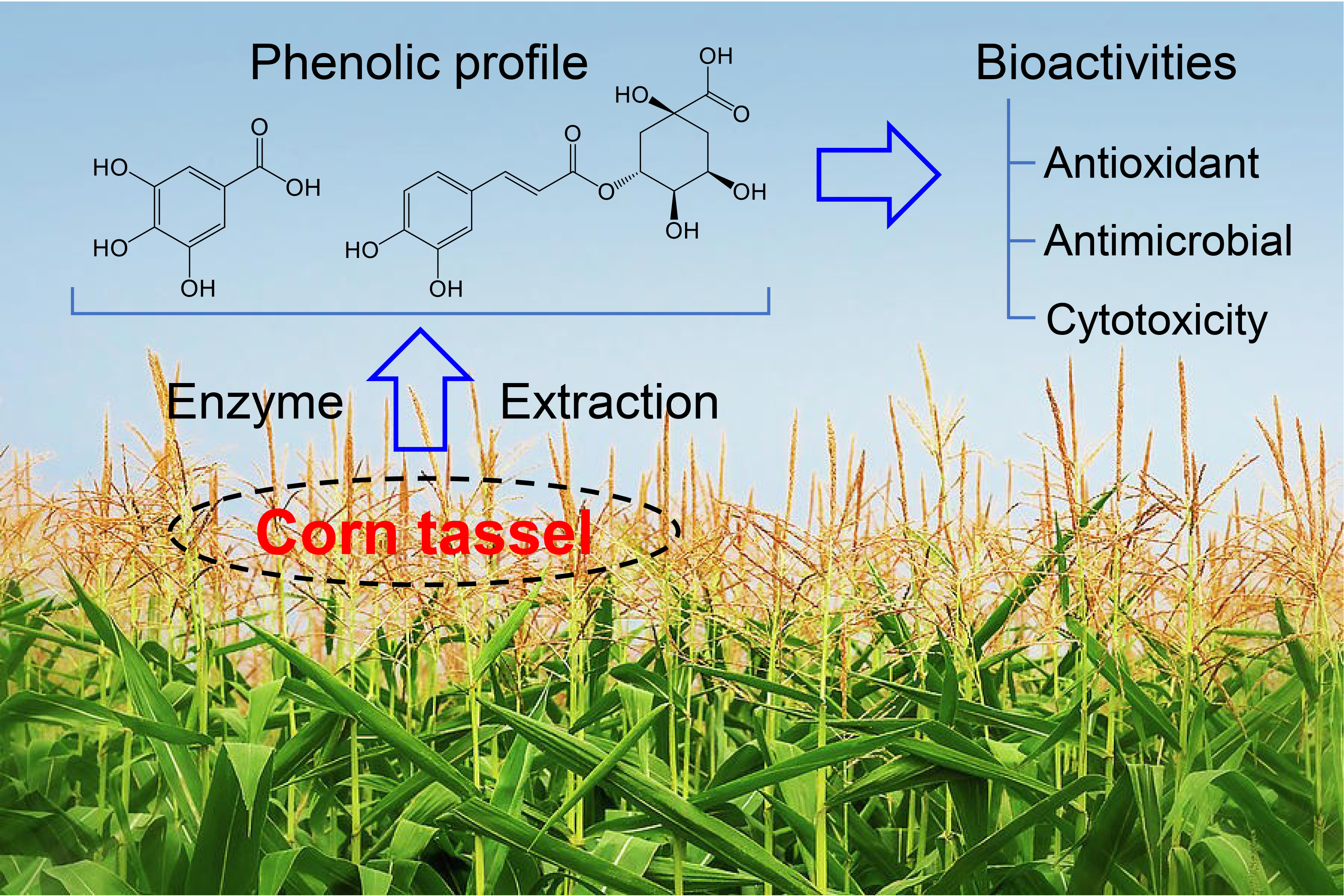
Phenolic Profiling and In-Vitro Bioactivities of Corn (Zea mays L.) Tassel Extracts by Combining Enzyme-Assisted Extraction

by
Nesren Elsayed
1,
Diaa A. Marrez
2,
Mohamed A. Ali
3,
Ahmed Ali Abd El-Maksoud
4,
Weiwei Cheng
5,* and
Tarek Gamal Abedelmaksoud
1,*
1
Food Science Department, Faculty of Agriculture, Cairo University, Giza 12613, Egypt
2
Food Toxicology and Contaminants Department, National Research Centre, Cairo 12622, Egypt
3
Biochemistry Department, Faculty of Agriculture, Cairo University, Giza 12613, Egypt
4
Department of Dairy Science, Faculty of Agriculture, Cairo University, Giza 12613, Egypt
5
Institute for Innovative Development of Food Industry, Institute for Advanced Study, Shenzhen University, Shenzhen 518060, China
*
Authors to whom correspondence should be addressed.
Foods 2022, 11(14), 2145; https://doi.org/10.3390/foods11142145
Submission received: 13 June 2022 / Revised: 29 June 2022 / Accepted: 7 July 2022 / Published: 20 July 2022

Abstract

:
In this work, enzyme-assisted extraction (EAE) of phenolic compounds from corn tassel using cellulase, protease, and their combination (1:1) was developed and optimized by central composite response surface methodology. The phenolic profile of obtained corn tassel extracts (CTE) was elucidated by high-performance liquid chromatography–diode array detection (HPLC–DAD) analysis, and their antioxidative, antimicrobial, and cytotoxic properties were evaluated in vitro. The results showed that CTE by EAE with combined enzymes had the highest total phenolic content (TPC). Under optimum enzymatic conditions, the experimental TPC values were 9.78, 8.45, and 10.70 mg/g, respectively, which were significantly higher than that of the non-enzymatic control (6.75 mg/g) (p < 0.05). Fourteen more phenolic compounds (13.80–1694.36 µg/g) were identified in CTE by EAE with the combined enzymes, and thus the antioxidant activity of that extract, determined by DPPH and ABTS radical scavenging method, was demonstrated to be stronger than that of the extracts by EAE with the single and ethanol extraction. Furthermore, this extract also showed remarkably better antimicrobial properties against all tested food-borne pathogenic bacteria and mycotoxigenic fungi than CTE by other extraction methods. CTE by EAE were nontoxic to normal lung fibroblast cell line (Wi-38) but cytotoxic to human colorectal and lung cancer cell lines (Caco-2 and A549), with IC50 values of 392.62–461.98 and 210.66–359.56 µg/mL, respectively, which indicated its potential anticancer properties. In conclusion, CTE by EAE, especially with the combined use of cellulase and protease, seems to hold promising potential for multifunctional application in food and pharma fields.

Graphical Abstract

1. Introduction

Corn (Zea mays L.) is one of the most extensively grown crops in the world, with an estimated global yield of 13.7 billion bushels [1]. Corn production in Egypt was 8 tons per ha [2]. Corn tassel is a waste part of the plant, and the chemical composition consists of carbohydrates 70.26%, ash 11.06%, fat 10.10%, and protein 6.26% [3]. In addition, corn tassel is considered an important source of valuable products such as volatile oils, lipids [4], phenolic compounds, and flavonoids such as quercetin, isorhamnetin, and kaempferol [5]. The main bioactive compounds in corn tassel are phenolic substances, which possess a variety of biological and physiological properties, including antioxidative, antibacterial, anticancer, anti-allergic, anti-atherogenic, anti-inflammatory, anti-thrombotic, cardioprotective, and vasodilatory actions [6]. To our knowledge, the most common method for extracting phenolic compounds is conventional solvent extraction. However, the disadvantage of solvent extraction is the loss of the phenolic compounds due to long extraction periods and some phenolic compounds’ degradation. In addition, conventional extraction requires a large amount of solvent, which is not economical. According to Pinelo et al. [7], various parameters influence extraction efficiency, including the type of solvent, solvent extraction, extraction time, extraction temperature, and solid-solvent ratio.
Recently, based on the avoidance of chemical ingredients’ side effects in foodstuffs, researchers have tried to replace conventional solvent extraction with green methods for the extraction of phytochemicals from plant sources such as enzymes, ultrasound, high hydrostatic pressure, etc. Enzymatic-assisted extraction as one of the green methods was applied to rupture the plant cell wall polysaccharides to enhance the release of bioactive compounds intermingled in plants.
Phenolic compounds in plant cell walls are both chemically diverse and botanically widespread. Therefore, in theory, the enzymatic treatment may extract more phenolic chemicals than solvent extraction. Many studies have also well used enzymes in the extraction of phenolic compounds [8,9]. However, until now, there are no reports available for using the enzymes to extract phenolic compounds from corn tassel. Furthermore, there is little information available concerning the bioactivities of corn tassel extract.
Therefore, the present work aims to improve the enzyme-assisted extraction process of phenolic components from corn tassels and assess the antioxidative, antimicrobial, and cytotoxic properties of extracts. To this purpose, a standard 2-factor central composite response surface design was adopted to optimize the phenolic recovery from corn tassel by enzyme-assisted extraction. The phenolic composition in corn tassel extract was determined by high-performance liquid chromatography–diode array detection (HPLC–DAD). Furthermore, the scavenging activities against DPPH and ABTS radicals, antimicrobial activities against food-borne pathogenic bacteria and mycotoxigenic fungi, and cytotoxic effects against cancer and normal cell lines were performed in our work. The results gathered in this paper would provide a solid foundation for the development of new value-added products from corn tassel, such as soft drinks and juices.

2. Materials and Methods

2.1. Chemicals

All reagents and chemicals used in this research were of analytical grade. Gallic acid, Folin-Ciocalteu reagent, ABTS 2,2′-azinobis (3-ethylbenzothiazoline-6-sulphonic acid), 2,2-diphenyl-1-picrylhydrazyl (DPPH), 3-(4,5-Dimethylthiazol-2-yl)-2,5-Diphenyltetrazolium Bromide (MTT), ethanol, sodium bicarbonate, sodium acetate, and butylated hydroxytoluene (BHT) were obtained from Sigma Chemical Co., Ltd. (St. Louis, MO, USA). Cellulase (Novozymes, Mumbai, India) (3500 U/g non-GMO) and Proteases (VemoZyme, Mumbai, India) (15,000 U/g) enzymes were obtained from Alfa-chemicals, El-Dokki-Giza, Egypt. Tryptic soy broth and Potato Dextrose agar were obtained from Conda Laboratories (Madrid, Spain).

2.2. Materials

Corn tassels were collected from the field of the Faculty of Agriculture, Cairo University, Egypt. Corn tassels were dried in an oven (Shel-lab, Cornelius, OR, USA) at 40 °C for 48 h until no further weight reduction was measurable, then subjected to grinding by an analytical mill to a size of 1 mm (Cole-Parmer, Vernon Hills, IL, USA), sieved up to 50 mesh and kept in the dark place at room temperature for analysis.

2.3. Conventional Extraction

The ethanolic solvent for the extraction of phenolic compounds was used. As described by Younis et al. [10], with some modifications, about one gram of dried corn tassel powder was added to 20 mL 60% (v/v) ethanol–water solutions and then subjected to a shaking incubator at room temperature for 24 h, followed by filtration through Whatman No. 4 filter paper to get pure extracts. The extracts were kept at −18 °C until further analysis.

2.4. Experimental Design and Statistical Analysis

Cellulase and protease were used either individually or their binary mixture (1:1) to extract phenolic compounds from corn tassels. The concentration of each enzyme (cellulase and protease) for extracting phenolic compounds was studied. Experimental variables were optimized to maximize the yield of extracted polyphenols using response surface methodology (RSM). A central composite response surface design generated from Design-Expert version 7.0.0 (Statease Inc., Minneapolis, MN, USA, Trial version) was used. The selected levels of incubation time (χ1) and enzyme concentration as U/g (χ2) for individual enzymes while pH (χ1) and Temperature (χ2) for mixed enzymes that result in the highest polyphenol content (data not shown) were 12, 24, 36, and 48 h; 500, 1000, 1500, 2000, and 2500 U/g; 4, 4.5, 5, 5.5, 6, 6.5, 7, and 7.5; and 35, 40, 45, 50, and 60 °C, respectively.
Browne–Forsythe and one-way analysis of variance (ANOVA) test with post hoc Tukey’s test (p < 0.05) were used to assess response variance homogeneity and significant effects among various treatments (combinations), respectively.
The response data were fitted to a second-order polynomial equation, which characterized the effect of the independent factors on the response as well as their combined effect on the response Y and determined the interrelationship between the test variables. To fit the experimental data, a reduced cubic model with linear, squared, and interaction factors was used. In the response surface analysis, the generalized second-order polynomial model was utilized, as given by (Equation (1)):
Y = ao + a1 χ1 + a2 χ2 + a12 χ1 χ2 + a11 χ12 + a22 χ22
where Y (i = 1–5) is the expected total phenolic content (TPC) response, ao the estimated regression coefficient of the fitted response at the central point of the model, a1, a2, and a3 the coefficient of regression for linear effect expressions, a11, a22, and a33 the quadratic effects, and a12, a13, and a23 the effects of interaction.
ANOVA was used to determine the statistical significance of the model and its various terms. In addition to R2, adjusted R2, and anticipated R2 values, the lack of fit test was employed to assess the adequacy of created models.

2.5. Enzyme-Assisted Extraction

The enzyme-assisted extraction was done as reported by Fernández et al. [11] with some adjustments. About 1 g corn tassel was extracted in 50 mL of buffer solution (0.1 M sodium acetate or 0.1 M sodium phosphate based on the pH of the used enzyme) containing enzyme under evaluation (cellulase, protease, and their mixture). Firstly, different incubation times (χ1) (12–60 h) and concentrations (χ2) (500–2500 U/g) for each enzyme (Cellulase and protease) were used. Secondly, the effect of temperature (χ1, 40 to 55 °C) and pH (χ2, 5 to 7) values of the cellulase–protease mixture with ratio (1:1) was also investigated. The hydrolysis conditions were chosen on the basis of the optimum temperature, pH, enzyme concentration, and incubation time for each enzyme as specified on the data sheets provided by the enzyme dealers. The extraction of corn tassel by 60% ethanol–water solution was accomplished in a precise shaking incubator (WiseCube WIS-20, Daihan scientific, Seoul, South Korea) with gentle agitation (200 rpm) in the dark. Finally, the sample was filtered utilizing Whatman No. 4 filter paper and stored at −18 °C in a brown flask till analysis.

2.6. Total Phenolic Compounds and HPLC Analysis

Total phenolic compounds of dried corn tassel extract were estimated by Folin–Ciocâlteu method based on Abedelmaksoud et al. [12], and results were expressed as mg Gallic acid equivalents (GAE)/g powder.
The phenolic composition of the optimal corn tassels extract was determined using an Agilent 1260 series HPLC system (Agilent Technologies Inc., Santa Clara, CA, USA) according to Elsayed, Hammad, and Abd El-Salam [10].

2.7. Antioxidant Activity

DPPH radical inhibition efficiency of the investigated Corn tassels extract was determined according to Elsayed et al. [13]. The absorbance of the samples was measured using the UV-visible spectrophotometer (Unico UV-2000, Dayton, NJ, USA) at 517 nm. Butylated hydroxytoluene was used as a standard.
ABTS radical scavenging activity was evaluated using the ABTS method [14]. The results are expressed as a percentage of quenched radicals according to the equation: ABTS scavenging activity (%) = (Ac − As)/Ac × 100, where Ac is the absorbance of ABTS, and As is the absorbance of the sample.

2.8. Antimicrobial Activity

Tested microorganism, the inhibitory effect of corn tassels extracts was examined against five strains of food-borne pathogenic bacteria. Two Gram-positive bacteria Bacillus cereus EMCC 1080, Staphylococcus aureus ATCC 13565, and three gram-negative bacteria Salmonella typhi ATCC 25566, Escherichia coli 0157 H7 ATCC 51659, and Pseudomonas aeruginosa NRRL B-272. Eight fungal species were used for antifungal assay, containing Aspergillus flavus NRR 3357, A. parasiticus SSWT 2999, A. ochraceus ITAL 14, A. niger IM I288550, A. westerdijikia CCT 6795, A. carbonarius ITAL 204, Fusarium proliferatum MPVP 328, and Penicillium verrucosum BFE 500.
The sensitivity test of corn tassels extracts was determined with various bacterial and fungal cultures using disc diffusion method by the Kirby–Bauer technique [15,16]. DMSO was a negative control, and ceftriaxone, commercial fungicide (miconazole nitrate, 1 mg mL−1), was used as a positive control for bacterial and fungal, then the inoculated plates were incubated at 37 °C for 24 h for bacterial and 25 °C for 24–48 h for fungal. At the end of the incubation period, inhibition zones were measured and expressed as the diameter of the clear zone with the diameter of the paper disc. All treatments consisted of three replicates, and the averages of the experimental results were determined.
Minimum inhibitory concentration (MIC) for corn tassel extracts was determined using the micro broth dilution methods as of Andrews [17]. Two-fold serial dilutions of the different extract concentrations ranging from 5.0 mg mL−1 to 10.0 µg mL−1 were used. Equal volumes of tested bacteria (108 cfu mL−1; 0.5 McFarland standards) were added to each. A 24 h culture of the tested bacterial species was diluted in 10 mL of tryptic soy broth (TSB) with reference to the 0.5 McFarland standard to achieve inoculation of well. MIC values were taken as the lowest concentration of the antimicrobial agent that inhibited bacterial growth after 24 h incubation at 37 °C.
MIC against fungi was performed by using the technique of Marrez and Sultan [18]. Corn tassel extracts at different concentrations were separately dissolved in 0.5 mL of 0.1% Tween 80 (Merck, Darmstadt, Germany), then mixed with 9.5 mL of melting, 45 °C, PDA and poured into a Petri dish (6 cm). The prepared plates were centrally inoculated with 3 µL of fungal suspension (108 cfu mL−1; 0.5 McFarland standards). The plates were incubated at 25 °C for 24–48 h. At the end of the incubation period, mycelial growth was monitored, and MIC was determined.

2.9. Cytotoxic Activity

The cytotoxic effects of corn tassel extracts by ethanol, cellulose, protease, and mixed enzymatic extraction on human cancerous and normal cell lines were examined by MTT assay according to Senthilraja and Kathiresan [19] against human colorectal carcinoma cell line (Caco-2), human Lung carcinoma cell line (A549), and human lung fibroblast normal cell line (Wi-38).

2.10. Statistical ANALYSIS

Chemical assays were performed in triplicate, and the results were represented as mean standard deviations. Statistical analysis was performed using the Originpro 9.6 software (OriginLab Corp., Northampton, MA, USA). One-way ANOVA was applied to analyze the significant differences by GraphPad Prism 5.02 software (GraphPad Prism Software Inc., San Diego, CA, USA). A p-value of <0.05 was considered statistically significant.

3. Results and Discussion

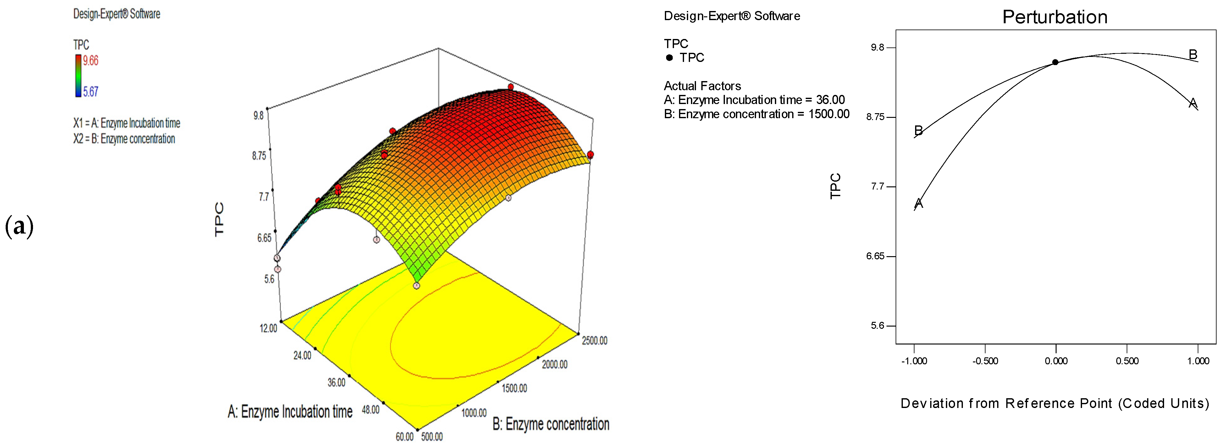
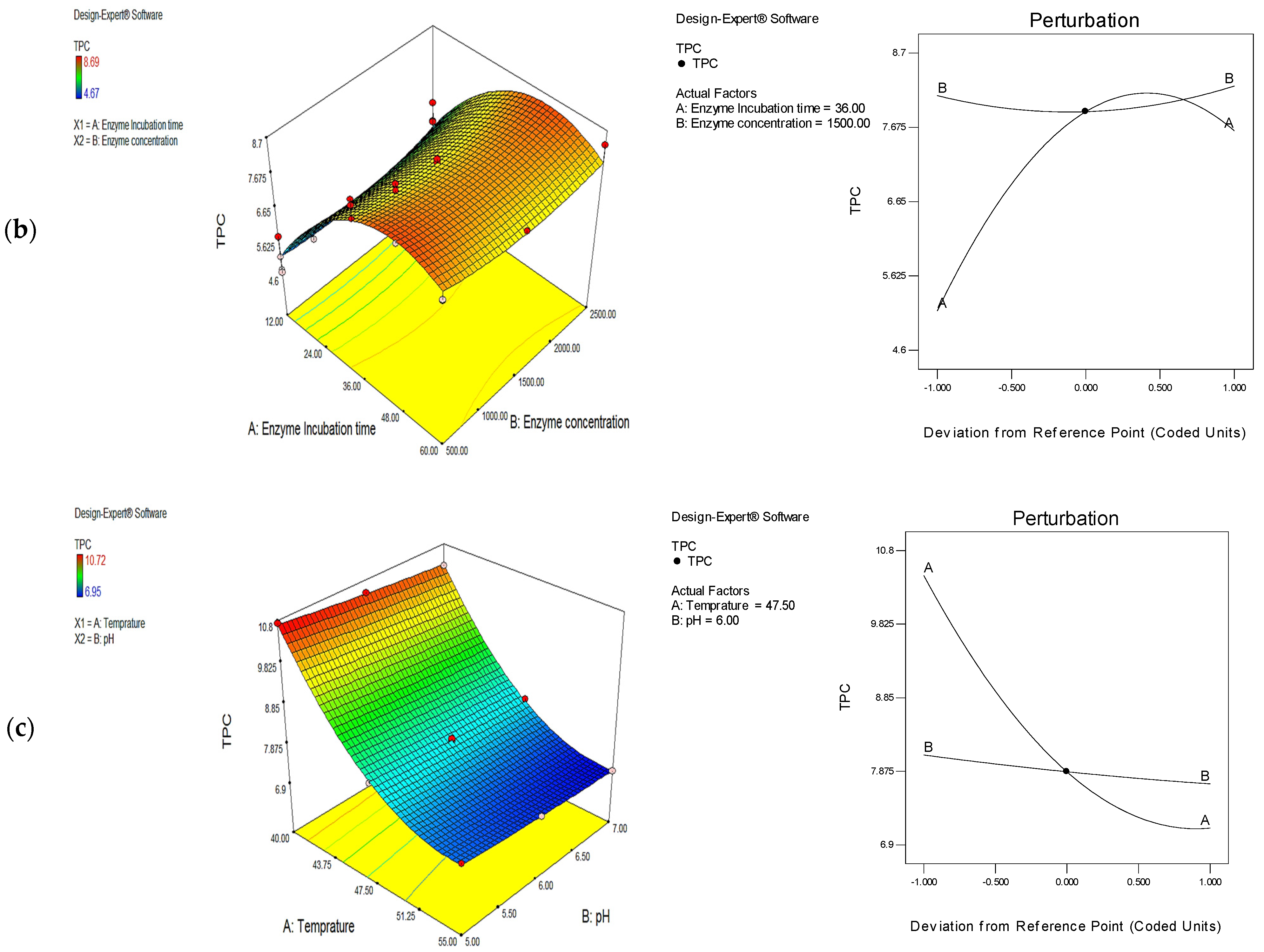
3.1. Optimization of Phenolic Extraction Conditions with the Use of Cellulase, Protease, and Their Mixture (1:1)

The utilization of enzymes for phenolic extraction from corn tassels was conducted using cellulase, protease, and their mixture (1:1). Firstly, different incubation times (χ1) and enzyme concentrations (χ2) for each enzyme (cellulase and protease) were used (Tables S1 and S2Figure 1a,b) for RSM to assess their impact on TPC values and optimize extraction parameter. The TPC value in corn tassel extract varied from 5.67 to 9.66 mg/g for cellulase treatments and from 4.67 to 8.69 mg/g for protease treatments (Tables S1 and S2). Then, the effects of temperature (χ1, from 40 to 55 °C) and pH (χ2, from 5 to 7) values of the enzyme mixture, including cellulose and protease with ratio (1:1) were also investigated (Table S3Figure 1c) for RSM to assess the impact of the mixture of enzymes on the TPC values and also optimize extraction parameters. The TPC value of corn tassel extract varied between 6.95 and 10.72 mg/g for the mixture treatment (Table S3). The influence of cellulase and protease concentrations on the extraction of phenolic compounds from corn tassels was very remarkable for extracting such compounds. It was observed that the extraction amounts of phenolic compounds in corn tassel extracts by enzyme-assisted method increased by 41.62% (cellulase), 25.62% (protease), and 59.11% (cellulase–protease mixture 1:1) compared to that in the control sample (6.75 mg/g) at the respective optimum condition was observed (Table 1).
Because the cell wall is composed of cellulose, hemicellulose, pectin, and protein, as well as phenolic compounds linked to available polysaccharides via hydrogen and hydrophobic bonds, hydrolyzing enzymes such as cellulase, pectinase, and hemicellulose can be used to destroy the cell wall structure. These enzymes can also be utilized to improve the penetrating capacity of cell walls, resulting in the release of phenolic compounds and a higher extraction yield of bioactive substances [20]. Another mechanism is most likely the enzyme’s direct action on the breakdown of ester or ether bonds between phenols and plant cell wall polymers [21]. Furthermore, cellulase impacts the cellulose found beneath the main layer and the midline lamella of plant cells. The main layer is made up of a stiff and strong cellulose skeleton that is embedded in a gel-like matrix of hemicellulose, pectin, and glycoprotein. All responses had a significant sum of squares and higher regression coefficients, indicating compliance with the variables set with a second-order polynomial (see Equations (2)–(4)). To obtain regression models, linear, interactive (2 FI), quadratic, and cubic models were fitted to the experimental data, and the results are shown in Table S4. From Table S4, the quadratic model was the best fit for modeling experimental data. Thus, the values of the quadratic model’s coefficient of determination (R2), modified coefficient of determination (Adj-R2), and expected coefficient of determination (Pred-R2) were among the highest and most significant (p < 0.001). The multiple regression equations generated in uncoded form between the various responses and process variables are shown below (Equations (2)–(4)):
TPC (Cellulase) = 9.57 + 0.76χ1 + 0.57χ2 − 0.15χ1χ2 − 1.47χ12 − 0.56χ22
TPC (Protease) = 7.89 + 1.24χ1 + 0.066χ2 − 0.18χ1χ2 − 1.50χ12 + 0.29χ22
TPC (mix) = 7.87 − 1.67χ1 − 0.19χ2 + 0.010χ1χ2 + 0.93χ12 + 0.031χ22
Appropriate model parameters (p < 0.05) are χ1 (enzyme incubation time), χ2 (enzyme concentration), χ1χ2, χ12, χ22 in the case of cellulase and protease treatments. While in case of mixed enzyme (1:1), χ1 (Temperature), χ2 (pH), χ1χ2, χ12, χ22, are significant model terms (p < 0.05). Regression coefficients magnitude designated maximum positive effect of mixed enzyme (TPC was 10.74 mg/g) followed by cellulase (TPC was 9.56 mg/g) and protease (TPC was 8.48 mg/g) (Table 1).
Figure 1a,b illustrated the TPC increased significantly with increasing incubation time (factor A) till 36 h and then decreased subsequently. It was also found that there was no significant effect for factor B (enzyme concentration) on the TPC in corn tassel extract by protease-assisted extraction (Figure 1b). Therefore, surface plots of the model equation, disturbance, and 3D response demonstrated that incubation time has a relatively bigger effect on TPC values for cellulase- or protease-assisted extraction than enzyme concentration. For enzyme-assisted extraction with the combined use of cellulase and protease, the effect of incubation temperature on the TPC in corn tassel extract was bigger than that of pH. The optimal combination of factors with the highest desirability value was chosen to be 1.00, 0.95, and 0.99 for cellulase, protease, and their mixture, respectively. The optimum conditions are presented in Table 1. Corn tassel extraction by solvent and enzyme-assisted method was conducted under these conditions in the lab, and the experimental response values matched the anticipated value using the model obtained for the optimized corn tassel to extract TPC. Hence, the fitted models were determined to be suitable for predicting the responses (Table 1).

3.2. Identification of the Phenolic Compounds in Corn Tassel Extract Obtained by Enzyme-Assisted Method

HPLC was used to identify the phenolic compounds in corn tassel extracts and evaluate for the first time the effect of the enzyme used on the composition of phenolic compounds. From Figure S1 and Table 2, the extraction technique has a slight influence on the varieties of phenolic components; 14 compounds were detected in corn tassel extract by cellulase–protease-mixture-assisted method and 12 compounds in the extract by cellulose- or protease-assisted method. Ellagic acid and taxifolin were not detected in the extract by cellulose-assisted extraction, while kaempferol and coumaric acid were not detected in the extract by protease-assisted extraction. The common phenolic compounds in corn tassel extracts obtained by three methods were listed as gallic acid, chlorogenic acid, caffeic acid, naringenin, rutin, ferulic acid, syringic acid, and catechin, and their contents except catechin and pyrocatechol for the sample from mixed-enzyme-assisted extraction were significantly higher than that for the sample from single-enzyme-assisted extraction (Table 2). The finding indicated that mixed-enzyme-assisted extraction could lead to an increase in the number of phenolic compounds in corn tassel extracts compared to cellulase/protease-assisted extraction. Cerda et al. [22] reported that the extraction by enzymes raises the number of phenolic compounds extracted in addition to the antioxidant activity, the mechanism of enzymatic extraction is the degradation of the network of polysaccharides in the plant cell wall where this network retains the phenolic compounds. Another mechanism of enzymatic extraction is direct catalysis causing distraction of the ether and/or ester bonds between the phenols and polymers of the plant cell wall [21].

3.3. Antioxidant Activity of Corn Tassel Extracts

The assays of DPPH and ABTS radical scavenging were used to evaluate the antioxidant capacity of corn tassel extracts from different extraction methods, and the results are shown in Table 3. The results presented significant variation in DPPH radical scavenging activity for the extracts from different extraction methods. The DPPH and ABTS radical scavenging activities were observed to be significantly higher in the corn tassel extracted by enzymes (83.20–92.98% and 88.70–95.18%, respectively) compared to ethanol extract (75.25% and 80.25%, respectively) and also BHT (87.69% and 83.74%, respectively). The extract obtained by mixed enzyme showed the highest antioxidant activity, followed by cellulose > protease > ethanol. Our results were similar to Cerda, Martínez, Soto, Poirrier, Perez-Correa, Vergara-Salinas, and Zúñiga [22], who reported that enzyme-assisted extraction raised the number of phenolic compounds in the extracts, also resulting in an increase in the antioxidant activity compared to the ethanolic extraction. de Vries and Visser [23] attributed the efficiency of extraction with enzymes to an increase in the antioxidant activity due to 90% of the plant cell wall being formed from polysaccharides, which consist of cellulose, hemicellulose, and pectin. Enzymatic analysis of the plant wall polysaccharides with certain enzymes led to a rise in the release of compounds combined with the cell wall components, and this is a good replacement for conventional extraction methods; this explains the reasons for the superiority of cellulase over protease in antioxidant activity. Additionally, Shahidi [24] found that the radical scavenging capacity of protease extracts could result from their ability to enhance the extraction capacity of phenolic compounds which was known as strong DPPH radical scavengers.

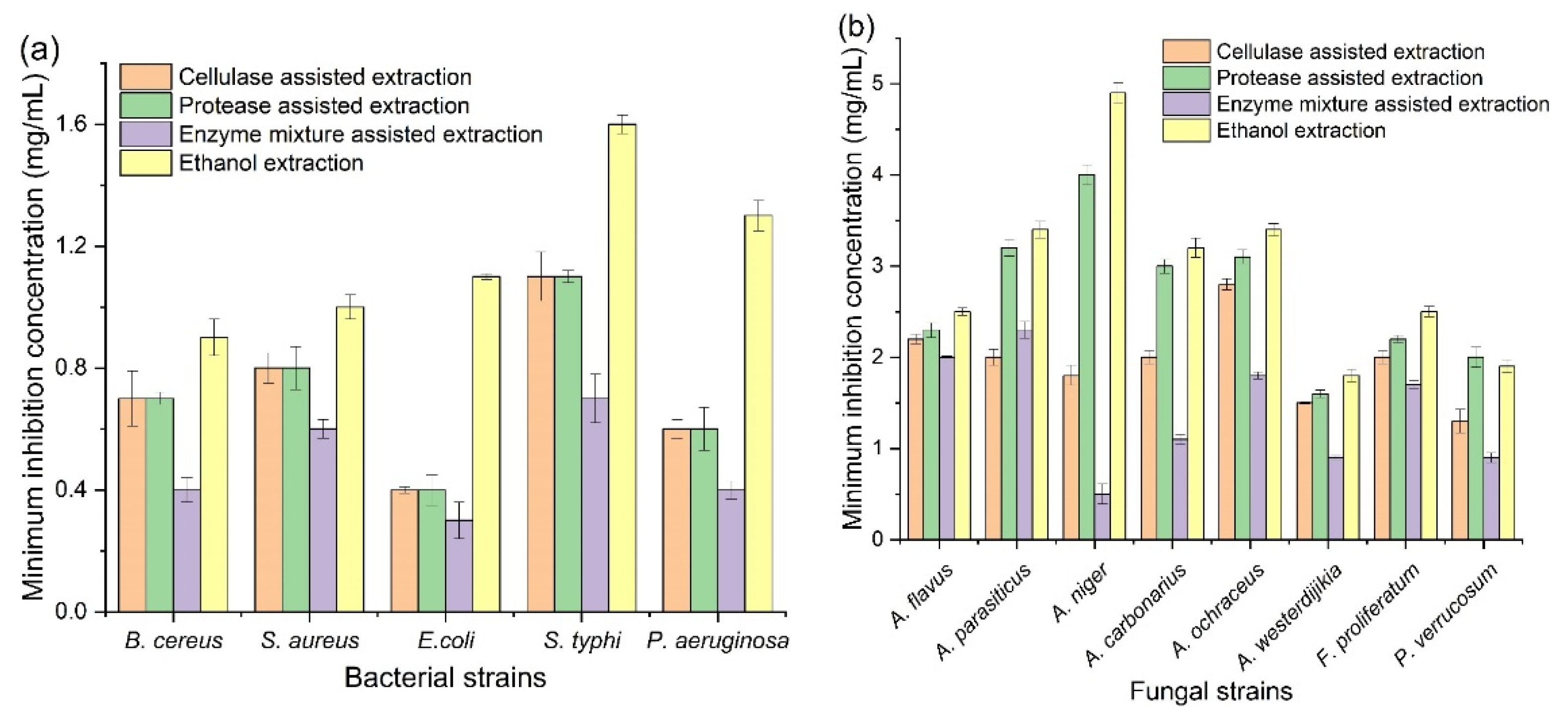
3.4. Antimicrobial Activity of Corn Tassel Extracts

The antibacterial activity of corn tassel extracts by ethanol, cellulose, protease, and their binary-mixture-assisted extraction against two Gram-positive and three Gram-negative bacteria of food borne pathogenic are shown in Table S5. The corn tassel extract from enzyme-mixture-assisted extraction had the highest inhibition zone (12.8, 11.5, 10.8, 10.5, and 10.2 mm, respectively) against all borne pathogenic bacteria (E. coli, P. aeruginosa, S. typhi, B. cereus, and S. aureus) compared to other extracts. In addition, the corn tassel extracts by ethanol had a relatively lower inhibition zone for all pathogens bacterial, with a diameter of the inhibition zones varying from 5.7 to 7.9 mm. The results also showed that the corn tassels extract by protease exhibited moderated inhibitory activity against B. cereus (8.7 mm) and S. aureus (8.8 mm) compared to the positive control. As shown in Figure 2a, the minimum inhibitory concentrations (MIC) of corn tassel extract from enzyme-mixture-assisted extraction against tested bacterial strains was observed to be 0.3–0.7 mg/mL, which were significantly lower than that of the extracts by cellulase- or protease-assisted extraction (1.3–2.8 and 1.6–3.2 mg/mL, respectively) and also ethanol extraction (1.8–4.9 mg/mL). The finding suggested that the antibacterial activity of corn tassel extract by mixed-enzyme-assisted extraction was stronger than that of the extracts by single-enzyme-assisted extraction and ethanol extraction.
Additionally, the inhibition zone diameter of corn tassel extracts against mycotoxigenic fungus is shown in Table S6. The corn tassel extracts had antifungal activity against all tested mycotoxigenic fungi. Meanwhile, the corn tassel extract by a cellulase–protease mixture showed the strongest antifungal activity, 11.0 mm against A. westerdijikia, followed by A. niger and P. verrucosum with inhibition zone 10.7 and 10.0 mm, respectively. The corn tassel extract by ethanol showed the lowest antifungal activity against all examined fungi, and the highest inhibition zones, 7.2 and 7.1 mm, were observed against A. carbonarius and A. westerdijikia, respectively, while the lowest activity was observed against A. flavus with inhibition zone 6.1 mm.
Minimum inhibitory concentrations of corn tassel extracts against mycotoxigenic fungi are shown in Figure 2b. The lowest MIC value was observed in corn tassel extracts by mixed-enzyme-assisted method, followed by corn tassel extracts by cellulase, protease, and ethanol extraction. The highest activity in corn tassel extracts by mixed-enzymes-assisted extraction and corn tassel extracts by cellulase were observed against A. niger and P. verrucosum with MIC values of 0.5 and 0.7 mg/mL, respectively; however, the corn tassel extract by ethanol and cellulase recorded the lowest activity against A. niger, with MIC values 4.9 and 4.00 mg/mL, respectively. The antimicrobial activity of corn tassels extracts could be attributed to the high phenolic compounds such as gallic acid, chlorogenic acid, and caffeic acid, which had higher contents in the extract by cellulase–protease-mixture-assisted extraction than that by other extraction methods (Table 2). The phenolic compounds could damage the bacterial cell by decaying the cell wall, stopping the turn of ion channels, and inhibiting the synthesis of adenosine triphosphate [25]. Accordingly, the antimicrobial activity of corn tassel extract by mixed-enzyme-assisted extraction was stronger than that of the extracts by single-enzyme-assisted extraction and ethanol extraction.

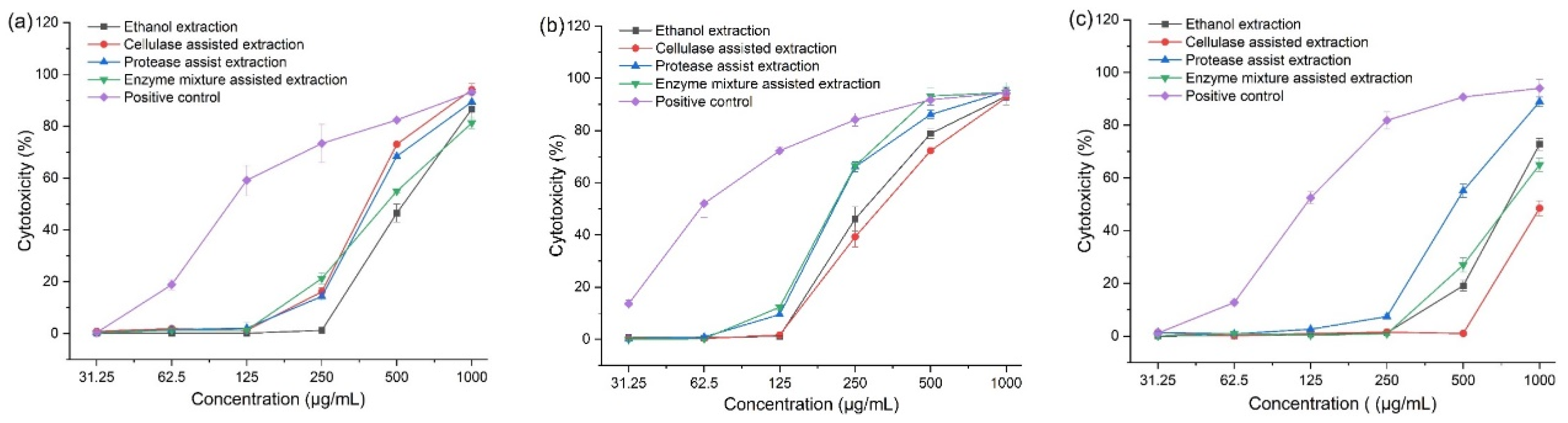
3.5. Cytotoxic Activity of Corn Tassel Extracts

Cytotoxicity was expressed as the percentage growth inhibition of Caco-2, A549, and Wi-38 cells treated with various doses of extracts (from 1000 to 31.25 μg/mL). Doxorubicin, an anticancer drug for hematologic and solid tumors, was used as the positive control. From Figure 3, some of the extracts caused cell cytotoxicity in a concentration-dependent manner. Probit analysis was carried out for IC50 determination, and the results are shown in Table 4. The mixed enzyme extract had the highest inhibitory activity against the Caco-2 cell line (IC50, 392.62 μg/mL) and A549 cell line (IC50, 210.66 μg/mL) compared to the extracts obtained by free enzyme and ethanol extraction, and a low toxicity effect on the Wi-38 cell line (IC50, 809.85 μg/mL). The observation indicated that corn tassel extract obtained by mixed-enzyme-assisted extraction was nontoxic to normal lung fibroblast cell line but cytotoxic to human colorectal and lung cancer cell lines with the dose-dependent effect. The antiproliferative activity of doxorubicin was found to be more potent against the Caco-2 cell line (IC50, 110.83 μg/mL) and A549 cell line (IC50, 61.42 μg/mL) and had the highest toxicity effect on the Wi-38 cell line (IC50, 122.72 μg/mL).
Chemotherapy is one of the most efficient cancer therapies, but its efficiency is limited due to significant drug-related side effects. Extended chemotherapy treatment reduces the immune system response, making patients more susceptible to various diseases and infections. Doxorubicin is a powerful anticancer medication that can be used to treat both hematologic and solid cancers. However, it has the potential to produce multi-organ toxicity in a variety of patients. Doxorubicin toxicity can be attributed to apoptosis, inflammation, and oxidative stress, among other mechanisms [26]. Plants have been reported to treat a variety of cancers and they provide leads for the creation of possible new drugs [27,28]. The most widely dispersed natural minor metabolites in plants are phenolic compounds. Flavonoids are a significant category of phenolics that play a key role in functional extracts [29,30]. Corn tassels are a valuable by-product and a valuable source of bioactive phytochemicals [31].
The variations observed among corn tassel extraction methods were found in HPLC and antioxidant activities results as mentioned above. The results showed that the effect of the extraction method was the main reason for the difference in the cytotoxicity. Corn tassel extract by enzyme-mixture-assisted method recorded the highest phenolic content and antioxidant capacity. Therefore, in previous cytotoxic screenings with different extracts, the score of cytotoxic responses of cellulase–protease mixture extract was higher than the ethanol, cellulase and protease extracts against Caco-2 and A549 cell lines. Phenolic compounds, which are isolated from dried corn tassel, are bioactive phytochemicals with antioxidant properties [3]. Additionally, the corn tassel consists of polysaccharide, saponin, and flavonoids, which can inhibit the proliferation of MGC80-3 gastric cancer cells in vitro. Furthermore, “Tasselin A”, a pure 4-hydroxyl-1-oxindole-3-acetic acid isolated from sweet corn tassels, was found to have a strong antioxidant potential [32,33]. Corn bioactive compounds also decreased levels of Ras protein and nitric oxide synthases (NOS) inflammatory enzymes and decreased mRNA expression of cytokines such as tumor necrosis factor-alpha (TNF-α) and interleukin 6 (IL-6) while increasing the free radical detoxifying enzymes mRNA expression. Corn extracts activated caspase-3 and lowered the levels of Ras protein in tumor cells at the molecular level [34,35,36,37].

4. Conclusions

The recovery of phenolic compounds from corn tassel was well improved by enzyme-assisted extraction compared to ethanolic extraction. Especially TPC in corn tassel extract was greatly enhanced by the combined use of cellulase and protease (1:1). Under the optimal enzyme pretreatment conditions obtained by RSM, the TPCs in corn tassel extracts by cellulase-, protease-, and their mixture-assisted extraction were up to 9.56, 8.45, and 10.70 mg/g, respectively. Furthermore, more varieties of phenolic compounds in corn tassel extract by mixed-enzyme-assisted extraction were quantitatively identified in corn tassel extract by mixed-enzyme-assisted extraction than that by single-enzyme-assisted extraction. Additionally, based on the DPPH and ABTS free radical scavenging assay, the antioxidant activity of corn tassel extract by mixed-enzyme-assisted extraction was significantly stronger than that of the single-enzyme-assisted extracts, ethanolic extract, and also BHT (p < 0.05). Corn tassel extract by mixed-enzyme-assisted extraction showed remarkably better antimicrobial properties against all tested food-borne pathogenic bacteria and mycotoxigenic fungi than CTE by other extraction methods. It was also confirmed that corn tassel extract obtained by mixed-enzyme-assisted extraction was nontoxic to normal lung fibroblast cell line (Wi-38) but cytotoxic to human colorectal and lung cancer cell lines (Caco-2 and A549) with the dose-dependent effect.

Supplementary Materials

The following supporting information can be downloaded at: https://www.mdpi.com/article/10.3390/foods11142145/s1, Table S1: Central composite design and response for TPC value in corn tassel extract by cellulase-assisted method; Table S2: Central composite design and response for TPC value in corn tassel extract by protease-assisted method; Table S3: Central composite design and response for TPC value in corn tassel extract by mixed enzyme-assisted method; Table S4: Regression coefficients of the fitted second-order polynomials representing the relationship between the responses and variables; Table S5: Antibacterial activity of corn tassels extracts by different extraction methods; Table S6: Antifungal activity of corn tassels extracts by different extraction methods; Figure S1: HPLC chromatograms for phenolic compounds in corn tassel extracts by enzyme-assisted method. A: Cellulase; B protease; C: cellulase-protease mixture (1:1).

Author Contributions

Methodology, data curation, writing—original draft, N.E.; writing—review and editing, D.A.M.; validation, writing—review and editing, methodology, M.A.A.; software, validation, formal analysis, A.A.A.E.-M.; supervision, funding acquisition, writing—review and editing, W.C.; conceptualization, supervision, writing—review and editing, T.G.A. All authors have read and agreed to the published version of the manuscript.

Funding

This work was financially supported by the National Natural Science Foundation of China (32001814 and 32172318) and the Key-Area Research and Development Program of Guangdong Province (2019B020212001).

Institutional Review Board Statement

Not applicable.

Informed Consent Statement

Not applicable.

Data Availability Statement

Data are contained within the article or Supplementary Materials.

Conflicts of Interest

The authors declare no conflict of interest.

References

  1. USDA National Agricultural Statistics Service. Crop Production 2019 Summary (January 2020). Available online: https://usda.library.cornell.edu/concern/publications/k3569432s (accessed on 12 January 2022).
  2. Knoema. Sugarcane Area Harvested. Available online: https://knoema.com/atlas/topics/Agriculture/Crops-Production-Area-Harvested/Sugar-cane-area-harvested (accessed on 28 March 2020).
  3. Mohsen, S.M.; Ammar, A.S. Total Phenolic Contents and Antioxidant Activity of Corn Tassel Extracts. Food Chem. 2009, 112, 595–598. [Google Scholar] [CrossRef]
  4. Buttery, R.G.; Ling, L.C.; Teranishi, R. Volatiles of Corn Tassels: Possible Corn Ear WORM Attractants. J. Agric. Food Chem. 1980, 28, 771–774. [Google Scholar] [CrossRef]
  5. Ceska, O.; Styles, E.D. Flavonoids from Zea mays Pollen. Phytochemistry 1984, 23, 1822–1823. [Google Scholar] [CrossRef]
  6. Balasundram, N.; Sundram, K.; Samman, S. Phenolic Compounds in Plants and Agri-Industrial by-Products: Antioxidant Activity, Occurrence, and Potential Uses. Food Chem. 2006, 99, 191–203. [Google Scholar] [CrossRef]
  7. Pinelo, M.; Rubilar, M.; Jerez, M.; Sineiro, J.; Núñez, M.J. Effect of Solvent, Temperature, and Solvent-to-Solid Ratio on the Total Phenolic Content and Antiradical Activity of Extracts from Different Components of Grape Pomace. J. Agric. Food Chem. 2005, 53, 2111–2117. [Google Scholar] [CrossRef] [PubMed]
  8. Drevelegka, I.; Goula, A.M. Recovery of Grape Pomace Phenolic Compounds through Optimized Extraction and Adsorption Processes. Chem. Eng. Process.-Process Intensif. 2020, 149, 107845. [Google Scholar] [CrossRef]
  9. Krakowska, A.; Rafińska, K.; Walczak, J.; Buszewski, B. Enzyme-assisted Optimized Supercritical Fluid Extraction to Improve Medicago Sativa Polyphenolics Isolation. Ind. Crops Prod. 2018, 124, 931–940. [Google Scholar] [CrossRef]
  10. Younis, M.I.; Xiaofeng, R.; Alzubaidi, A.K.; Mahmoud, K.F.; Altemimi, A.B.; Cacciola, F.; Raza, H.; Pratap-Singh, A.; Abedelmaksoud, T.G. Optimized Green Extraction of Polyphenols from Cassia javanica L. Petals for Their Application in Sunflower Oil: Anticancer and Antioxidant Properties. Molecules 2022, 27, 4329. [Google Scholar] [CrossRef]
  11. Fernández, K.; Vega, M.; Aspé, E. An Enzymatic Extraction of Proanthocyanidins from País Grape Seeds and Skins. Food Chem. 2015, 168, 7–13. [Google Scholar] [CrossRef]
  12. Abedelmaksoud, T.G.; Mohsen, S.M.; Duedahl-Olesen, L.; Elnikeety, M.M.; Feyissa, A.H. Optimization of Ohmic Heating Parameters for Polyphenoloxidase Inactivation in not-from-Concentrate Elstar Apple Juice using RSM. J. Food Sci. Technol. 2018, 55, 2420–2428. [Google Scholar] [CrossRef]
  13. Elsayed, N.; El-Din, H.S.; Altemimi, A.B.; Ahmed, H.Y.; Pratap-Singh, A.; Abedelmaksoud, T.G. In Vitro Antimicrobial, Antioxidant and Anticancer Activities of Egyptian Citrus Beebread. Molecules 2021, 26, 2433. [Google Scholar] [CrossRef] [PubMed]
  14. Arnao, M.B.; Cano, A.; Acosta, M. The Hydrophilic and Lipophilic Contribution to Total Antioxidant Activity. Food Chem. 2001, 73, 239–244. [Google Scholar] [CrossRef]
  15. Baur, A.W.; Kirby, W.M.M.; Sherris, J.C.; Turck, M. Antibiotic Susceptibility Testing by a Standardized Single Disk Method. Am. J. Clin. Pathol. 1966, 45, 493–496. [Google Scholar] [CrossRef]
  16. Marrez, D.A.; Naguib, M.M.; Sultan, Y.Y.; Higazy, A.M. Antimicrobial and Anticancer Activities of Scenedesmus Obliquus metabolites. Heliyon 2019, 5, e01404. [Google Scholar] [CrossRef] [PubMed] [Green Version]
  17. Andrews, J.M. Determination of Minimum Inhibitory Concentrations. J. Antimicrob. Chemoth. 2001, 48, 5–16. [Google Scholar] [CrossRef] [PubMed] [Green Version]
  18. Marrez, D.A.; Sultan, Y.Y. Antifungal Activity of the Cyanobacterium Microcystis Aeruginosa against Mycotoxigenic Fungi. J. Appl. Pharm. Sci. 2016, 11, 191–198. [Google Scholar] [CrossRef] [Green Version]
  19. Senthilraja, P.; Kathiresan, K. In Vitro Cytotoxicity MTT Assay In Vero, HepG2 and MCF-7 Cell Lines Study of Marine Yeast. J. Appl. Pharm. Sci. 2015, 5, 80–84. [Google Scholar] [CrossRef] [Green Version]
  20. Miron, T.; Herrero, M.; Ibáñez, E. Enrichment of Antioxidant Compounds from Lemon Balm (Melissa Officinalis) by Pressurized Liquid Extraction and Enzyme-Assisted Extraction. J. Chromatogr. A 2013, 1288, 1–9. [Google Scholar] [CrossRef] [Green Version]
  21. Pinelo, M.; Zornoza, B.; Meyer, A.S. Selective Release of Phenols from Apple Skin: Mass Transfer Kinetics during Solvent and enzyme-assisted extraction. Sep. Purif. Technol. 2008, 63, 620–627. [Google Scholar] [CrossRef]
  22. Cerda, A.; Martínez, M.E.; Soto, C.; Poirrier, P.; Perez-Correa, J.R.; Vergara-Salinas, J.R.; Zúñiga, M.E. The enhancement of Antioxidant Compounds Extracted from Thymus Vulgaris using Enzymes and the Effect of Extracting Solvent. Food Chem. 2013, 139, 138–143. [Google Scholar] [CrossRef]
  23. De Vries, R.P.; Visser, J. Aspergillus Enzymes involved in Degradation of Plant Cell Wall Polysaccharides. Microbiol. Mol. Biol. Rev. 2001, 65, 497–522. [Google Scholar] [CrossRef] [Green Version]
  24. Shahidi, F. Antioxidant Properties of Food Phenolics. Phenol. Food Nutraceut. 2004. Available online: https://www.researchgate.net/publication/287473903_Antioxidant_properties_of_food_phenolics (accessed on 28 March 2020).
  25. Kamonwannasit, S.; Nantapong, N.; Kumkrai, P.; Luecha, P.; Kupittayanant, S.; Chudapongse, N. Antibacterial Activity of Aquilaria Crassna Leaf Extract against Staphylococcus Epidermidis by Disruption of Cell Wall. Ann. Clin. Microbiol. Antimicrob. 2013, 12, 1–7. [Google Scholar] [CrossRef] [Green Version]
  26. Pugazhendhi, A.; Edison, T.N.J.I.; Velmurugan, B.K.; Jacob, J.A.; Karuppusamy, I. Toxicity of Doxorubicin (Dox) to Different Experimental Organ Systems. Life Sci. 2018, 200, 26–30. [Google Scholar] [CrossRef] [PubMed]
  27. Hu, Q.; Deng, Z. Protective Effects of Flavonoids from Corn Silk on Oxidative Stress Induced by Exhaustive Exercise in Mice. Afr. J. Biotechnol. 2011, 10, 3163–3167. [Google Scholar]
  28. Shallan, M.A.; Ali, M.A.; Meshrf, W.A.; Marrez, D.A. In Vitro Antimicrobial, Antioxidant and Anticancer Activities of Globe Artichoke (Cynara Cardunculus var. scolymus L.) Bracts and Receptacles Ethanolic Extract. Biocatal. Agric. Biotechnol. 2020, 29, 101774. [Google Scholar] [CrossRef]
  29. Cheynier, V.; Comte, G.; Davies, K.M.; Lattanzio, V.; Martens, S. Plant Phenolics: Recent Advances on Their Biosynthesis, Genetics, and Ecophysiology. Plant Physiol. Biochem. 2013, 72, 1–20. [Google Scholar] [CrossRef]
  30. Solaiman, M.A.; Ali, M.A.; Abdel-Moein, N.M.; Mahmoud, E.A. Synthesis of Ag-NPs Developed by Green-Chemically Method and Evaluation of Antioxidant Activities and Anti-inflammatory of Synthesized Nanoparticles against LPS-Induced NO in RAW 264.7 Macrophages. Biocatal. Agric. Biotechnol. 2020, 29, 101832. [Google Scholar] [CrossRef]
  31. Duangpapeng, P.; Lertrat, K.; Lomthaisong, K.; Paul Scott, M.; Suriharn, B. Variability in Anthocyanins, Phenolic Compounds and Antioxidant Capacity in the Tassels of Collected Waxy Corn Germplasm. Agronomy 2019, 9, 158. [Google Scholar] [CrossRef] [Green Version]
  32. Wille, J.; Berhow, A.; Bioactives, M. Derived from Ripe Corn Tassels: A possible New Natural Skin Whitener, 4-Hydroxy-1-Oxindole-3-Acetic Acid. Curr. Bioact. Compd. 2011, 7, 126–134. [Google Scholar] [CrossRef] [Green Version]
  33. Wang, L.-C.; Yu, Y.-Q.; Fang, M.; Zhan, C.-G.; Pan, H.-Y.; Wu, Y.-N.; Gong, Z.-Y. Antioxidant and Antigenotoxic Activity of Bioactive Extracts from Corn Tassel. J. Huazhong Univ. Sci. Technol. [Med. Sci.] 2014, 34, 131–136. [Google Scholar] [CrossRef]
  34. Fukamachi, K.; Imada, T.; Ohshima, Y.; Xu, J.; Tsuda, H. Purple Corn Color Suppresses Ras PROTEIN Level and Inhibits 7, 12-Dimethylbenz [a] Anthracene-Induced Mammary Carcinogenesis in the Rat. Cancer Sci. 2008, 99, 1841–1846. [Google Scholar] [PubMed]
  35. Guo, H.; Guan, H.; Yang, W.; Liu, H.; Hou, H.; Chen, X.; Liu, Z.; Zang, C.; Liu, Y.; Liu, J. Pro-apoptotic and Anti-proliferative Effects of Corn Silk Extract on Human Colon Cancer Cell Lines. Oncol. Lett. 2017, 13, 973–978. [Google Scholar] [CrossRef] [PubMed] [Green Version]
  36. Sánchez, J.G.; Arismendi, S.M.; Menacho, L.M.P. Characteristics and Functional Properties of Purple Corn (Zea mays L.) var. Subnigroviolaceo. Sci. Agropecu. 2014, 5, 211–217. [Google Scholar]
  37. Wu, T.; Guo, X.; Zhang, M.; Yang, L.; Liu, R.; Yin, J. Anthocyanins in black Rice, Soybean and Purple Corn Increase Fecal Butyric Acid and Prevent liver Inflammation in High Fat Diet-Induced Obese Mice. Food Funct. 2017, 8, 3178–3186. [Google Scholar] [CrossRef]
Figure 1. The response surface plots and perturbation plots of the effects of incubation time, enzyme concentration, incubation temperature, and pH on the TPC values in corn tassel extract by cellulase (a), protease (b), and their mixture (c) assisted extraction.
Foods 11 02145 g001aFoods 11 02145 g001b
Figure 2. Minimum inhibitory concentrations of corn tassel extracts by different extraction methods against food-borne pathogenic bacteria (a) and mycotoxigenic fungi (b).
Foods 11 02145 g002
Figure 3. Cytotoxicity of corn tassel extracts by different extraction methods in Caco-2 (a), A549 (b), and Wi-38 (c) cell lines. Positive control—doxorubicin.
Foods 11 02145 g003
Table 1. Predicted and actual values of TPC in corn tassel extracts by cellulase-, protease-, and their mixture (1:1)-assisted extraction.
Treatment Enzyme Incubation Time (h)Enzyme Concentration (U/g)TPC (mg/g)Desirability
Predicted ValueActual Value *
Cellulase39.321978.609.789.56 ± 0.11 b1.000
Protease47.38500.008.458.48 ± 0.12 c0.941
Enzyme mixture (40 °C, pH 5.0)--10.7010.74 ± 0.10 a0.995
Ethanol 60%---6.75 ± 0.11 d-
* Values are expressed as mean ± standard deviation (n = 3), and different letters represent a significant difference at p < 0.05. Exzyme mixture—cellulase and protease (1:1). -: not included values according to RSM outputs.
Table 2. Phenolic compounds in corn tassel extract by enzyme-assisted method.
Phenolic CompoundsCellulase (µg/g)Proteases (µg/g)Cellulase–Protease Mixture (µg/g)
Gallic acid967.88 ± 34.00314.87 ± 8.741694.36 ± 54.75
Chlorogenic acid659.8 ± 41.5697.83 ± 2.091172 ± 86.38
Caffeic acid370.48 ± 20.469.75 ± 0.63661.81 ± 32.40
Naringenin259.05 ± 20.55118.76 ± 2.24270.01 ± 28.44
Rutin181.24 ± 7.21159.92 ± 1.18211.24 ± 11.05
Ferulic acid26.61 ± 2.8720.39 ± 0.91180.39 ± 4.93
Syringic acid148.18 ± 6.0966.08 ± 1.09167.22 ± 5.13
Catechin198.41 ± 16.96114.62 ± 3.66134.23 ± 4.51
Ellagic acidnd6.99 ± 0.2974.95 ± 4.50
Methyl gallate26.18 ± 3.878.77 ± 0.8058.82 ± 5.39
Kaempferol6.31 ± 0.47nd40.86 ± 3.55
Coumaric acid36.91 ± 1.68nd36.83 ± 0.50
Pyrocatechol21.30 ± 1.5411.86 ± 0.8218.85 ± 0.50
Taxifolinnd6.11 ± 0.2913.80 ± 0.83
Values are expressed as mean ± standard deviation (n = 3). nd: not detected.
Table 3. In vitro antioxidant activity of the different corn tassel extracts from ethanol and enzyme-assisted extraction.
Corn Tassel ExtractAntioxidant Activity (%)
DPPH AssayABTS Assay
Ethanol extract75.25 ± 0.10 e 80.25 ± 0.16 e
Cellulose-assisted extract86.11 ± 0.12 c90.29 ± 0.14 b
Protease-assisted extract83.20 ± 0.13 d88.70 ± 0.18 c
Mixed-enzyme-assisted extract92.98 ± 0.09 a95.18 ± 0.15 a
BHT87.69 ± 0.14 b83.74 ± 0.17 d
Values are expressed as mean ± standard deviation (n = 3). Different letters within each column represent a significant difference at p < 0.05. Mixed enzyme—cellulase and protease (1:1).
Table 4. Cytotoxic effects of corn tassel extracts by different extraction methods against Caco2, A549, and Wi-38 cell lines.
TreatmentIC50 (µg/mL)
Caco2A549Wi-38
Ethanol extraction624.57 ± 17.66 a332.14 ± 11.73 a778.66 ± 61.38 b
Cellulase-assisted extraction410.51 ± 15.41c359.56 ± 18.11 a1073.50 ± 49.16 a
Protease-assisted extraction461.98 ± 18.87 b213.45 ± 13.66 b578.89 ± 17.77 c
Mixed-enzyme-assisted extract392.62 ± 24.3 c210.66 ± 8.9 b809.85 ± 37.25 b
Doxorubicin110.83 ± 6.82 d61.42 ± 5.79 c122.72 ± 7.8 d
Values are expressed as mean ± standard deviation (n = 3), and different letters whintin each column represent a significant difference at p < 0.05. IC50 is the concentration at which 50% of cell death occurs. Mixed enzyme—cellulase and protease (1:1).
Publisher’s Note: MDPI stays neutral with regard to jurisdictional claims in published maps and institutional affiliations.

