Article

Structure and Anti-Inflammatory Activity Relationship of Ergostanes and Lanostanes in Antrodia cinnamomea

1
Department of Food Science and Technology, National University of Singapore, 2 Science Drive 2, Singapore 117542, Singapore
2
AgriGADA Biotech Pte Ltd., 8 Eu Tong Sen Street #17–82, The Central, Singapore 059818, Singapore
3
Ph.D. Programme for Aging, College of Medicine, China Medical University, Taichun 40678, Taiwan
4
National University of Singapore (Suzhou) Research Institute, 377 Linquan Street, Suzhou 215123, China
*
Author to whom correspondence should be addressed.
Foods 2022, 11(13), 1831; https://doi.org/10.3390/foods11131831
Submission received: 23 May 2022 / Revised: 13 June 2022 / Accepted: 14 June 2022 / Published: 22 June 2022

Abstract

:
Antrodia cinnamomea is a precious edible mushroom originating from Taiwan that has been popularly used for adjuvant hepatoprotection and anti-inflammation; however, the chemical principle for its anti-inflammatory activity has not been elucidated, which prevents the quality control of related products. Using the RAW264.7 model for the anti-inflammatory activity assay as a guide, we reported the isolation and structural elucidation of three potent anti-inflammatory compounds from isolated ergostanes (16) and lanostanes (6). Their structures were elucidated on the basis of spectroscopic data analysis including NMR and HR-QTOF-MS. Particularly, the absolute configurations of (25R)-antcin K, (25R)-antcin A, versisponic acid D, and (25R)-antcin C were determined by single crystal X-ray diffraction (XRD). The representative and most promising compound antcin A was shown to suppress pro-inflammatory biomolecule release via the down-regulation of iNOS and COX-2 expression through the NF-κB pathway while the mRNA levels of IL-1β, TNF-α and IL-6 were also decreased. The high dependency on structural variation and activity suggests that there might be special biological targets for antcin A. Our work makes it possible to develop evidence-based dietary supplements from Antrodia cinnamomea based on anti-inflammatory constituents.

1. Introduction

Antrodia cinnamomea is a folk mushroom growing on the inner cavity of Cinnamomum kanehirae Hay, a member of the Lauraceae family only found in Taiwan [1,2,3,4]. Naturally, it is grown in the mountain ranges of Miaoli, Kaohsiung, Taitung and Taoyuan [5], and it is commonly known by its Chinese name as “Niu-chang chih” [1]. In its natural habitat, its fruiting body grows very slowly on its host tree, and it is difficult to cultivate in the greenhouse. Thus, the fungus is considered rare and expensive, and is therefore protected by the local government [6]. Historically, Antrodia cinnamomea was used by the aborigines as a treatment for food and alcohol intoxication. In recent times, it is sold in capsules and used in traditional Chinese medicine as a treatment for hypertension, diarrhoea, and other inflammatory disorders [7,8,9].
Current studies have shown that the fruiting bodies of Antrodia cinnamomea contain anti-oxidative, hepatoprotective, anti-cancer, and anti-inflammatory activities [10,11,12,13,14,15,16,17]. A study by Hsiao et al. [18] revealed that extracts of Antrodia cinnamomea were able to protect against oxidative stress by inhibiting nonenzymatic iron-induced lipid peroxidation in rat brain homogenates (IC50 = 3.1 mg/mL) and scavenged the stable free radical 1,1-diphenyl-2-picrylhydrazyl (DPPH). Another study by Rao et al. [11] showed the anti-cancer and anti-inflammatory properties of the fruiting bodies of Antrodia cinnamomea. The chloroform and methanol extracts were found to inhibit the excess production of inflammatory mediators such as nitric oxide. Antrodia cinnamomea extract was able to inhibit the LPS induction of cytokine, iNOS, and COX-2 expression by blocking NF-κB activation, thus reporting the first confirmation of its anti-inflammatory potential in vitro [12]. However, the existing studies report only limited activity in crude extract, which does not allow the establishment of bioactive molecules. Therefore, the objective of this study is to advance our understanding of the phytochemicals found in Antrodia cinnamomea that are responsible for its anti-inflammatory activity. The compounds we isolated are shown in Figure 1.

2. Results and Discussion

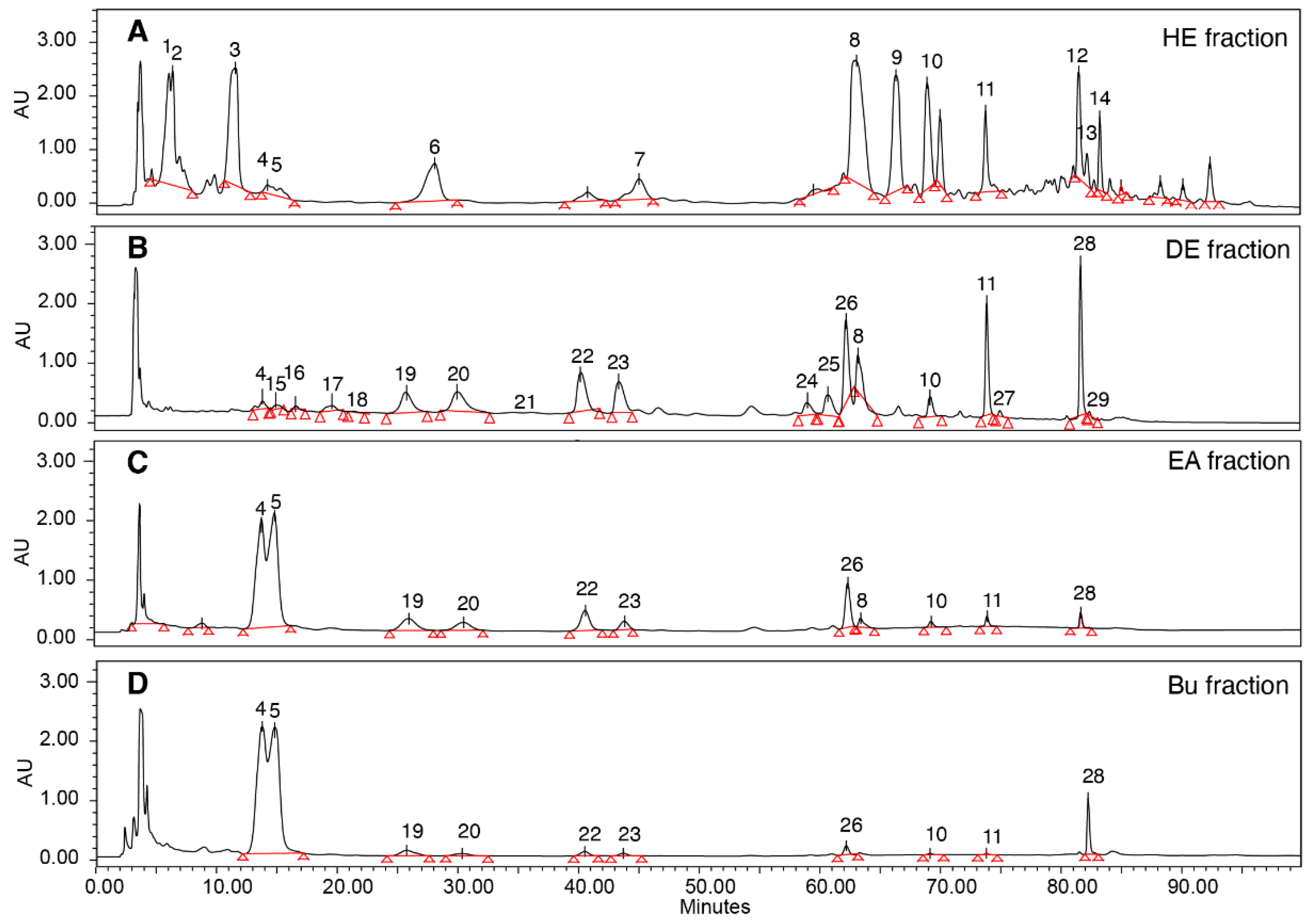
2.1. HPLC/LCMS Analysis of Bioactive Compounds from Antrodia cinnamomea

A. cinnamomea fruiting bodies were first extracted after using ethanol (95%) for 1 h to obtain crude extract (yield 19%). The crude extract was further fractionated by four solvents with increasing polarity. The hexane fraction yielded 7%, while the diethyl ether fraction gave 46%, showing high anti-inflammatory activity as measured by the RAW264.7 cell model. On the other hand, the ethyl acetate fraction (16%) and butanol fraction (21%) did not show measurable anti-inflammatory activity. Analytical HPLC-MS characterization of each fraction revealed that there were many compounds detected in the four fractions (Figure 2 and Table 1).
To further establish the structural and activity relationship, the hexane fraction and diethyl ether fraction were subjected to semipreparative HPLC isolation for each compound. Benzenoids and their derivatives were identified in the hexane-soluble fraction, while abundant triterpenoids including ergostane and lanostane types were detected in fractions with higher polarity. Ethyl acetate fraction and the Bu fraction shared a very similar HPLC fingerprint where compound 4 (25S-antcin K) and 5 (25R-antcin K) and a pair of C-25 epimers were dominant. The UHPLC/qTOF-MS of these crude fractions was also conducted for structural characterizations. The compounds with isomeric structures were differentiated by high-resolution mass spectrometry (HRMS) and their fragmentation patterns are summarized in Table 1.

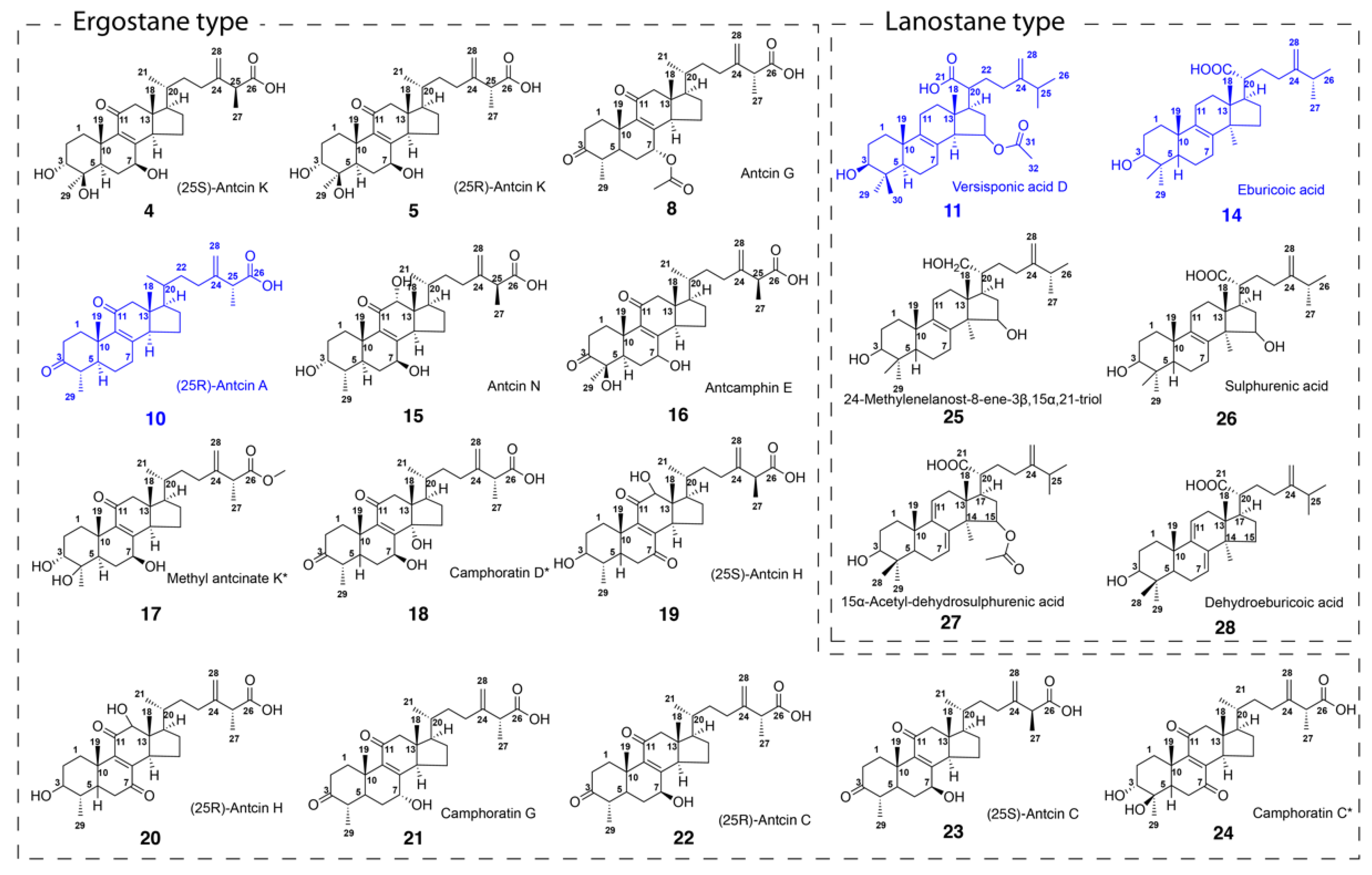
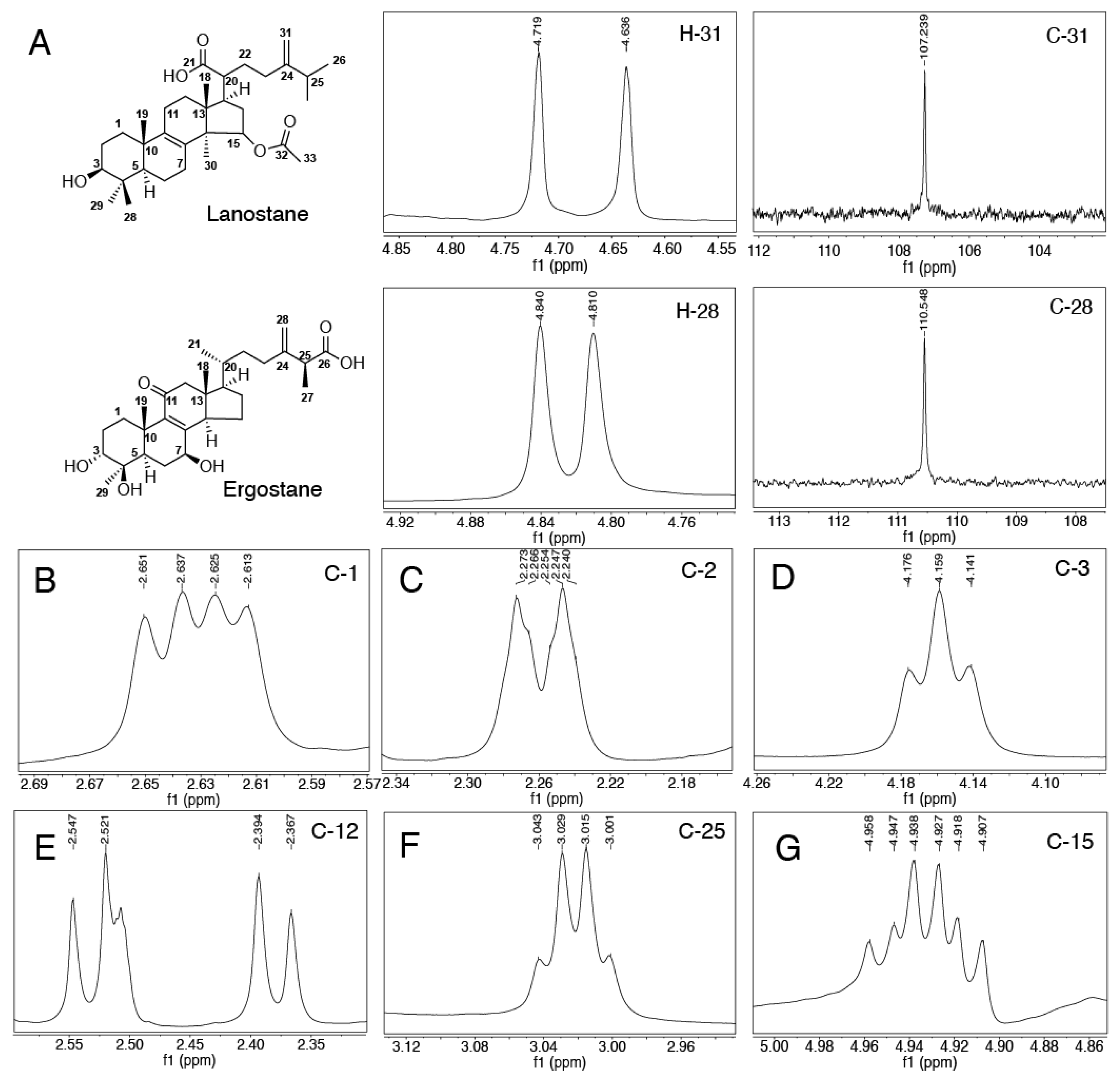
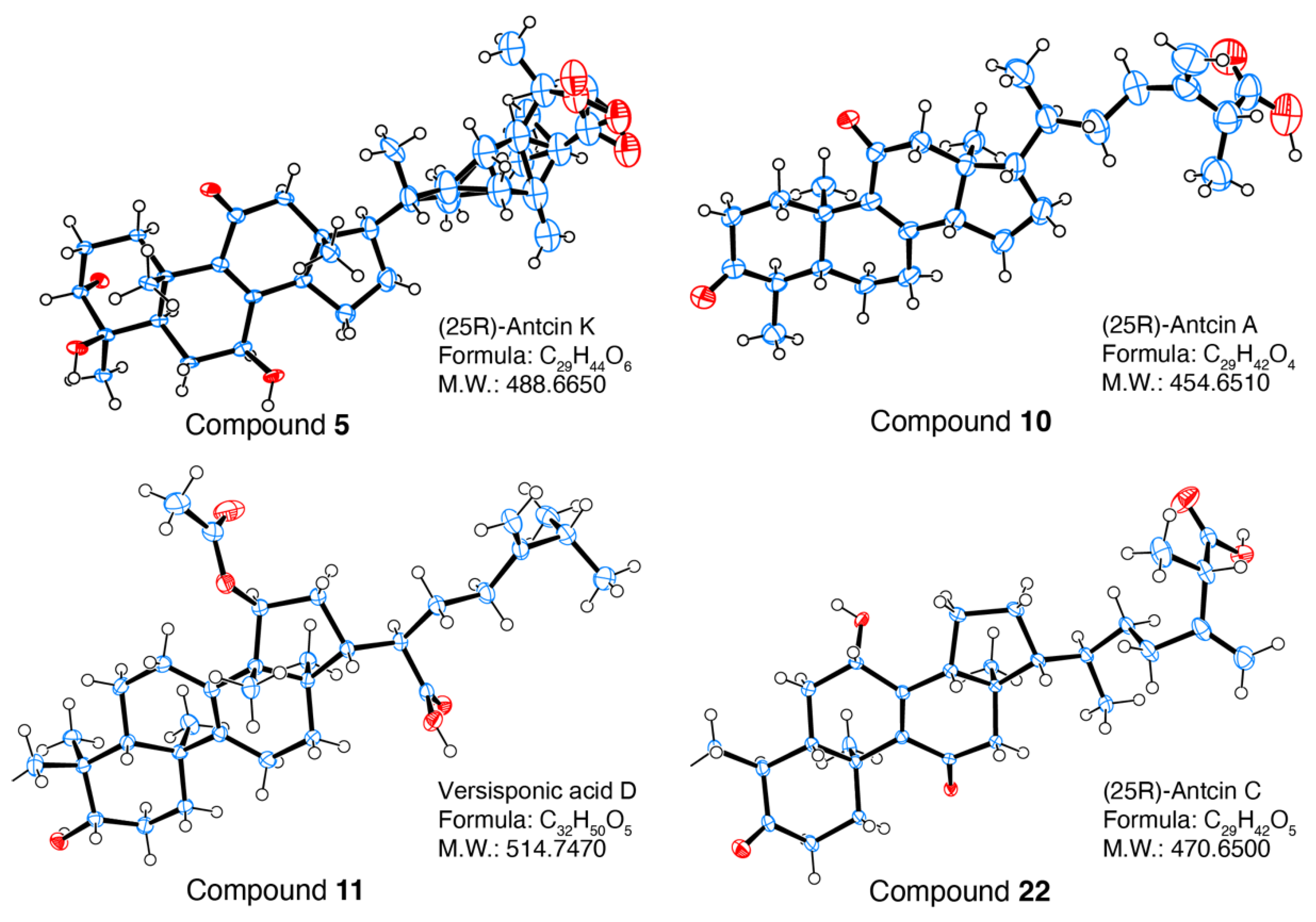
2.2. Structural Identification of Compounds 1-29 Isolated from Antrodia cinnamomea

Compounds 1, 2, 6, 7, 9, 12 and 13 were tentatively identified as benzenoids based on their MS fragmentations. Compounds 3, 4, 5, 8, 10 and 15-24 were identified as ergostane-type triterpenoids, while compounds 11, 14, 25-28 were identified as lanostane-type triterpenoids. Ergostanes and lanostane-type triterpenoids could be preliminary distinguished by their characteristic peaks of 1H NMR and 13C NMR signals, as shown in Figure 3A. The difference in the 1H chemical shift in the protons of C-28 in lanostane (0.083) was larger than that in ergostanes (0.03), while their 13C NMR signals had little difference (NMR spectra were listed in supplementary materials). The protons in C1 (Figure 3B) were located in the chemical shift at around 2.3 to 2.8 in DMSO-d6 with a doublet of doublets, and a similar coupling pattern was also observed in protons from C2 (Figure 3C). Hydroxyl substituents commonly appeared in C-3 (Figure 3D), C-5, C7 and C14 in different triterpenoids; they could be identified from the chemical shifts ranging from 3.2 to 4.8 as triplets. A doublet (J = 13.5 Hz,) was observed for protons on C12 (Figure 3E), which could be easily identified if C12 was not substituted. The signal for protons in C15 (Figure 3G) was split into a triplet by H in the methyl group of C30, and again into doublets by the H of C16, resulting in a ‘triplet of doublets’. C25 (Figure 3F), located between the vinyl group and the carboxyl group had a low file, and the methyl group accounted for the quadlets in a splitting pattern.
25S-Anctin K (4) and 25R-Anctin K (5) were obtained as a white, amorphous powder. They had similar polarity and could be separated by repeated semi-prep HPLC with different mobile phases. Their molecular formula was determined by HRMS with signals of m/z at 487.3058 [M-H]- and 487.3063, respectively, which is in accord with the calculated molecular weight value C29H44O6 (487.3065). The UV absorption maximum at 252 nm revealed the presence of an 8(9)-en-11-one moiety, which was confirmed by the carbon resonances at 201.2 (C-11), 154.0 (C-8), and 143.1 (C-9) ppm. The 1H NMR and 13C NMR spectra of 4 showed the presence of five methyl groups at δH 1.30 (d, J = 5.9 Hz, 3H), 1.15 (dd, J = 7.1, 3.3 Hz, 3H), 1.05 (s, 3H), 0.87 (d, J = 5.6 Hz, 3H), and 0.64 (s, 3H), while the 13C NMR spectra displayed 29 signals suggesting that 4 contained a 4-methylergosta-8(9)-ene-7,11-dione skeleton. However, the absolute configuration of 4 and 5 could not be confirmed, although they could be separated by HPLC at different retention times. After trying several solvent systems, the single crystal of 5 was obtained in MeOH:H2O = 90:10, and then the absolute configuration of 5 was determined as 25S by its X-ray crystallography data (Figure 4).
(25R)-antcin A (10) was obtained as a white powder. HRESIMS analysis established its molecular formula as C29H42O4 with an [M-H] anion at m/z 453.3007. In agreement, its 13C NMR spectrum displayed 29 carbon signals with δC at 212.3 (C-3), 199.4 (C-11), 175.7 (C-26), 157.8 (C-8), and 138.1 (C-9). The methyl signals for CH3-19 (1.25, s), CH3-27 (1.15, dd), CH3-29 (0.93, d), CH3-21 (0.87, d), and CH3-18 (0.65, s) suggested the structure of 10 was very similar to that of antcin C. The UV spectrum showed an absorption maximum at 253 nm, indicating the presence of an 8(9)-ene-11-one moiety. The only difference between antcin A and C was the hydroxyl group in C-6, which was supported by 1H NMR and 13C NMR. The absolute configuration of 10 was determined to be 25R by its X-ray crystallography data (CCDC# 2099280). (Figure 3).
Versisponic acid D (11) was isolated as a white powder. It had an [M-H] ion at m/z 527.3738 (calcd 527.3742) by HRESIMS analysis that corresponded to the molecular formula of C32H50O5, indicating that 11 is a triterpenoid. The chemical shift difference between H-28a (δH 1.13, d, J = 6.4 Hz) and H-28b (δH 1.13, d, J = 6.4 Hz) in 11 was significantly larger than that in 4, 5 and 10, indicating that 11 belongs to lanostane-type triterpenoids rather than the ergostane type. Its UV absorption maximum at 249 nm suggested the presence of an 8(9)-en-11-one moiety, which was confirmed by the carbon resonances at 26.26 (C-7), 132.40 (C-8), 135.80 (C-9), and 20.533 (C-11). The colourless needle single crystals were obtained from MeOH and acetone mixture (1:1), the absolute configuration of 11 was confirmed to be versisponic acid D by its X-ray crystallographic data (CCDC# 2099279) (Figure 3) for the first time.
(25R)-antcin C (22) and (25S)-antcin C (23) were identified as a pair of ergostane epimers with the same molecular formula of C29H42O5 by HRMS peaks at m/z 469.3319 and 469.2953, respectively. Their UV and NMR spectra were identical, although they showed different high-performance liquid chromatography (HPLC) retention. The 1H and 13C NMR spectra of 22 almost overlapped with those of 4, except for 3-hydroxyl in 4 instead of 3-ketone in 22, and one more 4-hydroxyl in 4. The absolute configuration of 22 was determined to be 25R by its X-ray crystallography data (CCDC#2099281) (Figure 3).

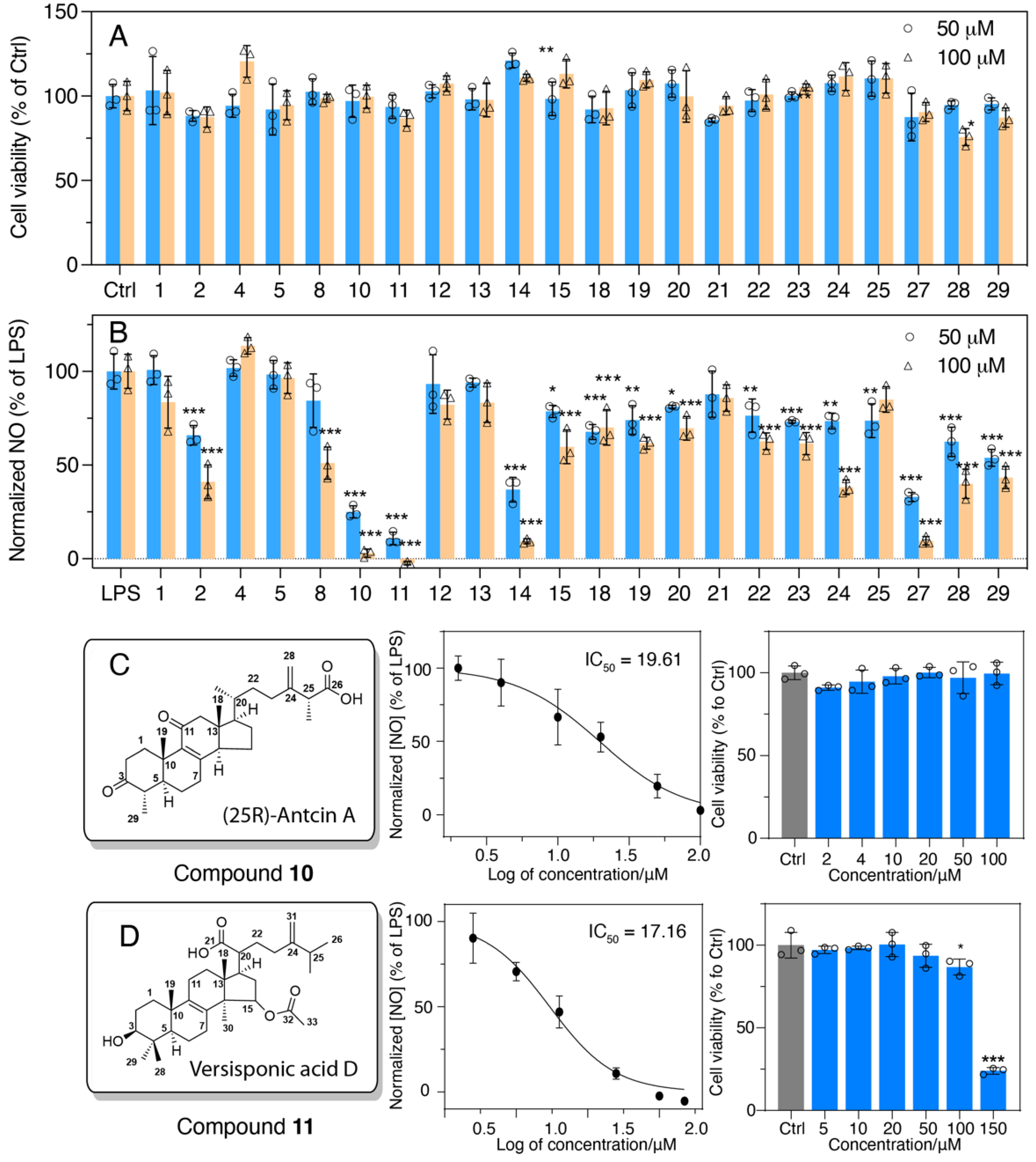
2.3. Evaluation of Anti-Inflammatory Activity of Major Compounds

The compounds with high abundancy were selected for the anti-inflammatory test using murine macrophage RAW 264.7 cells. Compounds with concentrations of 50 μM and 100 μM were tested preliminarily for their anti-inflammatory activities (Figure 4). Among these, 2 (H6), 8 (antcin G), 10 (25R-antcin A), 11 (versisponic acid D), 14 (eburicoic acid), 24 (antcamphin P/Q), 27 (15α-acetyl-dehydrosulphurenic acid), 28 (dehydroeburicoic acid) and 29 (structure unknown) exhibited significant anti-inflammatory activity compared to LPS-treated cells. Most bioactive compounds belong to the triterpenoid family, especially lanostane-type triterpenoids. All compounds tested did not show any significant cytotoxicity under the concentrations tested for their activity (Figure 5A). Remarkably, 4 (25S-antcin K) exhibited growth-promoting effects in contrast with the control cells.
Notably, 10 and 11 (10 μM) seemed to be the most conspicuous in relieving the inflammation stimulated by LPS (Figure 5B), and the half-inhibitory concentrations (IC50) of 10 and 11 were determined to be 19.61 ± 0.8 (Figure 5C) and 17.16 ± 1.0 (Figure 5D), respectively.
Antcins are dominant bioactive compounds found in A. cinnamomea, but only antcin A (10), a steroid-like compound, showed measurable anti-inflammatory activity. Its structure bears close similarity with that of glucocorticoid. Hence, it is conceivable that antcin A could bind the same biological target as the glucocorticoid receptor and activate anti-inflammatory responses or suppress inflammatory reaction [19]. In contrast, antcin C (22 and 23), H (19 and 20) and K (4 and 5) had hydroxyl at C-7 which prevented them from binding to the glucocorticoid receptor. Additionally, antcin K showed anti-inflammatory activity by inhibiting NF-κB activation and interleukin-8 production in LPS-induced human gastric cancer AGS cells [20], and we did not observe its activity in NO inhibition. The derivative of antcin K, methyl antcinate K (17), showed activity in NO suppression in LPS- and IFN-γ-induced BV-2 cell lines, which only comprised 10% and 5% of the NOS inhibition activity of L-NAME [21]. The other type of extracts, lanostanes, also exhibited inflammation inhibitory activities. For example, eburicoic acid (14) and dehydroeburicoic acid (28) significantly suppressed λ-carrageenan-simulated paw edema in mice by down-regulating mediators including NO, iNOS, COX-2, TNF-α, and IL-1β. Notably, the dehydrogenation slightly improved the activity of dehydroeburicoic acid [14]. The biological targets for 14 and 28 await to be determined.

3. Materials and Methods

3.1. General Experimental Procedures

UV–vis spectra were obtained from Shimadzu UV-2101 PC (Shimadzu Corporation, Kyoto, Japan) or Waters 2996 Photodiode Array Detector (PDA) (Waters Corporation, Milford, MA, USA). NMR spectral data were recorded on a Bruker Avance III 500 MHz spectrometer. High-resolution electrospray ionization mass spectrometry (HRESIMS) was performed on A 6530 Accurate-Mass LC–QTOF-MS device (Agilent Technologies, Santa Clara, CA, USA). The ACQUITY Arc System (Waters, Milford, MA, USA) was applied for the analytical determination of bioactive compounds from plant samples while the Waters semi-prep HPLC system with a 2998 PDA Detector was used for product separation.

3.2. Plant Material

The fruiting bodies of Antrodia cinnamomea were provided by AgriGADA Biotech Pte. Ltd. (Singapore).

3.3. Extraction and Isolation

Two hundred grams of air-dried fruiting bodies of Antrodia cinnamomea were ground into fine powder using a mini blender (DM-6, Youqi, Taiwan) and extracted with 95% ethanol at ambient temperature (3 × 2.0 L). The slurry was filtered and the filtrate was concentrated under reduced pressure to yield a dark-brown residue of 38.9 g as the crude extract, which was suspended in water (1.0 L) and extracted with hexane (HE), diethyl ether (DE), ethyl acetate (EA), and n-butanol (Bu), sequentially. The fractions were dissolved in DMSO (0.1 g/mL) for the screening of anti-inflammatory activity.
The crude fractions with promising activities were further separated by semi-prep HPLC using the Waters Purification System equipped with a 2998 PDA Detector and a 2707 AUTOSAMPLER. A HPLC column (Luna 5 µM C18(2) 100 Å, 250mm × 10 mm; Phenomenex) was employed with two solvent systems: H2O with 0.1% formic acid (A) and MeOH (B).

3.4. Characterization of Isolated Products from Antrodia cinnamomea

1H NMR spectra were recorded on commercial instruments operating at 400 and 500 MHz (1H) and 100 and 125 MHz (13C), respectively. Chemical shifts were reported in ppm from the solvent resonance as the internal standard (DMSO-d6), δ = 2.50). Spectra were reported as follows: chemical shift (δ ppm), multiplicity (s = singlet; d = doublet; t = triplet; q = quartet; hept = heptet; m = multiplet), coupling constants (Hz), and integration and assignment. 13C NMR spectra were recorded on commercial instruments (126 MHz). Chemical shifts were reported in ppm with the solvent resonance as the internal standard (DMSO-d, δ = 39.52). HRMS was recorded on a commercial apparatus (ESI source).

3.5. Cell Culture

The murine macrophage RAW 264.7 (TIB-71, ATCC) cells were maintained in high glucose Dulbecco’s Modified Eagle Medium (DMEM, Hyclone™, cytiva Pte. Ltd., Singapore), supplemented with 10% foetal bovine serum (FBS, Hyclone™, cytiva Pte. Ltd., Singapore) and 1% penicillin–streptomycin antibiotics (100 IU/mL penicillin and 100 µg/mL streptomycin, Gibco™, ThermoFisher Scientific Pte. Ltd., Singapore), and culture medium was changed every 2 days. Next, 25,000 cells were seeded into 96-well plates for nitric oxide production and the cytotoxicity test, and 750,000 cells were seeded into 6-well plates for protein and mRNA extraction. After cells were incubated at 37 °C with 5% CO2 overnight, they were pre-treated with crude fractions or isolated pure compounds for 4 h, and with (for nitric oxide production) or without (for cytotoxicity) 100 ng/mL lipopolysaccharide (LPS, Escherichia coli serotype 055:B5, Sigma Aldrich, Singapore) for another 20 h.

3.6. Cytotoxic Activities

Cytotoxicity was characterized by cell viability via Cell Counting Kit-8 (CCK-8 Dojindo, Kumamoto, Japan). After 24 h of treatment, 100 µL CCK-8 in DMEM (1:10) was added to cells following incubation at 37 °C with 5% CO2, and light was avoided for 45 min. Afterwards, the absorbance value was measured at 450 nm by a microplate reader (Tecan Infinite F200, Tecan Group Ltd., Singapore). The optical density was converted to the percentage of the control group, and all experiments were performed in three independent tests.

3.7. Nitric Oxide (NO) Inhibitory Assay

The effects of the compounds isolated from the A. cinnamomea extracts on NO production were measured indirectly by the analysis of nitrite levels in cell culture medium using the Greiss reagent assay (Promega, Singapore). Briefly, 50 µL of cell supernatant from A. cinnamomea extracts treated with RAW 264.7 cells were collected for NO production measurements following the manufacturer’s protocol. Absorbance value was measured on the microplate reader at 540 nm. The NO concentration was quantified using the nitrite standard curve (1.56–100 µM) and results were expressed as a percentage of inhibition relative to the LPS. Each treatment was carried out in triplicate. IC50 represents the concentrations at which 50% of the NO was inhibited by the extracts.

3.8. Statistical Analysis

Descriptive statistical analyses were performed using GraphPad Prism 8.0.2 for calculating the means and the standard error of the mean. Results are expressed as the mean ± standard deviation (SD). Data comparison was analysed by one-way analysis of variance (ANOVA). A Dunnett correction was selected for the post hoc test. p* < 0.05 was considered statistically significant.
Research manuscripts reporting large datasets that are deposited in a publicly available database should specify where the data have been deposited and provide the relevant accession numbers. If the accession numbers have not yet been obtained at the time of submission, please state that they will be provided during review. They must be provided prior to publication.
Interventionary studies involving animals or humans, and other studies that require ethical approval, must list the authority that provided approval and the corresponding ethical approval code.

4. Conclusions

Taken together, we have established the molecular basis for the anti-inflammatory activity of Antrodia cinnamomea, and identified that the less abundant terpenoids (4 and 5) are the active compounds. The anti-inflammatory activity of terpenoids is highly related to their structures. For lanostane-type terpenoids (with double bonds between C8 and C9), the substituents on C15 seemed to be important in the anti-inflammatory activities; compound 25 and compound 26, which had a hydroxyl group on C15 of the backbone, showed significantly lower activities than the rest of the compounds. While ergostane in type, the functional group (hydroxyl group, ester group, and ketone) occurring on C7 of the backbone was found to have a negative impact on anti-inflammatory activity. Our work serves as a foundation for the in-depth study of the health promotion activity of this treasured mushroom.

Supplementary Materials

The following supporting information can be downloaded at: https://www.mdpi.com/article/10.3390/foods11131831/s1.

Author Contributions

Conceptualization, X.Y. and D.H.; methodology, X.Y. and X.W.; software, J.L.; validation, K.-C.F., C.-L.K. and H.Z.; formal analysis, C.X.; investigation, P.X.; resources, P.X.; data curation, S.C.; writing—original draft preparation, X.Y.; writing—review and editing, D.H.; visualization, Y.C. and S.L.; supervision, X.Y.; project administration, D.H.; funding acquisition, D.H. All authors have read and agreed to the published version of the manuscript.

Funding

Authors thank funding of the project from a collaborative research project between NUS and AgriGADA (grant number R160-000-017-592).

Institutional Review Board Statement

Not applicable.

Informed Consent Statement

Not applicable.

Data Availability Statement

Data are contained within the article.

Conflicts of Interest

This work is funded by a Collaborative agreement between NUS and AgriGADA Pte Ltd. Michael Kuo Chang Fu is AGRIDAGA- director for R and D.

References

  1. Wu, S.-H.; Ryvarden, L. Antrodia camphorata (“niu-chang-chih”), new combination of a medicinal fungus in Taiwan. Bot. Bull. -Acad. Sin. Taipei 1997, 38, 273–276. [Google Scholar]
  2. Wang, H.-C.; Chu, F.-H.; Chien, S.-C.; Liao, J.-W.; Hsieh, H.-W.; Li, W.-H.; Lin, C.-C.; Shaw, J.-F.; Kuo, Y.-H.; Wang, S.-Y. Establishment of the metabolite profile for an Antrodia cinnamomea health food product and investigation of its chemoprevention activity. J. Agric. Food Chem. 2013, 61, 8556–8564. [Google Scholar] [CrossRef] [PubMed]
  3. Huang, Y.-L.; Chu, Y.-L.; Ho, C.-T.; Chung, J.-G.; Lai, C.-I.; Su, Y.-C.; Kuo, Y.-H.; Sheen, L.-Y. Antcin K, an active triterpenoid from the fruiting bodies of basswood-cultivated Antrodia cinnamomea, inhibits metastasis via suppression of integrin-mediated adhesion, migration, and invasion in human hepatoma cells. J. Agric. Food Chem. 2015, 63, 4561–4569. [Google Scholar] [CrossRef]
  4. Hu, P.-F.; Huang, J.; Chen, L.; Ding, Z.; Liu, L.; Molnár, I.; Zhang, B.-B. Oxidative stress induction is a rational strategy to enhance the productivity of Antrodia cinnamomea fermentations for the antioxidant secondary metabolite antrodin C. J. Agric. Food Chem. 2020, 68, 3995–4004. [Google Scholar] [CrossRef] [PubMed]
  5. Geethangili, M.; Tzeng, Y. Review of pharmacological effects of Antrodia Camphorata and its bioactive compounds. Evid. Based Complementary Altern. 2011, 2011, 110. [Google Scholar]
  6. Wen, C.-L.; Chang, C.-C.; Huang, S.-S.; Kuo, C.-L.; Hsu, S.-L.; Deng, J.-S.; Huang, G.-J. Anti-inflammatory effects of methanol extract of Antrodia cinnamomea mycelia both in vitro and in vivo. J. Ethnopharmacol. 2011, 137, 575–584. [Google Scholar] [CrossRef] [PubMed]
  7. Ao, Z.-H.; Xu, Z.-H.; Lu, Z.-M.; Xu, H.-Y.; Zhang, X.-M.; Dou, W.-F. Niuchangchih (Antrodia camphorata) and its potential in treating liver diseases. J. Ethnopharmacol. 2009, 121, 194–212. [Google Scholar] [CrossRef]
  8. Lin, T.-Y.; Chen, C.-Y.; Chien, S.-C.; Hsiao, W.-W.; Chu, F.-H.; Li, W.-H.; Lin, C.-C.; Shaw, J.-F.; Wang, S.-Y. Metabolite profiles for Antrodia cinnamomea fruiting bodies harvested at different culture ages and from different wood substrates. J. Agric. Food Chem. 2011, 59, 7626–7635. [Google Scholar] [CrossRef]
  9. Zhang, B.-B.; Hu, P.-F.; Huang, J.; Hu, Y.-D.; Chen, L.; Xu, G.-R. Current advances on the structure, bioactivity, synthesis, and metabolic regulation of novel ubiquinone derivatives in the edible and medicinal mushroom Antrodia cinnamomea. J. Agric. Food Chem. 2017, 65, 10395–10405. [Google Scholar] [CrossRef]
  10. Tazuke, S.; Kurihara, S.; Yamaguchi, H.; Ikeda, T. Photochemically triggered physical amplification of photoresponsiveness. J. Phys. Chem. 1987, 91, 249–251. [Google Scholar] [CrossRef]
  11. Rao, Y.K.; Fang, S.-H.; Tzeng, Y.-M. Evaluation of the anti-inflammatory and anti-proliferation tumoral cells activities of Antrodia camphorata, Cordyceps sinensis, and Cinnamomum osmophloeum bark extracts. J. Ethnopharmacol. 2007, 114, 78–85. [Google Scholar] [CrossRef] [PubMed]
  12. Hseu, Y.-C.; Wu, F.-Y.; Wu, J.-J.; Chen, J.-Y.; Chang, W.-H.; Lu, F.-J.; Lai, Y.-C.; Yang, H.-L. Anti-inflammatory potential of Antrodia camphorata through inhibition of iNOS, COX-2 and cytokines via the NF-κB pathway. Int. Immunopharmacol. 2005, 5, 1914–1925. [Google Scholar] [CrossRef] [PubMed]
  13. Kuo, Y.-H.; Lin, C.-H.; Shih, C.-C. Antidiabetic and antihyperlipidemic properties of a triterpenoid compound, dehydroeburicoic acid, from Antrodia camphorata in vitro and in streptozotocin-induced mice. J. Agric. Food Chem. 2015, 63, 10140–10151. [Google Scholar] [CrossRef] [PubMed]
  14. Deng, J.-S.; Huang, S.-S.; Lin, T.-H.; Lee, M.-M.; Kuo, C.-C.; Sung, P.-J.; Hou, W.-C.; Huang, G.-J.; Kuo, Y.-H. Analgesic and anti-inflammatory bioactivities of eburicoic acid and dehydroeburicoic acid isolated from Antrodia camphorata on the inflammatory mediator expression in mice. J. Agric. Food Chem. 2013, 61, 5064–5071. [Google Scholar] [CrossRef]
  15. Hsieh, Y.-H.; Chu, F.-H.; Wang, Y.-S.; Chien, S.-C.; Chang, S.-T.; Shaw, J.-F.; Chen, C.-Y.; Hsiao, W.-W.; Kuo, Y.-H.; Wang, S.-Y. Antrocamphin A, an anti-inflammatory principal from the fruiting body of Taiwanofungus camphoratus, and its mechanisms. J. Agric. Food Chem. 2010, 58, 3153–3158. [Google Scholar] [CrossRef]
  16. Chen, C.-C.; Liu, Y.-W.; Ker, Y.-B.; Wu, Y.-Y.; Lai, E.Y.; Chyau, C.-C.; Hseu, T.-H.; Peng, R.Y. Chemical characterization and anti-inflammatory effect of polysaccharides fractionated from submerge-cultured Antrodia camphorata mycelia. J. Agric. Food Chem. 2007, 55, 5007–5012. [Google Scholar] [CrossRef]
  17. Chen, L.-Y.; Sheu, M.-T.; Liu, D.-Z.; Liao, C.-K.; Ho, H.-O.; Kao, W.-Y.; Ho, Y.-S.; Lee, W.-S.; Su, C.-H. Pretreatment with an ethanolic extract of Taiwanofungus camphoratus (Antrodia camphorata) enhances the cytotoxic effects of amphotericin B. J. Agric. Food Chem. 2011, 59, 11255–11263. [Google Scholar] [CrossRef]
  18. Hsiao, G.; Shen, M.-Y.; Lin, K.-H.; Lan, M.-H.; Wu, L.-Y.; Chou, D.-S.; Lin, C.-H.; Su, C.-H.; Sheu, J.-R. Antioxidative and hepatoprotective effects of Antrodia camphorata extract. J. Agric. Food Chem. 2003, 51, 3302–3308. [Google Scholar] [CrossRef]
  19. Chen, Y.-c.; Liu, Y.-l.; Li, F.-y.; Chang, C.-I.; Wang, S.-y.; Lee, K.-y.; Li, S.-l.; Chen, Y.-p.; Jinn, T.-r.; Tzen, J.T. Antcin A, a steroid-like compound from Antrodia camphorata, exerts anti-inflammatory effect via mimicking glucocorticoids. Acta Pharmacol. Sin. 2011, 32, 904–911. [Google Scholar] [CrossRef]
  20. Geethangili, M.; Fang, S.-H.; Lai, C.-H.; Rao, Y.K.; Lien, H.-M.; Tzeng, Y.-M. Inhibitory effect of Antrodia camphorata constituents on the Helicobacter pylori-associated gastric inflammation. Food Chem. 2010, 119, 149–153. [Google Scholar] [CrossRef]
  21. Shen, C.-C.; Wang, Y.-H.; Chang, T.-T.; Lin, L.-C.; Don, M.-J.; Hou, Y.-C.; Liou, K.-T.; Chang, S.; Wang, W.-Y.; Ko, H.-C. Anti-inflammatory ergostanes from the basidiomata of Antrodia salmonea. Planta Med. 2007, 73, 1208–1213. [Google Scholar] [CrossRef] [PubMed]
Figure 1. Structures of phytochemicals isolated from cultured Antrodia cinnamomea fruiting bodies. The blue ones are potent anti-inflammatory agents. Note: the numbers correspond to the peak numbers in the HPLC chromatogram shown in Figure 2.
Foods 11 01831 g001
Figure 2. HPLC profiling and bioactivities of Antrodia cinnamomea fruiting bodies from different fractions. (A) Hexane; (B) diethyl ether; (C) ethyl acetate; and (D) n-butanol fractions. (HE: hexane; DE: diethyl ether; EA: ethyl acetate; Bu: n-BuOH).
Foods 11 01831 g002
Figure 3. Structural characterization of ergostane-type and lanostane-type triterpenoids. (A) 1H NMR resonance for H−3 and 13C NMR resonance for C-3. The characteristic NMR signal of C1 of (25S)-Antcin K is shown in (B), C2, C3, C12, C25 and C15 in (C), (D), (E), (F) and (G).
Foods 11 01831 g003
Figure 4. ORTFP drawing of 5, 10, 11 and 22.
Foods 11 01831 g004
Figure 5. Anti-inflammatory activities of bioactive compounds isolated from Antrodia cinnamomea. (A) Cell viability of cells treated only with compounds 1-29. Dose–response curve of normalized NO with the treatment of 100 ng/mL LPS with 10. (B) Nitric oxide production of RAW 264.7 cells treated with 100 ng/mL LPS and compounds 1-29 isolated from Antrodia cinnamomea. (C) Compound 10 and (D) 11 and their cytotoxicity. Results are shown as mean ± S.D, n = 3, p* < 0.05, p** < 0.01, p*** < 0.001 vs. LPS or control.
Foods 11 01831 g005
Table 1. Characterization data of phytochemicals in A. cinnamomea by UHPLC/qTOF-MS.
NotRMaxFormula[M-H]MS/MS of [M−H]Identification
(min)(nm)Pred.Meas.Δ(ppm)
16.355230C10H14O4197.0819197.0850.0011-2,3,4-trimethoxy-6-methylphenol
26.421230C24H19Na2O12-545.0677544.96730.1004118, 168, 396, 480Benzenoid (tentative)
311.564276C29H44O6487.3065487.30610.0004119, 247, 407, 425, 443Unknown
414.23252C29H44O6487.3065487.30580.0007259, 271, 342, 407, 443, 460(25S)-Antcin K
514.988252C29H44O6487.3065487.30630.0002259, 271, 342, 407, 443, 460(25R)-Antcin K
628.088284C29H29O9485.1218485.29040.1686272, 301, 425Benzenoid (tentative)
745.031265C25H23O10483.1297483.2762−0.1465193, 287, 337, 411, 421, 439Benzenoid (tentative)
863.086250C31H44O6511.3065511.34250.036395, 407, 451, 465Antcin G
968.933251C26H23O11452.0822452.08220126, 143, 262, 316Benzenoid (tentative)
1066.352253C29H42O4453.301453.30070.0003123, 257, 337, 393, 411(25R)-Antcin A
1170.011249C32H50O5527.3742527.37370.0005397. 467Versisponic acid D
1273.777244C35H29O9593.1812593.3732−0.1920265Benzenoid (tentative)
1381.509242C25H29O7467.1348467.3529−0.1095113, 227, 249, 385, 441Benzenoid (tentative)
1483.258245C29H41O5469.2959469.36840.0725113, 227, 281, 299, 385, 441Eburicoic acid
1513.935198C29H44O6487.3065487.30610.0004209, 407, 452Antcin N
1615.058198C29H42O6485.2909485.29030.0006149, 233, 408, 423, 441, 467Antcamphin E
1716.676198C30H46O6501.3222501.28540.0368325, 358, 395, 413, 439, 457, 483Methyl antcinate K *
1819.701198C29H42O6485.2909485.29060.0003137, 247, 289, 341, 407, 423, 441Camphoratin D *
1925.78256C29H42O6485.2909485.29030.0006149, 207, 233, 399, 423(25S)-Anctin H
2030.056256C29H42O6485.2909485.29020.0007193, 233, 397, 441(25R)-Anctin H
2134.119252C29H42O5469.2959469.29570.0002247, 301, 341, 407, 425Camphoratin G
2240.26255C29H42O5469.2959469.33190.036339, 395, 407, 425(25R)-Antcin C
2343.403255C29H42O5469.2959469.29530.0006219, 233, 272, 391, 425(25S)-Antcin C
2458.941273C29H42O6485.2909485.29060.0003233, 275, 397, 412, 423, 441Antcamphin P/Q
2560.664273C31H50O4485.3636485.29030.0733253, 339, 397, 405, 413, 44124-Methylenelanost-8-ene-3β,15α,21-triol
2662.16242C31H50O4485.3636485.29010.0735160, 369, 397, 423, 441Sulphurenic acid
2773.801242C33H50O5525.3585525.35770.0008233, 245, 397, 46715α-Acetyl-dehydrosulphurenic acid
2881.553240C31H49O3467.3531467.35260.0005156, 170, 339, 371Dehydroeburicoic acid
2981.965202C30H47O4469.3323469.36830.036339Unknown
Publisher’s Note: MDPI stays neutral with regard to jurisdictional claims in published maps and institutional affiliations.

