Article

Phenolic Profiles and Biological Activities of Extracts from Edible Wild Fruits Ehretia tinifolia and Sideroxylon lanuginosum

by
Imelda N. Monroy-García
1,
Irma Edith Carranza-Torres
1,2,
Pilar Carranza-Rosales
2,
María Oyón-Ardoiz
3,
Ignacio García-Estévez
3,
Jesús Fernando Ayala-Zavala
4,
Javier Morán-Martínez
5 and
Ezequiel Viveros-Valdez
1,*
1
Deparatmento de Química, Facultad de Ciencias Biológicas, Universidad Autónoma de Nuevo León, Av. Pedro de Alba S/N, San Nicolás de los Garza 66450, Nuevo León, Mexico
2
Centro de Investigación Biomédica del Noreste, Instituto Mexicano del Seguro Social, Jesús Dionisio González #501, Col. Independencia, Monterrey 64720, Nuevo León, Mexico
3
Grupo de Investigación en Polifenoles, Departamento de Química Analítica, Nutrición y Bromatología, Facultad de Farmacia, Campus Miguel de Unamuno, Universidad de Salamanca, E37007 Salamanca, Spain
4
Coordinación de Tecnología de Alimentos de Origen Vegetal, Centro de Investigación en Alimentación y Desarrollo A.C., Carretera Gustavo Enrique Astiazarán Rosas No. 46, Hermosillo 83304, Sonora, Mexico
5
Facultad de Medicina, Universidad Autónoma de Coahuila Unidad Torreón, Gregorio A. García No. 198, Torreón 27000, Coahuila, Mexico
*
Author to whom correspondence should be addressed.
Foods 2021, 10(11), 2710; https://doi.org/10.3390/foods10112710
Submission received: 14 October 2021 / Revised: 2 November 2021 / Accepted: 3 November 2021 / Published: 5 November 2021
(This article belongs to the Special Issue Food Natural Additives)

Abstract

:
Ehretia tinifolia Linnaeus (Boraginacea) and Sideroxylon lanuginosum Michaux (Sapotaceae) are wild fruits consumed in North America and are appreciated for their pleasant flavor and sweet taste. However, details regarding their composition and biological properties in the available literature are scarce. This study reports the phenolic composition, antioxidant, antiproliferative activities, and digestive enzymatic inhibition of amberlite-retained methanolic extracts from both fruits. Results revealed that these wild fruit extracts are rich in antioxidants. S. lanuginosum had lower phenolic but higher flavonoid contents (21.4 ± 1.5 mg GAE/100 g FW and 6.42 ± 0.9 mg CE/100 g FW) than E. tinifolia (64.7 ± 2.6 mg GAE/100 g FW and 5.1 ± 0.4 mg CE/100 g FW). HPLC-DAD-MS/MS analysis showed rosmarinic acid as a major polyphenol in E. tinifolia and quercetin glucoside in S. lanuginosum. Polyphenols content in E. tinifolia was related to a significant free radical scavenging ability: DPPH (EC50 = 0.32 ± 0.03 mg/mL), TEAC (4134 ± 9.7 μM TE/g dry extract), and hemolysis inhibition (IC50 = 58.55 ± 2.4 μg/mL). Both extracts were capable of inhibiting α-glucosidase, partially inhibiting α-amylase, and showed no inhibition against lipase, while showing antiproliferative activity against HeLa, HT-29 and MCF-7 cancer cell lines. Our study revealed that these wild fruit extracts are rich in health-beneficial phytochemicals and hold significant potential for elaborating functional foods.

Graphical Abstract

1. Introduction

In recent years, exploratory studies have been developed to identify endemic wild fruits as promising crops with high economic value [1]. Likewise, producing scientific evidence of the properties of these wild resources is highly relevant to evaluating their contribution to local biodiversity and assessing their effects on nutrition and human health [2,3]. Ehretia tinifolia Linnaeus (Boraginacea), commonly called pinguica and Sideroxylon lanuginosum Michaux (Sapotaceae), known as Gum Bully, Black Haw, or Coma, are wild fruits appreciated for their pleasant flavor and sweet taste and have been consumed in North America by Native Americans since pre-Columbian times. Thus, wild plants have an important role in indigenous peoples’ lives [4] as they supplement staple foods to provide a balanced diet to many populations in several regions across Mexico, mainly in the states of Michoacán, Nayarit, Nuevo León, San Luis Potosí, Sinaloa, Tamaulipas, and Veracruz. It has also been used in the southeastern USA, mostly used by the Kiowa and Comanche tribes [5]. E. tinifolia are small globoid yellow drupes of up to 8 mm diameter, with a sweet flavor and have been used as food and medicinal plant in several regions of Mexico and the USA [6,7,8,9]. Bromatological analysis of E. tinifolia has already been reported, and the content of total phenols in polar organic extracts (50 to 125 mg per 100 g of fresh fruit) is related to its antioxidant properties [10]. S. lanuginosum is also used in the north of Mexico and by native tribes of the USA as chewing gum [11]. However, there are no reports of the chemical composition or biological activity of this fruit. Therefore, expanding the chemical/biological knowledge of these species would add value to the possible production and commercialization of nutraceuticals or functional foods.
It is well known that fruits have antioxidants with a beneficial impact on human health. Phenolic compounds, ascorbate and carotenoids are the main antioxidants found in fruits [12]. The main phenolic components present in fruits are glycosylated flavones/flavonols, flavanones, anthocyanins, and phenolic acids [13]. Epidemiological studies and randomized clinical trials showed a strong association between polyphenol consumption and reduced risk of several chronic diseases, including cancer, diabetes, and inflammatory processes [14]. Dietary plant polyphenols modulated metabolism of carbohydrates and lipids, controlled hyperglycemia, dyslipidemia and insulin resistance, increased metabolism in adipose tissues, and alleviated oxidative stress and stress-sensitive signaling pathways and inflammatory processes in several in vitro, animal models and some human studies [12]. Furthermore, cancer prevention is one of the most documented biological properties of polyphenols. In cancer, these compounds induced apoptosis, reduced the number of tumors, inhibited angiogenesis, modulated multidrug resistance and antiproliferative activity [15]. Polyphenols also modulated the expression of cytochrome P-450 enzymes and activation of carcinogens [16].
Wild fruits with antioxidant activity have also become the object of an increasing number patent claims on behalf of the food and beverage industry. Wild rose fruit and jujube mixtures have been used to claim the formulation of an antioxidant beverage [17]. In addition, wild pawpaw has been used to formulate cakes [18]. At the same time, wild indigo fruit and wild blueberries have been used to formulate beers claiming to enhance vision and human immunity [19]. Wild rose fruit rich in polyphenols has been used to formulate cosmetics with skin repair activity [20]. Similarly, wild cherries were used to formulate beverages claiming to be useful on dietary therapies [21]. Additionally, Berbenol® (PharmExtrcta, Pontenure, Italy), a tablet formulation made from an extract of Berberis aristata D.C. and Silybum marianum (L.) Gaertn fruit is used to treat glycemia and lipid value alterations in patients with type 2 diabetes [22]. These patents evidence the potential economic impact of knowing the bioactive composition of wild fruits.
In this context, this study aimed to identify the phenolic profiles and contents from E. tinifolia and S. Lanoginosum from the north of Mexico and to evaluate the antiradical, antidiabetic and anticarcinogenic properties. This information would highlight their potential use in elaborating functional foods and preserving and revalorizing their use as traditional foods.

2. Materials and Methods

2.1. Plant Material

Ripe and healthy pinguicula and coma fruits were collected by hand from wild trees located in San Nicolas de los Garza, N.L. (northeast Mexico; Latitude: 25°44′31.9″ N; Longitude: 100°16′59.3″ W) and Higueras, N.L. (northeast Mexico; Latitude: 25°57′59.3″ N; Longitude: 100°00′52.5″ W), respectively, from May to August 2019. Fruits were immediately transported to the laboratory for processing. The authenticity of the collected species was confirmed by Dr. Marco Guzmán Lucio, taxonomist and responsible for the herbarium of the Faculty of Biological Sciences of the Autonomous University of Nuevo Leon.

2.2. Phytochemicals Extraction

The method used for phytochemicals extraction was reported previously by Viveros-Valdez, et al. [23]. E. tinifolia and S. lanuginosum fresh fruits were washed with distilled water, and seeds were removed manually. One hundred grams of each fruit pulp and peel were squeezed and mixed with 1 L of distilled water using a commercial blender. The mixtures were filtered using Whatman No.1 filter paper, and each juice was applied onto an Amberlite XAD-7 column (150 × 10 cm), which had been conditioned previously with 2 L of distilled water. The XAD-7 column was washed with distilled water (3 L) to eliminate proteins, carbohydrates, minerals and organic acids, while the retained phenolic compounds were eluted with methanol (MeOH) (2 L). Subsequently, the solvent was evaporated until dryness under reduced pressure and lyophilized for analysis. Yellow and purple extracts were obtained from E. tinifolia (w/w yield: 0.87%) and S. lanuginosum (w/w yield: 0.51%), respectively. Lyophilized extracts were stored at 4 ºC for a maximum period of 6 months.

2.3. Determination of Total Phenolic Content (TPC)

Total phenolic content was determined using the Folin-Ciocalteu reagent as described by Singleton and Rossi [24] with some modifications. Total phenolic content includes free and bound phenolics. The freeze-dried extracts were dissolved in distilled water to a concentration of 0.1 mg/mL. Then, 30 μL of each fruit extract, 150 μL of Folin-Ciocalteu reagent (1:10 v/v in distilled water) and 120 μL of an aqueous solution of Na2CO3 (7.5% w/v) were placed into a 96-well plate and incubated for 30 min at room temperature in darkness. Absorbance was measured at 765 nm (Agilent BioTek Epoch Microplate Spectrophotometer), using gallic acid solutions as standards (concentrations range of 10–1000 µg/mL in distilled water) (y = 0.0681x + 0.0732; R2 = 0.9848), and results were expressed as milligrams of gallic acid equivalents (GAE) per 100 g of fresh weight (FW). Data were reported as mean ± S.D. for at least three replications.

2.4. Determination of Total Flavonoid Content (TFC)

Total flavonoid content was determined using the aluminum chloride colorimetric assay [25]. Fruit extracts were diluted with MeOH (1 mg/mL). Then, 250 μL of fruit extract or standard solution of (+) Catechin (concentration range of 50–500 µg/mL of MeOH) was mixed with 1000 μL of distilled water and 75 μL of NaNO2 solution (7% w/v in distilled water). After 5 min at room temperature, 75 μL of AlCl3 aqueous solution (10% w/v) were added. One minute later, 500 μL of 1 M NaOH and 600 µL of distilled water were added and vigorously mixed. The absorbance in the reaction mixture was measured at 496 nm (Agilent BioTek Epoch Microplate Spectrophotometer). The values of the calibration curve are y = 0.0124x + 0.0173; R2 = 0.9995. Results were expressed as mg (+) Catechin Eq./100 g of fresh weight (FW). Data are reported as mean ± S.D. for at least three replications.

2.5. Antiradical and Antioxidant Assays

2.5.1. 2,2′-Diphenyl-1-Picrylhydrazyl Radical (DPPH) Assay

The ability of the phytochemical extracts to scavenge the 2,2′-diphenyl-1-picrylhydrazyl radical (DPPH) was assessed following the methodology proposed by Braca, et al. [26] with some modifications. Fruit extract (1 mg/mL of MeOH) was added to a 150 µM methanol solution of DPPH in a serial dilution 1:1 (v/v) ratio. Absorbance at 517 nm was determined after 30 min in darkness using a microplate spectrophotometer (Agilent BioTek Epoch) and converted into the percentage of antiradical activity (AA) using the following formula:
AA% = 100 − [(As − Ac) × 100/Ac]
where Ac and As are the absorbance of the control and samples, respectively. MeOH was used as a negative control and Trolox as the positive control. Mean values were obtained from triplicate experiments. The radical scavenging activities were expressed as the median effective concentration (EC50) (mg/mL). The EC50 was calculated from the log-dose inhibition curve obtained by a nonlinear regression algorithm.

2.5.2. Trolox Equivalent Antioxidant Capacity (TEAC) Assay

The procedure followed the method of Gupta et al. [27] with slight modifications. An aqueous solution of the radical was prepared with 7 mM ABTS and 2.45 mM potassium persulfate dissolved in distilled water and kept for 16 h at room temperature in darkness. The solution was then diluted by mixing ABTS●+ solution with deionized water to obtain an absorbance of 0.7 ± 0.02 units at 734 nm using a microplate reader (Agilent BioTek Epoch). In a 96-well microplate, 20 µL of fruit extracts were allowed to react with 200 µL of the ABTS●+ solution for 20 min in darkness. Then the absorbance was measured at 734 nm. The standard curve was linear between 25 and 600 mM Trolox (y = 0.1095x − 9.8189; R2 = 0.9521). Results are expressed in μmol of Trolox equivalents (TE)/g of dry extract.

2.5.3. Protective Effect on Human Erythrocytes

Hemolysis was induced by peroxyl radicals generated by AAPH (2-2′-Azobis (2-amidinopropane dihydrochloride) according to the methodology of Silva-Beltrán et al. [28]. Then, 5 mL of blood was obtained from healthy human volunteers by venipuncture and collected into tubes containing EDTA as an anticoagulant. Erythrocytes were separated from plasma by centrifugation at 1500 rpm for 10 min at room temperature and washed three times with five volumes of phosphate-buffered saline (PBS) (37 mM of NaCl, 2.7 mM of KCI, 8 mM of Na2HPO4, and 2 mM of KH2PO4 w/v) at pH 7.4. Later, erythrocytes were suspended in four volumes of PBS solution to obtain a density of 8 × 10⁹ cells/mL.
The addition of AAPH to the suspension of washed erythrocytes induces oxidation of membrane lipids and proteins, resulting in hemolysis. The erythrocyte suspension (250 µL) was mixed with 250 µL of fruit extracts dissolved in PBS at concentrations of 500, 750, 1000, 2000 µg/mL and 250 µL of 300 mM AAPH in PBS. The reaction mixture was shaken gently while being incubated at 37 °C for 3 h. After the incubation, the reaction mixture was diluted with eight volumes of PBS and centrifuged at 4000 rpm for 5 min. The supernatant’s absorbance was measured in a microplate reader (Agilent BioTek Epoch, Santa Clara, CA, United States) at 540 nm. Percent inhibition was calculated by the following equation:
% Inhibition = [AAAPH − As]/[AAAPH × 100]
where AAAPH is the absorbance of AAPH at 540 nm and As is the absorbance of the extracts at 540 nm. The extract concentration providing 50% inhibition (IC50) was also calculated from the dose-response curve obtained by plotting the percentage of hemolysis inhibition versus the extract concentration. Ascorbic acid was used as positive control and PBS as a negative control. Three independent experiments were used for these calculations.

2.6. Digestive Enzymes Inhibition

2.6.1. Inhibition of α-Glucosidase

The α-glucosidase inhibitory activity was evaluated according to the chromogenic method described by Kaskoos [29], with some modifications. Then, 50 µL of serial dilutions of 5 mg/mL of fruit extract dissolved in PBS solution (pH 6.8, DMSO 1%, v/v) was mixed with 50 µL of α-glucosidase (0.8 U/mL of PBS, pH 6.8). Then, the 96-well plate was incubated at 37 °C for 15 min. After that, 50 µL of 625 mM p-nitrophenyl-α-D-glucopyranoside (PNPG) solution was added to each well and incubated for another 15 min. Subsequently, 100 µL of 0.2 M Na2CO3 were added to stop the reaction and absorbance was measured at 405 nm using a UV-visible microplate reader (Agilent BioTek Epoch). The percent inhibition was calculated using the following formula:
% Activity = [Ac − As/Ac] × 100
where Ac and As are the absorbance of control and sample, respectively, and acarbose was used as positive control and PBS solution as a negative control. The half-maximal inhibitory concentration (IC50) was calculated using a logit analysis.

2.6.2. Inhibition of α-Amylase

The α-amylase inhibitory activity was evaluated according to Sudha et al. [30] with some modifications. α-Amylase (1 U/mL) dissolved in PBS (pH 6.8) was mixed with serial dilutions (with 5 mg/mL) of fruit extract dissolved in PBS solution (pH 6.8, DMSO 1%, v/v) in a 1:1 dilution (v/v) and incubated in a 96-well plate at 37 °C for 15 min. Then, 50 µL of 0.5% starch solution in PBS was added to each well, and the reaction was incubated for 20 min at 37 °C. The reaction was stopped with 20 µL of 1 M HCl, followed by the addition of 50 µL of iodine reagent (3 mM I2 and 30 mM KI), and absorbance was measured at 750 nm using a UV-visible microplate reader (Agilent BioTek Epoch). The percent inhibition was calculated using the following formula:
% Activity = [Ac − As/Ac] × 100
where Ac and As are the absorbance of control and sample, respectively, and acarbose was used as a positive control and PBS solution as a negative control. The IC50 values were calculated using a logit analysis.

2.6.3. Inhibition of Pancreatic Lipase

The pancreatic lipase inhibitory activity was determined according to Maqsood et al. [31] with slight modifications, using p-nitrophenyl palmitate (p-NPP) as a substrate. Under reaction conditions, the lipase enzyme hydrolyses p-NPP to release p-nitrophenol, a yellow-colored substance and can be measured at 410 nm. Pancreatic lipase (2.5 mg/mL) was dissolved in phosphate buffer solution PBS (60mM, pH 8). In a 96-well microplate, 100 μL of serial dilutions of fruit extract (5 mg/mL) or Orlistat was mixed with 30 μL of lipase solution. It was incubated for 15 min at 37 °C. Then, 10 μL substrate p-NPP (10 mM in DMSO) was added. After incubating the mixture for 30 min at 37 °C, its absorbance was measured at 405 nm in a microplate reader. The percent inhibition was calculated using the following formula:
% Activity = [Ac − As/Ac] × 100
where Ac and As are the absorbance of control and sample, respectively. The control contained all constituents except a test sample. Orlistat was used as a positive control. The IC50 values were calculated by logarithmic regression analysis.

2.7. Antiproliferative Activity

The antiproliferative effects of extracts of E. tinifolia and S. lanuginosum were assessed using the method described previously by Viveros-Valdez, et al. [32]. HeLa (ATCC® CCL-2), MCF-7 (ATCC® HTB-22) and HT-29 (ATCC® HTB-38) cancer cell lines were used. The cells were seeded in 25 cm2 tissue culture flasks in Dulbecco’s modified eagle medium (DMEM) mixed with Ham’s nutrients (Ham’s F-12) and supplemented with 10% fetal bovine serum (FBS), adjusting the pH to 7.2. A mixture of antibiotics composed of penicillin and streptomycin (10,000 IU/mL: 10,000 µg/mL; 1 mL of mixture/1 L of medium) was added. Cells were maintained at 37 °C in an incubator under a 5% CO2/95% air atmosphere at constant humidity. Cells were harvested by trypsinization, counted with a hemocytometer, and their viability was confirmed by Trypan Blue (0.4%). The different cancer cell lines were seeded with 5000 cells per well in a 96 well plate. After incubation for 24 h, 100 µL of fruit extracts (concentrations of 125, 500, 750, 1000, and 2000 µg/mL dissolved on PBS (pH 7.2) were added and incubated for another 24 h. 20 µL of Alamar Blue Invitrogen™ (Waltham, MA, USA) solution (10% v/v) was added to each well. The plate was incubated with agitation and the fluorescence was measured 4 h later in a fluorometer FLx800 Bio-Tek Instruments, INC (Waltham, MA, USA) (535 nm excitation and emission at 595 nm wavelength). Taxol was used as a positive control. The IC50 values were calculated by probit analysis.

2.8. HPLC–DAD-ESI-MS/MS Analysis of Phenolics Profiles

The phenolic composition was performed using a Hewlett-Packard 1100 Series liquid chromatograph (Agilent Technologies, Waldbronn, Germany) equipped with a Spherisorb S3 ODS-2 (80 Å, 3 mm, 4.6 mm × 150 mm) C-18 reversed-phase column (Waters Corporation, Milford, MA, USA) thermostatted at 35 °C. Optimization of the HPLC conditions was carried out for the analysis of these samples. The mobile phases employed were 0.1% (v/v) formic acid in water (solvent A) and 100% HPLC-grade acetonitrile (solvent B). The elution was performed at a flow rate of 0.5 mL/min, and the gradient was established as follows: from 0% to 10% B for 3 min, from 10% to 14.5% B for 34 min, from 14.5% to 20% B for 3 min, from 20% to 35% B for 15 min and from 35% to 60% B for 5 min. The absorption spectra were recorded between 220 and 600 nm, and detection was conducted at 250, 280, 330, and 370 nm as the preferred wavelengths. The HPLC system was coupled via the DAD outlet with a mass spectrometer API 3200 (AB Sciex LLC, Framingham, MA, USA) equipped with an electrospray ionization source and a triple quadrupole linear ion trap mass analyzer, controlled by the Analyst 5.1 software. Mass spectrometry detection was performed in negative mode as previously described by Cittadini et al. [33] for the analysis of flavanols and phenolic acids: declustering potential, 40 V, entrance potential, and ion spray voltage were set at 40 V, 7 V, and 5000 V, respectively whereas GS1 GS2 and curtain gas were set at 40 psi, 50 psi, and 20 psi, respectively, with collision gas as “high”. A full mass analysis (collision energy 10 V) and an MS2 analysis (collision energy 25 V) were performed. The phenolic characterization was analyzed in 1 mg of dry extract of each fruit resuspended in deionized water. Retention time, UV-vis spectra, parent ion, and fragmentation pattern data were used for compound identification. The differences in the relative abundance of the fragment ions obtained in MS/MS analysis of each compound were compared to the fragmentation patterns reported in the literature to differentiate between isomers.

2.9. Statistical Analysis

The values were expressed as mean ± SD (n = 3). The observed differences among means were performed using one-way analysis of variance (ANOVA), followed by Tukey’s pairwise comparison of means. The statistical analysis was carried out by one way analysis of variance using SPSS (version 18) statistical analysis program. Statistical significance was considered at p < 0.05.

3. Results and Discussion

3.1. Total Phenolic Content (TPC), Total Flavonoid Content (TFC), Antiradical, and Antioxidant Activity

The TPC was higher for E. tinifolia (64.7.4 ± 2.6 mg GAE/100 g F.W.) compared with S. lanuginosum (21.4 ± 1.5 mg GAE/100 g F.W.) (p < 0.05) (Table 1). E. tinifolia extract showed higher TPC than ethanolic extracts of wild fruits Prunus spinosa, Rosa canina and Rubus sanctus (0.29 ± 0.02, 0.23 ± 0.02 and 0.42 ± 0.02 mg GAE/g F.W., respectively) [34]. Otherwise, Pio-León et al. [10] evaluated the phenolics content of E. tinifolia fruits, and they found higher concentrations in the methanolic extract (125.45 ± 2.9 mg/100g F.W.) than in the ethanolic extract (50.25 ± 6.85 mg/100g F.W.). Schmeda-Hirschmann et al. [35] determined the TPC for Sideroxylon obtusifolium, and it was lower (4.71 ± 0.06 g GAE/kg F.W.) than S. lanuginosum in this study. The TPC on E. tinifolia is comparable with wild fruits as Eulychnia acida Phil. (80.6 ± 2.2 mg GAE/100 g F.W.) [36], Garcinia atrovidiris (68.45 ± 0.9 mg GAE/100g F.W.) and Durio zibenthinus (64.57 ± 3.43 mg GAE/100g F.W.) [37].
TFC was higher for S. lanuginosum (9.85 ± 1.6 mg CE/100 g F.W.) than E. tinifolia (6.4 ± 0.2 mg CE/100 g F.W.) (p < 0.05) (Table 1). Both fruit extracts showed higher TFC than Litchi chinensis (6 ± 1 mg CE/100g F.W.) and Citrus reticulata (4 ± 1 mg CE/100 g F.W.) and Persea americana (2 ± 1 mg CE/100g F.W.) [38]. However, the flavonoid content of different solvents extractions of Ceratonia siliqua fruits fluctuated between 0 to 98.7 ± 2.4 mg CE/100 g F.W. These results suggest that the total flavonoid content was strongly affected by the extraction [39]. Therefore, the low flavonoid contents obtained may be due to the extraction method.
The present work evaluated the antiradical and antioxidant activity of the samples using three different assays, including in vitro DPPH and TEAC assays and ex vivo inhibition of hemolysis in erythrocytes induced by AAPH. The antioxidant activity is summarized in Table 1. In all determinations, the most active extract was E. tinifolia. The antioxidant activity correlated with total phenolic compounds on this extract compared to S. lanuginosum. The DPPH antiradical activities of E. tinifolia (EC50 = 0.32 ± 0.03 mg/mL) and S. lanuginosum (EC50 = 0.48 ± 0.05 mg/mL) were higher than reported for other wild fruits. Pulp and peel extracts from Cydonia oblonga fruit showed DPPH free radical scavenging activities EC50 of 0.6 and 0.8 mg/mL, respectively [40] and XAD7 extract from Prumnopitys andina reported an EC50 of 0.93 ± 0.03 mg/mL [41]. For the TEAC assay, E. tinifolia showed a lower TEAC value (2454 μM/g) than S. lanuginosum (4134 μM/g) (p < 0.05). Both fruits presented high antiradical activity regarding other wild fruits such as Prunus espinosa (5080 μM/g), Rubus ulmifolius (4810 μM/g) and Arbutus unedo (4480 μM/g) [42].
The measurement of oxidative hemolysis in erythrocyte membranes represents a good model to study antioxidant and pro-oxidant compounds. AAPH was used as a peroxyl-radical generator to induce hemolysis in human erythrocytes. The protective capacity of E. tinifolia and S. lanuginosum extracts on human red blood cells, using AAPH radical showed values of IC50 of 58.55 μg/mL and 61.76 μg/mL, respectively (Table 1). Our results showed similar IC50 values compared to epicatechin (IC50 value of 42.3 μg/mL) [43], a known antioxidant flavonoid that has reported beneficial health effects [44,45]. Compared to other fruit extracts, such as Cydonia oblonga Miller (IC50 = 652 μg/mL) [40] and Mangifera indica L. (520 μg/mL shown 35% of hemolysis inhibition) [46] the extracts from E. tinifolia and S. lanuginosum presented higher antioxidant capacity.

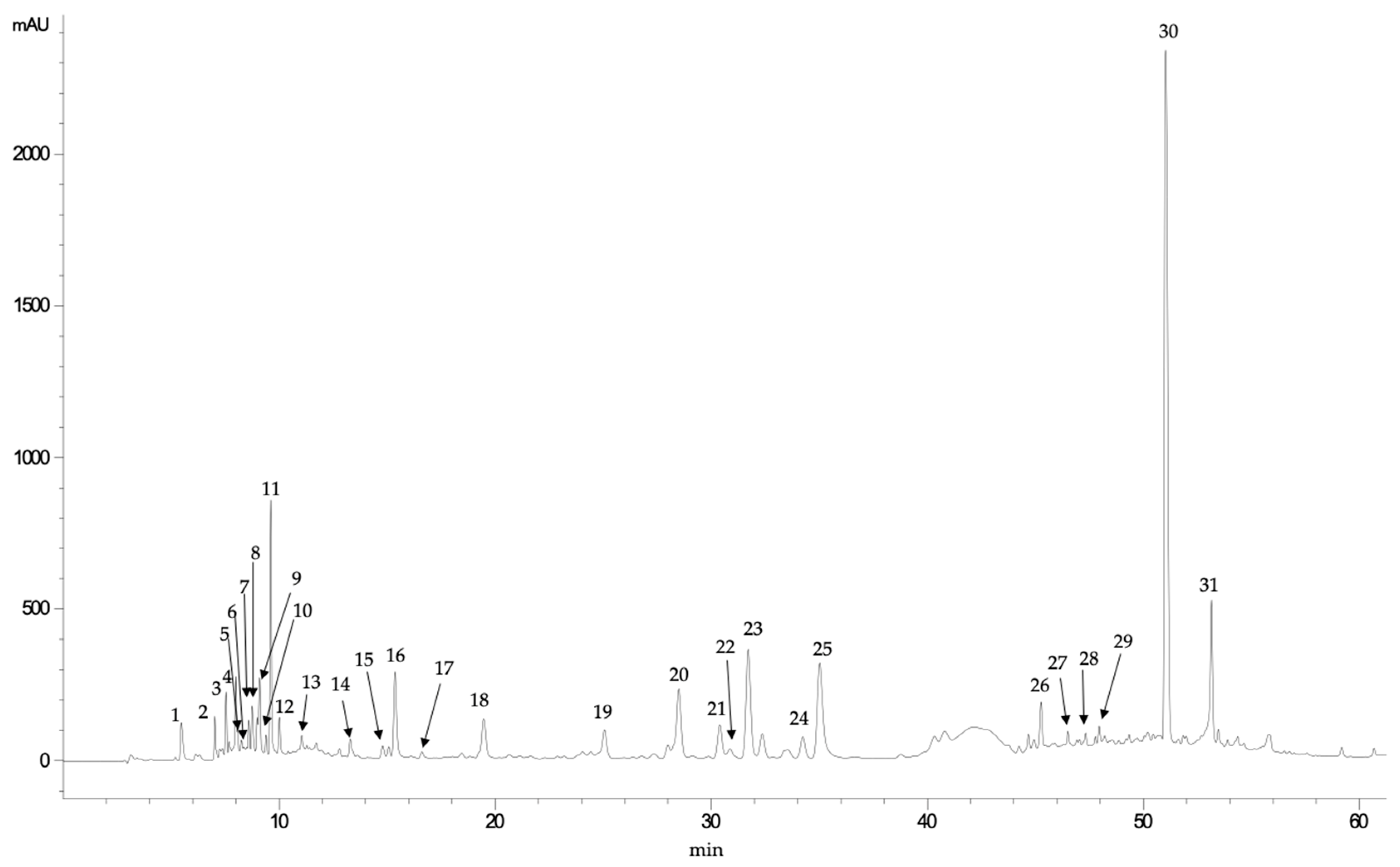
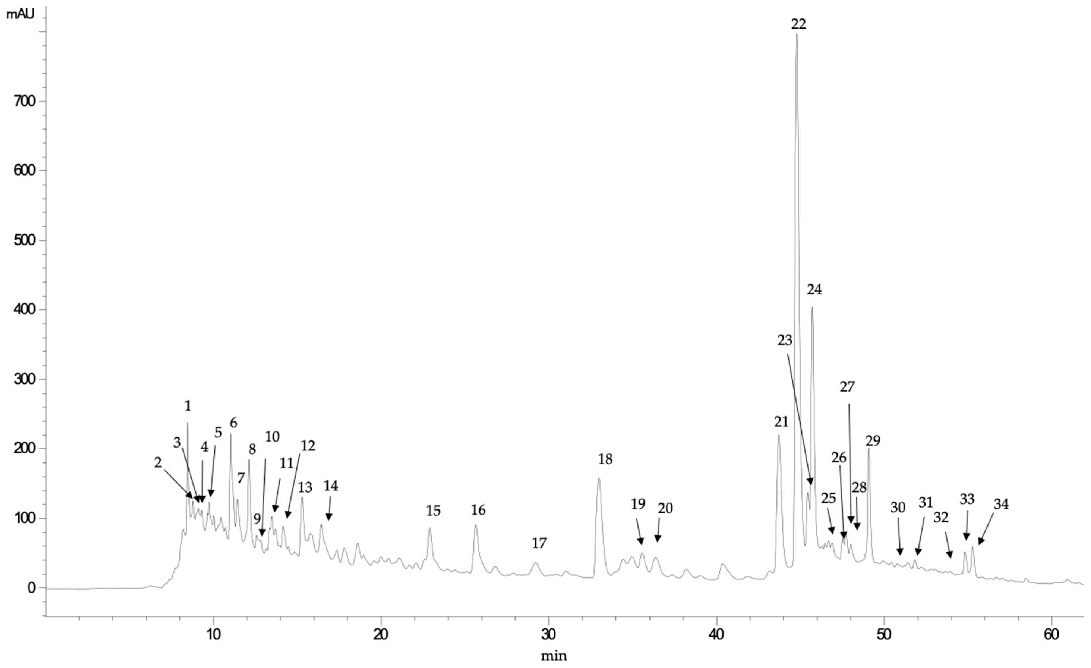
3.2. HPLC-DAD-MS/MS Analysis of Phenolic Profiles

E. tinifolia fruits (Figure 1) contained mainly derivatives of both hydroxybenzoic acids, such as gallic and syringic acids, and hydroxycinnamic acids, such as caffeic acid. However, it was observed that the most abundant compounds in E. tinifolia extract produced, in their mass analysis, pseudomolecular ions at m/z 359 and 537, which can be attributed to rosmarinic acid (RA) and different lithospermic acid derivatives, respectively (Table 2). Thus, compound 30, which showed a retention time of 51.02 min (Figure 1), was identified as RA, based on its parent ion (m/z 359) and its fragmentation pattern, which was as follows: fragment at m/z 359 corresponded to the RA radical ion [M-H-C18H16O8]; fragment at m/z 197, to the radical ion of 3,4-dihydroxyphenylactic acid [M-H-C9H10O5]; and fragment at m/z 179, to the radical ion of caffeic acid [M-H-C9H8O4]. Other fragment ions obtained for this compound were fragment at m/z 161 ([M-H-C9H7O3]), which can be attributed to acylonium ion and fragment at m/z 135, corresponding to [M-H-C8H6O2]. These same fragments were found in compounds 27, 28, and 29, except they showed other signals that corresponded to fragments not determined as sugars. Compounds 20 to 25 were identified as lithospermic acid derivatives, which were isomers since they showed the same pseudomolecular ion at m/z 625, with the main fragment ion detected at m/z 537 (C27H22O12), which can be identified as lithospermic acid. Moreover, the fragment ion at m/z 493 [M-H] can be formed by the loss of CO2 (44 Da) of the ion at m/z 537, whereas fragment ions at m/z 295 [M-H-CO2-C9H10O5] and m/z 312 [M-H-C9H8O4] were derived from the ion at m/z 493 due to the loss of C9H10O5 and C9H8O4 fragment ions. These fragments are those reported by Huang et al. [47] for lithospermic acid. Both RA and lithospermic acid derivatives could be relevant for the biological activity of these extracts since RA has been reported to be a compound with significant antioxidant and antineoplastic activity [32], and the lithospermic acid and its mono- and dimethyl esters are known to inhibit adenylate cyclase [48].
In the case of S. lanoginosum fruits (Figure 2), although different hydroxybenzoic acids (such us gallic and protocatechuic acids) and hydroxycinnamic acids (such as coumaric and ferulic acids) were identified, the main family of phenolic compounds identified was flavonols. Thus, mainly quercetin and myricetin glycosides and their corresponding non-glycoside forms were identified in these fruits (Table 3). The structure of flavonoids often results in substituents such as hydroxyl, methyl, and methoxyl groups. Therefore, in the MS/MS analysis, fragment ions are usually derived from the loss of CO (28 Da), H2O (18 Da) or CO2 (44 Da) molecules, as well as the fragment ions of substituents [49]. In addition, the retro-Diels–Alder (RDA) fragmentation is a common fragmentation pattern in flavonoids. Compound 34, which showed a pseudomolecular ion [M-H] at m/z 301, was identified as quercetin (C15H10O7). The fragment ion at m/z 273 [M-H-CO] was derived from the loss of a CO (28 Da) and that at m/z 179 ([M-H-C7H6O2]) can be explained by the RDA fragmentation. Subsequently, fragment ions at m/z 151 [M-H-C7H6O2-CO] originated from the ion at m/z 179 by the loss of a CO (28 Da) [46]. Compound 22 was the main compound, and it was identified as quercetin glucoside, showing a pseudomolecular ion at m/z 463, whose fragmentation produced the ion at m/z 301 [M -H C15H10O7] after the loss of a glucoside molecule (162 Da). Similarly, compounds 23 and 24 also showed fragment ions at m/z 301 in their MS/MS analysis, formed after losing a glucuronic acid (176 Da). In contrast, compounds 25 and 26 were identified as quercetin-pentoside because of detecting the same ion fragment (m/z 301) after the loss of 132 Da (which can be attributed to a pentose moiety). Quercetin derivatives and, mainly quercetin glucoside, can be relevant for the biological activity of S. lanoginosum fruits, since this compound has been shown to protect DNA and erythrocytes from oxidative damage and exhibited anticancer activity [50].

3.3. Digestive Enzymes Inhibition

3.3.1. Inhibition of α-Glucosidase

Currently, only a few α-glucosidase inhibitors, such as acarbose and voglibose, have been approved to treat diabetes, and their structures are mainly composed of sugar moieties [51]. Thus, many studies have been focused on searching for alternative α-glucosidase inhibitors with non-sugar core structure, particularly the polyphenols, due to their abundant availability in nature and their promising biological activities [52]. In the α-glucosidase assay, E. tinifolia and S. lanuginosum extracts showed high inhibitory activities with IC50 values of 0.17 and 0.21 mg/mL, respectively (Table 4). Under the same experimental conditions, the positive control acarbose presented an IC50 value of 0.13 mg/mL. Lopez-Martinez et al. [53] evaluated onions extracts for the inhibition of α-glucosidase activity and found that white variety inhibited more than 50% at 0.7 mg/mL. Hogan et al. [54] reported that red grape extract (1.5 mg/mL) inhibited 47% of α-glucosidase enzyme activity, and the inhibition potency was significantly higher than the white grape extract they tested (1.5 mg/mL), which only inhibited α-glucosidase activity by 39%. This research shows that an extract with more phenolics and anthocyanins is a better α-glucosidase inhibitor. Furthermore, it has been demonstrated that quercetin and kaempferol in S. lanuginosum and rosmarinic acid present on E. tinifolia are effective inhibitors of this enzyme [55,56]. Considering these compounds as the main polyphenols of extracts activity, the mechanism of α-glucosidase inhibition by wild fruit extracts could be due to the non-competitive or mixed-type interactions that polyphenols such as quercetin, kaempferol [57,58,59], and rosmarinic acid [60] maintain with the enzyme. Further studies must be done to determine the mechanism of action of S. lanuginosum and E. tinifolia extracts. These evidences emphasize the possible application of wild fruits or their derivatives in the design of diabetes treatments.

3.3.2. Inhibition of α-Amylase

α-Amylase enzyme is one of the key enzymes in the human digestive system, which degrades starch to monosaccharides and causes the rise of blood glucose [61]. Natural amylase inhibitors offer an attractive therapeutic approach to the treatment of postprandial hyperglycemia by decreasing glucose released from starch. Extracts of E. tinofolia and S. lanuginosum slightly inhibited amylase activity at concentrations of 5 mg/mL (30.95% and 26.06% respectively) with IC50 values > 5 mg/mL. In other studies, polyphenol-rich extracts from different types of berries inhibited α-amylase in vitro, and the most effective were from raspberry and rowanberry (IC50 values of 21 and 4.5 μg/mL, respectively). Thus, strawberry and raspberry extracts were more effective amylase inhibitors than blueberry and blackcurrant [62]. Other authors observed that the extent of inhibition of α-amylase was related to appreciable amounts of soluble tannins [62]. Our fruits extracts are poor in tannins content, and it may explain the low inhibitory enzymatic activity.

3.3.3. Inhibition of Pancreatic Lipase

Both fruit extracts showed no activity against lipase, while positive control Orlistat showed IC50 value of 0.04 μg/mL (Table 4). These results agree with a study of P. andina extracts that did not inhibit lipase activity [41]. The inhibition of lipase has been associated with higher content of tannins more than phenolic compounds [41]. This result could explain the absence of inhibition towards the lipase enzyme.

3.4. Antiproliferative Activity

Many studies reported the in vivo and in vitro effectiveness of phenolic compounds of fruits as anticancer. The bioactive compounds have significant anticancer effects through various complementary mechanisms of action, including the induction of metabolizing enzymes and the modulation of gene expression; and their impact on cell proliferation, apoptosis, and subcellular signaling pathway [63,64].
Antiproliferative activity of E. tinifolia and S. lanuginosum extracts were evaluated using three human cancer cell lines, HeLa (cervicouterine adenocarcinoma), MCF-7 (breast adenocarcinoma) and HT-29 (colon adenocarcinoma). Results showed that MCF-7 cell line was the most sensitive to the E. tinifolia extract, and the S. lanuginosum extract was against HT-29 cell line. The effect of E. tinifolia and S. lanuginosum extracts on cancer cell lines was dose-dependent and varied with the cell type and extract concentration. The concentration of 2 mg/mL of both extracts was the most effective. Nevertheless, E. tinifolia had the strongest effect on the three cell types. It revealed a marked decrease in the viability of the cancer cell lines after treatment. The 50% growth inhibition (IC50) of cells exposed to E. tinifolia was 0.99, 1.36 and 0.82 mg/mL, and for S. lanuginosum were 1.99, 3.22, and 1.97mg/mL for MCF-7, HeLa, and HT-29 cells, respectively (Table 4). It can be suggested that a wide range of phenolic compounds in the extract contributed to their cytotoxic activity. Al Hasani et al. [65] evaluated the antiproliferative activity for MCF-7 cells of Sideroxylon mascatense fruit extract and showed higher activity than E. tinifolia and S. lanuginosum extracts (IC50 value of 64 μg/mL). Though the extracts of Pyracantha coccinea (IC50 1.2 mg/mL) and Zosima absinthifolia (IC50 1.5 mg/mL) had less antiproliferative activity than E. tinifolia for HeLa cells [66,67]. The main phenol compound found on E. tinifolia extract was rosmarinic acid, and it could be responsible for the antiproliferative activity, as described by Niu et al. [68]. They compared the antiproliferative activity of Rosmarinus officinalis with rosmarinic acid and concluded that rosmarinic acid was more active than the plant extract against HeLa cells.
Breast cancer is the most frequently diagnosed cancer in women. An alternative strategy to reduce the risk of cancer is through dietary modification. E. tinifolia and S. lanuginosum extracts showed better antiproliferative activity against MCF-7 cells than an apple extract (IC50 = 70.7 mg/mL) [69]. Moreover, quercetin glucoside (QG) exhibited significant antiproliferative activity against MCF-7 cells. The EC50 value of QG in inhibiting MCF-7 cell growth was 46.4 μM [69]. This flavonoid is the most abundant phenolic compound in S. lanuginosum extract and could be responsible for inhibiting MCF-7 cells.
Phenolic compounds of E. tinifolia and S. lanuginosum inhibited proliferation of colon cancer cells HT-29. Similar effects have been shown of Borago extracts with CI50 values within the 250–300 μg/mL range after 72 h of cell exposure. Like in E. tinifolia, rosmarinic acid was the main phenolic compound detected in all Borago taxa [70]. In another study, the main phenolic compounds in Rabbiteye blueberry could inhibit HT-29 cancer cell proliferation and induce apoptosis. The phenolic acid fraction showed antiproliferation activities with an IC50 of ~1000 μg/ mL [71].

4. Conclusions

Sideroxylon lanuginosum (coma) presented the higher content of total flavonoid (21.4 ± 1.5 mg GAE/100g FW); the most abundant flavonoid detected was the quercetin glucoside. In comparison, Ehretia tinifolia (pinguica) had higher total phenol content (64.7 ± 2.6 mg GAE/100g FW), and the most abundant phenolic detected was rosmarinic acid. These major compounds could be related to the interesting biological activity of both extracts. E. tinifolia showed the best effect in antioxidant/antiradical activity both in vitro (EC50 = 0.32 ± 0.03 mg/mL; 4134 ± 9.7 μM TE/g dry extract), ex vivo assay (IC50 = 58.55 ± 2.4 μg/mL), and the inhibition of α-glucosidase (IC50 = 0.17 ± 0.1 mg/mL). Additionally, the extract of E. tinifolia showed a better effect on the growth inhibition of cancer cells, particularly against those of the human colon (IC50 = 0.82 ± 0.09 mg/mL) and breast cancer (IC50 = 0.99 ± 0.01 mg/mL). These results contribute to the scant information available on these wild fruits and contribute to revalue their use as a natural source of antioxidants with potential therapeutic application in diseases caused by free radicals. Further investigations using animal models are needed to confirm the beneficial effects of the studied extracts as supplementary treatments for oxidative diseases.

Author Contributions

Bioassays, I.N.M.-G.; Conceptualization, I.N.M.-G. and E.V.-V.; Antiproliferative methodology and supervision, I.E.C.-T. and P.C.-R.; Antioxidant assays and supervision, J.F.A.-Z. and J.M.-M.; Enzymatic inhibition assays and supervision, E.V.-V.; Phenolic composition, I.G.-E. and M.O.-A.; Project administration, E.V.-V.; I.N.M.-G., and E.V.-V. writing—review and editing. All authors have read and agreed to the published version of the manuscript.

Funding

This research study was funded by PAICYT-UANL: CN1581-21.

Acknowledgments

Author Imelda N. Monroy-García thanks CONACyT (Mexico) for her doctoral scholarship no.701994, MEXICO, and the Salamanca University (USAL, Spain) for technical support.

Conflicts of Interest

We declare that we have no conflict of interest.

References

  1. Jiménez-Aspee, F.; Thomas-Valdés, S.; Schulz, A.; Ladio, A.; Theoduloz, C.; Schmeda-Hirschmann, G. Antioxidant activity and phenolic profiles of the wild currant Ribes magellanicum from Chilean and Argentinean Patagonia. Food Sci. Nutr. 2015, 4, 595–610. [Google Scholar] [CrossRef] [Green Version]
  2. Turner, N.J.; Łuczaj, L.J.; Migliorini, P.; Pieroni, A.; Dreon, A.L.; Sacchetti, L.E.; Paoletti, M.G. Edible and Tended Wild Plants, Traditional Ecological Knowledge and Agroecology. Crit. Rev. Plant Sci. 2011, 30, 198–225. [Google Scholar] [CrossRef]
  3. Bvenura, C.; Sivakumar, D. The role of wild fruits and vegetables in delivering a balanced and healthy diet. Food Res. Int. 2017, 99, 15–30. [Google Scholar] [CrossRef] [PubMed]
  4. Hadjichambis, A.C.; Paraskeva-Hadjichambi, D.; Della, A.; Giusti, M.E.; De Pasquale, C.; Lenzarini, C.; Censorii, E.; Gonzales-Tejero, M.R.; Sanchez-Rojas, C.P.; Ramiro-Gutierrez, J.M.; et al. Wild and semi-domesticated food plant consumption in seven circum-Mediterranean areas. Int. J. Food Sci. Nutr. 2008, 59, 383–414. [Google Scholar] [CrossRef] [PubMed]
  5. Tardío, J.; Pardo-De-Santayana, M.; Morales, R. Ethnobotanical review of wild edible plants in Spain. Bot. J. Linn. Soc. 2006, 152, 27–71. [Google Scholar] [CrossRef]
  6. Miller, J.S. A Revision of the New World Species of Ehretia (Boraginaceae). Ann. Mo. Bot. Gard. 1989, 76, 1050. [Google Scholar] [CrossRef]
  7. Monroy-Ortiz, C.; Monroy, R. Las Plantas Compañeras de Siempre. La Experiencia en Morelos; Universidad Autónoma del Estado de Morelos: Cuernavaca, Mexico, 2006. [Google Scholar]
  8. Martínez, M. Catálogo de Nombres Vulgares y Científicos de Plantas Mexicanas; Fondo de Cultura Económica: Ciudad de México, México, 1979; pp. 93–1071. [Google Scholar]
  9. Benítez-Badillo, G.; Pulido-Salas, M.T.P.; Equihua-Samora, M. Árboles Multiusos Nativos de Veracruz para Reforestación, Restauración y Plantaciones; Instituto de Ecología, A.C., Sigolfo, Conafor: Xalapa, México, 2008; 108p. [Google Scholar]
  10. Pío-León, J.F.; Díaz-Camacho, S.P.; López, M.G.; Montes-Avila, J.; López-Angulo, G.; Delgado-Vargas, F. Physicochemical, nutritional, and antioxidant characteristics of the fruit of Ehretia tinifolia. Rev. Mex. Biodivers. 2012, 83, 273–280. [Google Scholar] [CrossRef]
  11. Cowan, R.S.; Little, E.L. The Audubon Society Field Guide to North American Trees: Eastern Region. TAXON 1981, 30, 548. [Google Scholar] [CrossRef]
  12. Bahadoran, Z.; Mirmiran, P.; Azizi, F. Dietary polyphenols as potential nutraceuticals in management of diabetes: A review. J. Diabetes Metab. Disord. 2013, 12, 43. [Google Scholar] [CrossRef] [Green Version]
  13. Tsao, R. Chemistry and Biochemistry of Dietary Polyphenols. Nutrients 2010, 2, 1231–1246. [Google Scholar] [CrossRef]
  14. Pandey, K.B.; Rizvi, S.I. Plant Polyphenols as Dietary Antioxidants in Human Health and Disease. Oxidative Med. Cell. Longev. 2009, 2, 270–278. [Google Scholar] [CrossRef] [Green Version]
  15. Yang, C.S.; Landau, J.M.; Huang, M.-T.; Newmark, H.L. Inhibition of Carcinogenesis by Dietary Polyphenolic Compounds. Annu. Rev. Nutr. 2001, 21, 381–406. [Google Scholar] [CrossRef] [Green Version]
  16. Moein, S. Polyphenols and cancer: A review. Mol. Med. J. 2015, 1, 6–12. [Google Scholar]
  17. Wu, X. Rosa Roxburghii tratt Fruit and Green Jujube Beverage and Preparation Method Thereof. CN106578777A, 26 April 2017. [Google Scholar]
  18. Wang, C.; Li, J.; Li, Q.; Song, Q. A Kind of Stauntonvine Pulp Fruitcake and Preparation Method Thereof. CN108094661A, 1 June 2018. [Google Scholar]
  19. Yichun Zhongzhi Dashanwang Liquor Industry Co Ltd. Wild Indigo Naturalis-Cowberry Beer. CN105524756A, 27 April 2016.
  20. Foshan Huili Cosmetics Science and Technology Co Ltd. Post-Solar Restorative Composition. CN108272729A, 13 July 2018.
  21. Liu, J. Wild Cherry Berry Diet Therapy Drink and Preparation Method Thereof. CN106983058A, 28 July 2017. [Google Scholar]
  22. Derosa, G.; Romano, D.; D’Angelo, A.; Maffioli, P. Berberis aristata/Silybum marianum fixed combination (Berberol®) effects on lipid profile in dyslipidemic patients intolerant to statins at high dosages: A randomized, placebo-controlled, clinical trial. Phytomedicine 2015, 22, 231–237. [Google Scholar] [CrossRef]
  23. Viveros-Valdez, E.; Jaramillo-Mora, C.; Oranday-Cárdenas, A.; Morán-Martínez, J.; Carranza-Rosales, P. Antioxidant, cytotoxic and alpha-glucosidase inhibition activities from the Mexican berry Anacahuita (Cordia boissieri). Arch. Latinoam. Nutr. 2016, 66, 211–218. [Google Scholar]
  24. Singleton, V.; Rossi, J. Colorimetry of Total Phenolic Compounds with Phosphomolybdic-Phosphotungstic Acid Reagents. Am. J. Enol. Vitic. 1965, 16, 144–158. [Google Scholar]
  25. Zhishen, J.; Mengcheng, T.; Jianming, W. The determination of flavonoid contents in mulberry and their scavenging effects on superoxide radicals. Food Chem. 1999, 64, 555–559. [Google Scholar] [CrossRef]
  26. Braca, A.; Sortino, C.; Politi, M.; Morelli, I.; Mendez, J. Antioxidant activity of flavonoids from Licania licaniaeflora. J. Ethnopharmacol. 2002, 79, 379–381. [Google Scholar] [CrossRef]
  27. Gupta, R.; Sharma, M.; Lakshmy, R.; Prabhakaran, D.; Reddy, K.S. Improved method of total antioxidant assay. Indian J. Biochem. Biophys. 2009, 46, 126–129. [Google Scholar]
  28. Silva-Beltrán, N.P.; Balderrama-Carmona, A.P.; López-Cuevas, O.; Portela-Márquez, M.A.; Umsza Guez, M.A.; López-Mata, M.A. Antioxidant and antimicrobial activity of Barchata (Zizhipus Obtusifolia). Revista Ciencias. 2019, 6, e523. [Google Scholar] [CrossRef]
  29. Kaskoos, R.A. In-vitro α-glucosidase inhibition and antioxidant activity of methanolic extract of Centaurea calcitrapa from Iraq. Am. J. Essent. Oil. Nat. Prod. 2013, 1, 122–125. [Google Scholar]
  30. Shuda, P.; Zinjarde, S.S.; Bhargava, S.Y.; Kumar, A.R. Potent α-amylase inhibitory activity of Indian Ayurvedic medicinal plants. BMC Complement. Altern. Med. 2011, 11, 5. [Google Scholar] [CrossRef] [Green Version]
  31. Maqsood, M.; Ahmed, D.; Atique, I.; Malik, W. Lipase inhibitory activity of Lagenaria siceraria fruit as a strategy to treat obesity. Asian Pac. J. Trop. Med. 2017, 10, 305–310. [Google Scholar] [CrossRef]
  32. Viveros-Valdez, E.; Rivas-Morales, C.; Oranday-Cárdenas, A.; Castro-Garza, J.; Carranza-Rosales, P. Antiproliferative Effect from the Mexican Poleo (Hedeoma drummondii). J. Med. Food 2010, 13, 740–742. [Google Scholar] [CrossRef]
  33. Cittadini, M.C.; García-Estévez, I.; Escribano-Bailón, M.T.; Rivas-Gonzalo, J.C.; Valentich, M.A.; Repossi, G.; Soria, E.A. Modulating activity of phenolic compounds from American plant infusions on fatty acid-related interleukin-6 release in glial cells. Nutr. Cancer 2018, 70, 267–277. [Google Scholar] [CrossRef] [PubMed]
  34. Turker, G. Free radical scavenging activity and phenolic content of edible wild fruits from Kazdagi (Ida Mountains), Turkey. J. Med. Plants Res. 2012, 6. [Google Scholar] [CrossRef]
  35. Schmeda-Hirschmann, G.; Feresin, G.; Tapia, A.; Hilgert, N.; Theoduloz, C. Proximate composition and free radical scavenging activity of edible fruits from the Argentinian Yungas. J. Sci. Food Agric. 2005, 85, 1357–1364. [Google Scholar] [CrossRef]
  36. Jiménez-Aspee, F.; Quispe, C.; Soriano, M.D.P.C.; Gonzalez, J.F.; Hüneke, E.; Theoduloz, C.; Schmeda-Hirschmann, G. Antioxidant activity and characterization of constituents in copao fruits (Eulychnia acida Phil., Cactaceae) by HPLC–DAD–MS/MSn. Food Res. Int. 2014, 62, 286–298. [Google Scholar] [CrossRef]
  37. Ikram, E.H.K.; Eng, K.H.; Jalil, A.M.M.; Ismail, A.; Idris, S.; Azlan, A.; Nazri, H.S.M.; Diton, N.A.M.; Mokhtar, R.A.M. Antioxidant capacity and total phenolic content of Malaysian underutilized fruits. J. Food Compos. Anal. 2009, 22, 388–393. [Google Scholar] [CrossRef]
  38. Recuenco, M.; Lacsamana, M.; Hurtada, W.; Sabularse, V. Total Phenolic and Total Flavonoid Contents of Selected Fruits in the Philippines. Philipp. J. Sci. 2016, 145, 275–281. [Google Scholar]
  39. Goulas, V.; Georgiou, E. Utilization of Carob Fruit as Sources of Phenolic Compounds with Antioxidant Potential: Extraction Optimization and Application in Food Models. Foods 2019, 9, 20. [Google Scholar] [CrossRef] [Green Version]
  40. Magalhães, A.S.; Silva, B.M.; Pereira, J.A.; Andrade, P.; Valentão, P.; Carvalho, M. Protective effect of quince (Cydonia oblonga Miller) fruit against oxidative hemolysis of human erythrocytes. Food Chem. Toxicol. 2009, 47, 1372–1377. [Google Scholar] [CrossRef] [PubMed]
  41. Jiménez-Aspee, F.; Theoduloz, C.; Pormetter, L.; Mettke, J.; Ávila, F.; Schmeda-Hirschmann, G. Andean Prumnopitys Andina (Podocarpacae) Fruit Extracts: Characterization of Secondary Metabolites and Potential Cytoprotective Effect. Molecules 2019, 24, 4028. [Google Scholar] [CrossRef] [PubMed] [Green Version]
  42. Ruíz-Rodriguez, B.M. Frutos Silvestres de uso Tradicional en la Alimentación: Evaluación de su Valor Nutricional, Compuestos Bioactivos y Capacidad Antioxidante. Ph.D. Thesis, Universidad Complutense de Madrid, Madrid, Spain, 2014. [Google Scholar]
  43. Mitjans, M.; Martínez, V.; del Campo, J.; Abajo, C.; Lozano, C.; Torres, J.; Vinardell, M.P. Novel epicatechin derivatives with antioxidant activity modulate interleukin-1β release in lipopolysaccharide-stimulated human blood. Bioorg. Med. Chem. Lett. 2004, 14, 5031–5034. [Google Scholar] [CrossRef]
  44. Prakash, M.; Basavaraj, B.V.; Murthy, K.C. Biological functions of epicatechin: Plant cell to human cell health. J. Funct. Foods 2018, 52, 14–24. [Google Scholar] [CrossRef]
  45. Shay, J.; Elbaz, H.; Lee, I.; Zielske, S.P.; Malek, M.H.; Hüttemann, M. Molecular Mechanisms and Therapeutic Effects of (−)-Epicatechin and Other Polyphenols in Cancer, Inflammation, Diabetes, and Neurodegeneration. Oxidative Med. Cell. Longev. 2015, 2015, 1–13. [Google Scholar] [CrossRef] [Green Version]
  46. Vega-Vega, V. Enriquecimento de la Capacidad Antioxidante y Proteción Antimicrobiana del Mango Fresco Cortado Aplicando Compuestos Fenólicos de sus Subproductos. Master’s Thesis, CIAD, Hermosillo, México, August 2011. [Google Scholar]
  47. Huang, G.; Liang, J.; Chen, X.; Lin, J.; Wei, J.; Huang, D.; Zhou, Y.; Sun, Z.; Zhao, L. Isolation and Identification of Chemical Constituents from Zhideke Granules by Ultra-Performance Liquid Chromatography Coupled with Mass Spectrometry. J. Anal. Methods Chem. 2020, 2020, 1–16. [Google Scholar] [CrossRef] [PubMed]
  48. Lu, Y.; Foo, L.Y. Polyphenolics of Salvia—A review. Phytochemistry 2002, 59, 117–140. [Google Scholar] [CrossRef]
  49. Zhang, W.-X.; Feng, M.; Miao, Y.-L.; Li, Y.-Y.; Tong, L.-G.; He, P.; Ni, Y. Analysis of chemical components of Huanbei Zhike Prescription based on UPLC-Q-TOF-MS/MS technology. China J. Chin. Mater. Med. 2019, 44, 3022–3034. [Google Scholar]
  50. Girish, T.; Kumar, K.A.; Rao, U.P. C- Glycosylated flavonoids from black gram husk: Protection against DNA and erythrocytes from oxidative damage and their cytotoxic effect on HeLa cells. Toxicol. Rep. 2016, 3, 652–663. [Google Scholar] [CrossRef] [PubMed] [Green Version]
  51. Yin, Z.; Zhang, W.; Feng, F.; Zhang, Y.; Kang, W. α-Glucosidase inhibitors isolated from medicinal plants. Food Sci. Hum. Wellness 2014, 3, 136–174. [Google Scholar] [CrossRef] [Green Version]
  52. Inthongkaew, P.; Chatsumpun, N.; Supasuteekul, C.; Kitisripanya, T.; Putalun, W.; Likhitwitayawuid, K.; Sritularak, B. α-Glucosidase and pancreatic lipase inhibitory activities and glucose uptake stimulatory effect of phenolic compounds from Dendrobium formosum. Rev. Bras. Farm. 2017, 27, 480–487. [Google Scholar] [CrossRef]
  53. López-Martínez, L.X.; Aguilar Cisneros, L.M.; Dublán-García, O. Actividad antioxidante e inhibidora de α-glucosidasa y α-amilasa de tres variedades de cebolla (Allium cepa L.). Nova Sci. 2014, 6, 234–347. [Google Scholar] [CrossRef] [Green Version]
  54. Hogan, S.; Zhang, L.; Li, J.; Sun, S.; Canning, C.; Zhou, K. Antioxidant rich grape pomace extract suppresses postprandial hyperglycemia in diabetic mice by specifically inhibiting alpha-glucosidase. Nutr. Metab. 2010, 7, 71. [Google Scholar] [CrossRef] [PubMed] [Green Version]
  55. Pereira, D.F.; Cazarolli, L.H.; Lavado, C.; Mengatto, V.; Figueiredo, M.S.R.B.; Guedes, A.; Pizzolatti, M.G.; Silva, F.R.M.B. Effects of flavonoids on α-glucosidase activity: Potential targets for glucose homeostasis. Nutrition 2011, 27, 1161–1167. [Google Scholar] [CrossRef] [PubMed] [Green Version]
  56. Lin, L.; Dong, Y.; Zhao, H.; Wen, L.; Yang, B.; Zhao, M. Comparative evaluation of rosmarinic acid, methyl rosmarinate and pedalitin isolated from Rabdosia serra (MAXIM.) HARA as inhibitors of tyrosinase and α-glucosidase. Food Chem. 2011, 129, 884–889. [Google Scholar] [CrossRef]
  57. Meng, Y.; Su, A.; Yuan, S.; Zhao, H.; Tan, S.; Hu, C.Y.; Deng, H.; Guo, Y. Evaluation of Total Flavonoids, Myricetin, and Quercetin from Hovenia dulcis Thunb. As Inhibitors of α-Amylase and α-Glucosidase. Plant Foods Hum. Nutr. 2016, 71, 444–449. [Google Scholar] [CrossRef]
  58. Şöhretoğlu, D.; Sari, S.; Barut, B.; Özel, A. Discovery of potent α-glucosidase inhibitor flavonols: Insights into mechanism of action through inhibition kinetics and docking simulations. Bioorg. Chem. 2018, 79, 257–264. [Google Scholar] [CrossRef]
  59. Liu, Y.; Zhan, L.; Xu, C.; Jiang, H.; Zhu, C.; Sun, L.; Sun, C.; Li, X. α-Glucosidase inhibitors from Chinese bayberry (Morella rubra Sieb. et Zucc.) fruit: Molecular docking and interaction mechanism of flavonols with different B-ring hydroxylations. RSC Adv. 2020, 10, 29347–29361. [Google Scholar] [CrossRef]
  60. Xngo, Y.L.; Chua, Y.L.N.A.L.S.; Ngo, Y.L. Anti-diabetic Activity of Rosmarinic Acid Rich Fractions from Orthosiphon stamineus. Curr. Enzym. Inhib. 2018, 14, 97–103. [Google Scholar] [CrossRef]
  61. Kotowaroo, M.I.; Mahomoodally, M.F.; Gurib-Fakim, A.; Subratty, A.H. Screening of traditional antidiabetic medicinal plants of mauritius for possibleα-amylase inhibitory effectsin vitro. Phytother. Res. 2006, 20, 228–231. [Google Scholar] [CrossRef] [PubMed]
  62. Grussu, D.; Stewart, D.; McDougall, G.J. Berry Polyphenols Inhibit α-Amylase In Vitro: Identifying Active Components in Rowanberry and Raspberry. J. Agric. Food Chem. 2011, 59, 2324–2331. [Google Scholar] [CrossRef]
  63. Subramaniam, S.; Selvaduray, K.R.; Radhakrishnan, A.K. Bioactive Compounds: Natural Defense against Cancer? Biomolecules 2019, 9, 758. [Google Scholar] [CrossRef] [PubMed] [Green Version]
  64. Karasawa, M.M.G.; Mohan, C. Fruits as Prospective Reserves of bioactive Compounds: A Review. Nat. Prod. Bioprospect. 2018, 8, 335–346. [Google Scholar] [CrossRef] [Green Version]
  65. Al-Attabi, Z.H.; Al Hasani, S.; Waly, M.; Rahman, M.S.; Tamimi, Y. Antioxidant and Antitumor Properties of Wild Blueberry (Sideroxylon mascatense): Effects of Drying Methods. Int. J. Nutr. Pharmacol. Neurol. Dis. 2021, 11, 71. [Google Scholar] [CrossRef]
  66. Vahabi, L.; Monajemi, R.; Hosseini, S.A. The Cytotoxic Effect of Methanolic Extract of Pyracanthacoccinea M. Roemer Fruit on Hela Cell Line, Antioxidant Capacities and Total Phenol Contents of Methanolic and Aquatic Extract of this Fruit. Biomed. Pharmacol. J. 2015, 8, 99–103. [Google Scholar] [CrossRef]
  67. Razavi, S.M.; Ghasemiyan, A.; Salehi, S.; Zahri, F. Screening of biological activity of Zosima absinthifolia fruits extracts. EurAsian J. Biosci. 2009, 25–28. [Google Scholar] [CrossRef]
  68. Nie, J.; Li, R.; Wang, Y.; Tan, J.; Tang, S.; Jiang, Z.-T. Antioxidant activity evaluation of rosemary ethanol extract and their cellular antioxidant activity toward HeLa cells. J. Food Biochem. 2019, 43, e12851. [Google Scholar] [CrossRef] [PubMed]
  69. Yang, J.; Liu, R.H. Synergistic Effect of Apple Extracts and Quercetin 3-β-d-Glucoside Combination on Antiproliferative Activity in MCF-7 Human Breast Cancer Cells In Vitro. J. Agric. Food Chem. 2009, 57, 8581–8586. [Google Scholar] [CrossRef]
  70. Lyashenko, S.; Fabrikov, D.; González-Fernández, M.J.; Gómez-Mercado, F.; Ruiz, R.L.; Fedorov, A.; de Bélair, G.; Urrestarazu, M.; Rodríguez-García, I.; Álvarez-Corral, M.; et al. Phenolic composition and in vitro antiproliferative activity of Borago spp. seed extracts on HT-29 cancer cells. Food Biosci. 2021, 42, 101043. [Google Scholar] [CrossRef]
  71. Yi, W.; Fischer, J.; Krewer, G.; Akoh, C.C. Phenolic Compounds from Blueberries Can Inhibit Colon Cancer Cell Proliferation and Induce Apoptosis. J. Agric. Food Chem. 2005, 53, 7320–7329. [Google Scholar] [CrossRef] [PubMed]
Figure 1. Chromatogram (registered at 280 nm) obtained in the analysis of phenolic compounds of pinguica (E. tinifolia).
Foods 10 02710 g001
Figure 2. Chromatogram (registered at 280 nm) obtained in the analysis of phenolic compounds of coma (S. lanoginosum).
Foods 10 02710 g002
Table 1. Total phenolic content, total flavonoid content, and antioxidant/antiradical capacities of Ehretia tinifolia and Sideroxylon laniginosum extracts.
SampleTPC
mg GAE/100g F.W.
TFC
mg CE/100g F.W.
TEAC
μmol TE/g
DPPH
EC50 mg/mL
Hemolytic Inhibition
IC50 (μg/mL)
S. lanuginosum21.4 ± 1.5 b6.42 ± 0.9 a4134 ± 97 a0.48 ± 0.05 a61.76 ± 7.9 b
E. tinifolia64.7 ± 2.6 a5.1 ± 0.4 b2454 ± 38 b0.32 ± 0.03 b58.55 ± 6.5 b
** Control0.013 ± 2 c289 ± 20 a
** Trolox was used on DPPH free radical scavenging assay and ascorbic acid on AAPH-induced hemolysis assay. n = 3, (a–c) on each column show significant differences among each determination, according to Tukey’s test (p < 0.05).
Table 2. Tentative identification of phenolic compounds of E. tinifolia fruit extract, determined by HPLC-DAD-MS/MS.
PeakRt (min)UV Max[M+H]+MS/MS FragmentsTentative Identification
15.46261297.4135Unknown
27.01281191.2173, 129, 111Quinic acid
37.53262373.0311, 285, 267, 249,
241, 227, 196
Unknown
47.98252, 275 (sh)282.5150, 133Unknown
58.07301331.3169, 125Galloil-glucose
68.58282507.2489, 459, 293, 233,
195, 131, 125,113
Caffeic acid derivative
78.74280, 314 (sh)165.0137Unknown
88.96279507.2233, 165, 150, 125Unknown
99.08276719.0515, 359, 197, 179,
135
Rosmarinic acid derivative
109.38272515.0269, 251, 225, 213,
179, 159, 135, 109
Unknown
119.60278359.0197, 179, 135Syringic acid hexoside
1210.00281521.0197, 179, 135Syringic acid dihexoside
1311.03272165.0150, 121Unknown
1413.28278, 320 (sh)401.0359, 341, 297, 197,
179,135
Unknown
1514.78278, 314365.4323, 262, 250Unknown
1615.06311, 292 (sh)373.0211, 179, 123Methyl rosmarinate
1715.35281, 330 (sh)567.0179, 135Caffeic acid derivative
1819.45291, 322179.0135Caffeic acid
1925.04276863.0701, 521, 359, 315,
297, 197, 135
Syringil-rosmarinic acid dihexoside
2028.48276695.5579, 554, 537, 493, 312,
295, 277, 203, 135
Lithospermic acid derivative
2130.38276695.5579, 554, 537, 493, 312,
295, 277, 203, 135
Lithospermic acid derivative
2231.69276695.5579, 554, 537, 493, 312,
295, 277, 203, 135
Lithospermic acid derivative
2332.35276695.5579, 554, 537, 493, 312,
295, 277, 203, 135
Lithospermic acid derivative
2434.22276695.5579, 554, 537, 493, 312,
295, 277, 203, 135
Lithospermic acid derivative
2535.01276695.5579, 554, 537, 493, 312,
295, 277, 203, 135
Lithospermic acid derivative
2645.25275, 330 (sh)597.4579, 509, 491, 355, 337,
329, 311, 293, 267, 239,
197, 179, 135, 109
Unknown
2746.49284, 320861.5843, 817, 655, 521, 501,
475, 457, 383, 359, 339,
323, 197, 179, 135
Rosmarinic acid derivative
2847.31285, 318521.0359, 197, 179, 161, 135Rosmarinic acid-hexoside
2947.94283, 327521.0359, 197, 179, 161, 135Rosmarinic acid-hexoside
3051.02330, 290 (sh)359.0197, 179, 161, 135Rosmarinic acid
3153.13278537.0493, 359, 295, 277, 203,
185, 159, 135, 109
Lithospermic acid A
sh = shoulder.
Table 3. Tentative identification of phenolic compounds of S. lanoginosum fruit extract by HPLC-DAD electrospray ionization (ESI)-MS/MS.
PeakRt (Min)UV Max[M+H]+MS/MS FragmentsTentative Identification
18.44272169.1125, 113Gallic acid
28.76245137.0 p-hydroxybenzoic acid
38.89278329.3167, 151, 109Unknown
49.30282331.2169, 125Unknown
59.74283315.2152, 108Unknown
611.02259-293153.1123, 109Protocatechuic acid
711.43286, 315 (sh)461.0351, 323, 248, 233, 193Ferulic acid derivative
812.11285, 324 (sh)463.3283, 272, 255, 175, 163Unknown
913.48295, 311487.4187, 163, 145, 119Coumaric dihexoside
1014.15256, 311 (sh)435.2241, 193, 153Ferulic acid derivative
1115.26255, 335311.3249, 231, 205, 187, 161, 14
147, 135, 121
Unknown
1215.82303421.2241Unknown
1316.41280417.1399, 227, 167, 153Unknown
1417.77281, 310 (sh)387.0163Coumaric acid derivative
1522.88267, 327 (sh)241.2197, 168, 141, 130Phenolic acid derivative
1625.63262, 331 (sh)295.0251, 189, 137, 121Phenolic acid derivative
1729.23307, 290 (sh)163.0119Coumaric acid
1832.95255, 351, 301 (sh)755.5609, 489, 355, 343, 325
301, 271, 179
Quercetin glucoside dirhamnoside
1934.82354, 300 (sh)479.0317, 287, 271, 179, 151Myricetin glucoside
2035.54254, 352, 302 (sh)771.0301Quercetin diglucoside rhamnoside
2143.66256, 353, 305 (sh)609.4343, 301, 271, 255, 179,
151
Quercetin neohesperidoside
2244.73256, 355, 301 (sh)463.4306, 301, 271, 255, 248
179, 151, 121
Quercetin glucoside
2345.40257, 342, 302 (sh)477.0301, 151Quercetin glucuronide
2445.68256, 354, 300 (sh)477.0301, 179, 151Quercetin glucuronide
2546.68267, 359 (sh)433.8301, 271, 179, 151Quercetin pentoside
2647.52255, 351433.9301, 271, 256, 180, 152Quercetin pentoside
2747.71264, 349447.9285, 256, 227, 151Kaempferol glucoside
2847.99284, 340 (sh)436.0346, 316, 274, 167, 123Unknown
2949.06257, 347447.0301, 273, 257, 179, 151Kaempferol glucoside
3050.85371317.0179, 151, 138Myricetin
3151.18327, 287 (sh)359.0197, 179, 161, 135Rosmarinic acid
3254.79264, 316, 356 (sh)609.8463, 301, 151Quercetin rutinoside
3355.25251, 330, 300 (sh)639.8477, 463, 316, 300β-hydroverbascoside
3457.00370, 300 (sh)301.2273, 229, 179, 161,
151, 121
Quercetin
sh = shoulder.
Table 4. Inhibition of digestive enzymatic and antiproliferative activity by E. tinifolia and S. lanuginosum fruit extracts.
SampleHalf Maximal Inhibitory Concentration [mg/mL]
α-Gluα-AmyLipaseMCF-7HeLaHT-29* RBCs
S. lanuginosum0.21 ± 0.3 a>5>51.99 ± 0.3 a3.22 ± 0.8 a1.97 ± 0.2 a>5
E. tinifolia0.17 ± 0.1 a,b>5>50.99 ± 0.01b1.36 ± 0.2 b0.82 ± 0.09 b>5
** Control0.13 ± 0.2 b0.97 ± 0.080.17 ± 0.200.013 ± 0.001 c0.011 ± 0.002 c0.015 ± 0.001 cND
* RBCs = Red blood cells = Erythrocytes. ** Acarbose was used on α-glucosidase and α-amylase assays, orlistat on Lipase inhibition and taxol against cancer cells lines. n = 3, literals (a–c) on each column show significant differences among treatments, according to Tukey’s test (p < 0.05). ND: not determined.
Publisher’s Note: MDPI stays neutral with regard to jurisdictional claims in published maps and institutional affiliations.

