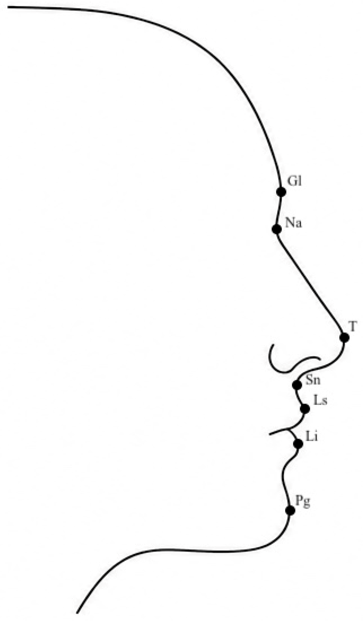
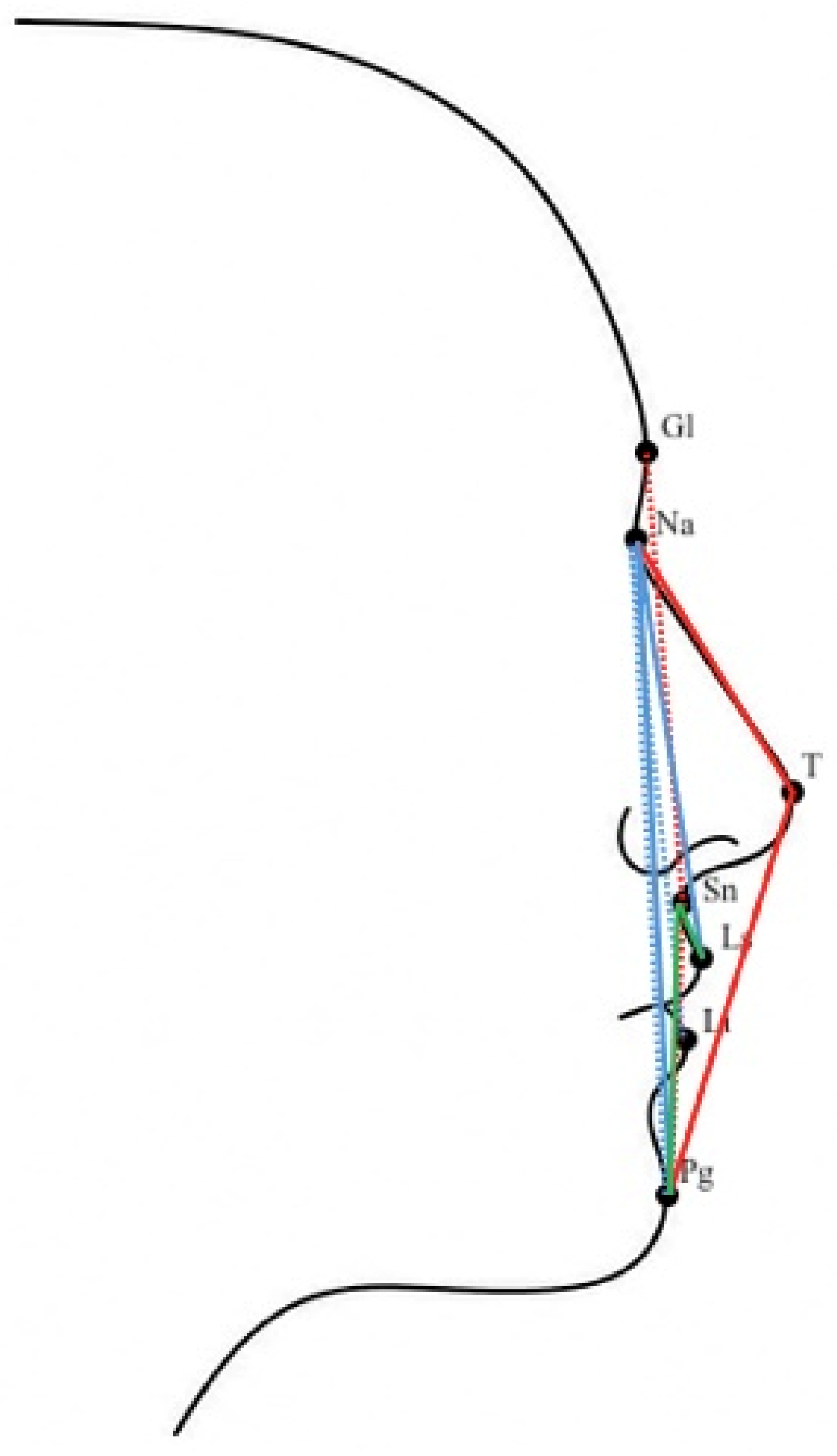
Evaluation of Facial Soft Tissue Angles in Adolescents with Angle Class I, II, and III Malocclusion Using Profile Image Analysis

Abstract
1. Introduction
2. Materials and Methods
2.1. Study Design and Participants
2.2. Ethical Consideration and Study Registration
2.3. Inclusion and Exclusion Criteria
2.4. Image Acquisition
2.5. Quantification of Image with Landmarks
2.6. Reproducibility and Error of Method Analysis
2.7. Statistical Analyses
3. Results
3.1. Angle Characteristics
3.2. Comparing Males and Females
3.3. Landmark Accuracy
4. Discussion
5. Conclusions
Author Contributions
Funding
Institutional Review Board Statement
Informed Consent Statement
Data Availability Statement
Conflicts of Interest
References
- Farkas, L.G. Anthropometry of the Head and Face, 2nd ed.; Raven Press: New York, NY, USA, 1994. [Google Scholar]
- Baysal, A.; Sahan, A.O.; Ozturk, M.A.; Uysal, T. Reproducibility and Reliability of Three-Dimensional Soft Tissue Landmark Identification. Angle Orthod. 2016, 86, 1004–1009. [Google Scholar] [CrossRef]
- Moon, S.; Mohamed, A.M.A.; He, Y.; Dong, W.; Chen, Y.; Yang, Y. Extraction vs. Nonextraction on Soft-Tissue Profile Change in Patients with Malocclusion: A Systematic Review and Meta-Analysis. BioMed Res. Int. 2021, 2021, 7751516. [Google Scholar] [CrossRef] [PubMed]
- Bergman, R.T.; Waschak, J.; Borzabadi-Farahani, A.; Murphy, N.C. Longitudinal Study of Cephalometric Soft Tissue Profile Traits. Angle Orthod. 2014, 84, 48–55. [Google Scholar] [CrossRef]
- Sharma, P.; Arora, A.; Valiathan, A. Age Changes of Jaws and Soft Tissue Profile. Sci. World J. 2014, 2014, 301501. [Google Scholar] [CrossRef] [PubMed]
- Chaconas, S.J. A Statistical Evaluation of Nasal Growth. Am. J. Orthod. 1969, 56, 403–414. [Google Scholar] [CrossRef]
- Wisth, P.J. Changes of the Soft Tissue Profile During Growth. Eur. J. Orthod. 2007, 29, 1114–1117. [Google Scholar] [CrossRef]
- Subtelny, J.D. A Longitudinal Study of Soft Tissue Facial Structures and Their Profile Characteristics, Defined in Relation to Underlying Skeletal Structures. Am. J. Orthod. 1959, 45, 481–507. [Google Scholar] [CrossRef]
- Vig, P.S.; Cohen, A.M. Vertical Growth of the Lips: A Serial Cephalometric Study. Am. J. Orthod. 1979, 75, 405–415. [Google Scholar] [CrossRef]
- Bergeron, L.; Tu, C.C.; Chen, Y.R. Single-Split Technique for Correction of Severe Facial Asymmetry. Plast. Reconstr. Surg. 2008, 122, 1535. [Google Scholar] [CrossRef]
- Cheong, Y.W.; Lo, L.J. Facial Assymetry: Etiology, Evaluation and Management. Chang Gung Med. J. 2011, 34, 341–351. [Google Scholar]
- Enlow, D.H.; Hans, M.G. Handbook of Facial Growth, 2nd ed.; WB Saunders: Philadelphia, PA, USA, 2008. [Google Scholar]
- Varatharaju, V.; Caflisch, M.; Soroken, C.; Kiliaridis, S.; Antonarakis, G.S. Does Age Influence Self-Perception of the Soft-Tissue Profile in Children? Am. J. Orthod. Dentofac. Orthop. 2021, 159, e207–e215. [Google Scholar] [CrossRef]
- Proffit, W.; Severt, T. The Prevalence of Facial Asymmetry in the Dentofacial Deformities Population at the University of North Carolina. Int. J. Adult Orthodon. Orthognath. Surg. 1997, 12, 171–175. [Google Scholar]
- Cenzato, N.; Nobili, A.; Maspero, C. Prevalence of Dental Malocclusions in Different Geographical Areas. Dent. J. 2021, 9, 117. [Google Scholar] [CrossRef]
- Björk, A.; Palling, M. Adolescent Age Changes in Sagittal Jaw Relation. Acta Odontol. Scand. 1955, 12, 201–232. [Google Scholar] [CrossRef] [PubMed]
- Grammer, K.; Thornhill, R. Human Facial Attractiveness and Sexual Selection. J. Comp. Psychol. 1994, 108, 233–242. [Google Scholar] [CrossRef] [PubMed]
- Jones, D.; Hill, K. Criteria of Facial Attractiveness in Five Populations. Hum. Nat. 1993, 4, 271–296. [Google Scholar] [CrossRef]
- Shaw, W.C.; Rees, G.; Dawe, M.; Charles, C.R. The Influence of Dentofacial Appearance on the Social Attractiveness of Young Adults. Am. J. Orthod. 1985, 87, 21–26. [Google Scholar] [CrossRef]
- Eubanks, R.J. Surgical Correction of Masseter Muscle Hypertrophy Associated with Unilateral Prognathism: Report of case. J. Oral Surg. 1957, 15, 66–69. [Google Scholar] [PubMed]
- Kronmiller, J.E. Development of Asymmetries. Semin. Orthod. 1998, 4, 134–137. [Google Scholar] [CrossRef]
- Aljehani, D.; Baeshen, H.A. Effectiveness of the American Board of Orthodontics Discrepency Index in Predicting Treatment Time. J. Contemp. Dent. Pract. 2018, 19, 647–650. [Google Scholar] [CrossRef]
- Shimizu, Y.; Tanikawa, C.; Kajiwara, T.; Nagahara, H.; Yamashiro, T. The Validation of Orthodontic Artificial Intelligence Systems that Perform Orthodontic Diagnoses and Treatment Planning. Eur. J. Orthod. 2022, 44, 436–444. [Google Scholar] [CrossRef]
- Albalawi, F.; Alamoud, K.A. Trends and Application of Artificial Intelligence Technology in Orthodontic Diagnosis and Treatment Planning—A Review. Appl. Sci. 2022, 12, 11864. [Google Scholar] [CrossRef]
- Kulikowski, C.A. An Opening Chapter of the First Generation of Artificial Intelligence in Medicine. Med. Inform. 2015, 10, 227–233. [Google Scholar] [CrossRef]
- Li, Z.; Liu, F.; Yang, W.; Peng, S.; Zhou, J. A Survey of Convolutional Neural Networks: Analysis, Applications, and Prospects. IEEE Trans. Neural Netw. Learn. Syst. 2022, 33, 6999–7019. [Google Scholar] [CrossRef] [PubMed]
- Liu, J.; Zhang, C.; Shan, Z. Application of Artificial Intelligence in Orthodontics: Current state and Future Perspectives. Healthcare 2023, 11, 2760. [Google Scholar] [CrossRef]
- Tanikawa, C.; Tan, T.J.; Takada, K. Facial Soft-Tissue Shape Changes after Fixed Edgewise Treatment with Premolar Extraction in Individual Artificial-Intelligence-Classified Facial Profile Patterns. BMC Oral Health 2024, 24, 740. [Google Scholar] [CrossRef]
- Sharif, M.S.; Abbod, M.; Amira, A.; Zaidi, H. Artificial Neural Network-Based System for PET Volume Segmentation. Int. J. Biomed. Imaging 2010, 2010, 105610. [Google Scholar] [CrossRef] [PubMed]
- Tomè, D.; Monti, F.; Baroffio, L.; Bondi, L.; Tagliasacchi, M.; Tubaro, S. Deep Convolutional Neural Networks for Pedestrian Detection. Signal Process. Image Commun. 2016, 47, 482–489. [Google Scholar] [CrossRef]
- Wang, X.L.; Liu, J.; Li, Z.Q.; Luan, Z.L. Application of Physical Examination Data on Health Analysis and Intelligent Diagnosis. BioMed. Res. Int. 2021, 2021, 8828677. [Google Scholar] [CrossRef] [PubMed]
- Monilla-Gonzalez, A.; Rovira-Calatayud, L.; Gustavo d’Oliveira, N.; Ustrell-Torrent, J.M. Artificial Intelligence in Orthodontics: Where are we now? A scoping Review. Orthod. Craniofac. Res. 2021, 24, 6–15. [Google Scholar] [CrossRef]
- Parisi, G.M.; Partipilo, E.; Gisondi, C.; Condrò, P.; Anastasio, M.D.; Brescia, A.V.; Docimo, R. Aesthetic Perception of Patient in Developmental Age in Interceptive Orthodontic Treatment. Eur. J. Paediatr. Dent. 2024, 25, 94–97. [Google Scholar] [CrossRef]
- Liu, L.; Liu, Y.; Guo, K.; Ma, H.; Yang, F. Soft and Hard Tissue Changes after Compensatory Treatment in Skeletal Class III Malocclusion. PLoS ONE 2025, 20, e0322551. [Google Scholar] [CrossRef]
- Children in Latvia Collection of Statistics. Central Statistical Bureau of Latvia: Riga, 2024. Available online: https://admin.stat.gov.lv/system/files/publication/2024-08/Nr_05_Berni_Latvija_2024_%2824_00%29_LV_EN.pdf (accessed on 18 May 2026).
- Björk, A. Variations in the Growth Pattern of the Human Mandible. J. Dent. Res. 1963, 42, 400–411. [Google Scholar] [CrossRef]
- Foley, T.F.; Mamandras, A.H. Facial Growth in Females 14 to 20 Years of Age. Am. J. Orthod. Dentofac. Orthop. 1992, 101, 248–254. [Google Scholar] [CrossRef] [PubMed]
- Bishara, S.E.; Jakobsen, J.R.; Treder, J.; Nowak, A. Arch Width Changes from 6 Weeks to 45 Years. Am. J. Orthod. Dentofac. Orthop. 1997, 111, 401–409. [Google Scholar] [CrossRef] [PubMed]
- Chew, M.T. Spectrum and Management of Dentofacial Deformities in a Multiethnic Asian Population. Angle Orthod. 2006, 76, 806–809. [Google Scholar] [CrossRef] [PubMed]
- Meyer-Marcotty, P.; Alpers, G.W.; Gerdes, A.B.M.; Stellzig-Eisenhauer, A. Impact of Facial Asymmetry in Visual Perception: A 3-dimensional data analysis. Am. J. Orthod. Dentofac. Orthop. 2010, 137, 168.e1–168.e8. [Google Scholar] [CrossRef]
- Patel, A.; Islam, S.M.S.; Murray, K.; Goonewardene, M.S. Facial Asymmetry Assessment in Adults Using 3D Imaging. Prog. Orthod. 2015, 16, 36. [Google Scholar] [CrossRef]
- Nanda, R.S.; Meng, H.; Kapila, S.; Goorhuis, J. Growth Changes in the Soft Tissue Facial Profile. Angle Orthod. 1990, 60, 177–190. [Google Scholar] [CrossRef]
- Shah, S.M.; Joshi, M.R. An Assessment of Asymmetry in the Normal Craniofacial Complex. Angle Orthod. 1978, 48, 141–148. [Google Scholar] [CrossRef]
- de Moraes, M.E.; Hollender, L.G.; Chen, C.S.; Moraes, L.C.; Balducci, I. Evaluating Craniofacial Asymmetry with Digital Cephalometric Images and Cone-beam Computed Tomography. Am. J. Orthod. Dentofac. Orthop. 2011, 139, e523–e531. [Google Scholar] [CrossRef] [PubMed]
- Miller, L.; Morris, D.O.; Berry, E. Visualizing Three-Dimensional Facial Soft-Tissue Changes Following Orthognathic Surgery. Eur. J. Orthod. 2007, 29, 14–20. [Google Scholar] [CrossRef] [PubMed]



| Age (Years) | Range | 12–16 | ||||
|---|---|---|---|---|---|---|
| Male | Female | All | ||||
| Class | Count | Age (Mean ± SD) | Count | Age (Mean ± SD) | Count | Age (Mean ± SD) |
| I | 35 | 13.63 ± 1.33 | 95 | 13.76 ± 1.27 | 130 | (13.72 ± 1.28) |
| II | 102 | 13.75 ± 1.33 | 212 | 13.60 ± 1.30 | 314 | (13.65 ± 1.31) |
| III | 22 | 13.64 ± 1.64 | 23 | 13.83 ± 1.03 | 45 | (13.84 ± 1.35) |
| Total | 159 | 13.74 ± 1.37 | 330 | 13.66 ± 1.27 | 489 | (13.69 ± 1.30) |
| p-Value | |||||
|---|---|---|---|---|---|
| Class | Na-T-Pg | Gl-Sn-Pg | Pg-Na-Ls | Pg-Na-Li | Pg-Sn-Ls |
| (Mean ± SD) | (Mean ± SD) | (Mean ± SD) | (Mean ± SD) | (Mean ± SD) | |
| I | 128.24 ± 4.08 | 169.42 ± 4.75 | 6.33 ± 2.14 | 4.05 ± 1.60 | 10.66 ± 5.95 |
| II | 125.89 ± 4.59 | 165.75 ± 5.22 | 7.72 ± 2.43 | 4.61 ± 2.02 | 13.16 ± 6.43 |
| III | 131.98 ± 5.06 | 173.34 ± 4.68 | 3.88 ± 2.21 | 3.24 ± 1.86 | 7.08 ± 5.28 |
| Total | 127.07 ± 4.87 | 167.42 ± 5.61 | 6.99 ± 2.60 | 4.33 ± 1.94 | 11.93 ± 6.48 |
| a ANOVA | p < 0.0001 | p < 0.0001 | p < 0.0001 | p < 0.0001 | p < 0.0001 |
| b t-test | |||||
| I vs. II | 2.165 × 10⁻⁷ * | 6.456 × 10⁻¹² * | 6.861 × 10⁻⁹ * | 0.002146 * | 0.0001083 * |
| I vs. III | 0.00003097 * | 0.000007028 * | 1.007 × 10⁻⁸ * | 0.01121 * | 0.0002857 * |
| II vs. III | 3.522 × 10⁻¹⁰ * | 1.1746× 10⁻¹⁴ * | 1.194 × 10⁻¹⁵ * | 0.00002525 * | 1.794 × 10⁻⁹ * |
| p-Value | ||||
|---|---|---|---|---|
| Face Angles | Class I | Class II | Class III | Total |
| Na-T-Pg | ||||
| Males | 129.15 ± 4.42 | 125.08 ± 4.87 | 132.63 ± 5.69 | 127.02 ± 5.61 |
| Females | 127.90 ± 3.91 | 126.27 ± 4.42 | 131.36 ± 4.42 | 127.10 ± 4.48 |
| ** t-test | 0.147 | 0.0383 * | 0.411 | 0.884 |
| Gl-Sn-Pg | ||||
| Males | 169.05 ± 4.16 | 164.20 ± 5.37 | 172.79 ± 5.05 | 166.45 ± 5.99 |
| Females | 169.56 ± 4.96 | 166.50 ± 4.99 | 173.86 ± 4.34 | 167.89 ± 5.37 |
| ** t-test | 0.5625 | 0.0003577 * | 0.4482 | 0.01072 * |
| Pg-Na-Ls | ||||
| Males | 5.80 ± 1.89 | 8.07 ± 2.53 | 3.90 ± 2.41 | 6.99 ± 2.83 |
| Females | 6.52 ± 2.20 | 7.55 ± 2.36 | 3.87 ± 2.05 | 7.00 ± 2.49 |
| ** t-test | 0.07148 | 0.08303 | 0.9561 | 0.9926 |
| Pg-Na-Li | ||||
| Males | 3.81 ± 1.35 | 4.87 ± 2.01 | 3.25 ± 1.90 | 4.41 ± 1.97 |
| Females | 4.13 ± 1.68 | 4.48 ± 2.01 | 3.23 ± 1.64 | 4.29 ± 1.94 |
| ** t-test | 0.2559 | 0.103 | 0.9734 | 0.5156 |
| Pg-Sn-Ls | ||||
| Males | 8.93 ± 4.90 | 13.04 ± 6.51 | 6.93 ± 5.68 | 11.29 ± 6.51 |
| Females | 11.29 ± 6.19 | 13.21 ± 6.41 | 7.23 ± 4.99 | 12.24 ± 6.45 |
| ** t-test | 0.02639 * | 0.8275 | 0.8532 | 0.1298 |
Disclaimer/Publisher’s Note: The statements, opinions and data contained in all publications are solely those of the individual author(s) and contributor(s) and not of MDPI and/or the editor(s). MDPI and/or the editor(s) disclaim responsibility for any injury to people or property resulting from any ideas, methods, instructions or products referred to in the content. |
© 2026 by the authors. Licensee MDPI, Basel, Switzerland. This article is an open access article distributed under the terms and conditions of the Creative Commons Attribution (CC BY) license.
Share and Cite
Cernova, K.; Abeltins, A.; Radzins, O.; Slaidina, A. Evaluation of Facial Soft Tissue Angles in Adolescents with Angle Class I, II, and III Malocclusion Using Profile Image Analysis. Dent. J. 2026, 14, 324. https://doi.org/10.3390/dj14060324
Cernova K, Abeltins A, Radzins O, Slaidina A. Evaluation of Facial Soft Tissue Angles in Adolescents with Angle Class I, II, and III Malocclusion Using Profile Image Analysis. Dentistry Journal. 2026; 14(6):324. https://doi.org/10.3390/dj14060324
Chicago/Turabian StyleCernova, Kristina, Andris Abeltins, Oskars Radzins, and Anda Slaidina. 2026. "Evaluation of Facial Soft Tissue Angles in Adolescents with Angle Class I, II, and III Malocclusion Using Profile Image Analysis" Dentistry Journal 14, no. 6: 324. https://doi.org/10.3390/dj14060324
APA StyleCernova, K., Abeltins, A., Radzins, O., & Slaidina, A. (2026). Evaluation of Facial Soft Tissue Angles in Adolescents with Angle Class I, II, and III Malocclusion Using Profile Image Analysis. Dentistry Journal, 14(6), 324. https://doi.org/10.3390/dj14060324