Detecting Residual Root Canal Filling Material After Retreatment: Cone-Beam Computed Tomography and Digital Microscopy Compared with Microcomputed Tomography

Abstract
1. Introduction
2. Materials and Methods
2.1. Study Design and Ethical Approval
2.2. Sample Size Calculation
2.3. Sample Selection and Standardization
2.4. Canal Preparation, Obturation, and Retreatment
2.5. Outcome Definition and Regional Standardization
2.6. CBCT and Micro-CT Acquisition
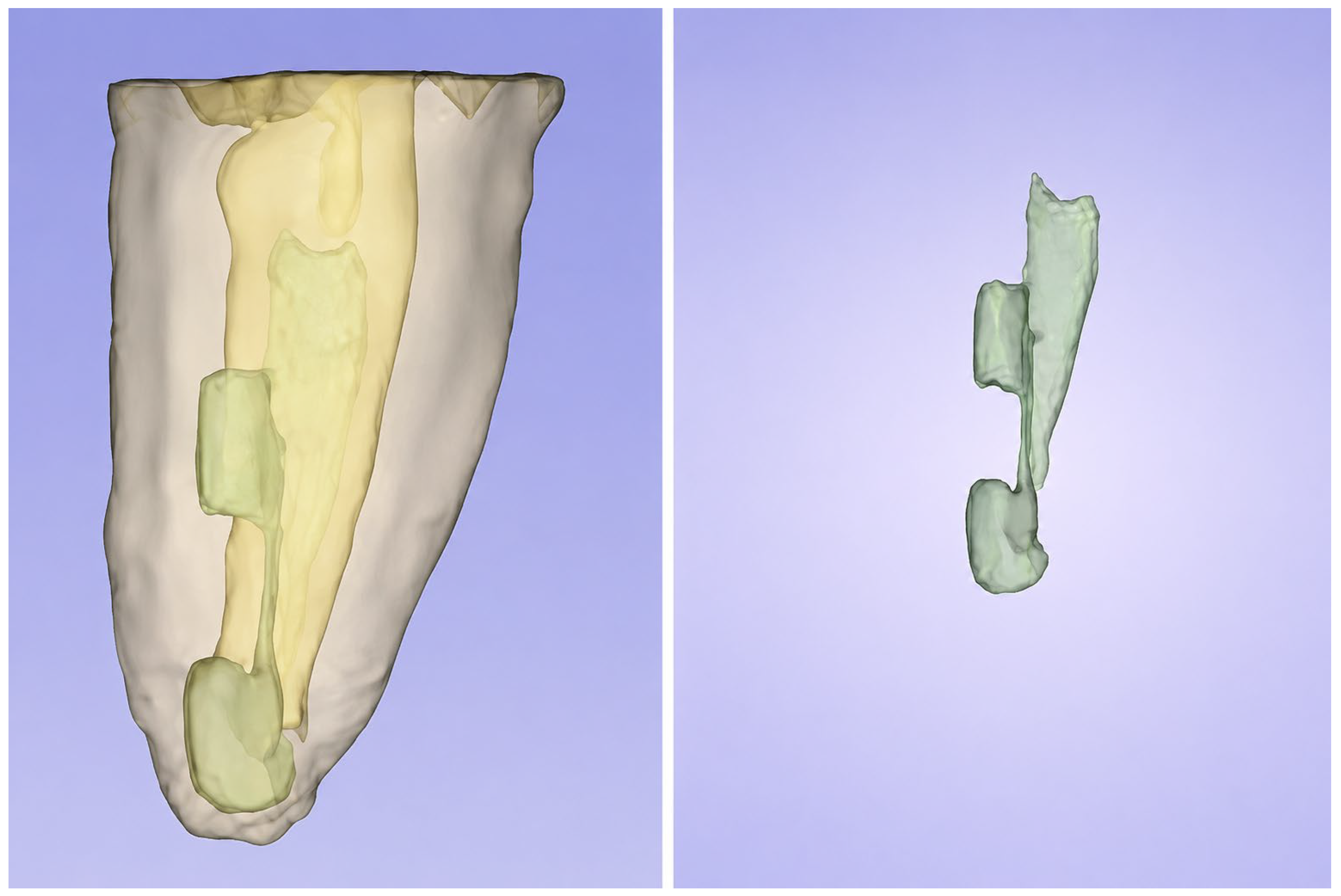
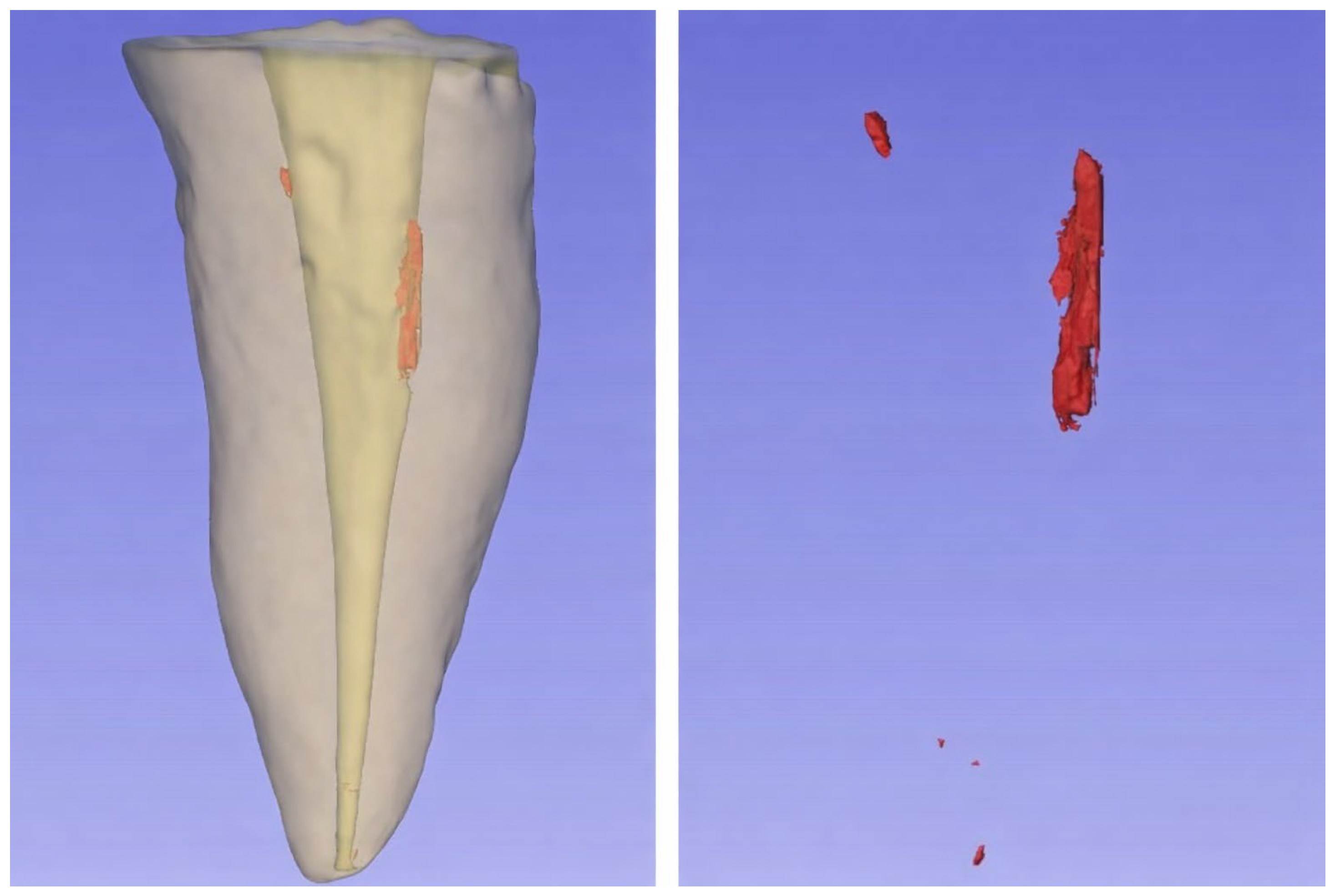
2.7. Image Processing and Volumetric Computation
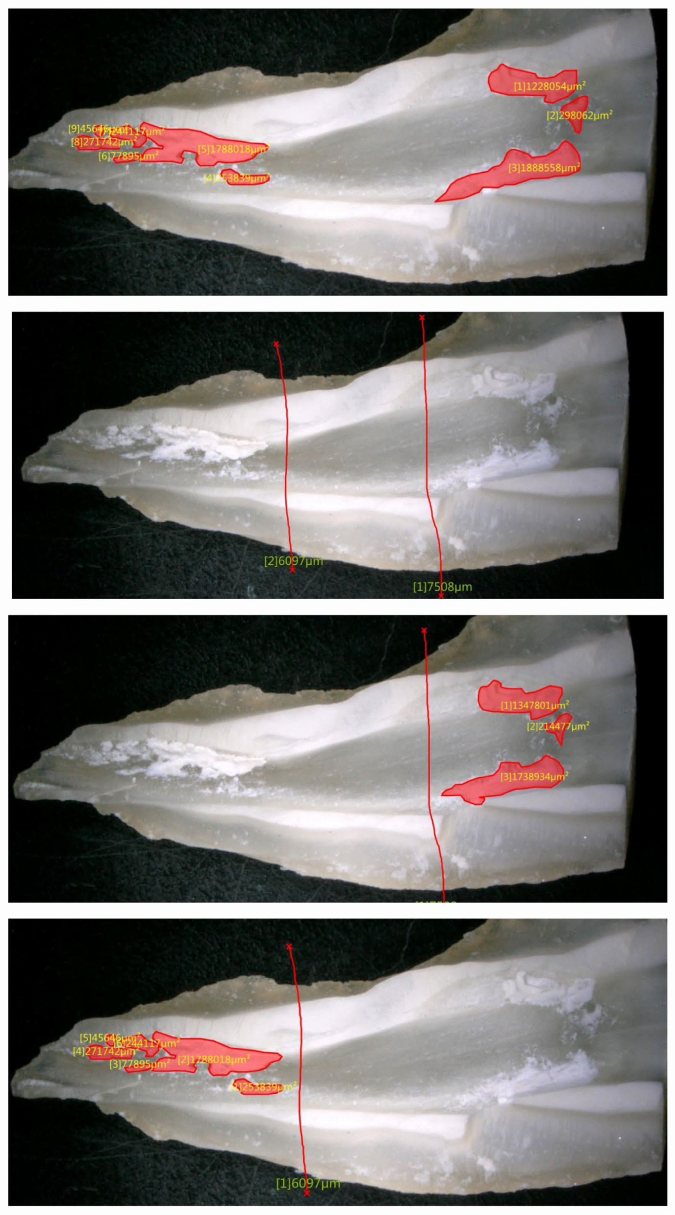
2.8. DGM and Surface Area Quantification
2.9. Examiner Calibration and Measurement Reliability, Scoring, and Statistical Analysis
3. Results
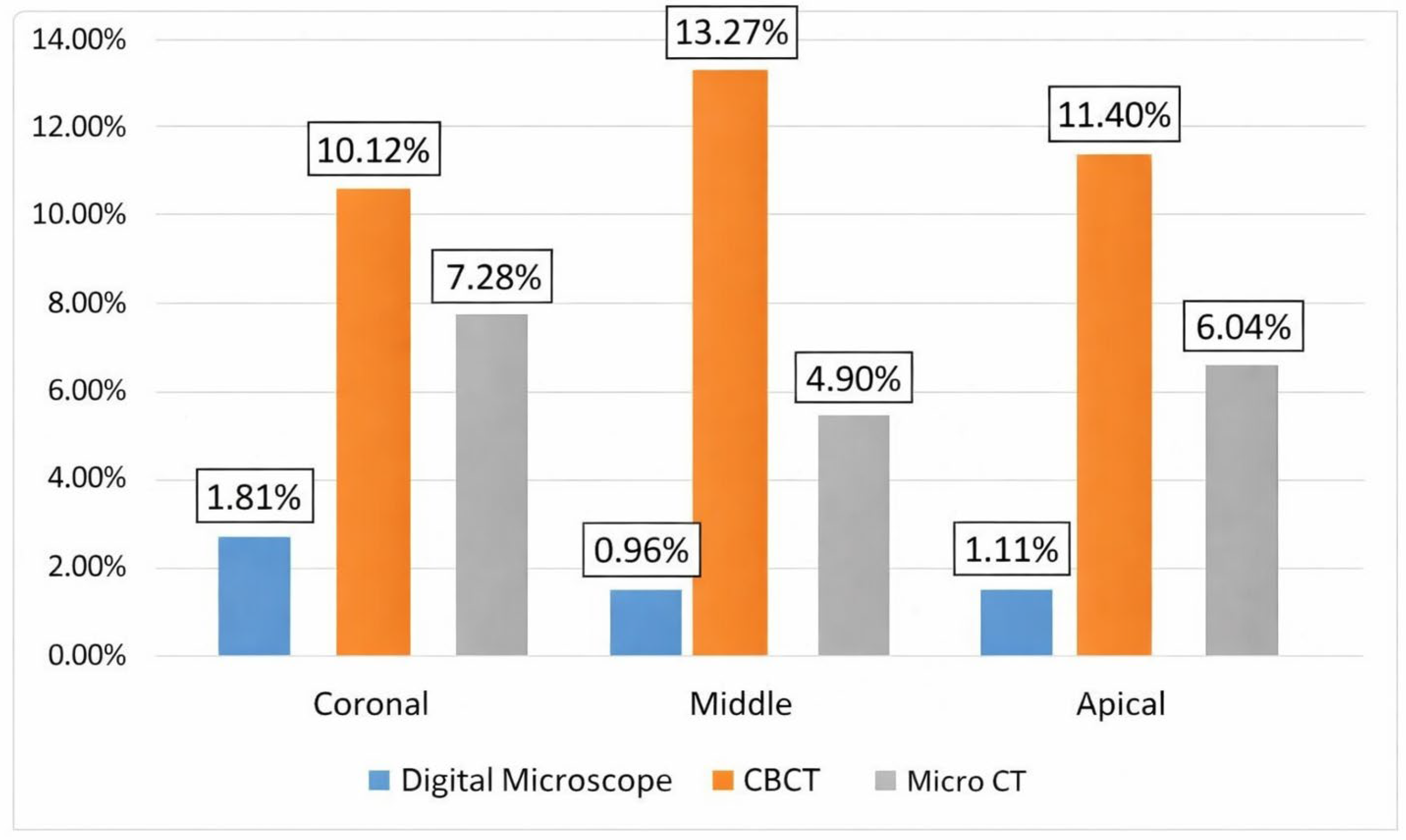
3.1. Method Specific Quantitative Measurements of Residual Obturation Material
3.2. Paired Volumetric Comparison Between CBCT and Micro-CT
3.3. Diagnostic Accuracy of CBCT for Detecting Residual Material Using Micro CT as Reference
3.4. Diagnostic Accuracy of Digital Microscopy for Detecting Residual Material Using Micro CT as Reference
4. Discussion
5. Clinical Relevance
- CBCT overestimates residual root canal filling material after retreatment. Clinicians should avoid making retreatment decisions based solely on CBCT-detected radiopacities, particularly in the middle and apical thirds, without correlating findings with clinical signs.
- Digital microscopy (or surgical operating microscopes) can reliably confirm the presence of residual material when visible, but cannot rule out remnants beneath the inspected surface.
- Micro-CT remains the gold standard for laboratory research; its volumetric accuracy cannot be matched by current clinical CBCT or surface microscopy.
- When comparing retreatment protocols in future studies, investigators should clearly specify whether they are measuring surface area or volume, as these are not interchangeable.
6. Take-Home Messages
- CBCT → sensitive but overestimates remnants → don’t retreat based on CBCT alone.
- Microscopy → specific but misses deep remnants → positive = retreat, negative = not conclusive.
- Micro-CT → research standard, not clinical.
7. Conclusions
Author Contributions
Funding
Institutional Review Board Statement
Informed Consent Statement
Data Availability Statement
Acknowledgments
Conflicts of Interest
References
- Nair, P.N.R. On the causes of persistent apical periodontitis: A review. Int. Endod. J. 2006, 39, 249–281. [Google Scholar] [CrossRef]
- Siqueira, J.F., Jr.; Rôças, I.N. Clinical implications and microbiology of bacterial persistence after treatment procedures. J. Endod. 2008, 34, 1291–1301.e3. [Google Scholar] [CrossRef]
- Silva, E.J.N.L.; Belladonna, F.G.; Zuolo, A.S.; Rodrigues, E.; Ehrhardt, I.C.; Souza, E.M.; De-Deus, G. Effectiveness of XP-endo Finisher and XP-endo Finisher R in removing root filling remnants: A micro-CT study. Int. Endod. J. 2018, 51, 86–91. [Google Scholar] [CrossRef] [PubMed]
- Mancino, D.; Kharouf, N.; Hemmerlé, J.; Haïkel, Y. Microscopic and chemical assessments of the filling ability in oval-shaped root canals using two different carrier-based filling techniques. Eur. J. Dent. 2019, 13, 166–171. [Google Scholar] [CrossRef]
- Sanz, J.L.; López-García, S.; Rodríguez-Lozano, F.J.; Melo, M.; Lozano, A.; Llena, C.; Forner, L. Cytocompatibility and bioactive potential of AH Plus Bioceramic Sealer: An in vitro study. Int. Endod. J. 2022, 55, 1066–1080. [Google Scholar] [CrossRef] [PubMed]
- Silva, E.J.N.L.; Versiani, M.A. The paradox of calcium silicate-based sealers: A reflection on bioactivity, solubility and clinical meaning. Int. Endod. J. 2026, 59, 550–555. [Google Scholar] [CrossRef]
- Kharouf, N.; Sauro, S.; Hardan, L.; Fawzi, A.; Suhanda, I.E.; Zghal, J.; Addiego, F.; Affolter-Zbaraszczuk, C.; Arntz, Y.; Ball, V.; et al. Impacts of Resveratrol and Pyrogallol on Physicochemical, Mechanical and Biological Properties of Epoxy-Resin Sealers. Bioengineering 2022, 9, 85. [Google Scholar] [CrossRef]
- Wahbi, E.; Achour, H.; Alsayed Tolibah, Y. Comparison between AH Plus sealer and Total Fill bioceramic sealer performance in previously untreated and retreatment cases of maxillary incisors with large-sized periapical lesion: A randomized controlled trial. BDJ Open 2024, 10, 73. [Google Scholar] [CrossRef]
- Sümbüllü, M.; Ali, A.; Bakhsh, A.; Arslan, H. Evaluation of the retreatability of bioceramic root canal sealers with various formulations in simulated grooves. PeerJ 2025, 13, e20398. [Google Scholar] [CrossRef]
- Alouda, M.; Akil, S.; Eid, A.; Cardinali, F.; Achour, H.; Haikel, Y.; Kharouf, N. Retreatment of Two Bioceramic Sealers Included Two Different Percentages of Calcium Silicate Using Two Endodontic File Systems: An In Vitro Study. Eur. J. Dent. 2026, 20, 77–85. [Google Scholar] [CrossRef]
- Farrayeh, A.; Akil, S.; Eid, A.; Macaluso, V.; Mancino, D.; Haïkel, Y.; Kharouf, N. Effectiveness of Two Endodontic Instruments in Calcium Silicate-Based Sealer Retreatment. Bioengineering 2023, 10, 362. [Google Scholar] [CrossRef]
- Nour, S.W.; Elgendy, A.A.E.; Bayoumi, A.A.; Baroudi, K.; Elsayed, M.A. Effect of Bioceramic-Based Obturation Techniques on the Amount of Residual Filling Material after Retreatment: An In Vitro Study. Eur. J. Dent. 2026. Online ahead of print. [Google Scholar]
- Zhekov, K.; Stefanova, V. Optimizing Nonsurgical Endodontic Retreatment: A 3D CBCT Quantification of Root Canal Bioceramic Filling Material Removal. Open Dent. J. 2024, 18, e18742106300149. [Google Scholar] [CrossRef]
- Colombo, J.; Rocha, D.; Limoeiro, A.; Nascimento, W.; Fontana, C.; Pelegrine, R.; Bueno, C.; De Martin, A. Micro-CT evaluation of sealers removal by reciprocal instrumentation followed by continuous ultrasonic irrigation in teeth with oval root canals. J. Clin. Exp. Dent. 2023, 15, e233–e238. [Google Scholar] [CrossRef]
- Obeid, M.; Zaghloul, M.E.S.; Abdelrahman, T.Y. Impact of LASER activated irrigation on the retrievability of Guttaflow bioseal (in vitro study). BDJ Open 2024, 10, 72. [Google Scholar] [CrossRef] [PubMed]
- Yang, X.; Wang, Y.; Ji, M.; Li, Y.; Wang, H.; Luo, T.; Gao, Y.; Zou, L. Microcomputed tomographic analysis of the efficiency of two retreatment techniques in removing root canal filling materials from mandibular incisors. Sci. Rep. 2023, 13, 2267. [Google Scholar] [CrossRef] [PubMed]
- Chauhan, R.; Tikku, A.; Chandra, A. Detection of residual obturation material after root canal retreatment with three different techniques using a dental operating microscope and a stereomicroscope: An in vitro comparative evaluation. J. Conserv. Dent. 2012, 15, 218–222. [Google Scholar] [CrossRef] [PubMed]
- Borges, C.C.; Estrela, C.; Decurcio, D.d.A.; Pécora, J.D.; Sousa-Neto, M.D.; Rossi-Fedele, G. Cone-beam and microcomputed tomography for the assessment of root canal morphology: A systematic review. Braz. Oral Res. 2020, 34, e056. [Google Scholar] [CrossRef]
- Ghavami-Lahiji, M.; Davalloo, R.T.; Tajziehchi, G.; Shams, P. Microcomputed tomography in preventive and restorative dental research: A review. Imaging Sci. Dent. 2021, 51, 341–350. [Google Scholar] [CrossRef]
- Syed, S.A.; Zafar, S.S.; Syed, F.A.; Iqbal, A.; Atif, S. Comparison of light microscopy and digital microscopy for learning oral pathology practicals among second year dental students. J. Dent. Sci. 2024, 19, 2135–2139. [Google Scholar] [CrossRef]
- Orhan, K.; Jacobs, R.; Celikten, B. Evaluation of Threshold Values for Root Canal Filling Voids in Micro-CT and Nano-CT Images. Scanning 2018, 2018, 9437569. [Google Scholar] [CrossRef]
- Jung, M.; Lommel, D.; Klimek, J. The imaging of root canal obturation using micro-CT. Int. Endod. J. 2005, 38, 617–626. [Google Scholar] [CrossRef]
- Lamira, A.; Mazzi-Chaves, J.F.; Nicolielo, L.F.P.; Leoni, G.B.; Silva-Sousa, A.C.; Silva-Sousa, Y.T.C.; Pauwels, R.; Buls, N.; Jacobs, R.; Sousa-Neto, M.D. CBCT-based assessment of root canal treatment using micro-CT reference images. Imaging Sci. Dent. 2022, 52, 245–258. [Google Scholar] [CrossRef]
- Alsayed Tolibah, Y.; Al-Shiekh, M.N.; Abbara, M.T.; Alhaji, M.; Aljabban, O.; Bshara, N. An epidemiological study of extracted mandibular premolars from adolescent patients in Damascus using two classification system analyzed with CBCT and digital periapical radiographs. BMC Oral Health 2025, 25, 1460. [Google Scholar] [CrossRef]
- Celikten, B.; Jacobs, R.; Vasconcelos, K.d.F.; Huang, Y.; Shaheen, E.; Nicolielo, L.F.P.; Orhan, K. Comparative evaluation of cone beam CT and micro-CT on blooming artifacts in human teeth filled with bioceramic sealers. Clin. Oral Investig. 2019, 23, 3267–3273. [Google Scholar] [CrossRef]
- Sharma, G.; Kakkar, P.; Vats, A. A comparative SEM investigation of smear layer remaining on dentinal walls by three rotary NiTi files with different cross-sectional designs in moderately curved canals. J. Clin. Diagn. Res. 2015, 9, ZC43–ZC47. [Google Scholar] [CrossRef]
- Rodrigues, C.T.; Jacobs, R.; Vasconcelos, K.F.; Lambrechts, P.; Rubira-Bullen, I.R.F.; Gaêta-Araujo, H.; Oliveira-Santos, C.; Duarte, M.A.H. Influence of CBCT-based volumetric distortion and beam hardening artifacts on the assessment of root canal filling quality in isthmus-containing molars. Dentomaxillofac. Radiol. 2021, 50, 20200503. [Google Scholar] [CrossRef] [PubMed]
- Celikten, B.; Jacobs, R.; Vasconcelos, K.D.; Huang, Y.; Nicolielo, L.F.P.; Orhan, K. Assessment of volumetric distortion artifact in filled root canals using different cone-beam computed tomographic devices. J. Endod. 2017, 43, 1517–1521. [Google Scholar] [CrossRef] [PubMed]
- Spin-Neto, R.; Gotfredsen, E.; Wenzel, A. Impact of voxel size variation on CBCT-based diagnostic outcome in dentistry: A systematic review. J. Digit. Imaging 2013, 26, 813–820. [Google Scholar] [CrossRef] [PubMed]
- Brüllmann, D.; Schulze, R.K. Spatial resolution in CBCT machines for dental/maxillofacial applications—What do we know today? Dentomaxillofac. Radiol. 2015, 44, 20140204. [Google Scholar] [CrossRef]
- Schulze, R.; Heil, U.; Groβ, D.; Bruellmann, D.D.; Dranischnikow, E.; Schwanecke, U.; Schoemer, E. Artifacts in CBCT: A review. Dentomaxillofac. Radiol. 2011, 40, 265–273. [Google Scholar] [CrossRef]
- Maret, D.; Peters, O.A.; Galibourg, A.; Dumoncel, J.; Esclassan, R.; Kahn, J.-L.; Sixou, M.; Telmon, N. Comparison of the accuracy of 3-dimensional cone-beam computed tomography and microcomputed tomography reconstructions by using different voxel sizes. J. Endod. 2014, 40, 1321–1326. [Google Scholar] [CrossRef] [PubMed]
- Patel, S.; Durack, C.; Abella, F.; Shemesh, H.; Roig, M.; Lemberg, K. Cone beam computed tomography in Endodontics—A review. Int. Endod. J. 2015, 48, 3–15. [Google Scholar] [CrossRef]
- Patel, S.; Horner, K. The use of cone beam computed tomography in endodontics. Int. Endod. J. 2009, 42, 755–756. [Google Scholar] [CrossRef]
- Khademi, A.; Saatchi, M.; Sheikhi, M.; Soltani, M.M.; Moradi, S. In vitro diagnostic accuracy and agreement of dental microscope and cone-beam computed tomography in comparison with microcomputed tomography for detection of the second mesiobuccal canal of maxillary first molars. Scanning 2022, 2022, 1493153. [Google Scholar] [CrossRef]
- Ordinola-Zapata, R.; Bramante, C.M.; Versiani, M.A.; Moldauer, B.I.; Topham, G.; Gutmann, J.L.; Nuñez, A.; Duarte, M.A.H.; Abella, F. Comparative accuracy of the clearing technique, CBCT, and micro-CT methods in studying the mesial root canal configuration of mandibular first molars. Int. Endod. J. 2017, 50, 90–96. [Google Scholar] [CrossRef]
- Pires, M.; Martins, J.N.; Pereira, M.R.; Vasconcelos, I.; da Costa, R.P.; Duarte, I.; Ginjeira, A. Diagnostic value of cone beam computed tomography for root canal morphology assessment—A micro-CT based comparison. Clin. Oral Investig. 2024, 28, 201. [Google Scholar] [CrossRef] [PubMed]
- Bai, B.; Tang, Y.; Wu, Y.; Pei, F.; Zhu, Q.; Zhu, P.; Gu, Y. Ex vivo detection of mandibular incisors’ root canal morphology using cone-beam computed tomography with 4 different voxel sizes and microcomputed tomography. BMC Oral Health 2023, 23, 656. [Google Scholar] [CrossRef]
- Baena-de la Iglesia, T.; Yañez-Vico, R.M.; Iglesias-Linares, A. Diagnostic performance of cone-beam computed tomography to diagnose in vivo/in vitro root resorption: A systematic review and meta-analysis. J. Evid. Based Dent. Pract. 2023, 23, 101803. [Google Scholar] [CrossRef] [PubMed]
- Haghanifar, S.; Moudi, E.; Mesgarani, A.; Bijani, A.; Abbaszadeh, N. A comparative study of cone-beam computed tomography and digital periapical radiography in detecting mandibular molars’ root perforations. Imaging Sci. Dent. 2014, 44, 115–119. [Google Scholar] [CrossRef][Green Version]
- Cohen, J.F.; A Korevaar, D.; Altman, D.G.; E Bruns, D.; A Gatsonis, C.; Hooft, L.; Irwig, L.; Levine, D.; Reitsma, J.B.; de Vet, H.C.W.; et al. STARD 2015 guidelines for reporting diagnostic accuracy studies: Explanation and elaboration. BMJ Open 2016, 6, e012799. [Google Scholar] [CrossRef]
- Kırıcı, D.; Demirbuga, S.; Karataş, E. Microcomputed tomographic assessment of the residual filling volume, apical transportation, and crack formation after retreatment with Reciproc and Reciproc Blue systems in curved root canals. J. Endod. 2020, 46, 238–243. [Google Scholar] [CrossRef] [PubMed]
- Santos-Junior, A.O.; Fontenele, R.C.; Neves, F.S.; Ali, S.; Jacobs, R.; Tanomaru-Filho, M. A unique AI-based tool for automated segmentation of pulp cavity structures in maxillary premolars on CBCT. Sci. Rep. 2025, 15, 5509. [Google Scholar] [CrossRef] [PubMed] [PubMed Central]
- Santos-Junior, A.O.; Fontenele, R.C.; Neves, F.S.; Tanomaru-Filho, M.; Jacobs, R. A novel artificial intelligence-powered tool for automated root canal segmentation in single-rooted teeth on cone-beam computed tomography. Int. Endod. J. 2025, 58, 658–671. [Google Scholar] [CrossRef] [PubMed] [PubMed Central]
- Karobari, M.I.; Adil, A.H.; Mathur, A.; Snigdha, N.T. Applications and clinical translation of artificial intelligence in CBCT-based detection of endodontic lesions: A scoping review. Oral Radiol. 2026, 42, 251–261. [Google Scholar] [CrossRef] [PubMed]


| Root Third | Digital Microscopy Mean (%) | Digital Microscopy SD (%) | Digital Microscopy Median (%) | CBCT Mean (%) | CBCT SD (%) | CBCT Median (%) | Micro-CT Mean (%) | Micro-CT SD (%) | Micro-CT Median (%) |
|---|---|---|---|---|---|---|---|---|---|
| Coronal | 1.81 | 1.7 | 1.52 | 10.12 | 5.37 | 10.2 | 7.28 | 5.54 | 5.04 |
| Middle | 0.96 | 1.27 | 0.34 | 13.27 | 9.44 | 13.1 | 4.9 | 5.08 | 3.27 |
| Apical | 1.11 | 1.35 | 0.74 | 11.4 | 7.65 | 14.2 | 6.04 | 5.29 | 3.83 |
| Root Third | CBCT Median Volume (%) | Micro-CT Median Volume (%) | p-Value |
|---|---|---|---|
| Coronal | 10.2 | 5.04 | 0.648 |
| Middle | 13.1 | 3.27 | 0.002 * |
| Apical | 14.2 | 3.83 | 0.199 |
| Third of the Root Canal | Scoring System | Sensitivity | Positive Predictive Value | Negative Predictive Value | Specificity | Accuracy | Agreement | Kappa Test | |
|---|---|---|---|---|---|---|---|---|---|
| Coronal | Score (0) | 50.0% (95%CI: 21.5–78.5%) (4/8) | 100.0% (4/4) (95%CI: 51.0–100.0%) | 63.6% (7/11) (95%CI: 35.4–84.8%) | 100.0% (7/7) (95%CI: 59.0–100.0%) | 73.3% (11/15) | 66.7% (10/15) | 42.2% (19/45) | −0.031 (95% CI: −0.27 to 0.21) |
| Score (1) | 66.7% (2/3) (95%CI: 20.8–93.9%) | 33.3% (2/6) (95%CI: 9.7–70.0%) | 88.9% (8/9) (95%CI: 56.5–98.0%) | 66.7% (8/12) (95%CI: 39.1–86.2%) | 66.7% (10/15) | ||||
| Score (2) | 100.0% (3/3) (95%CI: 43.9–100.0%) | 75.0% (3/4) (95%CI: 30.1–95.4%) | 100.0% (11/11) (95%CI: 74.1–100.0%) | 91.7% (11/12) (95%CI: 64.6–98.5%) | 93.3% (14/15) | ||||
| Score (3) | 100.0% (1/1) (95%CI: 20.7–100%) | 100.0% (1/1) (95%CI: 20.7–100.0%) | 100.0% (14/14) (95%CI: 78.5–100.0%) | 100.0% (14/14) (95%CI: 78.5–100.0%) | 100.0% (15/15) | ||||
| Score (4) | - | - | 100.0% (15/15) (95%CI: 79.6–100.0%) | 100.0% (15/15) (95%CI: 79.6–100.0%) | 100.0% (15/15) | ||||
| Middle | Score (0) | 33.3% (3/9) (95%CI: 12.1–64.6%) | 60.0% (3/5) (95%CI: 23.1–88.2%) | 40.0% (4/10) (95%CI: 16.8–68.7%) | 66.7% (4/6) (95%CI: 30.0–90.3%) | 46.7% (7/15) | 33.3% (5/15) | 0.09 (95% CI: −0.20 to 0.37) | |
| Score (1) | 25.0% (1/4) (95%CI: 4.6–69.9%) | 50.0% (1/2) (95%CI: 9.5–90.5%) | 76.9% (10/13) (95%CI: 49.7–91.8%) | 90.9% (10/11) (95%CI: 62.3–98.4%) | 73.3% (11/15) | ||||
| Score (2) | 50.0% (1/2) (95%CI: 9.5–90.5%) | 25.0% (1/4) (95%CI: 4.6–69.9%) | 90.9% (10/11) (95%CI: 62.3–98.4%) | 76.9% (10/13) (95%CI: 49.7–91.8%) | 73.3% (11/15) | ||||
| Score (3) | - | 0.0% (0/1) (95%CI: 0.0–79.3%) | 100.0% (14/14) (95%CI: 78.5–100.0%) | 93.3% (14/15) (95%CI: 70.2–98.8%) | 93.3% (14/15) | ||||
| Score (4) | - | 0.0% (0/3) (95%CI: 0.0–56.1%) | 100.0% (12/12) (95%CI: 75.8–100.0%) | 80.0% (12/15) (95%CI: 54.8–93.0%) | 80.0% (12/15) | ||||
| Apical | Score (0) | 25.0% (2/8) (95%CI: 7.2–59.1%) | 40.0% (2/5) (95%CI: 11.8–76.9%) | 40.0% (4/10) (95%CI: 16.8–68.7%) | 57.1% (4/7) (95%CI: 25.0–84.2%) | 40.0% (6/15) | 26.6% (4/15) | 0.54 (95% CI: 0.22 to 0.86) | |
| Score (1) | 0.0% (0/4) (95%CI: 0.0–60.2%) | 0.0% (0/1) (95%CI: 0.0–79.3%) | 71.4% (10/14) (95%CI: 45.4–88.3%) | 90.9% (10/11) (95%CI: 62.3–98.4%) | 66.7% (10/15) | ||||
| Score (2) | 66.7% (2/3) (95%CI: 20.8–93.9%) | 28.6% (2/7) (95%CI: 8.2–64.1%) | 87.5% (7/8) (95%CI: 52.9–97.8%) | 58.3% (7/12) (95%CI: 32.0–80.7%) | 60.0% (9/15) | ||||
| Score (3) | - | 0.0% (0/1) (95%CI: 0.0–79.3%) | 100.0%% (14/14) (95%CI: 78.5–100.0%) | 93.3% (14/15) (95%CI: 70.2–98.8%) | 93.3% (14/15) | ||||
| Score (4) | - | 0.0% (0/1) (95%CI: 0.0–79.3%) | 100.0% (14/14) (95%CI: 78.5–100.0%) | 93.3% (14/15) (95%CI: 70.2–98.8%) | 93.3% (14/15) | ||||
| Third of the Root Canal | Scoring System | Sensitivity | Positive Predictive Value | Negative Predictive Value | Specificity | Accuracy | Agreement | Kappa Test | |
|---|---|---|---|---|---|---|---|---|---|
| Coronal | Score (0) | 100.0% (8/8) (95%CI: 67.6–100.0%) | 57.1% (8/14) (95%CI: 32.6–78.6%) | 100.0% (1/1) (95%CI: 20.7–100.0%) | 14.3% (1/7) (95%CI: 2.6–51.3%) | 60.0% (9/15) | 53.3% (8/15) | 55.5% (25/45) | NA (no variability in data) |
| Score (1) | 0.0% (0/3) (95%CI: 0.0–70.8%) | 0.0% (0/1) (95%CI: 0.0–79.3%) | 78.6% (11/14) (95%CI: 52.4–92.4%) | 91.7% (11/12) (95%CI: 64.6–98.5%) | 73.3% (11/15) | ||||
| Score (2) | 0.0% (0/3) (95%CI: 0.0–70.8%) | - | 80.0% (12/15) (95%CI: 54.8–93.0%) | 100.0% (12/12) (95%CI: 75.8–100.0%) | 80.0% (12/15) | ||||
| Score (3) | 0.0% (0/1) (95%CI: 0.0–79.3%) | - | 93.3% (14/15) (95%CI: 70.2–98.8%) | 100.0% (14/14) (95%CI: 78.5–100.0%) | 93.3% (14/15) | ||||
| Score (4) | - | - | 100.0% (15/15) (95%CI: 79.6–100.0%) | 100.0% (15/15) (95%CI: 79.6–100.0%) | 100.0% (15/15) | ||||
| Middle | Score (0) | 100.0% (9/9) (95%CI: 70.1–100.0%) | 60.0% (9/15) (95%CI: 35.8–80.2%) | - | 0.0% (0/6) (95%CI: 0.0–39.3%) | 60.0% (9/15) | 60.0% (9/15) | NA (no variability in data) | |
| Score (1) | 0.0% (0/4) (95%CI: 0.0–60.2%) | - | 73.3% (11/15) (95%CI: 48.1–89.1%) | 100.0% (11/11) (95%CI: 74.1–100.0%) | 73.3% (11/15) | ||||
| Score (2) | 0.0% (0/2) (95%CI: 0.0–65.8%) | - | 86.7% (13/15) (95%CI: 62.1–96.3%) | 100.0% (13/13) (95%CI: 77.2–100.0%) | 86.7% (13/15) | ||||
| Score (3) | - | - | 100.0% (15/15) (95%CI: 79.6–100.0%) | 100.0% (15/15) (95%CI: 79.6–100.0%) | 100.0% (15/15) | ||||
| Score (4) | - | - | 100.0% (15/15) (95%CI: 79.6–100.0%) | 100.0% (15/15) (95%CI: 79.6–100.0%) | 100.0% (15/15) | ||||
| Apical | Score (0) | 53.3% (8/8) (95%CI: 30.1–75.2%) | 100.0% (8/8) (95%CI: 67.6–100.0%) | - | - | 53.3% (8/15) | 53.3% (8/15) | 0.045 (95%CI: −0.05 to 0.14) | |
| Score (1) | 100.0% (4/4) (95%CI: 51.0–100.0%) | 100.0% (4/4) (95%CI: 51.0–100.0%) | 100.0% (11/11) (95%CI: 74.1–100.0%) | 100.0% (11/11) (95%CI: 74.1–100.0%) | 100.0% (15/15) | ||||
| Score (2) | 100.0% (3/3) (95%CI: 43.9–100.0%) | 100.0% (3/3) (95%CI: 43.9–100.0%) | 100.0% (12/12) (95%CI: 75.8–100.0%) | 100.0% (12/12) (95%CI: 75.8–100.0%) | 100.0% (15/15) | ||||
| Score (3) | - | - | 100.0% (15/15) (95%CI: 79.6–100.0%) | 100.0% (15/15) (95%CI: 79.6–100.0%) | 100.0% (15/15) | ||||
| Score (4) | - | - | 100.0% (15/15) (95%CI: 79.6–100.0%) | 100.0% (15/15) (95%CI: 79.6–100.0%) | 100.0% (15/15) | ||||
Disclaimer/Publisher’s Note: The statements, opinions and data contained in all publications are solely those of the individual author(s) and contributor(s) and not of MDPI and/or the editor(s). MDPI and/or the editor(s) disclaim responsibility for any injury to people or property resulting from any ideas, methods, instructions or products referred to in the content. |
© 2026 by the authors. Licensee MDPI, Basel, Switzerland. This article is an open access article distributed under the terms and conditions of the Creative Commons Attribution (CC BY) license.
Share and Cite
Alouda, M.; Akil, S.; Abbara, M.T.; Eid, A.; Almasri, I.-A.; Alsayed Tolibah, Y.; Baghdadi, Z.D. Detecting Residual Root Canal Filling Material After Retreatment: Cone-Beam Computed Tomography and Digital Microscopy Compared with Microcomputed Tomography. Dent. J. 2026, 14, 318. https://doi.org/10.3390/dj14060318
Alouda M, Akil S, Abbara MT, Eid A, Almasri I-A, Alsayed Tolibah Y, Baghdadi ZD. Detecting Residual Root Canal Filling Material After Retreatment: Cone-Beam Computed Tomography and Digital Microscopy Compared with Microcomputed Tomography. Dentistry Journal. 2026; 14(6):318. https://doi.org/10.3390/dj14060318
Chicago/Turabian StyleAlouda, Mohamad, Samar Akil, Mohammad Tamer Abbara, Ammar Eid, Imad-Addin Almasri, Yasser Alsayed Tolibah, and Ziad D. Baghdadi. 2026. "Detecting Residual Root Canal Filling Material After Retreatment: Cone-Beam Computed Tomography and Digital Microscopy Compared with Microcomputed Tomography" Dentistry Journal 14, no. 6: 318. https://doi.org/10.3390/dj14060318
APA StyleAlouda, M., Akil, S., Abbara, M. T., Eid, A., Almasri, I.-A., Alsayed Tolibah, Y., & Baghdadi, Z. D. (2026). Detecting Residual Root Canal Filling Material After Retreatment: Cone-Beam Computed Tomography and Digital Microscopy Compared with Microcomputed Tomography. Dentistry Journal, 14(6), 318. https://doi.org/10.3390/dj14060318