Differential Attachment of Engineered Oral Soft Tissues to Implant Surfaces
 , ,
, , 
Abstract
1. Introduction
2. Materials and Methods
2.1. Cell Sources and Culture Conditions
2.2. Test Materials
- Gamma-sterilized, sandblasted, and acid-etched titanium–zirconium alloy rods (TiZr-SLA).
- Gamma-sterilized, machined titanium–zirconium alloy rods (TiZr-M).
- Ethylene oxide–sterilized, machined zirconia rods (ZrO2-M).
- Ethylene oxide–sterilized, polished zirconia rods (ZrO2-P).
- Steam-sterilized, machined polyether ether ketone rods (PEEK-M). PEEK rods were evaluated in their machined state (PEEK-M), reflecting the as-supplied finish and aligning with a previous pilot study in which surface roughness parameters for the same PEEK-M rods were reported. This approach allowed direct comparison across studies and provided insight into the soft tissue response to PEEK under a relatively rough topographical condition [20,21].
- Commercially available sterile titanium healing abutments (HA-Ti) with 4.5 mm height and 5.2 mm diameter.
- Commercially available sterile zirconia healing abutments (HA-ZrO2) with 4.5 mm height and 5.2 mm diameter.
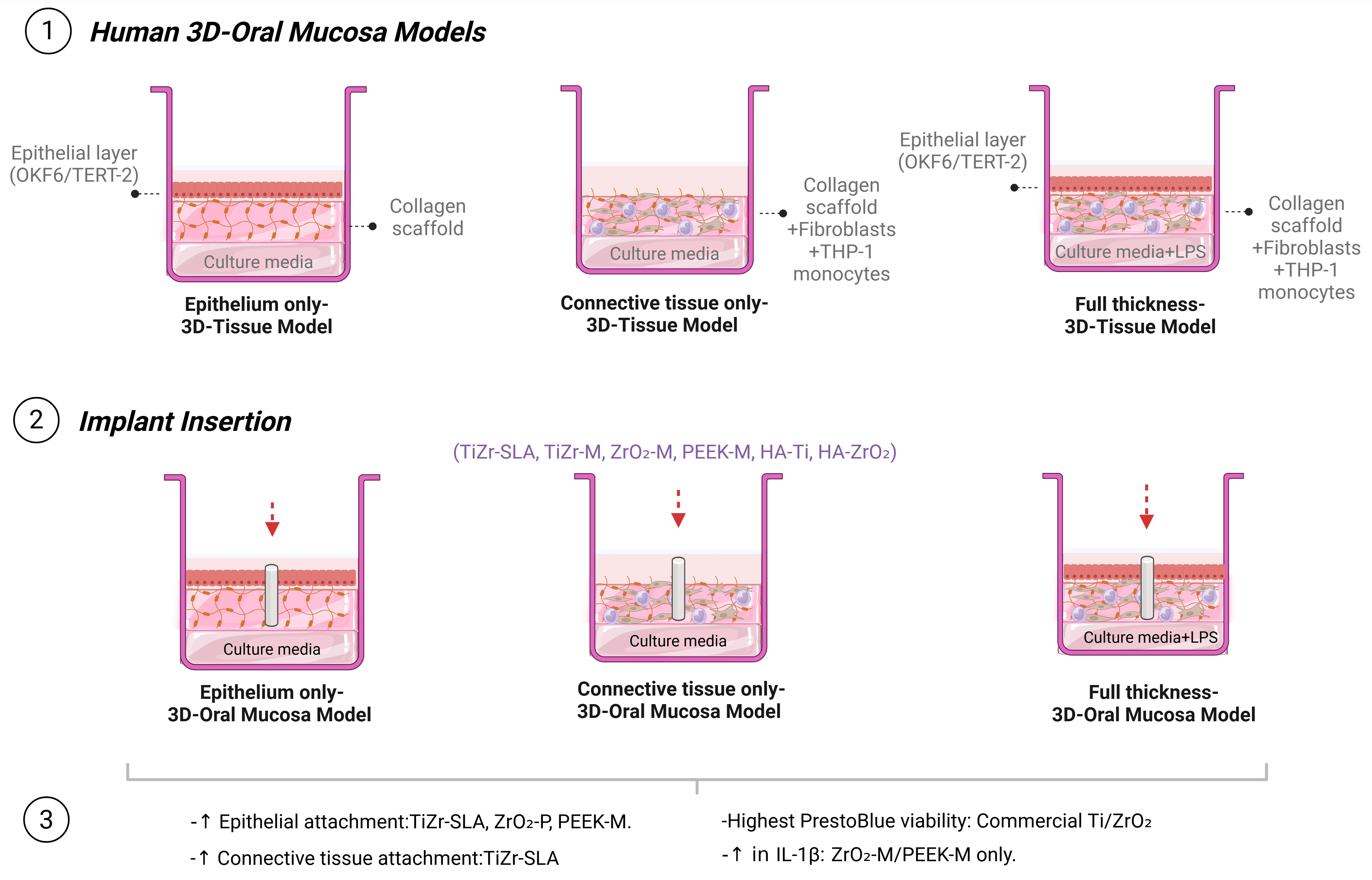
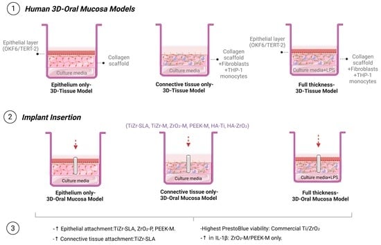
- Stage 1: Full-thickness oral mucosal models were prepared for histological assessment and inflammatory response testing, including TiZr-SLA, TiZr-M, ZrO2-M, PEEK-M, and control groups (n = 6).
- Stage 2: Split-thickness epithelium–connective tissue models were developed to evaluate differential soft tissue attachment across TiZr-SLA, TiZr-M, ZrO2-M, ZrO2-P, and PEEK-M groups (n = 6).
- Stage 3: Full-thickness oral mucosal models were generated for comparative analysis with commercially available abutments, encompassing all material groups listed above (n = 6).
2.3. 3D Oral Mucosa Models
2.3.1. Epithelium-Only Models
2.3.2. Connective Tissue-Only Models
2.3.3. Full-Thickness Oral Mucosal Models
2.4. Implant and Abutment Insertion
2.5. Induction of Inflammation
2.6. Multiple Endpoint Analyses
2.6.1. Tissue Viability Pull Test
2.6.2. Histological Processing
2.6.3. Scanning Electron Microscopy
2.6.4. Measurement of the Release of Pro-Inflammatory Markers
2.6.5. Statistical Analysis
3. Results
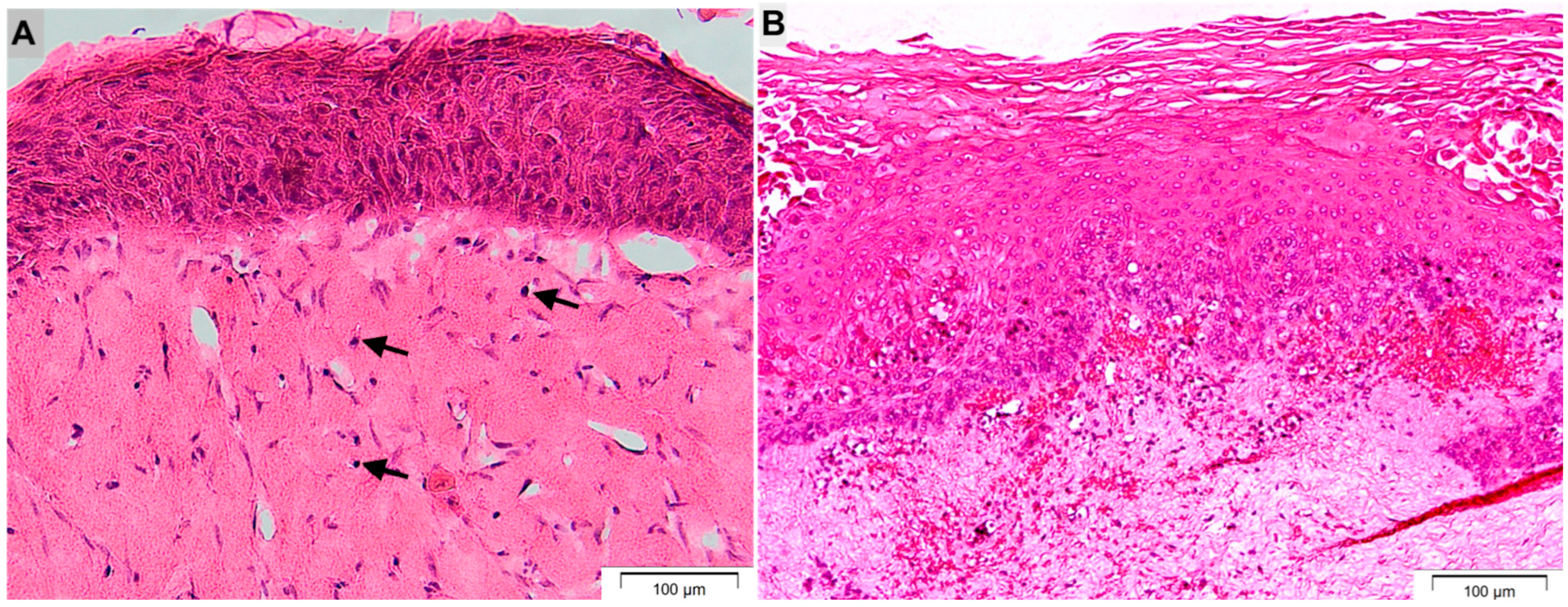
3.1. Tissue-Engineered Oral Mucosa Recapitulates Native Gingival Histology with Stratified Epithelium and Viable Fibroblast/THP-1 Connective Tissue
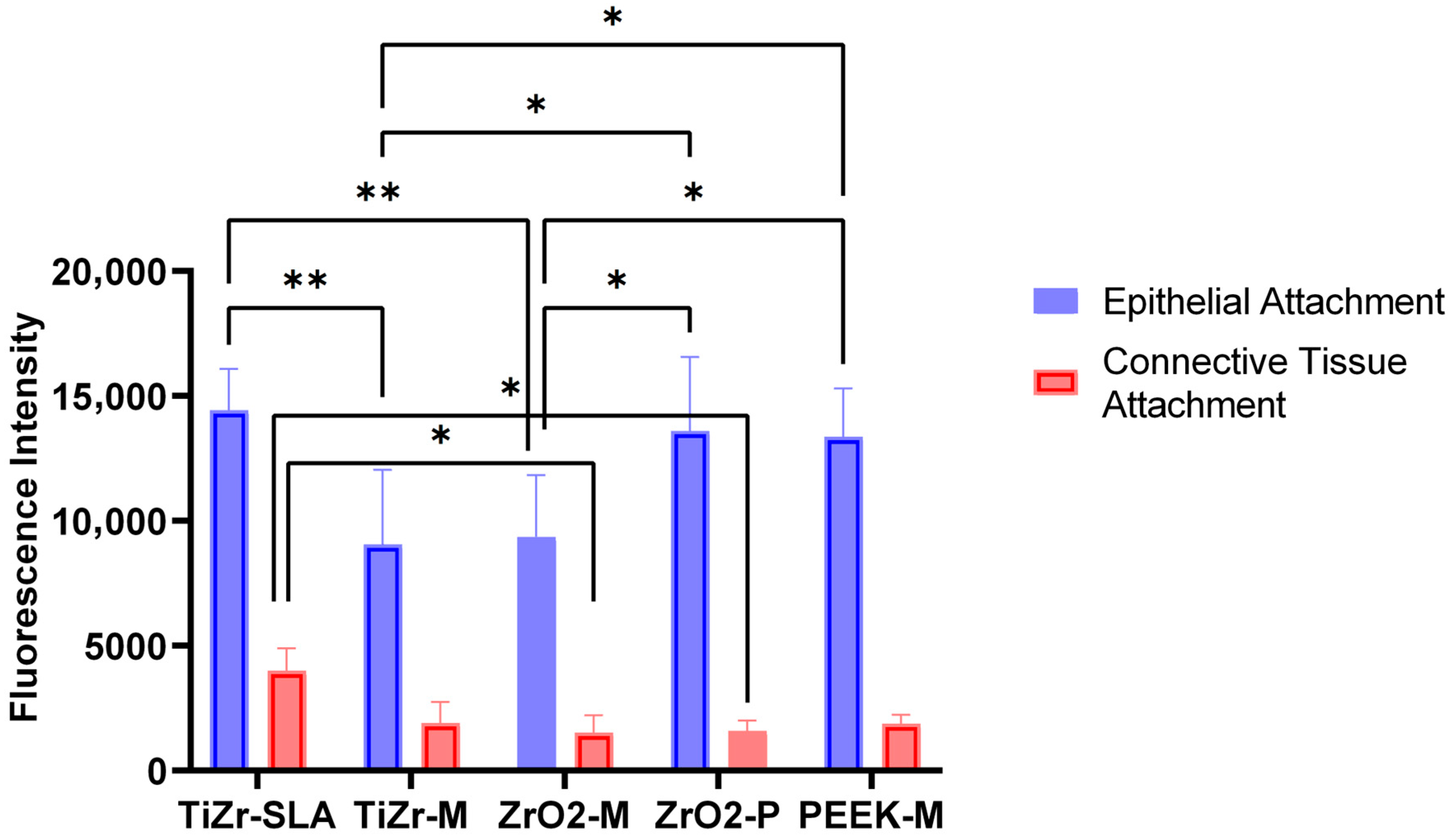
3.2. TiZr-SLA Surfaces Promote Superior Epithelial and Connective Tissue Attachment Compared with Machined and Polished Alternatives
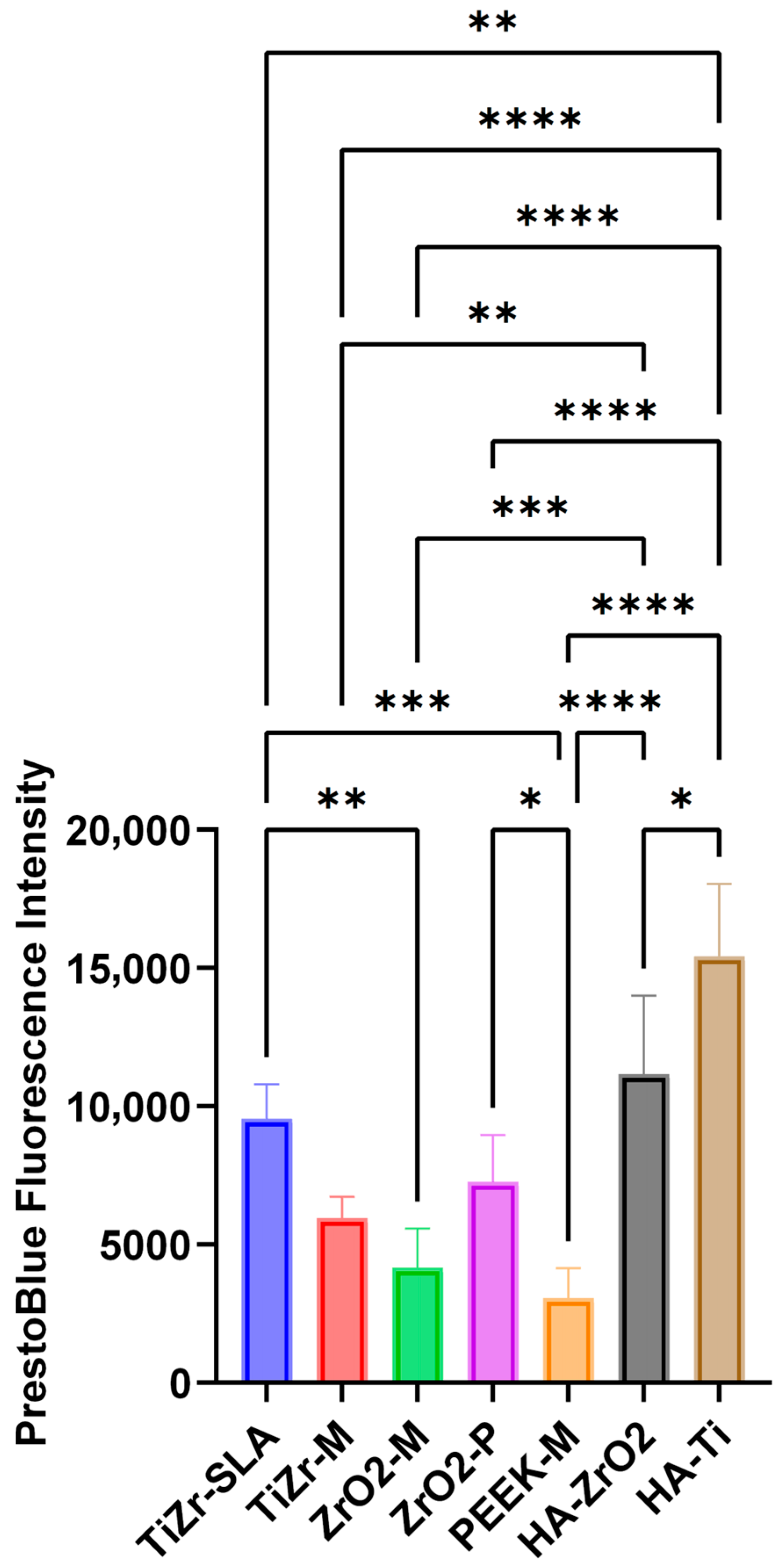
3.3. Commercial Titanium and Zirconia Healing Abutments Exhibit Highest Overall Soft Tissue Viability Post-Pull Test Versus Experimental Rods
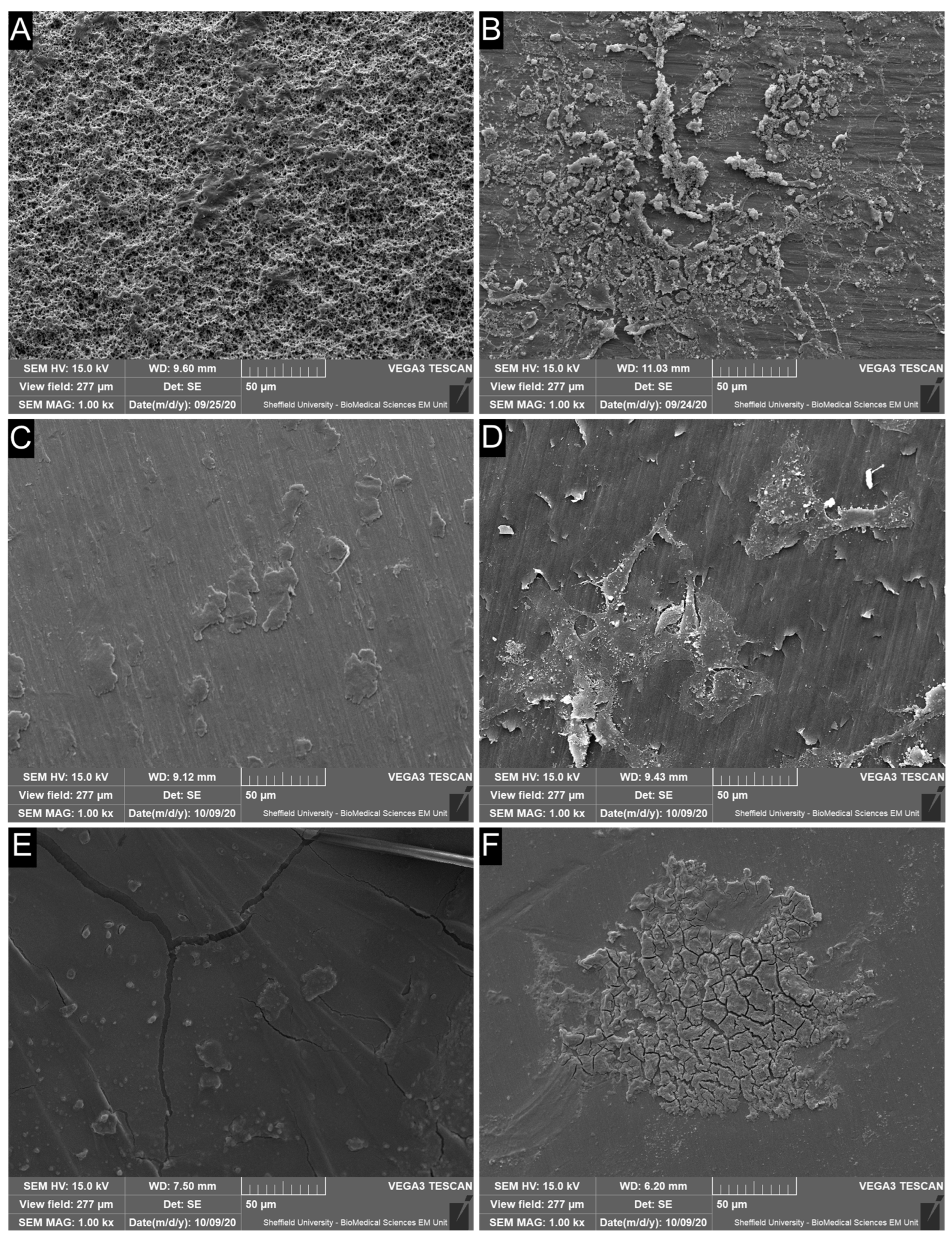
3.4. SEM Reveals Extensive Cell Retention on Rough TiZr-SLA Surfaces with Patchier Coverage on Machined TiZr-M and PEEK-M Implants
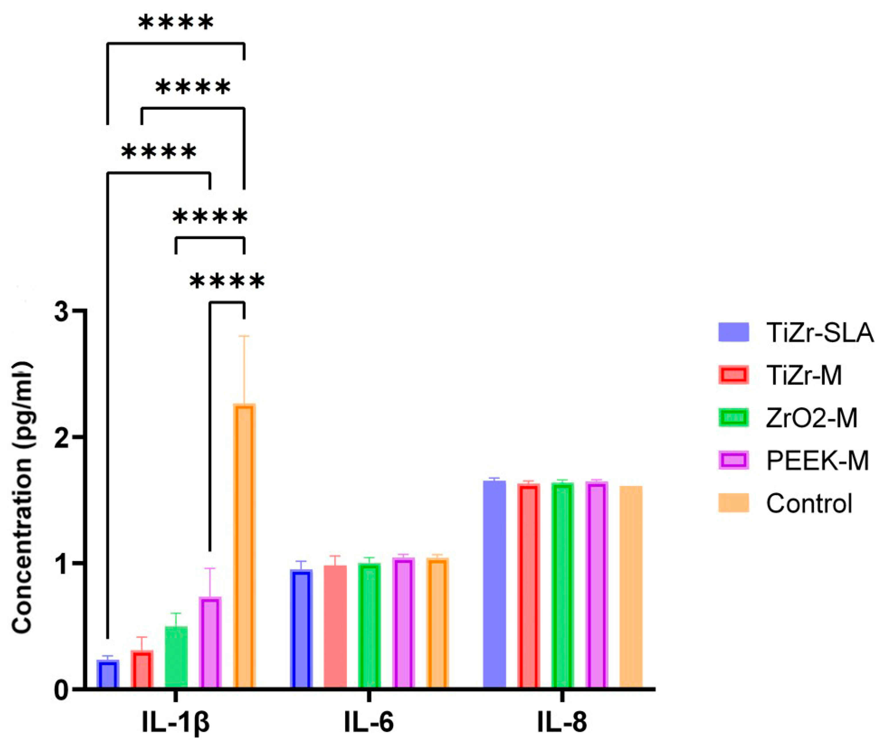
3.5. ZrO-M and PEEK-M Implant Surfaces Induce Significantly Higher IL-1β Release, Promoting Pro-Inflammatory Responses in 3D Oral Mucosal Models
4. Discussion
5. Conclusions
Author Contributions
Funding
Institutional Review Board Statement
Informed Consent Statement
Data Availability Statement
Acknowledgments
Conflicts of Interest
References
- Chai, W.L.; Razali, M.; Moharamzadeh, K.; Zafar, M.S. The hard and soft tissue interfaces with dental implants. In Dental Implants: Materials, Coatings, Surface Modifications and Interfaces with Oral Tissue; Zafar, M.S., Khurshid, Z., Eds.; Woodhead Publishing: Cambridge, UK; Elsevier: Amsterdam, The Netherlands, 2020; pp. 173–201. [Google Scholar]
- Rompen, E.; Domken, O.; Degidi, M.; Pontes, A.E.; Piattelli, A. The effect of material characteristics, of surface topography and of implant components and connections on soft tissue integration: A literature review. Clin. Oral Implant. Res. 2006, 17, 55–67. [Google Scholar] [CrossRef]
- Schupbach, P.; Glauser, R. The defense architecture of the human periimplant mucosa: A histological study. J. Prosthet. Dent. 2007, 97, S15–S25. [Google Scholar] [CrossRef]
- Koidou, V.P.; Argyris, P.P.; Skoe, E.P.; Mota Siqueira, J.; Chen, X.; Zhang, L.; Hinrichs, J.E.; Costalonga, M.; Aparicio, C. Peptide coatings enhance keratinocyte attachment towards improving the peri-implant mucosal seal. Biomater. Sci. 2018, 6, 1936–1945. [Google Scholar] [CrossRef]
- Giannasi, C.; Pagni, G.; Polenghi, C.; Niada, S.; Manfredi, B.; Brini, A.T.; Rasperini, G. Impact of Dental Implant Surface Modifications on Adhesion and Proliferation of Primary Human Gingival Keratinocytes and Progenitor Cells. Int. J. Periodontics Restor. Dent. 2018, 38, 127–135. [Google Scholar] [CrossRef]
- Pandoleon, P.; Bakopoulou, A.; Papadopoulou, L.; Koidis, P. Evaluation of the biological behaviour of various dental implant abutment materials on attachment and viability of human gingival fibroblasts. Dent. Mater. 2019, 35, 1053–1063. [Google Scholar] [CrossRef] [PubMed]
- Pham, M.H.; Haugen, H.J.; Rinna, A.; Ellingsen, J.E.; Reseland, J.E. Hydrofluoric acid treatment of titanium surfaces enhances the proliferation of human gingival fibroblasts. J. Tissue Eng. 2019, 10, 2041731419828950. [Google Scholar] [CrossRef] [PubMed]
- Gibbs, S.; Roffel, S.; Meyer, M.; Gasser, A. Biology of soft tissue repair: Gingival epithelium in wound healing and attachment to the tooth and abutment surface. Eur. Cell Mater. 2019, 38, 63–78. [Google Scholar] [CrossRef]
- Canullo, L.; Annunziata, M.; Pesce, P.; Tommasato, G.; Nastri, L.; Guida, L. Influence of abutment material and modifications on peri-implant soft-tissue attachment: A systematic review and meta-analysis of histological animal studies. J. Prosthet. Dent. 2020, 125, 426–436. [Google Scholar] [CrossRef] [PubMed]
- Menini, M. Influence of modified titanium abutment surface on peri-implant soft tissue behavior: A systematic review of in vitro studies. Int. J. Oral Maxillofac. Implant. 2020, 35, 503–519. [Google Scholar] [CrossRef]
- Moharamzadeh, K.; Colley, H.; Murdoch, C.; Hearnden, V.; Chai, W.L.; Brook, I.M.; Thornhill, M.H.; Macneil, S. Tissue-engineered oral mucosa. J. Dent. Res. 2012, 91, 642–650. [Google Scholar] [CrossRef]
- Barker, E.; AlQobaly, L.; Shaikh, Z.; Franklin, K.; Moharamzadeh, K. Implant Soft-Tissue Attachment Using 3D Oral Mucosal Models-A Pilot Study. Dent. J. 2020, 8, 72. [Google Scholar] [CrossRef]
- Chai, W.L.; Moharamzadeh, K.; Brook, I.M.; Emanuelsson, L.; Palmquist, A.; van Noort, R. Development of a novel model for the investigation of implant-soft tissue interface. J. Periodontol. 2010, 81, 1187–1195. [Google Scholar] [CrossRef]
- Chai, W.L.; Moharamzadeh, K.; van Noort, R.; Emanuelsson, L.; Palmquist, A.; Brook, I.M. Contour analysis of an implant–soft tissue interface. J. Periodontal Res. 2013, 48, 663–670. [Google Scholar] [CrossRef] [PubMed]
- Gavriiloglou, M.; Hammad, M.; Iliopoulos, J.M.; Layrolle, P.; Apatzidou, D.A. Bioengineering the junctional epithelium in 3d oral mucosa models. J. Funct. Biomater. 2024, 15, 330. [Google Scholar] [CrossRef]
- Ingendoh-Tsakmakidis, A.; Mikolai, C.; Winkel, A.; Szafranski, S.P.; Falk, C.S.; Rossi, A.; Walles, H.; Stiesch, M. Commensal and pathogenic biofilms differently modulate peri-implant oral mucosa in an organotypic model. Cell Microbiol. 2019, 21, e13078. [Google Scholar] [CrossRef] [PubMed]
- Shang, L.; Deng, D.; Buskermolen, J.K.; Janus, M.M.; Krom, B.P.; Roffel, S.; Waaijman, T.; van Loveren, C.; Crielaard, W.; Gibbs, S. Multi-species oral biofilm promotes reconstructed human gingiva epithelial barrier function. Sci. Rep. 2018, 8, 16061. [Google Scholar] [CrossRef] [PubMed]
- Roffel, S.; Wu, G.; Nedeljkovic, I.; Meyer, M.; Razafiarison, T.; Gibbs, S. Evaluation of a novel oral mucosa in vitro implantation model for analysis of molecular interactions with dental abutment surfaces. Clin. Implant. Dent. Relat. Res. 2019, 21, 25–33. [Google Scholar] [CrossRef]
- Malekahmadi, B.; Kheirmand-Parizi, M.; Mikolai, C.; Winkel, A.; Rahim, M.I.; Doll-Nikutta, K.; Kampmann, A.; Gellrich, N.-C.; Wirth, D.; Menzel, H. Establishment of a three-dimensional in vitro peri-implant bone-mucosa composite model. BMC Oral Health 2026, 26, 91. [Google Scholar] [CrossRef]
- Calvo-Correa, M.; Benitez-Forero, J.; Cortés-Rodríguez, C. Surface roughness analysis (RA) in the machining of Poly-Ether-Keton (PEEK) for tailored implants. Rev. Mex. Ing. Biomédica 2017, 38, 517–523. [Google Scholar][Green Version]
- Faadhila, A.; Taufiqurrakhman, M.; Katili, P.A.; Rahman, S.F.; Lestari, D.C.; Whulanza, Y. Optimizing PEEK implant surfaces for improved stability and biocompatibility through sandblasting and the platinum coating approach. Front. Mech. Eng. 2024, 10, 1360743. [Google Scholar] [CrossRef]
- Kawakami, K.; Fukuda, T.; Toyoda, M.; Nakao, Y.; Hayashi, C.; Watanabe, Y.; Aoki, T.; Shinjo, T.; Iwashita, M.; Yamashita, A. Research Article Luteolin Is a Potential Immunomodulating Natural Compound against Pulpal Inflammation. BioMed Res. Int. 2024, 2024, 8864513. [Google Scholar] [CrossRef] [PubMed]
- Canullo, L.; Rasperini, G. Preservation of peri-implant soft and hard tissues using platform switching of implants placed in immediate extraction sockets: A proof-of-concept study with 12- to 36-month follow-up. Int. J. Oral Maxillofac. Implant. 2007, 22, 995–1000. [Google Scholar]
- Hermann, J.S.; Buser, D.; Schenk, R.K.; Cochran, D.L. Crestal bone changes around titanium implants. A histometric evaluation of unloaded non-submerged and submerged implants in the canine mandible. J. Periodontol. 2000, 71, 1412–1424. [Google Scholar] [CrossRef]
- Berglundh, T.; Lindhe, J.; Ericsson, I.; Marinello, C.P.; Liljenberg, B.; Thomsen, P. The soft tissue barrier at implants and teeth. Clin. Oral Implant. Res. 1991, 2, 81–90. [Google Scholar] [CrossRef]
- Araujo, M.G.; Lindhe, J. Peri-implant health. J. Clin. Periodontol. 2018, 45, S230–S236. [Google Scholar] [CrossRef]
- Atsuta, I.; Ayukawa, Y.; Kondo, R.; Oshiro, W.; Matsuura, Y.; Furuhashi, A.; Tsukiyama, Y.; Koyano, K. Soft tissue sealing around dental implants based on histological interpretation. J. Prosthodont. Res. 2016, 60, 3–11. [Google Scholar] [CrossRef]
- Chai, W.L.; Brook, I.M.; Emanuelsson, L.; Palmquist, A.; van Noort, R.; Moharamzadeh, K. Ultrastructural analysis of implant-soft tissue interface on a three dimensional tissue-engineered oral mucosal model. J. Biomed. Mater. Res. A 2012, 100, 269–277. [Google Scholar] [CrossRef]
- Buser, D.; Weber, H.P.; Donath, K.; Fiorellini, J.P.; Paquette, D.W.; Williams, R.C. Soft tissue reactions to non-submerged unloaded titanium implants in beagle dogs. J. Periodontol. 1992, 63, 225–235. [Google Scholar] [CrossRef]
- Cochran, D.L.; Hermann, J.S.; Schenk, R.K.; Higginbottom, F.L.; Buser, D. Biologic width around titanium implants. A histometric analysis of the implanto-gingival junction around unloaded and loaded nonsubmerged implants in the canine mandible. J. Periodontol. 1997, 68, 186–198. [Google Scholar] [CrossRef] [PubMed]
- Berglundh, T.; Lindhe, J.; Jonsson, K.; Ericsson, I. The topography of the vascular systems in the periodontal and peri-implant tissues in the dog. J. Clin. Periodontol. 1994, 21, 189–193. [Google Scholar] [CrossRef] [PubMed]
- Schierano, G.; Ramieri, G.; Cortese, M.; Aimetti, M.; Preti, G. Organization of the connective tissue barrier around long-term loaded implant abutments in man. Clin. Oral Implant. Res. 2002, 13, 460–464. [Google Scholar] [CrossRef] [PubMed]
- Glauser, R.; Schüpbach, P.; Gottlow, J.; Hämmerle, C.H. Peri-implant soft tissue barrier at experimental one-piece mini-implants with different surface topography in humans: A light-microscopic overview and histometric analysis. Clin. Implant. Dent. Relat. Res. 2005, 7, s44–s51. [Google Scholar] [CrossRef]
- Altmann, B.; Kohal, R.J.; Steinberg, T.; Tomakidi, P.; Bachle-Haas, M.; Wennerberg, A.; Att, W. Distinct cell functions of osteoblasts on UV-functionalized titanium- and zirconia-based implant materials are modulated by surface topography. Tissue Eng. Part C Methods 2013, 19, 850–863. [Google Scholar] [CrossRef]
- Hermann, J.S.; Cochran, D.L.; Buser, D.; Schenk, R.K.; Schoolfield, J.D. Biologic Width around one-and two-piece titanium implants: A histometric evaluation of unloaded nonsubmerged and submerged implants in the canine mandible. Clin. Oral Implant. Res. 2001, 12, 559–571. [Google Scholar] [CrossRef]
- Abrahamsson, I.; Berglundh, T.; Glantz, P.; Lindhe, J. The mucosal attachment at different abutments. An experimental study in dogs. J. Clin. Periodontol. 1998, 25, 721–727. [Google Scholar] [CrossRef]
- Welander, M.; Abrahamsson, I.; Berglundh, T. The mucosal barrier at implant abutments of different materials. Clin. Oral Implant. Res. 2008, 19, 635–641. [Google Scholar] [CrossRef]
- Vigolo, P.; Givani, A.; Majzoub, Z.; Cordioli, G. A 4-year prospective study to assess peri-implant hard and soft tissues adjacent to titanium versus gold-alloy abutments in cemented single implant crowns. J. Prosthodont. 2006, 15, 250–256. [Google Scholar] [CrossRef]
- Abrahamsson, I.; Cardaropoli, G. Peri-implant hard and soft tissue integration to dental implants made of titanium and gold. Clin. Oral Implant. Res. 2007, 18, 269–274. [Google Scholar] [CrossRef]
- Linkevicius, T.; Apse, P. Influence of abutment material on stability of peri-implant tissues: A systematic review. Int. J. Oral Maxillofac. Implant. 2008, 23, 449–456. [Google Scholar]
- Yang, Y.; Zhou, J.; Liu, X.; Zheng, M.; Yang, J.; Tan, J. Ultraviolet light-treated zirconia with different roughness affects function of human gingival fibroblasts in vitro: The potential surface modification developed from implant to abutment. J. Biomed. Mater. Res. Part B Appl. Biomater. 2015, 103, 116–124. [Google Scholar] [CrossRef] [PubMed]
- Schwarz, F.; Herten, M.; Sager, M.; Wieland, M.; Dard, M.; Becker, J. Histological and immunohistochemical analysis of initial and early subepithelial connective tissue attachment at chemically modified and conventional SLA titanium implants. A pilot study in dogs. Clin. Oral Investig. 2007, 11, 245–255. [Google Scholar] [CrossRef]
- Linares, A.; Munoz, F.; Permuy, M.; Dard, M.; Blanco, J. Soft tissue histomorphology at implants with a transmucosal modified surface. A study in minipigs. Clin. Oral Implant. Res. 2015, 26, 996–1005. [Google Scholar] [CrossRef]
- Skirbutis, G.; Dzingutė, A.; Masiliūnaitė, V.; Šulcaitė, G.; Žilinskas, J. A review of PEEK polymer’s properties and its use in prosthodontics. Stomatologija 2017, 19, 19–23. [Google Scholar]
- Kwan, J.C.; Kwan, N. Clinical Application of PEEK as a Provisional Fixed Dental Prosthesis Retained by Reciprocated Guide Surfaces of Healing Abutments During Dental Implant Treatment. Int. J. Oral Maxillofac. Implant. 2021, 36, 581–586. [Google Scholar] [CrossRef]
- Nothdurft, F.P.; Fontana, D.; Ruppenthal, S.; May, A.; Aktas, C.; Mehraein, Y.; Lipp, P.; Kaestner, L. Differential Behavior of Fibroblasts and Epithelial Cells on Structured Implant Abutment Materials: A Comparison of Materials and Surface Topographies. Clin. Implant. Dent. Relat. Res. 2015, 17, 1237–1249. [Google Scholar] [CrossRef]
- Al Rezk, F.; Trimpou, G.; Lauer, H.C.; Weigl, P.; Krockow, N. Response of soft tissue to different abutment materials with different surface topographies: A review of the literature. Gen. Dent. 2018, 66, 18–25. [Google Scholar] [PubMed]
- Mladenova, I.; Song, X.; Nica, C.; Sculean, A.; Asparuhova, M.B. In Vitro Evidence for Oral Soft Tissue Regenerative Effects of a Polynucleotide–Hyaluronan Formulation. Clin. Exp. Dent. Res. 2025, 11, e70253. [Google Scholar] [CrossRef] [PubMed]
- Frisch, E.; Clavier, L.; Belhamdi, A.; Vrana, N.E.; Lavalle, P.; Frisch, B.; Heurtault, B.; Gribova, V. Preclinical in vitro evaluation of implantable materials: Conventional approaches, new models and future directions. Front. Bioeng. Biotechnol. 2023, 11, 1193204. [Google Scholar] [CrossRef]
- Wang, P.; Qian, H.; Xiao, M.; Lv, J. Role of signal transduction pathways in IL-1β-induced apoptosis: Pathological and therapeutic aspects. Immun. Inflamm. Dis. 2023, 11, e762. [Google Scholar] [CrossRef] [PubMed]
- Boronat-Catala, M.; Catala-Pizarro, M.; Bagan Sebastian, J.V. Salivary and crevicular fluid interleukins in gingivitis. J. Clin. Exp. Dent. 2014, 6, e175–e179. [Google Scholar] [CrossRef] [PubMed]
- Degidi, M.; Artese, L.; Scarano, A.; Perrotti, V.; Gehrke, P.; Piattelli, A. Inflammatory infiltrate, microvessel density, nitric oxide synthase expression, vascular endothelial growth factor expression, and proliferative activity in peri-implant soft tissues around titanium and zirconium oxide healing caps. J. Periodontol. 2006, 77, 73–80. [Google Scholar] [CrossRef]
- Sanz-Sánchez, I.; Sanz-Martín, I.; Carrillo de Albornoz, A.; Figuero, E.; Sanz, M. Biological effect of the abutment material on the stability of peri-implant marginal bone levels: A systematic review and meta-analysis. Clin. Oral Implant. Res. 2018, 29, 124–144. [Google Scholar] [CrossRef]
- Nascimento, C.; Pita, M.S.; Santos Ede, S.; Monesi, N.; Pedrazzi, V.; Albuquerque Junior, R.F.; Ribeiro, R.F. Microbiome of titanium and zirconia dental implants abutments. Dent. Mater. 2016, 32, 93–101. [Google Scholar] [CrossRef] [PubMed]
- Roehling, S.; Astasov-Frauenhoffer, M.; Hauser-Gerspach, I.; Braissant, O.; Woelfler, H.; Waltimo, T.; Kniha, H.; Gahlert, M. In Vitro Biofilm Formation on Titanium and Zirconia Implant Surfaces. J. Periodontol. 2017, 88, 298–307. [Google Scholar] [CrossRef] [PubMed]
- Bostanci, N.; Meier, A.; Guggenheim, B.; Belibasakis, G.N. Regulation of NLRP3 and AIM2 inflammasome gene expression levels in gingival fibroblasts by oral biofilms. Cell. Immunol. 2011, 270, 88–93. [Google Scholar] [CrossRef]
- Guarnieri, R.; Reda, R.; Di Nardo, D.; Miccoli, G.; Pagnoni, F.; Zanza, A.; Testarelli, L. Expression of IL-1β, IL-6, TNF-α, and a-MMP-8 in sites with healthy conditions and with periodontal and peri-implant diseases: A case-control study. J. Dent. Res. Dent. Clin. Dent. Prospect. 2024, 18, 135. [Google Scholar] [CrossRef]
- Cionca, N.; Hashim, D.; Cancela, J.; Giannopoulou, C.; Mombelli, A. Pro-inflammatory cytokines at zirconia implants and teeth. A cross-sectional assessment. Clin. Oral Investig. 2016, 20, 2285–2291. [Google Scholar] [CrossRef]
- Alves, C.H.; Russi, K.L.; Rocha, N.C.; Bastos, F.; Darrieux, M.; Parisotto, T.M.; Girardello, R. Host-microbiome interactions regarding peri-implantitis and dental implant loss. J. Transl. Med. 2022, 20, 425. [Google Scholar] [CrossRef] [PubMed]



Disclaimer/Publisher’s Note: The statements, opinions and data contained in all publications are solely those of the individual author(s) and contributor(s) and not of MDPI and/or the editor(s). MDPI and/or the editor(s) disclaim responsibility for any injury to people or property resulting from any ideas, methods, instructions or products referred to in the content. |
© 2026 by the authors. Licensee MDPI, Basel, Switzerland. This article is an open access article distributed under the terms and conditions of the Creative Commons Attribution (CC BY) license.
Share and Cite
Jalaleddine, N.; Barker, E.; Franklin, K.; Jamal, M.; Atieh, M.A.; Baqain, Z.H.; Moharamzadeh, K. Differential Attachment of Engineered Oral Soft Tissues to Implant Surfaces. Dent. J. 2026, 14, 150. https://doi.org/10.3390/dj14030150
Jalaleddine N, Barker E, Franklin K, Jamal M, Atieh MA, Baqain ZH, Moharamzadeh K. Differential Attachment of Engineered Oral Soft Tissues to Implant Surfaces. Dentistry Journal. 2026; 14(3):150. https://doi.org/10.3390/dj14030150
Chicago/Turabian StyleJalaleddine, Nour, Emilia Barker, Kirsty Franklin, Mohamed Jamal, Momen A. Atieh, Zaid H. Baqain, and Keyvan Moharamzadeh. 2026. "Differential Attachment of Engineered Oral Soft Tissues to Implant Surfaces" Dentistry Journal 14, no. 3: 150. https://doi.org/10.3390/dj14030150
APA StyleJalaleddine, N., Barker, E., Franklin, K., Jamal, M., Atieh, M. A., Baqain, Z. H., & Moharamzadeh, K. (2026). Differential Attachment of Engineered Oral Soft Tissues to Implant Surfaces. Dentistry Journal, 14(3), 150. https://doi.org/10.3390/dj14030150