Abstract
Objective: The primary goal of this investigation was to ascertain the efficacy of the CALM® motion artifact reduction algorithm in diminishing motion-induced blurriness in Cone Beam Computed Tomography [CBCT] images. The assessment was conducted through Fractal Dimension [FD] analysis of the trabecular bone. Methods and Materials: A desiccated human mandible was subjected to Planmeca ProMax 3D® scanning under eight distinct protocols, marked by variations in motion presence [at 5, 10, and 15 degrees] and the deployment of CALM®. In every scan, five distinct regions of interest [ROIs] were designated for FD analysis, meticulously avoiding tooth roots or cortical bone. The FD was computed employing the box-counting method with Image-J 1.53 software. Results: Our findings reveal that a 5-degree motion does not significantly disrupt FD analysis, while a 10-degree motion and beyond exhibit statistical differences and volatility among the sites and groups. A decreased FD value, signifying a less intricate or “rough” bone structure, correlated with amplified motion blurriness. The utilization of CALM® software seemed to counteract this effect in some instances, reconciling FD values to those akin to the control groups. Nonetheless, CALM®’s efficacy differed across sites and motion degrees. Interestingly, at one site, CALM® application in the absence of motion resulted in FD values considerably higher than all other groups. Conclusion: The study indicates that motion, particularly at 10 degrees or more, can considerably impact the FD analysis of trabecular bone in CBCT images. In some situations, the CALM® motion artifact reduction algorithm can alleviate this impact, though its effectiveness fluctuates depending on the site and degree of motion. This underscores the necessity of factoring in motion and the employment of artifact reduction algorithms during the interpretation of FD analysis outcomes in CBCT imaging. More research is necessary to refine the application of such algorithms and to comprehend their influence on different sites under varying motion degrees.
1. Introduction
Bone strength is closely tied to complex bone architecture [1]. Fractal Dimension analysis [FD] is a mathematical method employed to measure bone quality by quantifying complex bone morphology and irregular structures, often used in the assessment of conditions like osteoporosis [2,3]. White and Rudolph [3] demonstrated a significant difference in trabecular bone patterns between patients with osteoporosis and healthy individuals. FD decreases with lower bone density and increases with higher density, as observed through dual-energy X-ray absorptiometry [4].
Nevertheless, FD’s effectiveness as a predictor of osteoporosis using Cone Beam Computed Tomography [CBCT] is disputed due to inconsistencies and low accuracy in some studies [2,5,6]. However, it is crucial in assessing initial implant stability and the subsequent success of prosthetic treatments [7,8]. Comparisons of FD analysis on CBCT and digital panoramic radiographs show no significant differences [9]. CBCT’s popularity has risen for its wide range of applications, including dento-maxillofacial structure assessments and prosthetic planning [10,11,12], and for examining trabecular changes in Temporomandibular joints [TMJs] [13].
Despite these advancements, the impact of exposure parameters and artifacts on FD analysis through CBCT remains a concern. While variations in kV and mAs do not significantly affect the results, voxel size variations can affect trabecular structure analysis [14]. Motion artifacts, common in children or people with unstable conditions like Parkinson’s disease, can complicate interpretation [15,16]. Despite these challenges, FD’s use in maxillofacial conditions remains widely studied [1,2,3,5,6,13,17,18]. Motion artifacts can significantly affect the interpretation of the images, leading to the development of motion artifact reduction algorithms like CALM® in Planmeca Promax 3D [15].
The effectiveness of such algorithms in restoring fine details, such as trabecular bone patterns, is crucial for accurate FD analysis. However, to our knowledge, no studies have yet quantitively evaluated the effectiveness of motion artifact reduction algorithms like CALM® in reducing motion unsharpness as measured through FD analysis.
Therefore, this study aimed to evaluate the effectiveness of CALM® in reducing motion unsharpness as measured through fractal analysis. We hypothesized that the application of CALM® would significantly reduce motion unsharpness, thereby improving the accuracy of FD analysis of the trabecular bone using CBCT images.
Our null hypothesis is that there is no significant difference in the FD analysis of CBCT images with and without the application of CALM®. Therefore, different degrees of motion artifacts at 5, 10, and 15 degrees should not affect FD analysis on CBCT images after the application of CALM® software.
2. Materials and Methods
A dry human mandible (Figure 1) was scanned using the Planmeca ProMax 3D® system (Planmeca, Helsinki, Finland), which employed a field of view [FOV] of 10 × 6 cm, set at 90 kV and 10 mA, with an exposure duration of 15 s. The scans achieved a high-definition resolution with a voxel size of 150 µm. Eight distinct scanning protocols were tested to evaluate the influence of movement and the use of the Correction Algorithm for Latent Movement [CALM®]. The first group was scanned without any motion or CALM®. In the second group, scans were conducted without motion but with CALM® activated. For the third and fourth groups, a 5-degree motion was introduced, with the third group scanned without CALM® and the fourth with CALM®. The fifth and sixth groups involved a 10-degree motion, with the fifth group lacking CALM® and the sixth utilizing CALM®. Lastly, the seventh and eighth groups underwent a 15-degree motion, with the seventh group scanned without CALM® and the eighth group with CALM®. This comprehensive setup enabled a thorough investigation into the effects of motion and the application of CALM® on the image quality.
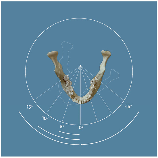
Figure 1.
A dry human mandible was scanned using the Planmeca ProMax 3D® system with a 10 × 6 cm field of view [FOV], operating at 90 kV, 10 mA, for 15 s, and a voxel size of 150 µm [high definition]. The scanning protocols were: Group 1 with no motion and no CALM®, Group 2 with no motion but with CALM®, Group 3 with 5° motion and no CALM®, Group 4 with 5° motion and CALM®, and Group 5 with 10° motion and no CALM®.
All motions were induced clockwise with a rotating device using a remote control and returned to the initial position. One person induced the motion to ensure accuracy and reproducibility. To control for variability in motion introduction, we manually controlled the motion using a remote control. We acknowledge the potential variability introduced by this method and suggest that future studies employ automated motion control systems to enhance reproducibility and reduce variability.
A pilot study was carried out using identical parameters, involving five scans per group and three site measurements. Based on the pilot study’s results with 15 samples per group, mean and standard deviations were calculated. Power analysis, conducted using GPower software version 3.1.2 (Heinrich-Heine-Universität Düsseldorf, Düsseldorf, Germany), yielded an effect size of 0.2976 with an alpha error of 0.05, and which required 90% power. This necessitated a total sample size of 216 distributed across 8 groups, resulting in 27 per subgroup. Thus, 80 scans, 10 per group, were obtained.
To control variables, all exposure parameters and mandible positions were fixed. The mandible was secured during image acquisition to limit motion. The final 80 scans, excluding the pilot, were captured in a single day, maintaining uniform positioning and slice selection. For the fractal analysis, five regions of interest [ROIs] were selected in each scan. These regions were the right molar [site #1] and premolar [site #2] regions, anterior [site #3] region, and left premolar [site #4] and molar [site #5] regions. Each ROI was a square of 128 × 128 pixels, chosen to avoid tooth roots or cortical bone. The ROIs were selected by a trained dental radiologist with over ten years of experience in CBCT imaging and fractal analysis.
The fractal dimension [FD] was calculated using the box-counting method. This method involves overlaying the image with a grid of boxes and counting the number of boxes that contain part of the image. This process is repeated with different box sizes, and the FD is calculated as the slope of the line when plotting the logarithm of the box size against the logarithm of the box count (Figure 2). This calculation was performed using Image-J 1.53 software (National Institutes of Health, Bethesda, MD, USA), following the method described by Magat et al., 2022 [9].
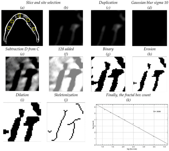
Figure 2.
Image analysis: One slice [#210] was selected apical to the root apices. Five areas of interest were chosen: right and left premolars, right and left molars, and anterior. (a,b) Region of interest [ROI] with the same axial slice number is used for reproducibility purposes. (c,d) The cropped ROI was duplicated (c) and then blurred with a Gaussian filter (d). (e,f) The blurred image was subtracted from the original image (e), and 128 was added to the result at each pixel location (f). (g) The resultant image was converted to binary, to set the image into trabeculae and marrow spaces. (h,i) The binary image was eroded and then dilated to reduce the noise before skeletonization. (j) The skeletonized image, which corresponds to trabeculae, was used for fractal analysis. A single ImageJ macro was used for all measurements to reproduce exact locations and measurements.
In the fractal analysis, five site measurements were performed for each group (Figure 3). The mean, standard deviation, and standard error were calculated, and a 95% confidence interval was used. A One-Way ANOVA with Post-Hoc Tukey HSD Test was used for intergroup comparisons for each site. Groups 1 and 2 were used as a baseline, to which all other groups were compared.
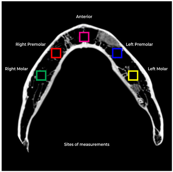
Figure 3.
Five site measurements were performed for each group.
3. Results
At Site 1 [Right Molar (RM)], the difference between Group 7 [15D no CALM®] and Group 1 was statistically significant [p = 0.005]. Group 7 shows significantly lower FD values. All other groups were not significantly different when compared to group 1 as well as Group 2. CALM® appears to have restored the values of FD [Table 1 and Figure 4].

Table 1.
FD values for Site 1 [Right Molars] including the Mean, Std. Deviation, Std. Error, and 95% Confidence Interval. It shows p values compared to Groups 1 and 2.
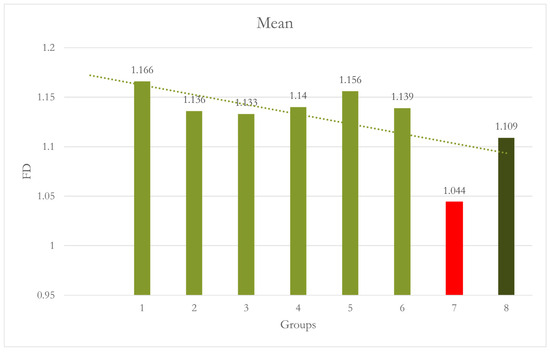
Figure 4.
Graph shows Group 7 [15º No CALM®]: motion caused significantly lower FD values than Group 1 [p = 0.005].
At Site 2 [RPM], Group 5, Group 6, and Group 8 exhibited significantly lowered FD values compared to both Groups 1 [p = 0.002, p = 0.002, and p < 0.001, respectively] and Group 2 [p = 0.009, p = 0.009, and p = 0.002, respectively]. The 15° motion in Group 7 did not significantly affect FD. However, Group 8 [15° with CALM®] had significantly lower FD values than Groups 1 and 2 [Table 2 and Figure 5].
Table 2.
FD values for Site 2 [right premolars] including the Mean, Std. Deviation, Std. Error, and 95% Confidence Interval. It shows p values compared to Groups 1 and 2.
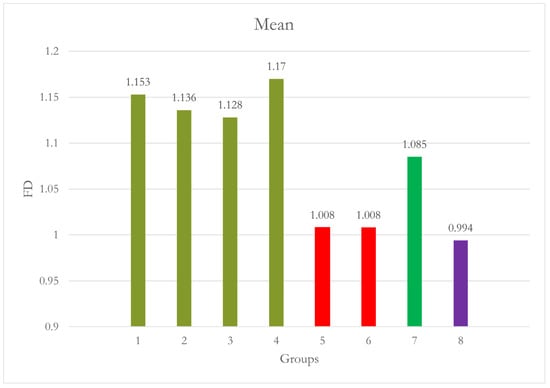
Figure 5.
Group 5 [10° No CALM®] and Group 6 [10° with CALM®] 10° motion with or without CALM® caused significantly lower FD values than Groups 1 and 2. A 15° motion in Group 7 did not significantly affect FD. However, Group 8 [15° with CALM®] had significantly lower FD values than Groups 1 and 2.
At Site 3 [anterior], the difference between Group 1 and Group 7 was statistically highly significant [p < 0.001], and Group 7 and Group 2 were statistically significant [p = 0.001]. Group 7 showed significantly lowered FD values. CALM® seems to have restored the values of FD values in Group 8 [Table 3 and Figure 6].
Table 3.
FD values for Site 3 [anterior] including the Mean, Std. Deviation, Std. Error, and 95% Confidence Interval. It shows p values compared to Groups 1 and 2.
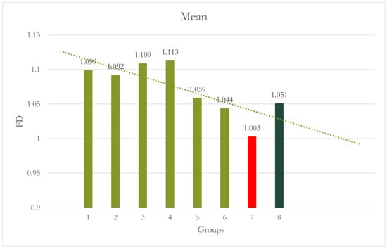
Figure 6.
Group 7 [15° No CALM®]: motion caused significantly lower FD values than Groups 1 and 2. Group 8 [15° with CALM®]: CALM® restored the values of FD.
At Site 4 [Left premolar (LPM)], Group 6 showed a significant difference when compared to both Group 1 [p = 0.007] and Group 2 [p = 0.017]. Combining motion and CALM® caused significantly higher FD values than Groups 1 and 2. Although the values of Groups 3, 4, 5, and 8 are also higher than Groups 1 and 2, they are not statistically significant [Table 4 and Figure 7].
Table 4.
FD values for Site 4 [left premolars] including the Mean, Std. Deviation, Std. Error, and 95% Confidence Interval. It shows p values compared to Groups 1 and 2.
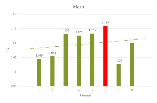
Figure 7.
Group 7 [15° No CALM®]: motion caused significantly lower FD values than Groups 1 and 2. Group 8 [15° with CALM®]: CALM® restored the values of FD.
At Site 5 [LM], Group 1 and Group 2 were significantly different from each other [p = 0.040], while all other groups were not significantly different from Group 1. When compared with Group 2, all the groups showed significant differences, with Group 5 and Group 8 showing highly significant differences [p < 0.001]. Using CALM® alone in Group 2 caused significantly higher FD values than all the other groups [Table 5 and Figure 8]. A summary of all the groups’ results is shown in Figure 9.
Table 5.
FD values for Site 5 [left molar] including the Mean, Std. Deviation, Std. Error, and 95% Confidence Interval. It shows p values compared to Groups 1 and 2.
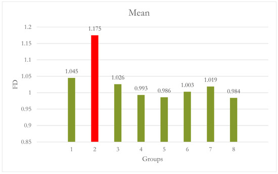
Figure 8.
Group 2 [0° with CALM®]: using CALM® alone caused significantly higher FD values than all other groups. Motion did not significantly affect the FD at this site, although it seems to have caused a slight decrease in FD values.

Figure 9.
Graph shows a summary of the results of all groups.
The results show that a 5-degree motion did not significantly affect the FD analysis, while 10-degree motion and higher showed statistical differences and variability between the sites and groups. A lower FD value, indicating less complexity or “roughness” in the bone structure, was associated with greater motion unsharpness. The application of CALM® software appeared to mitigate this effect in some cases, restoring FD values closer to those of the baseline groups.
However, the effectiveness of CALM® varied between sites and degrees of motion. Notably, at Site 5, the application of CALM® in Group 2 [no motion] resulted in significantly higher FD values than all other groups, suggesting that CALM® may affect the measurements even in the absence of motion.
Additional scans were acquired to further investigate these results. These scans confirmed that the application of CALM® could exaggerate the effect of motion, leading to significantly lower FD values, particularly in the presence of a 15-degree counterclockwise motion.
These additional measurement results in Table 6 show that Group 5 has a significantly lower value than Group 1 [p = 0.005] and Group 2 [p = 0.008]. The results show that CALM® in Group 2 without motion produces no significant effect. A 15° motion in Group 3 reduced FD, but the values were not statistically significant. CALM® in Group 4 exaggerated the effect of motion and produced significant results. Group 1, Group 2, and Group 3 were not significantly different. Also, the difference between Groups 3 and 4 was not statistically significant. These results agree with the previous findings at Sites 1, 2, and 3, except the scan used for these measurements used a counterclockwise motion direction.
Table 6.
FD values for Site 5 [left molar] with additional scans and measurements, including the Mean, Std. Deviation, Std. Error, and 95% Confidence Interval. It shows p values compared to Groups 1 and 2.
4. Discussion
This study aimed to evaluate the effectiveness of the CALM® motion artifact reduction algorithm in reducing motion unsharpness in CBCT images, as measured through fractal dimension [FD] analysis. Several factors were considered as potential causes of those variations. The first is the sensitivity of fractal dimension analysis. Normal FD for healthy bone ranges from 1.1 to 2.68 [9,19,20,21]. According to Magat et al. [9], the variation was mainly due to fractal analysis [FA] rather than the result of different materials, strategies, or anatomic sites used for the studies.
The second factor is the variation in density and grayscale values of the dry mandible used in the study. Perrotti et al. [22] stated that “The more the bone was compact, the higher were FD values”. Additionally, “The increase in the values of the FD strongly correlated with the increase of the percentage of the bone trabeculae observed in the histological slides” [22]. Hua et al. [4] also showed a significant drop in FD with decreased bone density measured on dual-energy X-ray absorptiometry. Both studies indicate that any slight change in including or excluding trabecular bone during FD analysis may change the results.
Southard et al. [19] found a direct relationship between the FD measurements and bone density. Hence, increasing the trabecular bone density translates to higher FD values. However, this study was performed on plain radiographs that were digitized. Therefore, the effect of some parameters, such as voxel size, could not be appreciated. However, other studies showed no correlation between grayscale values and FD [9,17].
Pauwels et al. [14] studied the effect of exposure parameters on bone structure analysis. The study included combinations of kV, mAs, and voxel sizes. Their study concluded that kV above 90 shows significant results with only bone volume per total volume [BV/TV], which is used to assess bone strength by analyzing bone microstructure. Additionally, kV did not affect other trabecular bone analyses, such as FD. In comparison, voxel size was the major factor and significantly affected trabecular bone analysis. The results show a decrease in the FD at larger voxel sizes, equal to or higher than 160 µm. The current study keeps the voxel size to 150 µm [HD] to provide optimal resolution and acceptable noise levels. This may partially undermine the clinical effect of noise from soft tissue.
Some studies revealed that moderate variants in noise level, either due to soft tissue emulators or water, did not significantly impact bone structure parameters on cone beam CT [14,23,24,25]. These studies also elaborated on the variability in the clinical images where the motion artifacts might cause blurring or unsharpness.
Several studies [14,24,26] concluded that kV did not significantly affect bone structural analysis, except for BV/TV values. On the other hand, voxel sizes significantly affect bone structural analysis, such as FD. This is because fine details are lost at larger voxel sizes due to lower spatial resolution [14,27,28].
Therefore, we fixed all previous parameters and focused on the effect of motion on FD. We found that motion and motion correction software affect FD. Additionally, this effect is partially unpredictable, at least in our experiment. Therefore, it is crucial to consider all parameters affecting FD before implementing clinical applications.
The third factor is the direction and timing of motion relative to the scan acquisition. The motion degrees and time of induction were controlled by the researcher using a remote control. This manual control may have resulted in the variability of the motion effect. Additionally, except for Site 5, only a clockwise direction was used in this study. These factors can be better controlled in future studies using a fully automated system.
The fourth factor is CALM®. Rigid body movements, such as translation and rotation, are the most common movement in the head and neck [15]. According to Hernandez et al. [15], there are two common ways to detect and correct motion: using head tracking devices or using the motion artifacts metric [MAM] optimization algorithm. CALM®, [proprietary for Planmeca Promax 3D®], seems to be of the MAM type. It does not need extra head devices and can be applied before or after scanning with a push of a button. In general, the MAM algorithm works on enhancing image sharpness with regularization terms that estimate the motion during reconstruction. This algorithm corrects sharpness, while the trabecular bone pattern has fine details that require high resolution.
Our study showed that CALM® affected FD analysis. It restored the FD of Group 8 at Sites 1 and 2. It failed at Site 2 in Groups 6 and 8. It exaggerated the motion effect at Site 2 in Group 8 and Site 5 in Group 4. It significantly increased the FD in Group 2 at Site 5.
In this study, we used an image-J macro to precisely reproduce the measurements. This macro does not correct for motion. If there is slight inclusion or exclusion in the trabecular bone, it may affect the measurements. This factor is also complicated by the fact that FA itself is variable and technique sensitive [9]. Furthermore, a significant correlation between the changes in bone density and FD values has been documented in the literature [22,23]. All these factors complicate FD measurements and result in variable values.
Several studies have attempted to correlate trabecular bone patterns with osseous diseases/conditions. White and Rudolph [3] were among the first authors correlating FD with altered bone trabeculae in osteoporotic subjects. By contrast, Sindeaux et al. [29] found a correlation between osteoporosis and FD of the cortex of the mandible, not the trabecular bone FD. Alman et al. [30] found that FD can be possibly utilized as a discriminator for people with low bone mineral density on dental radiographs. However, there is documented literature opposing using FD as a screening or adjunctive tool to refer or diagnose patients with osteoporosis [5]. A systematic review and meta-analysis by Franciotti et al. [31] demonstrated heterogeneity in the literature and low reliability in using FD for osteoporosis identification.
Recent studies [32,33] showed that FD could be a useful descriptor for medication-induced osteonecrosis of the jaw [MRONJ]. This could help in the assessment of the disease. Kato et al. [34] assessed the complexity of fibrous dysplasia and ossifying fibroma on CBCT using FD. They discovered fibrous dysplasia might have a significantly more complex structure, represented by higher FD values, than ossifying fibroma.
In general, our research showed that motion beyond 5 degrees led to significant variations in fractal dimension [FD] results, based on site and degree of motion, and CALM® use. The CALM® software generally mitigated motion unsharpness, but its effectiveness varied. At Site 5, CALM® notably increased FD values in a no-motion scenario, suggesting potential effects on measurements without motion.
Despite statistically significant FD differences under various motion conditions, and with or without CALM®, the clinical significance of these findings may depend on the specific context and how motion unsharpness affects CBCT image interpretation.
More research is needed to optimize artifact reduction algorithms like CALM® and to comprehend their impact under different motion scenarios. Further studies could also leverage automated systems for more consistent motion control.
5. Limitations
This study has several limitations that should be considered when interpreting the results. One of the primary limitations is the manual control of motion using a remote control, which could have introduced variability in the motion effect. This may limit the precision of the results and does not fully represent the range of motion that could occur in a real-world clinical setting. Future studies could benefit from using a fully automated system to control motion more consistently and explore the effects of motion in different directions. Lastly, the study primarily used a clockwise direction of motion, which may not fully represent the range of motion that could occur in a real-world clinical setting.
6. Conclusions
The study findings suggest that motion, particularly at 10 degrees and higher, can significantly affect the fractal dimension analysis of trabecular bone in CBCT images, leading to lower FD values that indicate greater motion unsharpness. The application of the CALM® motion artifact reduction algorithm can mitigate this effect in some cases, restoring FD values closer to those of baseline scans without motion. However, the effectiveness of CALM® varies depending on the site and degree of motion, and in some cases, it may affect the measurements even in the absence of motion. These findings highlight the importance of considering motion and the use of artifact reduction algorithms when interpreting FD analysis results in CBCT imaging. Further research is needed to optimize the use of such algorithms and to understand their impact on different sites and under varying degrees of motion.
Author Contributions
Conceptualization, Y.H.K. and A.Z.A.; methodology, Y.H.K., A.Z.A. and H.G.; software, Y.H.K. and A.Z.A.; validation, T.M.-Z., R.A.K. and W.M.; formal analysis, Y.H.K.; investigation, Y.H.K.; resources, Y.H.K.; data curation, Y.H.K.; writing—original draft preparation, Y.H.K. and A.Z.A.; writing—review and editing, T.M.-Z., R.A.K. and W.M.; visualization, H.G.; supervision, H.G. and A.Z.A.; project administration, Y.H.K.; funding acquisition, Y.H.K. All authors have read and agreed to the published version of the manuscript.
Funding
This research received no external funding.
Institutional Review Board Statement
The desiccated human mandible used in this study was obtained from a commercially available source adhering to ethical guidelines for the use of human tissues. No living human subjects were involved, and hence, the study was exempt from formal ethical approval.
Data Availability Statement
The data presented in this study are available on request from the corresponding author. The data are not publicly available due to privacy.
Acknowledgments
We would like to thank Nicolas Shinas for helping with scan acquisition.
Conflicts of Interest
The authors declare no conflicts of interest.
References
- Sanchez-Molina, D.; Velazquez-Ameijide, J.; Quintana, V.; Arregui-Dalmases, C.; Crandall, J.R.; Subit, D.; Kerrigan, J.R. Fractal Dimension and Mechanical Properties of Human Cortical Bone. Med. Eng. Phys. 2013, 35, 576–582. [Google Scholar] [CrossRef] [PubMed]
- Sánchez, I.; Uzcátegui, G. Fractals in Dentistry. J. Dent. 2011, 39, 273–292. [Google Scholar] [CrossRef] [PubMed]
- White, S.C.; Rudolph, D.J. Alterations of the Trabecular Pattern of the Jaws in Patients with Osteoporosis. Oral Surg. Oral Med. Oral Pathol. Oral Radiol. Endodontology 1999, 88, 628–635. [Google Scholar] [CrossRef] [PubMed]
- Hua, Y.; Nackaerts, O.; Duyck, J.; Maes, F.; Jacobs, R. Bone Quality Assessment Based on Cone Beam Computed Tomography Imaging. Clin. Oral Implant. Res. 2009, 20, 767–771. [Google Scholar] [CrossRef] [PubMed]
- Carvalho, B.F.; De Castro, J.G.K.; De Melo, N.S.; De Souza Figueiredo, P.T.; Moreira-Mesquita, C.R.; De Paula, A.P.; Sindeaux, R.; Leite, A.F. Fractal Dimension Analysis on CBCT Scans for Detecting Low Bone Mineral Density in Postmenopausal Women. Imaging Sci. Dent. 2022, 52, 53. [Google Scholar] [CrossRef] [PubMed]
- Mostafa, R.A.; Arnout, E.A.; Fotouh, M.M.A.E. Feasibility of Cone Beam Computed Tomography Radiomorphometric Analysis and Fractal Dimension in Assessment of Postmenopausal Osteoporosis in Correlation with Dual X-Ray Absorptiometry. Dentomaxillofacial Radiol. 2016, 45, 20160212. [Google Scholar] [CrossRef] [PubMed]
- Chrcanovic, B.R.; Albrektsson, T.; Wennerberg, A. Bone Quality and Quantity and Dental Implant Failure: A Systematic Review and Meta-Analysis. Int. J. Prosthodont. 2017, 30, 219–237. [Google Scholar] [CrossRef] [PubMed]
- Voumard, B.; Maquer, G.; Heuberger, P.; Zysset, P.K.; Wolfram, U. “Peroperative Estimation of Bone Quality and Primary Dental Implant Stability”. J. Mech. Behav. Biomed. Mater. 2019, 92, 24–32. [Google Scholar] [CrossRef] [PubMed]
- Magat, G.; Oncu, E.; Ozcan, S.; Orhan, K. Comparison of Cone-Beam Computed Tomography and Digital Panoramic Radiography for Detecting Peri-Implant Alveolar Bone Changes Using Trabecular Micro-Structure Analysis. J. Korean Assoc. Oral Maxillofac. Surg. 2022, 48, 41–49. [Google Scholar] [CrossRef] [PubMed]
- Tibúrcio-Machado, C.S.; Michelon, C.; Zanatta, F.B.; Gomes, M.S.; Marin, J.A.; Bier, C.A. The Global Prevalence of Apical Periodontitis: A Systematic Review and Meta-analysis. Int. Endod. J. 2021, 54, 712–735. [Google Scholar] [CrossRef]
- Antony, D.P.; Thomas, T.; Nivedhitha, M.S. Two-Dimensional Periapical, Panoramic Radiography Versus Three-Dimensional Cone-Beam Computed Tomography in the Detection of Periapical Lesion After Endodontic Treatment: A Systematic Review. Cureus 2020, 12, e7736. [Google Scholar] [CrossRef] [PubMed]
- PradeepKumar, A.R.; Shemesh, H.; Nivedhitha, M.S.; Hashir, M.M.J.; Arockiam, S.; Maheswari, T.N.U.; Natanasabapathy, V. Diagnosis of Vertical Root Fractures by Cone-Beam Computed Tomography in Root-Filled Teeth with Confirmation by Direct Visualization: A Systematic Review and Meta-Analysis. J. Endod. 2021, 47, 1198–1214. [Google Scholar] [CrossRef]
- Kocak, A.T.Ö.; Bulut, D.G. Measurement of the Trabecular Bone Structure of the TMJ Region in Patients with Transverse Maxillary Deficiency: A CBCT Fractal Analysis Study. Oral Surgery, Oral Med. Oral Pathol. Oral Radiol. 2021, 132, 352–360. [Google Scholar] [CrossRef]
- Pauwels, R.; Faruangsaeng, T.; Charoenkarn, T.; Ngonphloy, N.; Panmekiate, S. Effect of Exposure Parameters and Voxel Size on Bone Structure Analysis in CBCT. Dentomaxillofacial Radiol. 2015, 44, 20150078. [Google Scholar] [CrossRef] [PubMed]
- Hernandez, D.; Eldib, M.E.; Hegazy, M.A.A.; Cho, M.H.; Cho, M.H.; Lee, S.Y. A Head Motion Estimation Algorithm for Motion Artifact Correction in Dental CT Imaging. Phys. Med. Biol. 2018, 63, 065014. [Google Scholar] [CrossRef] [PubMed]
- Spin-Neto, R.; Wenzel, A. Patient Movement and Motion Artefacts in Cone Beam Computed Tomography of the Dentomaxillofacial Region: A Systematic Literature Review. Oral Surg. Oral Med. Oral Pathol. Oral Radiol. 2016, 121, 425–433. [Google Scholar] [CrossRef] [PubMed]
- Magat, G.; Sener, S.O. Evaluation of Trabecular Pattern of Mandible Using Fractal Dimension, Bone Area Fraction, and Gray Scale Value: Comparison of Cone-Beam Computed Tomography and Panoramic Radiography. Oral Radiol. 2018, 35, 35–42. [Google Scholar] [CrossRef]
- Kato, C.N.; Barra, S.G.; Tavares, N.P.; Amaral, T.M.; Brasileiro, C.B.; Mesquita, R.A.; Abreu, L.G. Use of Fractal Analysis in Dental Images: A Systematic Review. Dentomaxillofacial Radiol. 2020, 49, 20180457. [Google Scholar] [CrossRef]
- Southard, T.E.; Southard, K.A.; Jakobsen, J.R.; Hillis, S.L.; Najim, C.A. Fractal Dimension in Radiographic Analysis of Alveolar Process Bone. Oral Surg. Oral Med. Oral Pathol. Oral Radiol. Endodontology 1996, 82, 569–576. [Google Scholar] [CrossRef]
- Bianchi, A.E.; Dolci, G., Jr.; Sberna, M.T.; Sanfilippo, S. Factors affecting bone response around loaded titanium dental implants: A literature review. J. Appl. Biomater. Biomech. 2005, 3, 135–140. [Google Scholar]
- Geraets, W.G.; Van Der Stelt, P.F. Fractal Properties of Bone. Dentomaxillofac Radiol. 2000, 29, 144–153. [Google Scholar] [CrossRef] [PubMed]
- Perrotti, V.; Iezzi, G.; De Sanctis, A.; Pasculli, A.; Piattelli, A.; Aprile, G. Correlation Between Bone Density and Fractal Dimension: A Pilot Study. Nonlinear Phenom. Complex Syst. 2020, 23, 130–132. [Google Scholar] [CrossRef]
- Hsu, J.-T.; Wang, S.-P.; Huang, H.-L.; Chen, Y.-J.; Wu, J.; Tsai, M.-T. The Assessment of Trabecular Bone Parameters and Cortical Bone Strength: A Comparison of Micro-CT and Dental Cone-Beam CT. J. Biomech. 2013, 46, 2611–2618. [Google Scholar] [CrossRef]
- Van Dessel, J.; Huang, Y.; Depypere, M.; Rubira-Bullen, I.; Maes, F.; Jacobs, R. A Comparative Evaluation of Cone Beam CT and Micro-CT on Trabecular Bone Structures in the Human Mandible. Dentomaxillofacial Radiol. 2013, 42, 20130145. [Google Scholar] [CrossRef]
- Ibrahim, N.; Parsa, A.; Hassan, B.; Van Der Stelt, P.; Aartman, I.H.A.; Wismeijer, D. The Effect of Scan Parameters on Cone Beam CT Trabecular Bone Microstructural Measurements of the Human Mandible. Dentomaxillofacial Radiol. 2013, 42, 20130206. [Google Scholar] [CrossRef]
- Pauwels, R.; Silkosessak, O.; Jacobs, R.; Bogaerts, R.; Bosmans, H.; Panmekiate, S. A Pragmatic Approach to Determine the Optimal kVp in Cone Beam CT: Balancing Contrast-to-Noise Ratio and Radiation Dose. Dentomaxillofacial Radiol. 2014, 43, 20140059. [Google Scholar] [CrossRef] [PubMed]
- Pauwels, R.; Beinsberger, J.; Stamatakis, H.; Tsiklakis, K.; Walker, A.; Bosmans, H.; Bogaerts, R.; Jacobs, R.; Horner, K. Comparison of Spatial and Contrast Resolution for Cone-Beam Computed Tomography Scanners. Oral Surg. Oral Med. Oral Pathol. Oral Radiol. 2012, 114, 127–135. [Google Scholar] [CrossRef]
- Pauwels, R.; Araki, K.; Siewerdsen, J.H.; Thongvigitmanee, S.S. Technical Aspects of Dental CBCT: State of the Art. Dentomaxillofacial Radiol. 2015, 44, 20140224. [Google Scholar] [CrossRef] [PubMed]
- Sindeaux, R.; Figueiredo, P.T.d.S.; de Melo, N.S.; Guimarães, A.T.B.; Lazarte, L.; Pereira, F.B.; de Paula, A.P.; Leite, A.F. Fractal Dimension and Mandibular Cortical Width in Normal and Osteoporotic Men and Women. Maturitas 2014, 77, 142–148. [Google Scholar] [CrossRef]
- Alman, A.C.; Johnson, L.R.; Calverley, D.C.; Grunwald, G.K.; Lezotte, D.C.; Hokanson, J.E. Diagnostic Capabilities of Fractal Dimension and Mandibular Cortical Width to Identify Men and Women with Decreased Bone Mineral Density. Osteoporos. Int. 2011, 23, 1631–1636. [Google Scholar] [CrossRef]
- Franciotti, R.; Moharrami, M.; Quaranta, A.; Bizzoca, M.E.; Piattelli, A.; Aprile, G.; Perrotti, V. Use of Fractal Analysis in Dental Images for Osteoporosis Detection: A Systematic Review and Meta-Analysis. Osteoporos. Int. 2021, 32, 1041–1052. [Google Scholar] [CrossRef] [PubMed]
- Bachtler, R.; Walter, C.; Schulze, R.K.W. Fractal Dimension in CBCT Images as Predictor for MRONJ: A Retrospective Cohort Study. Clin. Oral Investig. 2020, 25, 2113–2118. [Google Scholar] [CrossRef] [PubMed]
- Torres, S.; Chen, C.; Leroux, B.; Lee, P.; Hollender, L.; Schubert, M. Fractal Dimension Evaluation of Cone Beam Computed Tomography in Patients with Bisphosphonate-Associated Osteonecrosis. Dentomaxillofacial Radiol. 2011, 40, 501–505. [Google Scholar] [CrossRef]
- Kato, C.d.N.A.d.O.; Barra, S.G.; Abreu, L.G.; Machado, V.C.; Pinheiro, J.d.J.V.; Henriques, J.A.S.; Castro, W.H.; Brasileiro, C.B.; Mesquita, R.A. Fractal Analysis of Fibrous Dysplasia and Ossifying Fibroma in 2D and 3D CBCT Images. J. Oral Maxillofac. Surgery, Med. Pathol. 2022, 34, 791–799. [Google Scholar] [CrossRef]
Disclaimer/Publisher’s Note: The statements, opinions and data contained in all publications are solely those of the individual author(s) and contributor(s) and not of MDPI and/or the editor(s). MDPI and/or the editor(s) disclaim responsibility for any injury to people or property resulting from any ideas, methods, instructions or products referred to in the content. |
© 2024 by the authors. Licensee MDPI, Basel, Switzerland. This article is an open access article distributed under the terms and conditions of the Creative Commons Attribution (CC BY) license (https://creativecommons.org/licenses/by/4.0/).