


The Use of Enamel Matrix Derivative during Surgical Therapy for Peri-Implantitis: A Case Series



Abstract
1. Introduction
2. Materials and Methods
Statical Analysis
3. Results
4. Discussion
5. Conclusions
Author Contributions
Funding
Institutional Review Board Statement
Informed Consent Statement
Data Availability Statement
Conflicts of Interest
References
- Koop, R.; Merheb, J.; Quirynen, M. Periodontal regeneration with enamel matrix derivative in reconstructive periodontal therapy: A systematic review. J. Periodontol. 2012, 83, 707–720. [Google Scholar] [CrossRef] [Scilit] [PubMed]
- Lang, N.P.; Berglundh, T.; Working Group 4 of Seventh European Workshop on Periodontology. Periimplant diseases: Where are we now?—Consensus of the Seventh European Workshop on Periodontology. J. Clin. Periodontol. 2011, 38 (Suppl. S11), 178–181. [Google Scholar] [CrossRef] [Scilit] [PubMed]
- Schwarz, F.; Derks, J.; Monje, A.; Wang, H.L. Peri-implantitis. J. Periodontol. 2018, 89 (Suppl. S1), S267–S290. [Google Scholar] [PubMed]
- Roos-Jansaker, A.M.; Lindahl, C.; Renvert, H.; Renvert, S. Nine- to fourteen-year follow-up of implant treatment. Part II: Presence of peri-implant lesions. J. Clin. Periodontol. 2006, 33, 290–295. [Google Scholar] [CrossRef] [Scilit] [PubMed]
- Derks, J.; Schaller, D.; Håkansson, J.; Wennström, J.L.; Tomasi, C.; Berglundh, T. Effectiveness of Implant Therapy Analyzed in a Swedish Population: Prevalence of Peri-implantitis. J. Dent. Res. 2016, 95, 43–49. [Google Scholar] [CrossRef] [Scilit]
- Shimchuk, A.A.; Weinstein, B.F.; Daubert, D.M. The impact of a change in classification criteria on the prevalence of peri-implantitis: A cross-sectional analysis. J. Periodontol. 2021, 92, 1339–1346. [Google Scholar] [CrossRef] [Scilit]
- Listl, S.; Frühauf, N.; Dannewitz, B.; Weis, C.; Tu, Y.K.; Chang, H.J.; Faggion, C.M., Jr. Cost-effectiveness of non-surgical peri-implantitis treatments. J. Clin. Periodontol. 2015, 42, 470–477. [Google Scholar] [CrossRef] [Scilit]
- Wilson, T.G., Jr. A New Minimally Invasive Approach for Treating Peri-Implantitis. Clin. Adv. Periodontics 2019, 9, 59–63. [Google Scholar] [CrossRef] [Scilit]
- Froum, S.J.; Froum, S.H.; Rosen, P.S. A Regenerative Approach to the Successful Treatment of Peri-implantitis: A Consecutive Series of 170 Implants in 100 Patients with 2- to 10-Year Follow-up. Int. J. Periodontics Restor. Dent. 2015, 35, 857–863. [Google Scholar] [CrossRef] [Scilit]
- Grunder, U.; Hurzeler, M.B.; Schupbach, P.; Strub, J.R. Treatment of ligature-induced peri-implantitis using guided tissue regeneration: A clinical and histologic study in the beagle dog. Int. J. Oral. Maxillofac. Implants 1993, 8, 282–293. [Google Scholar]
- Hurzeler, M.B.; Quinones, C.R.; Morrison, E.C.; Caffesse, R.G. Treatment of peri-implantitis using guided bone regeneration and bone grafts, alone or in combination, in beagle dogs. Part 1: Clinical findings and histologic observations. Int. J. Oral. Maxillofac. Implants 1995, 10, 474–484. [Google Scholar] [PubMed]
- Lorenzoni, M.; Pertl, C.; Keil, C.; Wegscheider, W.A. Treatment of peri-implant defects with guided bone regeneration: A comparative clinical study with various membranes and bone grafts. Int. J. Oral. Maxillofac. Implants 1998, 13, 639–646. [Google Scholar] [PubMed]
- Miron, R.J.; Dard, M.; Weinreb, M. Enamel matrix derivative, inflammation and soft tissue wound healing. J. Periodontal Res. 2015, 50, 555–569. [Google Scholar] [CrossRef] [Scilit] [PubMed]
- Arweiler, N.; Auschill, T.; Donos, N.; Sculean, A. Antibacterial effect of an enamel matrix protein derivative on in vivo dental biofilm vitality. Clin. Oral Investig. 2002, 6, 205–209. [Google Scholar] [CrossRef] [Scilit] [PubMed]
- Faramarzi, M.; Goharfar, Z.; Pourabbas, R.; Kashefimehr, A.; Shirmohmmadi, A. Microbiological and clinical effects of enamel matrix derivative and sustained-release micro-spherical minocycline application as an adjunct to non-surgical therapy in peri-implant mucosal inflammation. J. Korean Assoc. Oral Maxillofac. Surg. 2015, 41, 181–189. [Google Scholar] [CrossRef] [Scilit] [PubMed]
- Schwarz, F.; Rothamel, D.; Herten, M.; Sculean, A.; Scherbaum, W.; Becker, J. Effect of enamel matrix protein derivative on the attachment, proliferation, and viability of human SaOs(2) osteoblasts on titanium implants. Clin. Oral Investig. 2004, 8, 165–171. [Google Scholar]
- Qu, Z.; Andrukhov, O.P.; Laky, M.; Ulm, C.; Matejka, M.; Dard, M.; Rausch-Fan, X. Effect of enamel matrix derivative on proliferation and differentiation of osteoblast cells grown on the titanium implant surface. Oral Surg. Oral Med. Oral Pathol. Oral Radiol. Endod. 2011, 111, 517–522. [Google Scholar] [CrossRef] [Scilit]
- Mercado, F.; Hamlet, S.; Ivanovski, S. Regenerative surgical therapy for peri-implantitis using deproteinized bovine bone mineral with 10% collagen, enamel matrix derivative and Doxycycline—A prospective 3-year cohort study. Clin. Oral Implant. Res. 2018, 29, 583–591. [Google Scholar] [CrossRef] [Scilit]
- Isehed, C.; Holmlund, A.; Renvert, S.; Svenson, B.; Johansson, I.; Lundberg, P. Effectiveness of enamel matrix derivative on the clinical and microbiological outcomes following surgical regenerative treatment of peri-implantitis. A randomized controlled trial. J. Clin. Periodontol. 2016, 43, 863–873. [Google Scholar] [CrossRef] [Scilit]
- Isehed, C.; Svenson, B.; Lundberg, P.; Holmlund, A. Surgical treatment of peri-implantitis using enamel matrix derivative, an RCT: 3- and 5-year follow-up. J. Clin. Periodontol. 2018, 45, 744–753. [Google Scholar]
- Agha, R.A.; Sohrabi, C.; Mathew, G.; Franchi, T.; Kerwan, A.; O’Neill, N. The PROCESS 2020 Guideline: Updating Consensus Preferred Reporting of CasESeries in Surgery (PROCESS) Guidelines. Int. J. Surg. 2020, 84, 231–235. [Google Scholar] [CrossRef] [Scilit] [PubMed]
- John, M.T.; Michalowicz, B.S.; Kotsakis, G.A.; Chu, H. Network meta-analysis of studies included in the Clinical Practice Guideline on the nonsurgical treatment of chronic periodontitis. J. Clin. Periodontol. 2017, 44, 603–611. [Google Scholar] [CrossRef] [Scilit]
- Jemt, T.; Eriksson, J. Bone Loss Before and After Peri-implantitis Surgery: A 7-Year Retrospective Observational Study. Int. J. Oral Maxillofac. Implant. 2021, 36, 1199–1210. [Google Scholar] [CrossRef] [Scilit] [PubMed]
- Berglundh, T.; Wennstrom, J.L.; Lindhe, J. Long-term outcome of surgical treatment of peri-implantitis. A 2-11-year retrospective study. Clin. Oral. Implants Res. 2018, 29, 404–410. [Google Scholar] [CrossRef] [Scilit] [PubMed]
- Wilson, T.G., Jr. Bone loss around implants-is it metallosis? J. Periodontol. 2021, 92, 181–185. [Google Scholar] [CrossRef] [Scilit]
- Wilson, T.G., Jr. The positive relationship between excess cement and peri-implant disease: A prospective clinical endoscopic study. J. Periodontol. 2009, 80, 1388–1392. [Google Scholar] [CrossRef] [Scilit]
- Wilson, T.G., Jr.; Valderrama, P.; Burbano, M.; Blansett, J.; Levine, R.; Kessler, H.; Rodrigues, D.C. Foreign bodies associated with peri-implantitis human biopsies. J. Periodontol. 2015, 86, 9–15. [Google Scholar] [CrossRef] [Scilit]
- Linkevicius, T.; Puisys, A.; Vindasiute, E.; Linkeviciene, L.; Apse, P. Does residual cement around implant-supported restorations cause peri-implant disease? A retrospective case analysis. Clin. Oral. Implants Res. 2013, 24, 1179–1184. [Google Scholar] [CrossRef] [Scilit]
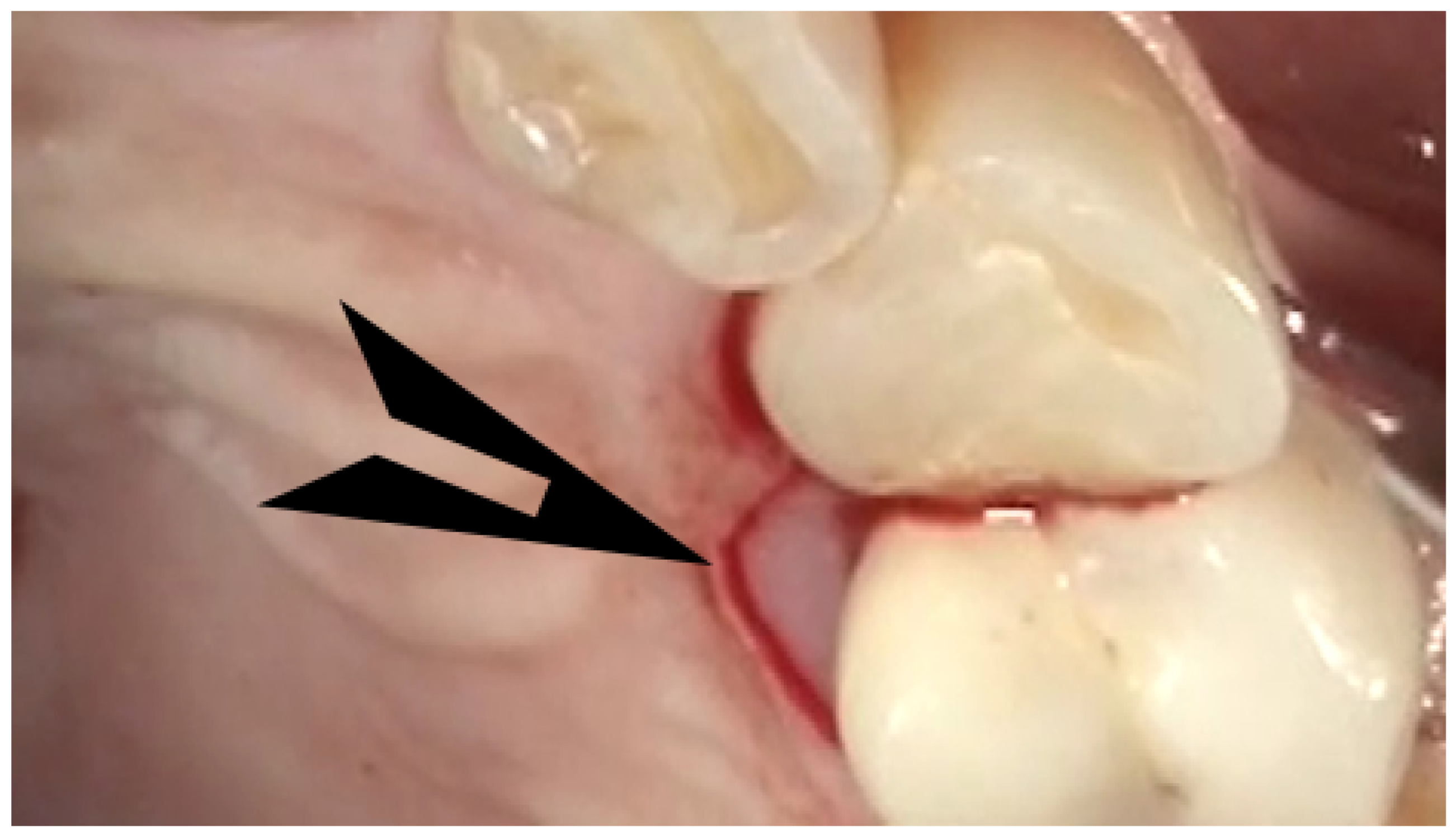
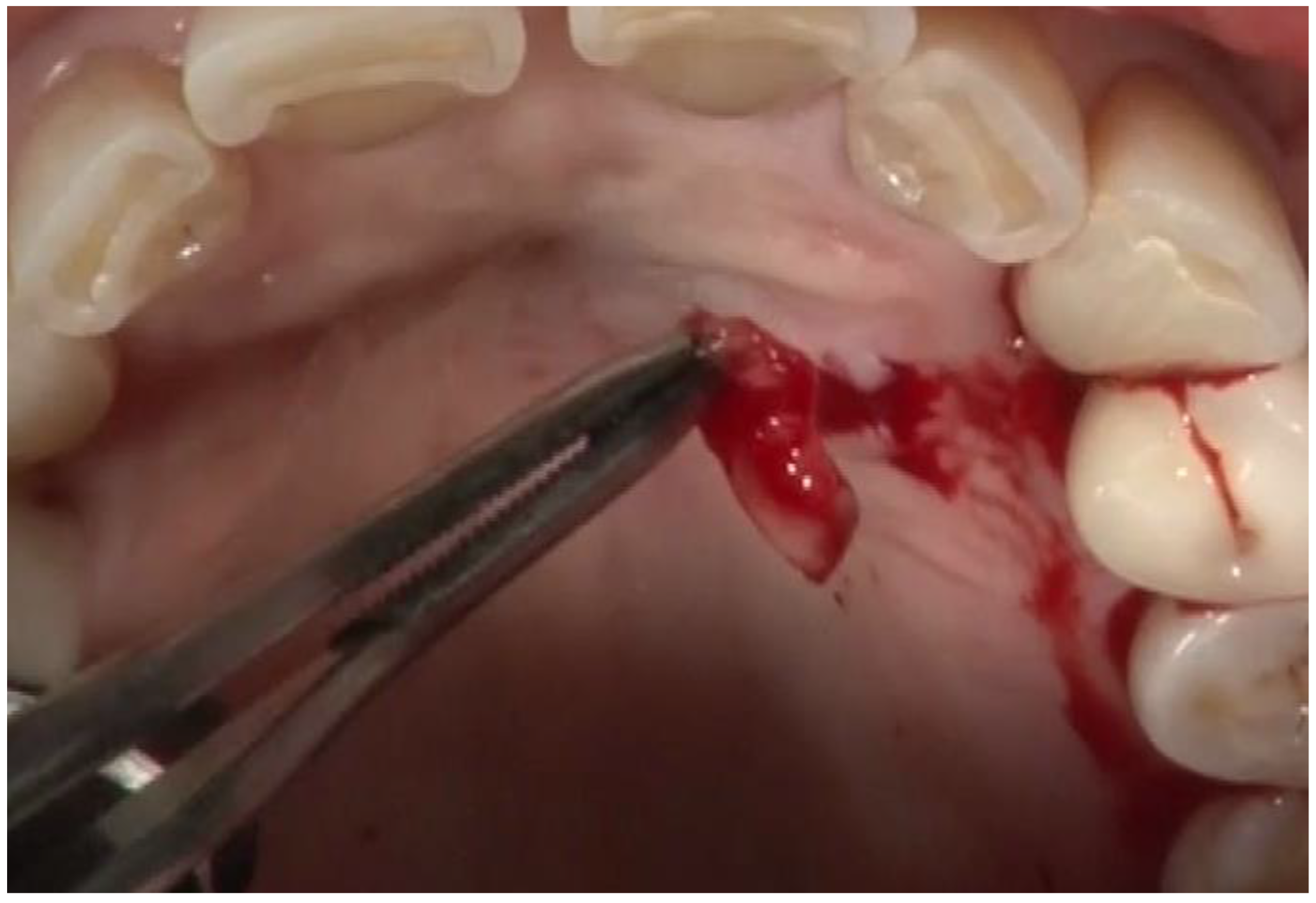
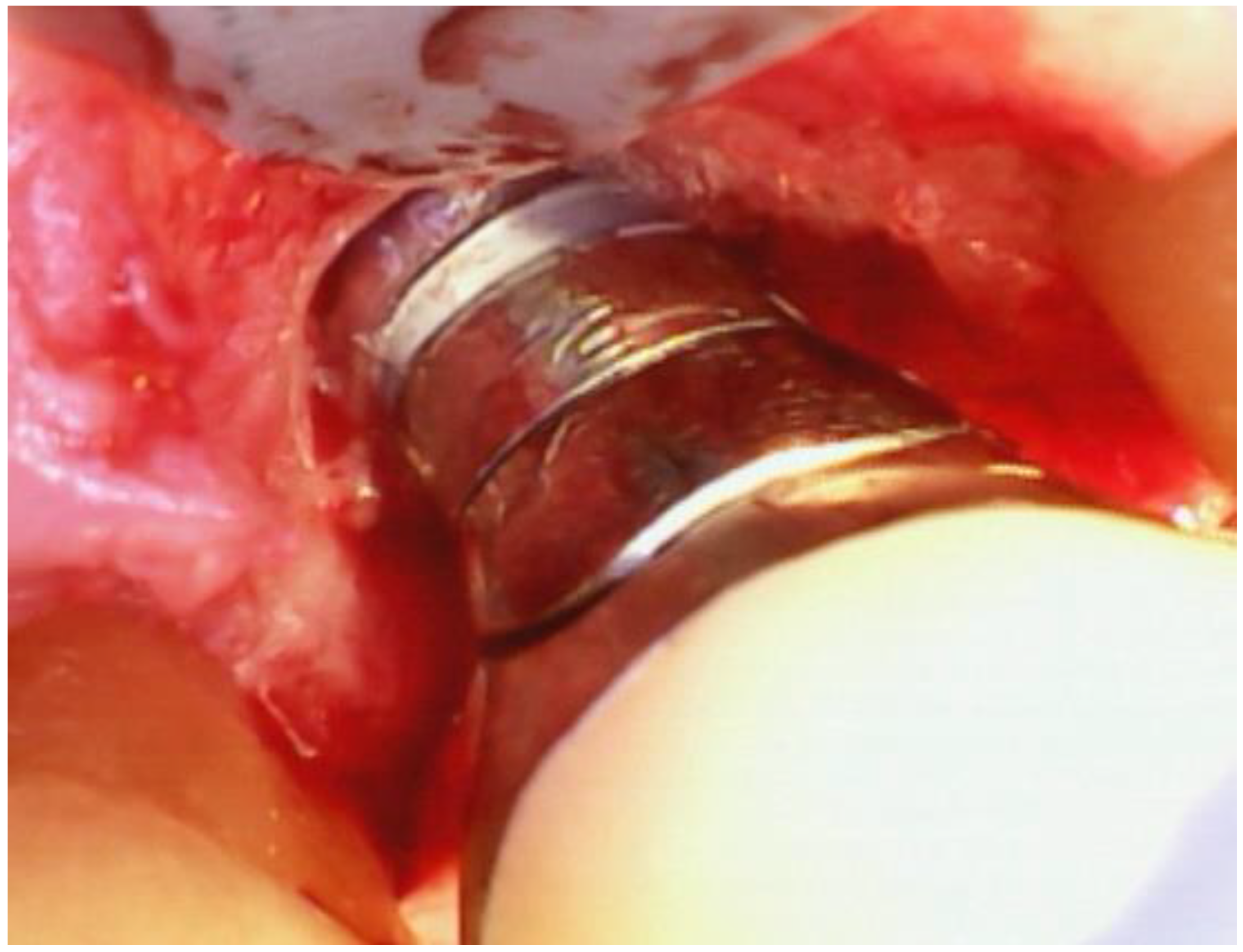

| n | Mean | SD | Median | Min | Max | p | |
|---|---|---|---|---|---|---|---|
| Mean Initial Probing Depth | 25 | 4.56 | 1.01 | 4.6 | 3.1 | 8 | <0.001 |
| Mean Final Probing Depth | 25 | 2.62 | 1.00 | 2.5 | 1.2 | 5.5 | |
| Change in Mean Probing Depth | 25 | 1.94 | 1.18 | 2.1 | -0.5 | 4.4 | |
| Deepest Initial Probing Depth | 25 | 6.50 | 0.94 | 6 | 5 | 8 | <0.001 |
| Deepest Final Probing Depth | 25 | 3.38 | 1.43 | 3 | 1.5 | 8 | |
| Change in Deepest Probing Depth | 25 | 3.12 | 1.45 | 3.5 | −1 | 5 | |
| Initial Bleeding on Probing | 25 | 100.0% | 0.0% | 100.0% | 100.0% | 100.0% | <0.001 |
| Final Bleeding on Probing | 25 | 29.3% | 45.5% | 0.0% | 0.0% | 100.0% | |
| Reduction in Bleeding on Probing | 25 | 73.6% | 43.9% | 100.0% | 0.0% | 100.0% |
| Characteristic | % | Characteristic | % |
|---|---|---|---|
| Immediate Implant? | Crown/Implant Stability? | ||
| Immediate | 93.5% (29/31) | No | 8.8% (3/34) |
| Not an immediate | 6.5% (2/31) | Yes | 91.2% (31/34) |
| Cemented or Screwed Prosthesis? | Initial radiograph? | ||
| Cemented | 88.2% (30/34) | No | 0.0% (0/34) |
| Screwed | 11.8% (4/34) | Yes | 100.0% (34/34) |
| Nightguard? | Final radiograph? | ||
| No Nightguard | 67.6% (23/34) | No | 20.6% (7/34) |
| Nightguard | 32.4% (11/34) | Yes | 79.4% (27/34) |
| Diabetes Mellitus? | Implant Failure? | ||
| No Diabetes Mellitus | 94.1% (32/34) | Survival | 94.1% (32/34) |
| Diabetes Mellitus | 5.9% (2/34) | Failure | 5.9% (2/34) |
| Smoker? | EMD + Bone? | ||
| Non-Smoker | 91.2% (31/34) | EMD Only | 57.6% (19/33) |
| Smoker | 8.7% (3/34) | EMD + Bone | 42.4% (14/33) |
| Implantoplasty? | Implant Site Type | ||
| No | 32.4% (11/34) | Upper Molar | 26.5% (9/34) |
| Yes | 67.6% 23/34) | Upper Premolar | 11.8% (4/34) |
| Osseous Reduction? | Upper Cuspid | 5.9% (2/34) | |
| No | 88.2% (30/34) | Upper Incisor | 11.8% (4/34) |
| Yes | 11.8% (4/34) | Lower Molar | 26.5% (9/34) |
| Minimally Invasive? | Lower Premolar | 11.8% (4/34) | |
| No | 41.2% (14/34) | Lower Cuspid | 2.9% (1/34) |
| Yes | 58.8% (20/34) | Lower Incisor | 2.9% (1/34) |
| Videoscope used? | Implant Length | ||
| No | 58.8% (20/34) | Mean ± SD | 9.97 ± 1.90 |
| Yes | 41.2% (14/34) | Median | 10 |
| Soft tissue Graft? | Range | 6 to 13 | |
| No | 76.5% (26/34) | Implant Diameter | |
| Yes | 23.5% (8/34) | Mean ± SD | 4.62 ± 1.43 |
| Bone Graft? | Median | 4.3 | |
| No | 67.6% (23/34) | Range | 3.3 to 11.5 |
| Yes | 32.4% (11/34) | ||
| Membrane? | |||
| No | 67.6% (23/34) | ||
| Yes | 32.4% (11/34) |
| Variable | Change in Mean PD ± SE | p | Change in Deepest PD ± SE | p | OR for BOP Change (95% CI) | p |
|---|---|---|---|---|---|---|
| Tooth Type | ||||||
| Incisor | 3.03 ± 0.54 | - | 3.40 ± 0.48 | - | NE | - |
| Cuspid | 2.62 ± 0.45 | 0.279 | 4.24 ± 0.26 | 0.007 | ||
| Premolar | 1.96 ± 0.24 | 0.071 | 2.76 ± 0.48 | 0.346 | ||
| Molar | 1.58 ± 0.25 | 0.014 | 3.11 ± 0.38 | 0.640 | ||
| Non-Molar or Molar | ||||||
| Non-Molar | 2.41 ± 0.27 | - | 3.15 ± 0.35 | - | 1.00 | |
| Molar | 1.67 ± 0.26 | 0.022 | 3.17 ± 0.38 | 0.958 | 1.64 (0.24 to 11.4) | 0.616 |
| Arch | ||||||
| Mandibular | 1.98 ± 0.22 | - | 3.24 ± 0.35 | - | 1.00 | |
| Maxillary | 2.05 ± 0.32 | 0.845 | 3.10 ± 0.35 | 0.803 | 0.30 (0.05 to 1.85) | 0.189 |
| Smoking Status | ||||||
| Non-Smoker | 1.99 ± 0.22 | - | 3.18 ± 0.27 | - | 1.00 | |
| Smoker | 2.39 ± 0.90 | 0.672 | 2.92 ± 1.15 | 0.844 | 0.60 (0.02 to 19.0) | 0.798 |
| Diabetes Mellitus | ||||||
| No | 1.99 ± 0.23 | - | 3.15 ± 0.28 | - | NE | - |
| Yes | 2.58 ± 0.06 * | 0.327 | 3.48 ± 0.07* | 0.415 | ||
| Cemented or Screwed | ||||||
| Cemented | 1.97 ± 0.25 | - | 3.02 ± 0.30 | - | NE | - |
| Screwed | 2.36 ± 0.11 | 0.261 | 4.11 ± 0.39 | 0.128 | ||
| Nightguard | ||||||
| No | 1.98 ± 0.28 | - | 3.09 ± 0.38 | - | NE | - |
| Yes | 2.09 ± 0.35 | 0.813 | 3.32 ± 0.30 | 0.658 | ||
| % Bone Loss | ||||||
| <30% | 1.83 ± 0.36 | - | 3.27 ± 0.39 | - | 1.00 | |
| 30% to <50% | 2.22 ± 0.23 | 0.293 | 3.49 ± 0.17 | 0.574 | 0.49 (0.05 to 4.65) | 0.534 |
| 50% and Greater | 1.73 ± 0.63 | 0.902 | 2.29 ± 0.84 | 0.284 | 0.11 (0.01 to 1.05) | 0.056 |
| Implantoplasty | ||||||
| No | 2.23 ± 0.27 | - | 3.67 ± 0.35 | - | 1.00 | |
| Yes | 1.90 ± 0.28 | 0.338 | 2.88 ± 0.33 | 0.137 | 0.77 (0.11 to 5.35) | 0.788 |
| Osseous Reduction | ||||||
| No | 1.99 ± 0.23 | - | 3.08 ± 0.30 | - | 1.00 | |
| Yes | 2.22 ± 0.38 | 0.579 | 3.73 ± 0.24 | 0.151 | 0.89 (0.08 to 10.4) | 0.925 |
| Minimally Invasive | ||||||
| No | 1.84 ± 0.31 | - | 3.35 ± 0.43 | - | 1.00 | |
| Yes | 2.17 ± 0.27 | 0.410 | 3.02 ± 0.33 | 0.534 | 0.52 (0.07 to 3.80) | 0.491 |
| Videoscope | ||||||
| No | 2.03 ± 0.32 | - | 3.08 ± 0.41 | - | 1.00 | |
| Yes | 2.00 ± 0.27 | 0.941 | 3.26 ± 0.35 | 0.746 | 2.17 (0.30 to 15.6) | 0.423 |
| CT Graft | ||||||
| No | 2.09 ± 0.30 | - | 3.00 ± 0.38 | - | 1.00 | |
| Yes | 1.82 ± 0.36 | 0.636 | 3.67 ± 0.34 | 0.278 | 2.11 (0.15 to 29.4) | 0.540 |
| Bone Graft | ||||||
| No | 2.10 ± 0.24 | - | 3.17 ± 0.30 | - | 1.00 | |
| Yes | 1.87 ± 0.40 | 0.605 | 3.15 ± 0.57 | 0.971 | 2.62 (0.25 to 27.5) | 0.366 |
| Membrane | ||||||
| No | 2.16 ± 0.28 | - | 3.27 ± 0.28 | - | 1.00 | |
| Yes | 1.69 ± 0.32 | 0.294 | 2.94 ± 0.60 | 0.654 | 0.93 (0.18 to 4.68) | 0.929 |
| Stability (crown/implant) | ||||||
| Stable | 2.00 ± 0.24 | - | 3.04 ± 0.28 | - | NE | - |
| Unstable | 2.16 ± 0.09 & | 0.583 | 4.46 ± 0.40 & | 0.153 | ||
| EMD (with/without bone) | ||||||
| EMD only | 1.98 ± 0.28 | - | 3.17 ± 0.35 | - | 1.00 | |
| EMD with bone | 2.03 ± 0.33 | 0.904 | 3.15 ± 0.45 | 0.973 | 4.75 (0.49 to 46.5) | 0.119 |
Disclaimer/Publisher’s Note: The statements, opinions and data contained in all publications are solely those of the individual author(s) and contributor(s) and not of MDPI and/or the editor(s). MDPI and/or the editor(s) disclaim responsibility for any injury to people or property resulting from any ideas, methods, instructions or products referred to in the content. |
© 2023 by the authors. Licensee MDPI, Basel, Switzerland. This article is an open access article distributed under the terms and conditions of the Creative Commons Attribution (CC BY) license (https://creativecommons.org/licenses/by/4.0/).
Share and Cite
Wilson, T.G., Jr.; Harrel, S.K.; Nunn, M.E. The Use of Enamel Matrix Derivative during Surgical Therapy for Peri-Implantitis: A Case Series. Dent. J. 2024, 12, 11. https://doi.org/10.3390/dj12010011
Wilson TG Jr., Harrel SK, Nunn ME. The Use of Enamel Matrix Derivative during Surgical Therapy for Peri-Implantitis: A Case Series. Dentistry Journal. 2024; 12(1):11. https://doi.org/10.3390/dj12010011
Chicago/Turabian StyleWilson, Thomas G., Jr., Stephen K. Harrel, and Martha E. Nunn. 2024. "The Use of Enamel Matrix Derivative during Surgical Therapy for Peri-Implantitis: A Case Series" Dentistry Journal 12, no. 1: 11. https://doi.org/10.3390/dj12010011
APA StyleWilson, T. G., Jr., Harrel, S. K., & Nunn, M. E. (2024). The Use of Enamel Matrix Derivative during Surgical Therapy for Peri-Implantitis: A Case Series. Dentistry Journal, 12(1), 11. https://doi.org/10.3390/dj12010011


