Article

Synthesis and In Vitro (Anticancer) Evaluation of η6-Arene Ruthenium Complexes Bearing Stannyl Ligands

1
Maastricht Science Programme, Faculty of Humanities and Science, Maastricht University, Kapoenstraat 2, P.O. Box 616, 6200 MD Maastricht, The Netherlands
2
Department of Chemistry, University of Cape Town, Rondebosch 7701, South Africa
3
Institut für Anorganische Chemie, Technische Universität Graz, Stremayrgasse 9, A-8010 Graz, Austria
4
Institut des Sciences et Ingénierie Chimiques, Ecole Polytechnique Fédérale de Lausanne (EPFL), CH­1015 Lausanne, Switzerland
5
Biosorption and Water Research Laboratory Department of Chemistry, Vaal University of Technology, Private Bag X021, Andries Potgieter Blvd, Vanderbijlpark 1911, South Africa
*
Author to whom correspondence should be addressed.
Inorganics 2017, 5(3), 44; https://doi.org/10.3390/inorganics5030044
Submission received: 13 June 2017 / Revised: 5 July 2017 / Accepted: 11 July 2017 / Published: 13 July 2017
(This article belongs to the Special Issue Tumor Inhibiting Metal Complexes)

Abstract

:
Treatment of the known half-sandwich complexes of the type [(η6-C6H6)RuCl2(P(OR)3)] (R = Me or Ph) with SnCl2 yielded three new half-sandwich ruthenium complexes (C1C3): [(η6-C6H6)RuCl(SnCl3)(P(OMe)3)] (C1), [(η6-C6H6)RuCl(SnCl3)(P(OPh)3)] (C2) and the bis-stannyl complex [(η6-C6H6)Ru(SnCl3)2(P(OMe)3)] (C3) by facile insertion of SnCl2 into the Ru–Cl bonds. Treatment of the known complexes [(η6-C6H6)RuCl(SnCl3)(PPh3)] and [(η6-C6H6)RuCl2(PPh3)] with 4-dimethylaminopyridine (DAMP) and ammonium tetrafluoroborate afforded the complex salts: [(η6-C6H6)Ru(SnCl3)(PPh3)(DAMP)]+BF4 (C4) and [(η6-C6H6)RuCl(PPh3)(DAMP)]+BF4 (C5) respectively. Complexes C1C5 have been fully characterized by spectroscopic means (IR, UV–vis, multinuclear NMR, ESI–MS) and their thermal behaviour elucidated by thermal gravimetric analysis (TGA). Structural characterization by single crystal X-ray crystallography of the novel complex C2 and [(η6-C6H6)RuCl2(P(OPh)3)], the latter having escaped elucidation by this method, is also reported. Finally, the cytotoxicity of the complexes was determined on the A2780 (human ovarian cancer), A2780cisR (human ovarian cis-platin-resistant cancer), and the HEK293 (human embryonic kidney) cell lines and discussed, and an attempt is made to elucidate the effect of the stannyl ligand on cytotoxicity.

Graphical Abstract

1. Introduction

Since the discovery of the anti-cancer properties of cis-platin, [cis-PtCl2(NH3)2] and related complexes [1,2,3,4], research directed towards the development of new metal-containing anticancer drugs has made staggering advances [5,6,7,8,9,10,11,12,13,14,15]. Metals other than platinum are worth investigating in the search for new classes of metallodrugs with high efficacy and fewer side effects. The ongoing search for new metallodrugs has led to the discovery of several ruthenium-based drugs: NAMI-A and KP1019, both of which have completed phase I clinical trials, as well as RAPTA-C (Chart 1) [16,17,18,19,20,21,22,23]. In addition, ruthenium(II)-arene complexes are also considered promising drug candidates, owing to their demonstrated low toxicity and high antitumor activity [18,19,20,21,22,23,24,25,26,27,28,29,30]. The bioavailability of these compounds is controlled by the arene moieties facilitating the outreach in the intracellular region given their hydrophobic nature [29].
A particularly interesting class of compounds in this regard are the easily accessible half-sandwich ruthenium(II) complexes of the type [(η6-C6H6)RuCl2(PR3)] (R = Aryl, O-alky, O-Aryl). Facile reaction of the arene ruthenium dimer [(η6-C6H6)Ru(μ-Cl)Cl]2 with strong σ-donor ligands, such as phosphines or phosphites, promote the cleavage of the Ru(II) dimer yielding half-sandwich Ru(II)-arene phosphine complexes [31,32]. A stable phosphine complex, reported in the 1970s [(η6-C6H6)RuCl2(PPh3)] [31,32], which is obtained in high yields as a product via a reaction of the afore-mentioned ruthenium dimer with triphenylphosphine. Similarly the phosphite derivatives [(η6-C6H6)RuCl2(P(OMe)3)] and [(η6-C6H6)RuCl2(P(OPh)3)] are afforded by reaction of [(η6-C6H6)Ru(μ-Cl)Cl]2 with trimethyl phosphite and triphenyl phosphite in an analogous fashion [32,33,34]. Surprisingly, despite the fact that these easily accessible phosphite complexes have been known since the early 1970s, they have not undergone rigorous in vitro cytotoxic testing with respect to cancer cell lines. This encouraged us to prepare and evaluate their cytotoxic activity. Moreover, to the best of our knowledge, the complex [(η6-C6H6)RuCl2(P(OPh)3)] has also not been structurally characterised by single crystal X-ray diffraction analysis, which prompted us to carry out such an investigation, and this is also reported herein.
The reaction of half-sandwich ruthenium(II) arene complexes [(η6-C6H6)RuCl2(PR3)] (R = aryl or O-Aryl, O-alkyl) with SnCl2 is also known to yield a Ru(II) complex exhibiting a strong covalent Ru-Sn bond via facile insertion of the SnCl2 moiety into the Ru–Cl bond [35,36]. While the reaction of SnX2 (X = halide) with other metals, such as palladium and platinum, has been extensively studied [37,38], the analogous reaction with ruthenium derivatives has received far less attention. The addition of trichlorostannyl ligands to the coordination sphere of the ruthenium centre is known to enhance the anticancer properties of the complexes from earlier investigations [39], possibly due to the enhanced σ-donor properties of the ligand, which might facilitate and promote the binding of the agent to potential biomolecular targets. Although there is known to be an increase in cytotoxicity, only a few examples of this class, i.e., those bearing stannyl groups, have been tested.
In this work we report the synthesis and characterisation of a series of complexes of formula [(η6-C6H6)RuX(SnCl3)(P(OR)3)] (X = Cl, SnCl3 and R = Me, Ph), and some cationic derivatives [(η6-C6H6)RuX(PPh3)(DAMP)]BF4 (X = SnCl3, Cl), with a view of attempting to delineate the effect of a trichlorostannyl group on cytotoxicity against several cancer cell-lines. Hence, the cytotoxicity of these new complexes against A2780 and A2780cisR (cis-platin resistant) human ovarian carcinoma cells and non-cancerous HEK293 embryonic kidney cells are reported, along with the known complexes [(η6-C6H6)RuCl2(PPh3)], [(η6-C6H6)RuCl2(P(OPh)3)] and [(η6-C6H6)RuCl(SnCl3)(PPh3)], was determined.

2. Results and Discussion

2.1. Synthesis of the Complexes

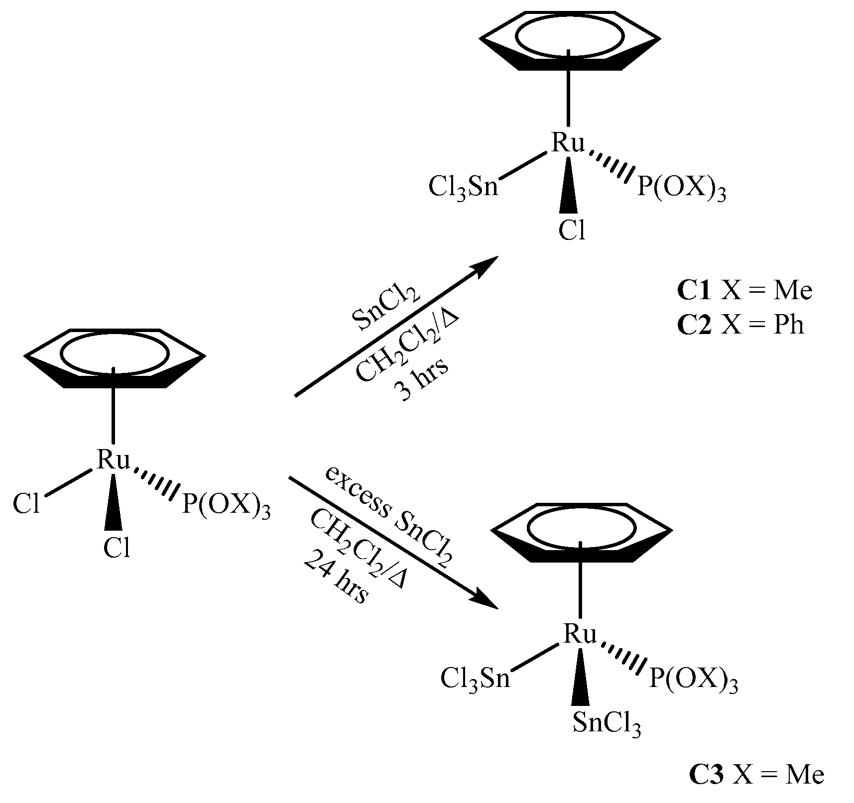
The reaction of the known complexes [(η6-C6H6)RuCl2(P(OX)3)] (X = Me, Ph) with 1.1 equivalents of anhydrous SnCl2 in dichloromethane under reflux affords the complexes [(η6-C6H6)RuCl(SnCl3)(P(OX)3)] (X = Me C1; X = Ph, C2), which were isolated in 64% and 69% yield (Scheme 1), respectively. The reaction of [(η6-C6H6)Ru(SnCl3)Cl(P(OMe)3)] with a large excess of SnCl2 in refluxing dichloromethane for 24 h affords the bis-(trichlorostannyl) complex C3 in 34% yield. The latter complex can also be prepared directly starting from [(η6-C6H6)RuCl2(P(OMe)3)] with a 20-fold molar excess of SnCl2 in dichloromethane affording similar yields. Complex C3, owing to the presence of an additional SnCl3 moiety is less solubility in dichlormethane or chloroform than C1 and C2, which are highly soluble in these solvents.
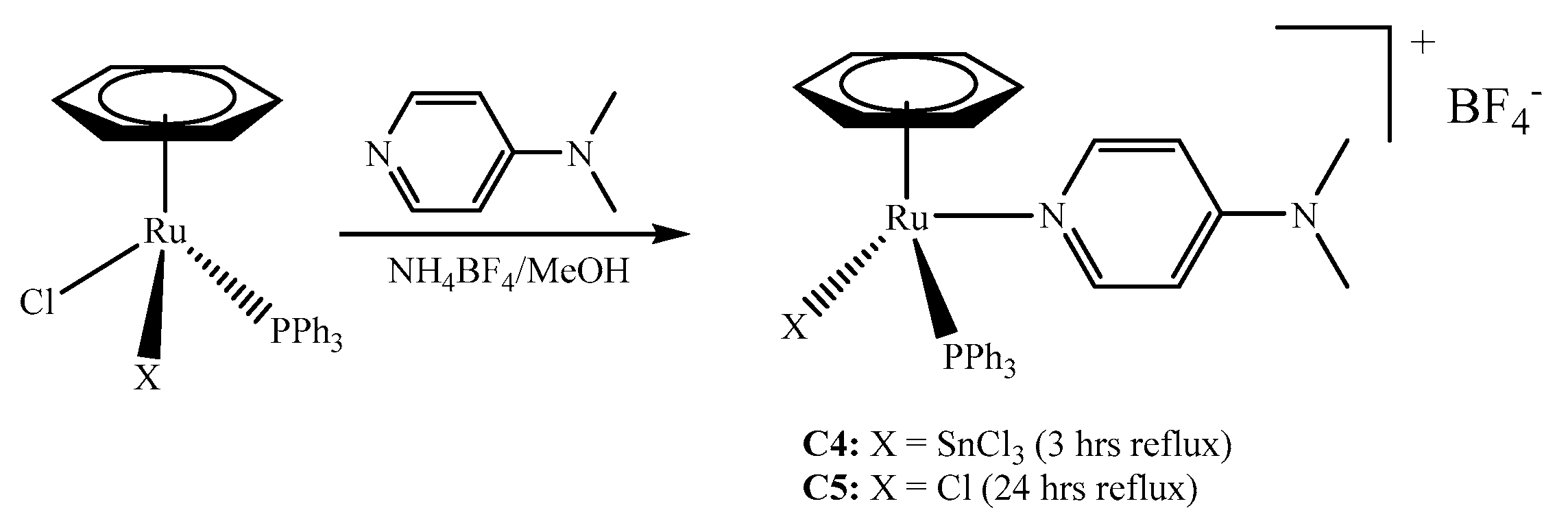
Reaction of [(η6-C6H6)RuClX(PPh3)] (X = Cl, X= SnCl3) with 1.1 equivalents of 4-dimethylaminopyridine (DMAP) and 1.1 equivalent of ammonium tetrafluoroborate in refluxing methanol affords the complex ionic salts C4 and C5 (Scheme 2), both of which are fully characterised by spectroscopic and analytical methods. All complexes C1C5 exhibit reasonable thermal stability as evidenced by decomposition temperatures in excess of 100 °C.

2.2. Spectroscopic Characterisation

Complexes C1C3 all exhibit an upfield shifted resonance signal, for the arene protons associated with the η6-coordinated ring, in the 1H NMR spectra: δ = 6.31 ppm (C1 and C3), 5.82 (C2). In complexes C1 and C3, a doublet is observed in the 1H NMR spectrum corresponding to the P(OMe)3 groups due to coupling to the phosphorus atom: 3J(H,P): C1: 12.0 Hz, C3: 12.3 Hz.
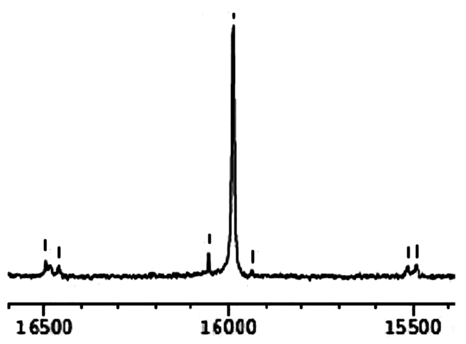
Complexes C1C3 exhibit singlet resonance signals in their 31P{1H} NMR spectra: (C1: δ = 131.2, C2: δ = 122.1, C3: δ = 136.5 ppm). Notaby, the presence of both 119Sn and 117Sn satellites, flanking the main resonance signals in all three complexes (C1C3) are visible in these spectra due to 2J(Sn,P) coupling. The presence of the Sn satellites in the 31P{1H} NMR spectra suggest, that in DMSO(dimethyl sulfoxide), the complexes are stable and dynamic SnCl3 exchange is unlikely to occur. The formation, in DMSO solutions, of [(η6-C6H6)Ru(SnCl3)(DMSO)(PR3)]+Cl can be ruled out for the mono-insertion products C1 and C2 over the time periods of the NMR measurements in DMSO-d6 (12 h). The cationic complexes C4 and C5 exibit dramatically shielded chemical shift positions in their respective 31P{1H} NMR spectra (C4: δ = 26.7, C5: δ = 36.0 ppm) compared with the neutral complexes C1C3, owing to their cationic nature. Unfortunately 119Sn NMR spectroscopy could not be carried out on the tin compounds due to the lack of a suitable probe in our laboratories.
The 31P{1H} NMR spectrum of complex C1 is shown in Figure 1. Both 119Sn and 117Sn satellites are visible, along with rotational side-bands on the main signal, the latter of which is typical in solution 31P NMR spectra.
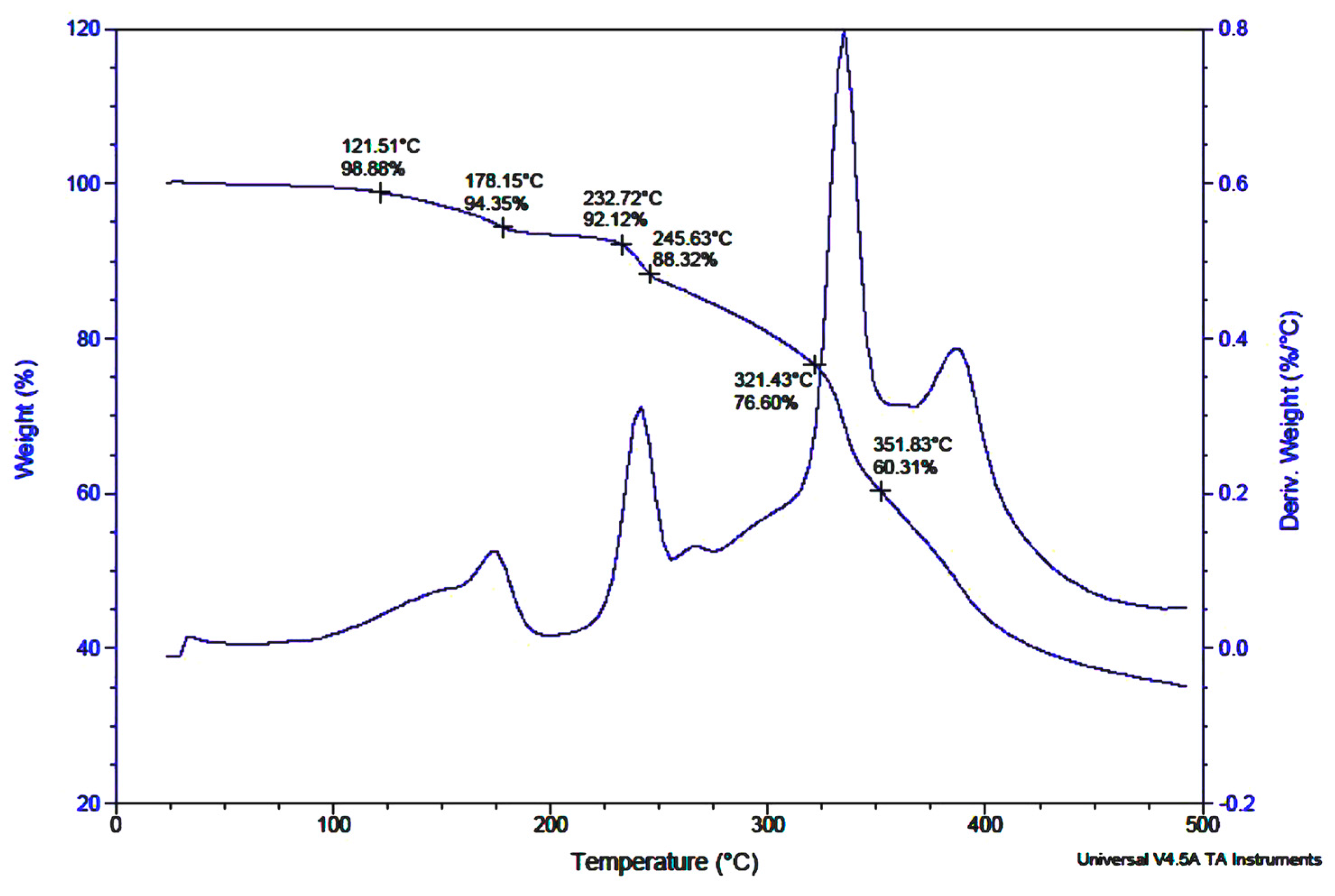
Inspection of the experimental solution UV–vis spectra of the complexes C1C3 reveal that, for the bis-trichlorostannyl complex C3, a much higher wavelength of absorption (λ = 459 nm) is observed compared to C1: λ = 348 and C2: λ = 351 nm, indicating pertubation in the electronic situation upon bis SnCl2-insertion. This is most likely due to the enhanced σ-donor capacity of SnCl3 vs. Cl. For the ionic complex the UV–vis spectra reveal absorptions at λ = 364 (C4) and λ = 335 nm (C5), comparable to that of C1 and C2. All complexes were also subjected to a TGA analysis to obtain information on their thermal behaviour and stability. In all cases the complexes are thermally robust with the first onset of mass loss occurring well in excess of 100 °C: (C1: 122 °C, C2: 186 °C, C3: 223 °C, C4: 184 °C, and C5: 190 °C), which is in accord with the melting point (decomposition temperature) determinations. An exact assignment of the mode of decomposition, i.e., according to which fragments are lost at which temperature was undertaken, and in all cases one decomposition step can be tentatively traced to the loss of the η6 coordinated ring. Figure 2 shows the TGA trace of complex C1. The approximately 12% mass loss can be roughly correlated to the loss of the arene ring.

2.3. X-ray Crystallography

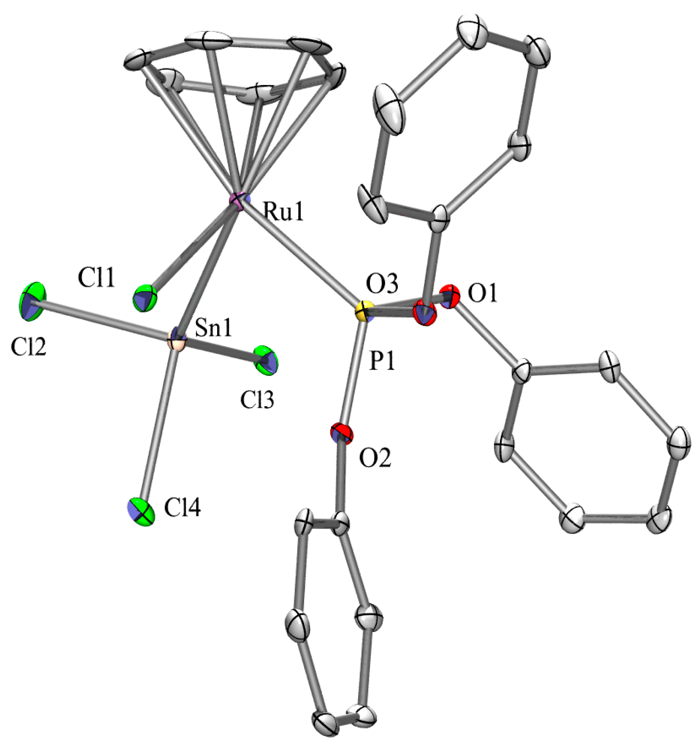
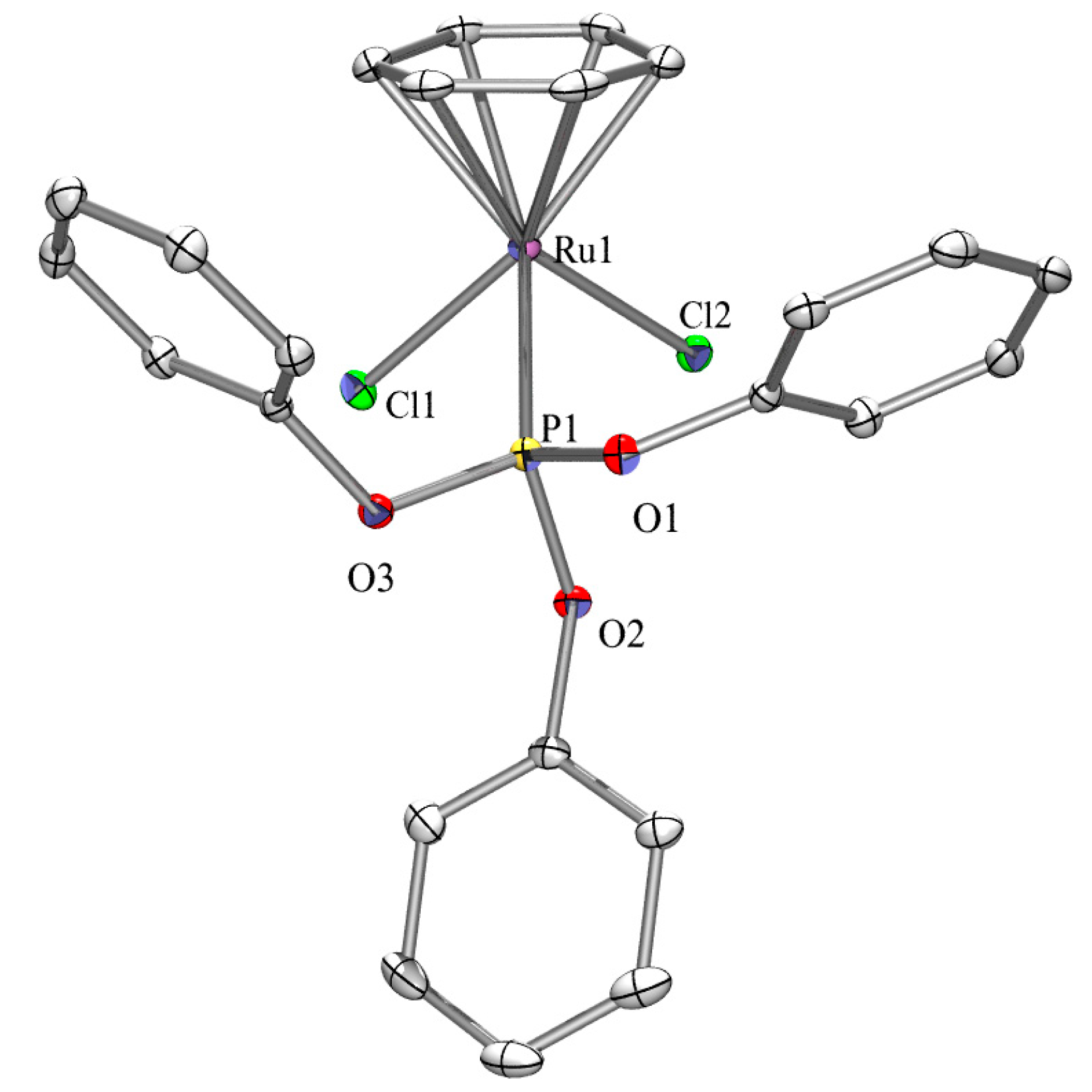
Single crystals of complex C2 and [(η6-C6H6)RuCl2(P(OPh)3)] were obtained and single crystal X-ray diffraction studies were undertaken and their structures are shown in Figure 3 and Figure 4, respectively with selected metric parameters provide in the figure captions (other bond angles and lengths are available in the supporting information). It is somewhat surprising that the complex [(η6-C6H6)RuCl2(P(OPh)3)] has eluded structural characterisation by X-ray diffraction, despite being reported in the 1970s.
Both complexes exhibit the typical piano-stool geometry with the metal centre being coordinated by the arene in η6 fashion. Complex C2 exhibits a Ru–Sn bond length of 2.5686(5) Å, which is comparable to known similar complexes featuring Ru–Sn single bonds, for example: [(η6-C6H6)RuCl(SnCl3)(PPh)3]: 2.5977(14) Å and [(η6-p-cymene)RuCl(SnCl3)(PPh)3]: 2.5830(9) Å (see the X-ray structures in Ref. [35]) respectively. Previous structural investigations into complexes of the type [(η6-C6H6)RuCl2(PR3)] (R = alkyl, aryl) are ubiquitous, but only three examples of previously structurally-characterised complexes of the type [(η6-C6H6)RuCl2(P(OR)3)] (i.e., arene phosphite complexes) exist [40,41,42] making the structural elucidation of both C2 and [(η6-C6H6)RuCl2P(OPh)3] of some interest.

2.4. Cytotoxicity Studies

The antiproliferative activity of the neutral complexes C1C3, cationic complexes C4 and C5 and the three known compounds [(η6-C6H6)RuCl2(PPh3)], [(η6-C6H6)RuCl2(P(OPh)3)], and [(η6-C6H6)RuCl(SnCl3)(PPh3)] were investigated in vitro against human ovarian cancer cells A2780 and the A2780cisR variant with aquired cis-platin resistance, as well as against non-cancerous human embryonic kidney (HEK293) cells (Table 1). The cytotoxicity of the latter three complexes has not been reported previously and are shown together with cis-platin for comparison (Table 2). IC50 values of the compounds were determined after exposure of the cells to the compounds for 72 h using the MTT assay.
Complexes C1 and C3 with trimethylphosphite ligands did not induce cytotoxicity even at concentrations as high as 500 µM and 200 µM, respectively, whereas all complexes with triphenylphosphite or triphenylphosphine ligands exhibit considerable cytotoxicity in A2780, A2780cisR and HEK293 cells. This is somewhat surprising as the presence of the SnCl3 moiety would have been expected to enhance the cytotoxic effect of the complex (see above). In case of complex C3, this may be due to its rather low solubility due to the presence of two trichlorostannyl groups attached to the Ru centre. Notably, the cationic complexes C4 and C5 display IC50 values in the low micromolar concentration range and, compared to cis-platin, showed even high efficacy in A2780cisR cells. Whereas complex C5 bearing a chloride ligand showed similar activity in all three cell lines, complex C4 with the chlorine replaced by the SnCl3 moiety, showed slight cancer cell selectivity. This phenomenon was not observed for [(η6-C6H6)RuCl2(PPh3)] and [(η6-C6H6)RuCl(SnCl3)(PPh3)], where the tin congener induced generally a two-fold higher cytotoxicity, but did not contribute to cancer cell selectivity. In contrast, the complexes with triphenylphosphite ligands [(η6-C6H6)RuCl2(P(OPh)3)] and its tin congener C2 show the opposite behaviour with C2 being >20-fold less potent than [(η6-C6H6)RuCl2(P(OPh)3)].
Overall looking at these results in totality and attempting to delineate the effect of the trichlorostannyl group on cytotoxicity is not straightforward as obviously solubility plays a key role which might offset the otherwise enhanced cytotoxic activity. Comparison of ionic complexes C4 and C5 which have similar solubilities clearly do demonstrate, however, on average, an increase in cytotxicity in the presence of SnCl3 vs. Cl (except for HEK293). This does suggest that the SnCl3 ligand is useful in this regard, but complex C3, for example, bearing two SnCl3 ligands exhibits very low activity which is driven by its insolubility, thereby potentially offsetting any enhanced efficacy in its cytotoxic effects. We are currently preparing more related complexes to attempt to delineate these effects more closely.

3. Experimental

3.1. General Procedures

All manipulations were performed in air as the Ru(II) complexes are stable towards air and moisture. All starting materials and solvents were obtained commercially (Strem, Sigma-Aldrich, Zwijndrecht, Netherlands) and used as received. [(η6-C6H6)RuCl2(PPh3)], [(η6-C6H6)RuCl2(P(OPh)3)], and [(η6-C6H6)RuCl(SnCl3)(PPh3)] were prepared according to published procedures [31,32,33,34,35,36], and the complexes obtained were characterised by 1H and 31P NMR spectroscopy and checked against literature data. NMR spectra were recorded on a Bruker Ultrashield 300 (Karlsruhe, Germany), IR spectra on a Shimadzu MIRacle IR (ATR, Kyoto, Japan), UV–vis on a Shimadzu UV 3600 (Kyoto, Japan), and TGA spectra were recorded on a TGA Q-500 (Maastricht, Netherlands) at the University of Maastricht Brightlands Campus, Netherlands. Electrospray (ESI) mass spectrometry experiments were conducted on BRUKER—Ion Trap MS (Karlsruhe, Germany) in positive mode (+) at the University of Neuchâtel, Switzerland. The following abbreviations apply to the intensity of peaks found within the spectra (IR): v: very strong; s: strong; m: medium; and w: weak. For NMR peaks obtained for the non-deuterated residue in the deuterated solvent were used as the internal reference points for the spectra (reference peak: DMSO-d6, 1H 2.49 ppm; 13C 39.5 ppm, CHCl3-d1, 1H 7.26 ppm; and 13C 77.2 ppm). All signals have been recorded using their appropriate chemical shift (δ in ppm), multiplicity, integral ratio, and coupling constants [Hz]. The following abbreviations apply to the signal multiplicity of peaks within spectra: s = singlet, d = doublet, t = triplet, and m = multiplet.

3.2. Synthesis of the Complexes

3.2.1. Synthesis of [(η6-C6H6)RuCl(SnCl3)(P(OMe)3)] (C1)

[(η6-C6H6)RuCl2(P(OMe)3)] (0.500 g, 1.340 mmol) and 1.1 equivalents of anhydrous SnCl2 (0.279 g, 1.474 mmol) were dissolved in 30 mL of dichloromethane and heated under reflux for 3 h. The reaction mixture was cooled to room temperature and filtered to remove excess SnCl2.The bright orange solution was evaporated to dryness in vacuo and afforded a scarlet powder which was subsequently washed with n-hexane (3 × 10 mL) and dried under reduced pressure. Yield 64%. m.p.: 103 °C dec. FTIR: v (cm−1): 3075 (w), 2959 (w), 2843 (vw), 1458 (w), 1439 (m), 1260 (m), 1177 (w), 1153 (vw), 1063 (m), 1005 (vs), 922 (w), 866 (w), 791 (vs), 752 (s), 706 (m), 662 (m), 608 (w), 542 (w). 1H NMR: (300.1 MHz, DMSO-d6, δ, ppm): 6.31 (s, 6H, C6H6), 3.82 (d, 3J(H,P) = 12.0 Hz, 9H, P(OMe)3). 13C NMR: (75.5 MHz, DMSO-d6, δ, ppm): 92.4 (d, 2J(C,P) = 3.9 Hz, C6H6), 54.7 (d, 2J(C,P) = 6.2 Hz, P(OMe)3). 31P NMR: (121.5 MHz, DMSO-d6, δ, ppm): 131.2 (s, 2J(119Sn,P) = 1004.90 Hz; 2J(117Sn,P) = 949.80 Hz). TGA: (Weight % decrease): 121.51–178.15 °C (4.53%), 178.15–232.72 °C (2.23%), 232.72–245.63 °C (3.80%), 245.63–321.43 °C (11.72%) 321.43–351.83 °C (15.77%). UV–vis (nm)/dichloromethane: 347.5, 451.0. EI-MS (CH3CN): m/z 339.0 [M − SnCl3]+, 353.4, 381.4, 397.3, 414.9, 426.0, 463.0, 481.0, 522.0, 537.0, 554.6, 582.7 [M + Na]+ (other higher mass unassignable fragments present).

3.2.2. Synthesis of [(η6-C6H6)RuCl(SnCl3)(P(OPh)3)] (C2)

Complex (C2) was synthesized in an analogous fashion as for (C1) starting from [(η6-C6H6)RuCl2(P(OPh)3)]: (0.500 g, 0.892 mmol) and SnCl2 (0.186 g, 0.981 mmol). Bright-orange crystals. Yield 69%. m.p.: 197 °C dec. FTIR: v (cm−1): 3075 (vw), 296 3(vw), 1583 (m), 1481 (s), 1437 (w), 1260 (w), 1206 (m), 1182 (s), 1173 (s), 1152 (s), 1022 (m), 945 (s), 922 (s), 908 (s), 891 (s), 822 (vs), 800 (m), 766 (vs), 688 (s), 601 (m). 1H NMR: (300.1 MHz, DMSO-d6, δ, ppm): 7.50 (d, 3J(H,H) = 7.4 Hz, 6H, P(OPh3)), 7.35 (d, 3J(H,H) = 7.4 Hz, 3H, P(OPh)3), 7.34 (br s, 6H, P(OPh)3), 5.84 (br s, 6H, C6H6), 13C NMR: (75.5 MHz, DMSO-d6, δ, ppm): 151.0 (d, 1J(C,P)= 10.8 Hz, C1, P(OPh)3), 130.5 (s, C2,6, P(OPh)3), 126.4 (s, C4, P(OPh)3), 121.9 (d, xJ(C,P) = 4.2 Hz, C3,5, P(OPh)3), 92.9 (d, 2J(C,P) = 3.8 Hz, C6H6), 31P NMR: (300.1 MHz, DMSO-d6, δ, ppm): 122.1 (s, 3J(119Sn,P) = 989.0 Hz, 2J(117Sn,P) = 946.6 Hz). TGA: (Weight % decrease): 185.99–202.83 °C (4.52%), 202.83–347.70 °C (18.50%), 347.70–393.60 °C (30.06%). UV–vis (nm)/dichloromethane: 351.1, 453.0. ESI–MS (CH3CN/MeOH): m/z 123.2, 353.4, 381.4, 449.3, 516.5, 599.1, 683.4, 711.0, 739.0, 767.7 [M + Na]+ (other higher mass fragments present).

3.2.3. Synthesis of [(η6-C6H6)Ru(SnCl3)2(P(OMe)3)] (C3)

The complex C1 was weighed into a flask (0.900 g, 1.200 mmol) along with a twenty-fold molar excess of SnCl2 (4.560 g, 24.000 mmol) in dichloromethane (100 mL) and refluxed for 20 h. During this time the reaction turned a lemon-yellow colour. The reaction solution was filtered to remove unreacted SnCl2 and the solvent of the filtrate removed in vacuo affording a pineapple-yellow waxy solid, which was washed with Et2O (3 × 10 mL) and the washings discarded. The compound can also be prepared directly from [η6-(C6H6)RuCl2(P(OMe)3)] with addition of a large excess of SnCl2 (20 molar equiv.) in dichloromethane with reflux for 24 h and isolation as describe above. Yield 34%. m.p.: 146 °C dec. Conductivity (DMSO): (μS·cm−1, 21 °C, 0.5 mg·mL1): 0.2. FTIR: v (cm−1): 3078 (vw), 2961 (vw), 1441 (w), 1260 (m), 1173 (w), 1088 (m), 1009 (vs), 922 (w), 864 (w), 787 (vs), 733 (s), 704 (m), 662 (w), 648 (w), 608 (vw), 561 (w). 1H NMR: (300.1 MHz, DMSO-d6, δ, ppm): 6.31 (br s, 6H, C6H6), 3.72 (d, 3J(H,P) = 12.3 Hz, 9H, P(OMe)3), 13C NMR: (75.5 MHz, DMSO-d6, δ, ppm): 94.0 (d, 2J(C,P) = 4.1 Hz, C6H6), 54.5 (d, 2J(C,P) = 6.6 Hz, P(OMe)3), 31P NMR: (121.5 MHz, DMSO-d6, δ, ppm): 136.5 (s, 2J(119Sn,P) = 741.7 Hz; 2J(117Sn,P) = 722.1 Hz). TGA: (Weight % decrease): 223.14–273.12 °C (12.70%), 273.12–317.27 °C (2.02%), 317.27–336.42 °C (11.56%), 336.42–394.73 °C (31.77%). UV–vis: (nm)/dichloromethane: 459. ESI–MS (CH3CN/MeOH): m/z 477.3, 541.2, 610.2, 615.2, 631.1, 684.2, 689.1, 698.6, 705.1, 718.5, 747.0, 758.2, 779.1 [M + Na]+ (other higher mass unassignable fragments also present).

3.2.4. Synthesis of [(η6-C6H6)Ru(SnCl3)(PPh3)(DMAP)]+BF4 (C4)

[(η6-C6H6)RuCl(SnCl3)(PPh3)] (0.30 g, 0.60 mmol) in 25 mL of methanol was stirred for ca. 5 min. 1.1 molar equivalents of 4-dimethylaminopyridine (DMAP) (0.060 g, 0.66 mmol) was added to the mixture and stirred for 30 min and 1.1 equivalent of ammonium tetrafluoroborate (0.05 g, 0.66 mmol) was added to the solution and the mixture heated at reflux overnight. The distinct orange solution was evaporated to dryness and afforded a dark red powder, which was subsequently washed with n-hexane and dried under reduced pressure. Yield 96%. m.p.: 119 °C dec. FTIR: v (cm−1): 3479 (w), 3317 (w), 3055 (w), 2960 (w), 2823 (w), 1647 (m), 1616 (w), 1560 (m), 1480 (w), 1433 (m), 1419 (m), 1404 (m), 1250 (m), 1217 (m), 1186 (w), 1087 (s), 1060 (s), 1026 (vs), 997 (m), 941 (w), 819 (m), 798 (s), 748 (s), 723 (m), 696 (s), 659 (m). 1H NMR: (300.1 MHz, CDCl3, δ, ppm): 8.02 (br s, 2H, C5H4N(N(CH3)2)) 7.77-7.38 (m, 15H, PPh3), 6.76 (br s, 2H, C5H4N(N(CH3)2), 5.40 (s, 6H, C6H6), 3.25 (s, 6H, N(CH3)2). 13C NMR: (75.5 MHz, CDCl3, δ, ppm): (low field signals expected for DMAP not visible, nor C1 of PPh3), 134.2 (d, xJ(C,P) = 10.7 Hz, C2,6, PPh3) 131.0 (br s, C4, PPh3), 128.2 (d, xJ(C,P) = 9.6 Hz, C3,5, PPh3), 106.9 (s, C5H4N(N(CH3)2), 89.2 (d, 2J(C,P) = 3.6 Hz, C6H6), 40.3 (s, N(CH3)2). 31P NMR: (121.5 MHz, CDCl3, δ, ppm): 26.7 (s). TGA: (Weight % decrease): 183.46–242.84 °C (5.7%), 242.84–277.79 °C (4.77%), 277.79–337.59 °C (26.75%), 337.59–404.13 °C (18.85%). UV–vis: (nm)/dichloromethane: 364.5. ESI–MS (CH3CN): m/z 123.2 [DMAP + H]+, 401.1, 479.0, 599.1, 635.0, 738.0, 785.0 (other unassignable higher mass fragments also present).

3.2.5. Synthesis of [(η6-C6H6)RuCl(PPh3)(DMAP)]+BF4 (C5)

Complex (C5) was synthesized in an analogous fashion as (C4), except [(η6-C6H6)RuCl2(PPh3)] was used as starting material. The work up and isolation procedure is analogous. Yield 24%. m.p.: 176 °C dec. FTIR: v (cm−1): 2898 (w), 2983 (w), 1622 (m), 1614 (m), 1588 (m), 1537 (m), 1481 (m), 1435 (m), 1406 (m), 1386 (m), 1230 (m), 1089 (s), 1055 (vs), 1018 (vs), 999 (s), 808 (m), 748 (s), 694 (vs). 1H NMR: (300.1 MHz, CDCl3, δ, ppm): 8.21 (d, 3J(H,H) = 6.1 Hz, C5H4N(N(CH3)2)), 7.38–7.73 (m, 15H, PPh3), 6.26 (d, 3J(H,H) = 6.21 Hz, C5H4N(N(CH3)2)), 5.76 (s, 6H, C6H6), 2.94 (s, 6H, N(CH3)2). 13C NMR: (75.5 MHz, CDCl3, δ, ppm): (low field signals expected for DMAP not visible), 154.5 (s, C1, PPh3), 133.8 (d, xJ(C,P) = 10.5, C2,6, PPh3), 131.1 (br s, C4, PPh3), 128.8 (d, xJ(C,P) = 9.6, C3,5, PPh3), 108.3 (s, C5H4N(N(CH3)2), 90.5 (d, 2J(C,P) = 3.0 Hz, C6H6), 39.1 (s, C5H4N(N(CH3)2)). 31P NMR: (121.5 MHz, CDCl3, δ, ppm): 36.0. TGA: (Weight % decrease): 190.65–214.81 °C (12.58%), 214.81–263.54 °C(11.91%), 263.54–273.12 °C (3.86%), 273.12–398.48 °C (16.12%). UV–vis: (nm)/dichloromethane: 335.0. ESI–MS (CH3CN): m/z 477.0, 553.0, 599.0 [M − BF4]+ (no other fragments visible).

3.3. Crystallographic Structure Determination

Crystals of X-ray diffraction quality were obtained by slow evaporation of a dichloromethane-diethyl ether 1:1 mixture of (C2) and [(η6-C6H6)RuCl2(P(OPh)3)] at room temperature using a vial with a narrow opening. For X-ray structure analyses the crystals are mounted onto the tip of glass fibers, and data collection was performed with a BRUKER-AXS SMART APEX CCD diffractometer using graphite-monochromated Mo Kα radiation (0.71073 Å) (Table 2). The data were reduced to Fo2 and corrected for absorption effects with SAINT [43] and SADABS [44,45], respectively. The structures were solved by direct methods and refined by full-matrix least-squares method (SHELXL97) [46]. If not noted otherwise all non-hydrogen atoms were refined with anisotropic displacement parameters. All hydrogen atoms were located in calculated positions to correspond to standard bond lengths and angles. All diagrams are drawn with 30% probability thermal ellipsoids and all hydrogen atoms were omitted for clarity. Figures of solid state molecular structures were generated using Ortep-3 as implemented in WINGX [47] and rendered using POV-ray 3.6 [48].

3.4. Cell Cultures and Cytotoxicity Measurements

Human A2780 and A2780cisR ovarian carcinoma cells were obtained from the European Collection of Authenticated Cell Cultures (ECACC, Salisbury, UK) and non-cancerous HEK293 cells were obtained from ATCC (Sigma, St. Gallen, Switzerland). A2780 were routinely grown in RPMI (Roswell Park Memorial Institute) medium: 1640 GlutaMAX (Lifetechnologies, Zug, Switzerland), while HEK293 were maintained in DMEM medium (Dulbecco's modified media), both containing 10% heat-inactivated fetal bovine serum (FBS, Pan Biotech, Aidenbach, Germany) and 1% antibiotics (penicillin/streptomycin), at a humidified atmosphere with 5% CO2 at 37 °C. Cytotoxicity was determined using the MTT assay (MTT = 3-(4,5-dimethyl-2-thiazolyl)-2,5-diphenyl-2H-tetrazolium bromide). Cells were seeded in 96-well plates as monolayers with 100 μL of cell solution per well and were pre-incubated for 24 h in the cell culture medium. Compounds were prepared as DMSO stock solutions that were dissolved in the culture medium and two-fold serially diluted to the appropriate concentration to give a final DMSO concentration of maximum 0.5%. 100 μL of the compound solution were added to each well and the plates were incubated for 72 h. Subsequently, MTT (5 mg/mL solution, 20 μL per well) was added to the cells and the plates were incubated for another 4 h. The culture medium was aspirated, and the purple formazan crystals formed by the mitochondrial dehydrogenase activity of vital cells were dissolved in DMSO (100 μL). The optical density, directly proportional to the number of surviving cells, was quantified at 590 nm using a multiwell plate reader (Molecular Devices). The fraction of surviving cells was calculated from the absorbance of untreated control cells. The IC50 values for the inhibition of cell growth were determined by fitting the plot of the logarithmic percentage of surviving cells against the logarithm of the drug concentration using a linear regression function. Evaluation is based on means (±SD) from at least two independent experiments, each comprising four tests per concentration level.

4. Conclusions

A series of novel, neutral, and cationic η6-arene ruthenium(II) complexes, some bearing one or two trichlorostannyl groups, have been synthesized, characterized, and tested in vitro for antiproliferative activity against human ovarian cancer cells and a non-tumorigenic cell line. Complexes C1 and C3 exhibit rather poor cyctotoxic activity, whilst complex C2 exhibits moderate activity. The lack of potency of complexes C1 and C3 may be linked to solubility in aqueous media, despite the presence of stannyl ligands expected to enhance the cytotoxicity. The ionic complexes C4 and C5 are cytotoxic, with an activity similar to cis-platin, with C4 even showing a degree of cancer cell selectivity. We are currently attempting to further delineate the effect of the SnCl3 moiety on related complexes, taking solubility into consideration, and will report these endeavours in due course.

Supplementary Materials

The following are available online at www.mdpi.com/2304-6740/5/3/44/s1, A PDF document with the details of the X-ray crystallographic analysis is available. Crystallographic data (excluding structure factors) for the structures of compounds (C2) and [(η6-C6H6)RuCl2(P(OPh)3)] reported in this paper are deposited with the Cambridge Crystallographic Data Center as supplementary publication no. CCDC-1555463 (C2) and 1555464 ([(η6-C6H6)RuCl2(P(OPh)3)]). Copies of data can be obtained free of charge at: http://www.ccdc.cam.ac.uk/products/csd/request/.

Acknowledgments

Burgert Blom thanks the Maastricht Science Programme and Maastricht University for support in carrying out this research. Bruno Therrien (Université de Neuchâtel) is gratefully acknowledged for submitting samples for mass spectrometry on the complexes and for very useful discussions and suggestions during the preparation of the manuscript.

Author Contributions

Olivier Renier, Connor Deacon-Price, Joannes E. B. Peters, Kunsulu Nurekeyeva, Catherine Russon, Simba Dyson: Carried out the synthesis and characterisation. Siyabonga Ngubane, Haleden Chiririwa: Assisted with interpretation of data and writing. Judith Baumgartner: X-ray structural investigations. Paul J. Dyson, Tina Riedel: Performed cytotoxic testing and wrote that part of the manuscript. Burgert Blom: Initiated, coordinated and supervised research, wrote most of the manuscript except cytotoxic part, corresponding author.

Conflicts of Interest

The authors declare no conflict of interest.

References

  1. Rosenberg, B.; Vancamp, L. Platinum compounds: A new class of potent antitumour agents. Nature 1969, 222, 385–386. [Google Scholar] [CrossRef] [PubMed]
  2. Lippman, A.J.; Helson, C.; Helson, L.; Krakoff, I.H. Clinical trials of cis-diamminedichloroplatinum (nsc-119875). Cancer Chemother. Rep. 1973, 57, 191–200. [Google Scholar] [PubMed]
  3. Siddik, Z.H. Cisplatin: Mode of cytotoxic action and molecular basis of resistance. Oncogene 2003, 22, 7265–7279. [Google Scholar] [CrossRef] [PubMed]
  4. Galanski, M.; Jakupec, M.A.; Keppler, B.K. Update of the preclinical situation of anticancer platinum complexes: Novel design strategies and innovative analytical approaches. Curr. Med. Chem. 2005, 12, 2075–2094. [Google Scholar] [CrossRef] [PubMed]
  5. Abid, M.; Shamsi, F.; Azam, A. Ruthenium complexes: An emerging ground to the development of metallopharmaceuticals for cancer therapy. Mini Rev. Med. Chem. 2016, 16, 772–786. [Google Scholar] [CrossRef] [PubMed]
  6. Ott, I.; Gust, R. Non Platinum Metal Complexes as Anti-cancer Drugs. Arch. Der Pharm. 2007, 340, 117–126. [Google Scholar] [CrossRef] [PubMed]
  7. Momekov, G.; Bakalova, A.; Karaivanova, M. Novel approaches towards development of non-classical platinum-based antineoplastic agents: Design of platinum complexes characterized by an alternative DNA-binding pattern and/or tumor-targeted cytotoxicity. Curr. Med. Chem. 2005, 12, 2177–2191. [Google Scholar] [CrossRef] [PubMed]
  8. Xin Zhang, C.; Lippard, S. New metal complexes as potential therapeutics. Curr. Opin. Chem. Biol. 2003, 7, 481–489. [Google Scholar]
  9. Wu, Q.; Fan, C.; Chen, T.; Liu, C.; Mei, W.; Chen, S.; Wang, B.; Chen, Y.; Zheng, W. Microwave-assisted synthesis of arene ruthenium(II) complexes that induce s-phase arrest in cancer cells by DNA damage-mediated p53 phosphorylation. Eur. J. Med. Chem. 2013, 63, 57–63. [Google Scholar] [CrossRef] [PubMed]
  10. Sava, G.; Bergamo, A. Ruthenium-based compounds and tumour growth control. Int. J. Oncol. 2000, 17, 353–418. [Google Scholar] [CrossRef] [PubMed]
  11. Sava, G.; Bergamo, A. Ruthenium Drugs for Cancer Chemotherapy: An Ongoing Challenge to Treat Solid Tumours. In Platinum and Other Heavy Metal Compounds in Cancer Chemotherapy: Molecular Mechanisms and Clinical Applications; Bonetti, A., Leone, R., Muggia, F.M., Howell, S.B., Eds.; Humana Press: Totowa, NJ, USA, 2009; pp. 57–66. [Google Scholar]
  12. Barry, N.P.; Sadler, P.J. 100 Years of metal coordination chemistry: From Alfred Werner to anticancer metallodrugs. Pure Appl. Chem. 2014, 86, 1897–1910. [Google Scholar] [CrossRef]
  13. Mjos, K.D.; Orvig, C. Metallodrugs in medicinal inorganic chemistry. Chem. Rev. 2014, 114, 4540–4563. [Google Scholar] [CrossRef] [PubMed]
  14. Esteban Leon, I.; Fernando Cadavid-Vargas, J.; Laura Di Virgilio, A.; Beatriz Etcheverry, S. Vanadium, ruthenium and copper compounds: A new class of nonplatinum metallodrugs with anticancer activity. Curr. Med. Chem. 2017, 24, 112–148. [Google Scholar] [CrossRef] [PubMed]
  15. Brabec, V.; Hrabina, O.; Kasparkova, J. Cytotoxic platinum coordination compounds. DNA binding agents. Coord. Chem. Rev. In Press. [CrossRef]
  16. Hartinger, C.G.; Jakupec, M.A.; Zorbas-Seifried, S.; Groessl, M.; Egger, A.; Berger, W.; Zorbas, H.; Dyson, P.J.; Keppler, B.K. Kp1019, a new redox-active anticancer agent-preclinical development and results of a clinical phase I study in tumor patients. Chem. Biodivers. 2008, 5, 2140–2155. [Google Scholar] [CrossRef] [PubMed]
  17. Hartinger, C.G.; Zorbas-Seifried, S.; Jakupec, M.A.; Kynast, B.; Zorbas, H.; Keppler, B.K. From bench to bedside—Preclinical and early clinical development of the anticancer agent indazolium trans-[tetrachlorobis(1H-indazole)ruthenate(III)] (kp1019 or ffc14a). J. Inorg. Biochem. 2005, 100, 891–904. [Google Scholar] [CrossRef] [PubMed]
  18. Pettinari, R.; Petrini, A.; Marchetti, F.; Pettinari, C.; Riedel, T.; Therrien, B.; Dyson, P.J. Arene-ruthenium(II) complexes with bioactive ortho-hydroxydibenzoylmethane ligands: Synthesis, structure, and cytotoxicity. Eur. J. Inorg. Chem. 2017, 12, 1800–1806. [Google Scholar] [CrossRef]
  19. Scolaro, C.; Geldbach, T.J.; Rochat, S.; Dorcier, A.; Gossens, C.; Bergamo, A.; Cocchietto, M.; Tavernelli, I.; Sava, G.; Rothlisberger, U.; et al. Influence of Hydrogen-Bonding Substituents on the Cytotoxicity of RAPTA Compounds. Organometallics 2006, 25, 756–765. [Google Scholar] [CrossRef]
  20. Scolaro, C.; Chaplin, A.B.; Hartinger, C.G.; Bergamo, A.; Cocchietto, M.; Keppler, B.K.; Sava, G.; Dyson, P.J. Tuning the hydrophobicity of ruthenium(II)-arene (RAPTA) drugs to modify uptake, biomolecular interactions and efficacy. Dalton Trans. 2007, 43, 5065–5072. [Google Scholar] [CrossRef] [PubMed]
  21. Scolaro, C.; Bergamo, A.; Brescacin, L.; Delfino, R.; Cocchietto, M.; Laurenczy, G.; Sava, G.; Dyson, P.J. In Vitro and In Vivo Evaluation of Ruthenium(II)-Arene PTA Complexes. J. Med. Chem. 2005, 48, 4161–4171. [Google Scholar] [CrossRef] [PubMed]
  22. Weiss, A.; Ding, X.; van Beijnum, J.R.; Wong, I.; Wong, T.J.; Berndsen, R.H.; Dormond, O.; Dallinga, M.; Shen, L.; Schingemann, R.O.; et al. Rapid optimization of drug combinations for the optimal angiostatic treatment of cancer. Angiogenesis 2015, 18, 233–244. [Google Scholar] [CrossRef] [PubMed]
  23. Allardyce, C.S.; Dyson, P.J.; Ellis, D.J.; Heath, S.L. [Ru(η-cymene)Cl(PTA)] (PTA = 1,3,5-triaza-7-phosphatricyclo-[3.3.1.1]decane): A water soluble compound that exhibits pH dependent DNA binding providing selectivity for diseased cells. Chem. Commun. 2001, 15, 1396–1397. [Google Scholar] [CrossRef]
  24. Pettinari, R.; Marchetti, F.; Petrini, A.; Pettinari, C.; Lupidi, G.; Fernández, B.; Diéguez, A.R.; Santoni, G.; Nabissi, M. Ruthenium(II)-arene complexes with dibenzoylmethane induce apoptotic cell death in multiple myeloma cell lines. Inorg. Chim. Acta 2017, 454, 139–148. [Google Scholar] [CrossRef]
  25. Kurzwernhart, A.; Kandioller, W.; Enyedy, É.A.; Novak, M.; Jakupec, M.A.; Keppler, B.K.; Hartinger, C.G. 3-hydroxyflavones vs. 3-hydroxyquinolinones: Structure–activity relationships and stability studies on Ru(II) (arene) anticancer complexes with biologically active ligands. Dalton Trans. 2013, 42, 6193–6202. [Google Scholar] [CrossRef] [PubMed]
  26. Singh, A.K.; Pandey, D.S.; Xu, Q.; Braunstein, P. Recent advances in supramolecular and biological aspects of arene ruthenium (II) complexes. Coord. Chem. Rev. 2014, 270, 31–56. [Google Scholar] [CrossRef]
  27. Betanzos-Lara, S.; Liu, Z.; Habtemariam, A.; Pizarro, A.M.; Qamar, B.; Sadler, P.J. Organometallic ruthenium and iridium transfer-hydrogenation catalysts using coenzyme NADH as a cofactor. Angew. Chem. Int. Ed. 2012, 51, 3897–3900. [Google Scholar] [CrossRef] [PubMed]
  28. Carrión, M.C.; Sepúlveda, F.; Jalón, F.A.; Manzano, B.R.; Rodríguez, A.M. Arene-ruthenium (II) complexes containing bispyrazolylmethane ligands: Effect of the ligand substituents on the formation of an isomer and on the fluxional behaviour. Eur. J. Inorg. Chem. 2013, 217–227. [Google Scholar] [CrossRef]
  29. Singh, S.K.; Pandey, D.S. Multifaceted half-sandwich arene-ruthenium complexes: Interactions with biomolecules, photoactivation, and multinuclearity approach. RSC Adv. 2014, 4, 1819–1840. [Google Scholar] [CrossRef]
  30. Therrien, B. Functionalised η6-arene ruthenium complexes. Coord. Chem. Rev. 2009, 253, 493–519. [Google Scholar] [CrossRef]
  31. Robertson, D.R.; Stephenson, T.; Arthur, T. Cationic, neutral and anionic complexes of ruthenium(II) containing η6-arene ligands. J. Organomet. Chem. 1978, 162, 121–136. [Google Scholar] [CrossRef]
  32. Zelonka, R.A.; Baird, M.C. Benzene complexes of ruthenium(II). Can. J. Chem. 1972, 50, 3063–3072. [Google Scholar] [CrossRef]
  33. Zelonka, R.; Baird, M. Reactions of π-benzeneruthenium(II) complexes with alkylating reagents. J. Organomet. Chem. 1972, 44, 383–389. [Google Scholar] [CrossRef]
  34. Bennett, M.A.; Smith, A.K. Arene ruthenium(II) complexes formed by dehydrogenation of cyclohexadienes with ruthenium (III) trichloride. J. Chem. Soc. Dalton Trans. 1974, 2, 233–241. [Google Scholar] [CrossRef]
  35. Therrien, B.; Thai, T.-T.; Freudenreich, J.; Süss-Fink, G.; Shapovalov, S.S.; Pasynskii, A.A.; Plasseraud, L. Bimetallic ruthenium–tin chemistry: Synthesis and molecular structure of arene ruthenium complexes containing trichlorostannyl ligands. J. Organomet. Chem. 2010, 695, 409–414. [Google Scholar] [CrossRef]
  36. Novák, M.; Bouška, M.; Dostál, L.; Lutter, M.; Jurkschat, K.; Turek, J.; De Proft, F.; Růžičková, Z.; Jambor, R. Role of the trichlorostannyl ligand in tin–ruthenium arene complexes: Experimental and computational studies. Eur. J. Inorg. Chem. 2017, 2017, 1292–1300. [Google Scholar] [CrossRef]
  37. Thoonen, S.H.; Lutz, M.; Spek, A.L.; Deelman, B.-J.; van Koten, G. Synthesis of novel trialkyl (trichlorostannyl) platinum(IV) complexes through SnCl2 insertion into the Pt–Cl bond. Organometallics 2003, 22, 1156–1159. [Google Scholar] [CrossRef]
  38. Blom, B. Reactivity of Ylenes at Late Transition Metal Centres. Ph.D. Thesis, Bonn University, Bonn, Germany, 2011. [Google Scholar]
  39. Gras, M.; Therrien, B.; Süss-Fink, G.; Casini, A.; Edafe, F.; Dyson, P.J. Anticancer activity of new organo-ruthenium, rhodium and iridium complexes containing the 2-(pyridine-2-yl) thiazole N,N-chelating ligand. J. Organomet. Chem. 2010, 695, 1119–1125. [Google Scholar] [CrossRef]
  40. Joslin, E.E.; McMullin, C.L.; Gunnoe, T.B.; Cundari, T.R.; Sabat, M.; Myers, W.H. Coordination chemistry of 4-methyl-2,6,7-trioxa-1-phosphabicyclo[2,2,1]heptane: Preparation and characterization of Ru(II) complexes. Inorg. Chem. 2012, 51, 4791–4801. [Google Scholar] [CrossRef] [PubMed]
  41. Meijboom, R.; Muller, A.; Roodt, A. (η6-Benzene)dichloro­[tris­(2-isopropyl­phen­yl) phosphite]­ruthenium(II) dichloro­methane solvate. Acta Crystallogr. Sect. E 2006, 62, m1866–m1868. [Google Scholar] [CrossRef]
  42. Fernandez-Zumel, M.A.; Lastra-Barreira, B.; Scheele, M.; Diez, J.; Crochet, P.; Gimeno, J. Chiral phosphonite, phosphite and phosphoramidite η6-arene-ruthenium(II) complexes: Application to the kinetic resolution of allylic alcohols. Dalton Trans. 2010, 39, 7780–7785. [Google Scholar] [CrossRef] [PubMed]
  43. SAINTPLUS: Software Reference Manual; Version 6.45; Bruker-AXS: Madison, WI, USA, 1997–2003.
  44. Blessing, R. An empirical correction for absorption anisotropy. Acta Crystallogr. Sect. A 1995, 51, 33–38. [Google Scholar] [CrossRef]
  45. Sheldrick, G.M. SADABS; Version 2.10; Bruker-AXS Inc.: Madison, WI, USA, 2003. [Google Scholar]
  46. Sheldrick, G. A short history of SHELX. Acta Crystallogr. Sect. A 2008, 64, 112–122. [Google Scholar] [CrossRef] [PubMed]
  47. Farrugia, L. WinGX and ORTEP for Windows: An update. J. Appl. Crystallogr. 2012, 45, 849–854. [Google Scholar] [CrossRef]
  48. POVRAY 3.6. Persistence of Vision Pty. Ltd.: Williamstown, Victoria, Australia, 2004. Available online: http://www.povray.org/download/ (accessed on 9 July 2008).
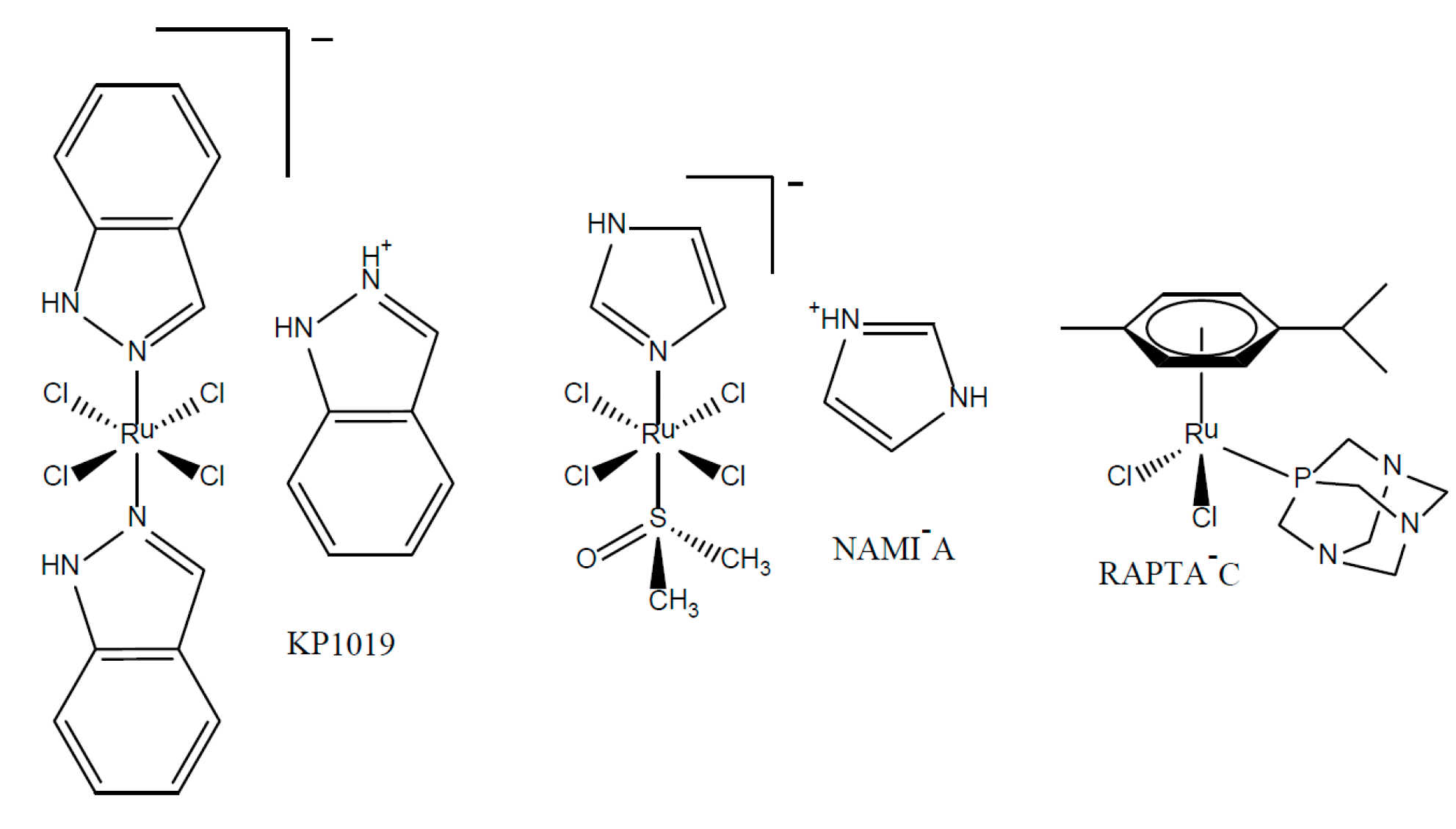
Chart 1. Examples of anti-cancer ruthenium-based agents.
Inorganics 05 00044 ch001
Scheme 1. Synthesis of mono(trichlorostannyl) complexes C1 and C2 and di(trichlorostannyl) complex C3.
Inorganics 05 00044 sch001
Scheme 2. Synthesis of the cationic complexes C4 and C5.
Inorganics 05 00044 sch002
Figure 1. The 31P NMR spectrum of complex C1 in which the main resonance signal is flanked with 117Sn (inner) and 119Sn (outer) satellites.
Inorganics 05 00044 g001
Figure 2. TGA trace of complex C1 with the onset of decomposition occuring at 121.51 °C.
Inorganics 05 00044 g002
Figure 3. ORTEP view of (C2) with atom-labelling scheme and thermal ellipsoids drawn at the 50% probability level. Selected bond distances (Å) and bond angles (°): Ru(1)–Sn(1) = 2.5686(5), Ru(1)–Cl(1) = 2.3919(10), Ru(1)–P(1) = 2.2.242(12). P(1)–Ru(1)–Cl(2) = 90.90(2), P(1)–Ru(1)–Cl(1) = 81.72(2), and Cl(2)–Ru(1)–Cl(1) = 87.49(2).
Inorganics 05 00044 g003
Figure 4. ORTEP view of [(η6-C6H6)RuCl2P(OPh)3] with atom-labelling scheme and thermal ellipsoids drawn at the 50% probability level. Selected bond distances (Å) and bond angles (°): C(1)–Ru(1) = 2.191(5), C(2)–Ru(1) = 2.254(5), C(3)–Ru(1) = 2.175(5), C(4)–Ru(1) = 2.246(5), C(5)–Ru(1) = 2.245(5), C(6)–Ru(1) = 2.249(5), C(1–6)–Ru(1)–P(1) = 117.49(18), P(1)–Ru(1)–Cl(1) = 85.71(4), P(1)–Ru(1)–Sn(1) = 86.84(3), and Cl(1)–Ru(1)–Sn(1) = 83.67(3).
Inorganics 05 00044 g004
Table 1. In vitro cytotoxicity of complexes against selected tumour cell lines after 72 h drug exposure.
CompoundIC50 (µM) a
A2780A2780cisRHEK293
C1>500>500>500
C251.5 ± 0.151.5 ± 0.125.0 ± 6.2
C3>200>200>200
C43.4 ± 0.44.1 ± 0.914.2 ± 4.9
C56.5 ± 0.97.1 ± 1.63.2 ± 0.3
[(η6-C6H6)RuCl2(PPh3)]30.5 ± 0.727.0 ± 5.627.8 ± 6.8
[(η6-C6H6)RuCl2(P(OPh)3)]2.3 ± 0.023.2 ± 0.41.0 ± 0.5
[(η6-C6H6)RuCl(SnCl3)(PPh3)]12.4 ± 0.412.5 ± 3.04.9 ± 0.1
cis-platin1.1 ± 0.414.4 ± 2.110.6 ± 1.4
a IC50 values (µM) are presented as mean ±SD of two or more independent experiments. The sign (>) indicates that IC50 value was not obtained up to given concentration.
Table 2. Crystal data, details of data collection and structure refinement parameters for (C2) and [(η6-C6H6)RuCl2{P(OPh)3}].
ComplexC2[(η6-C6H6)RuCl2{P(OPh)3}]
Empirical formulaC24H21Cl4PRuSnC24H21Cl2O3PRu
Formula weight749.94560.35
T/K100(2) K446(2) K
λ/Å Crystal system0.71073 Orthorhombic0.71073 Orthorhombic
Space groupP2(1)2(1)2(1)Pbca
a8.7164(17)17.371(3)
b16.420(3)14.997(3)
c18.514(4)17.551(3)
α(deg.)9090
β(deg.)9090
γ(deg.)9090
V(Å3)2649.9(9)4572.6(14)
Z48
Densitycalc(mg·m−3)1.8801.628
Absorption coefficient (mm−1)2.0011.013
F(000)14642256
Crystal size (mm)0.42 × 0.08 × 0.060.38 × 0.36 × 0.16
Theta range for data collection (deg.)1.66–26.332.14–26.34
Limiting indices−10 ≤ h ≤ 10, −16 ≤ k ≤ 20, −23 ≤ l ≤23−21 ≤h ≤21, −18 ≤ k ≤18, −21 ≤ l ≤ 21
Reflections Collected/Unique17168/5368 [R(int) = 0.0409]34691/4657 [R(int) = 0.0339]
Completeness of theta max.26.33 (99.6%)26.34 (99.9%)
Absorption correctionSADABSSADABS
Refinement methodFull-matrix least-squares on F2Full-matrix least-squares on F2
Data/Restraints/Parameters5368/0/3084657/0/280
Goodness-of-fit on F2 (GOF)1.0421.117
Largest diff. peak and hole (e·Å−3)0.805 and −0.4710.566 and −0.404