Article

Kinetics-Controlled Simple Method for the Preparation of Au@Ag Hierarchical Superstructures for SERS Analysis

1
Institute of Fire Safety Materials, School of Materials Science and Engineering, NingboTech University, Ningbo 315100, China
2
Department of Materials Science and Engineering and California NanoSystems Institute, University of California, Los Angeles, CA 90095, USA
*
Authors to whom correspondence should be addressed.
Inorganics 2025, 13(6), 191; https://doi.org/10.3390/inorganics13060191
Submission received: 6 April 2025 / Revised: 28 May 2025 / Accepted: 5 June 2025 / Published: 7 June 2025

Abstract

:
Silver nanostructures exhibit exceptional surface-enhanced Raman scattering (SERS) performance due to their strong plasmonic resonance. However, their practical applications are often hindered by structural instability, leading to deformation and performance degradation. In this study, we developed a kinetics-controlled synthetic strategy to fabricate gold-encapsulated silver (Au@Ag) hierarchical superstructures (HSs) with enhanced SERS activity and stability. By leveraging polyvinylpyrrolidone (PVP) as a surface modifier and precisely regulating the introduction rate of reaction precursors, we achieved meticulous control over the galvanic replacement kinetics, thereby preserving the structural integrity of pre-synthesized Ag HSs during the formation of Au@Ag HSs. The resulting well-defined Au@Ag HSs demonstrated superior SERS performance, achieving a detection limit of 10−9 M for crystal violet (CV) while exhibiting outstanding signal reproducibility (relative standard deviation, RSD = 11.60%). This work provides a robust and scalable approach to designing stable, high-efficiency SERS-active nanostructures with broad potential in analytical and sensing applications.

1. Introduction

Surface-enhanced Raman spectroscopy (SERS) harnesses plasmonic resonances to significantly amplify vibrational signal detection, offering enhanced sensitivity, expedited analysis, and precise molecular identification [1]. It is widely acknowledged that the efficacy of SERS is heavily dependent on the ability of various materials to enhance Raman signals, including noble metals [2,3], carbon materials [4,5], transition metal oxides [6], quantum dots, etc. [7]. Among these, silver (Ag), particularly in its complex architectural forms, manifests the most potent enhancement effects owing to its robust surface plasmon resonance activity [8]. Compared to traditional shapes, complex structures, such as star-shaped or dendritic forms, demonstrate superior enhancement capabilities, which is attributed to their larger surface area and enhanced adsorption properties [9], which provide an abundance of adsorption sites and a proliferation of hotspots—regions where the electromagnetic field intensity significantly surpasses that of the surrounding areas [10]. Nevertheless, the intricate structures of Ag are often challenged by stability issues, which can lead to deformation and a subsequent decline in their exceptional enhancement capabilities [11].
To address these issues, researchers have developed various optimization strategies to enhance the stability and SERS performance. These include the fabrication of high-density Ag nanoparticles (NPs) arrays [12], surface modification [13], and the creation of composite SERS substrates [14]. These strategies can alter the electronic structure and chemical stability, thereby improving oxidation resistance. However, these strategies may also involve complex fabrication processes, the introduction of materials with poor inherent SERS performance that can negatively impact the overall SERS activity, and high costs that hinder large-scale fabrication and application. Relatively, depositing Au on Ag surfaces to create core–shell structures is an effective strategy [15]. It not only serves as a protective layer to prevent Ag oxidation [16] but also, more importantly, combines the properties of Au and Ag to generate a stronger surface plasmon resonance, thereby enhancing the SERS signal [17]. The main strategies for preparing gold-coated silver include in situ reduction [18], seed-mediated growth [19], and metal deposition methods [20]. While these methods can effectively synthesize Au@Ag structures, they are typically limited to conventional Ag morphologies such as spheres [21], wires [22], and plates [23,24]. Extending these approaches to fabricate architecturally complex Ag@Au nanomaterials often proves challenging. Conspicuously, unlike the relatively straightforward preparation of silver-coated Au structures [25], the synthesis of gold-coated Ag structures while preserving the template’s intricate Ag architecture presents significant difficulties due to Ag’s higher chemical reactivity than Au [26,27]. In most cases, the high reactivity of Ag compared to Au often leads to morphological damage of Ag during the preparation process, significantly affecting the material’s inherent SERS activity. Developing more effective methods to fabricate complex gold-coated Ag structures and to demonstrate their structure-related high SERS activity is of great significance, yet it faces considerable challenges at this stage.
In this work, we demonstrate a kinetics-controlled yet simple synthetic approach for fabricating well-defined gold-encapsulated silver hierarchical superstructures (Au@Ag HSs, as illustrated in Scheme 1). Through meticulous manipulation of reaction kinetics enabled by PVP-mediated surface modification and precise control over precursor introduction rates, we successfully preserve the structural integrity of preformed silver templates while achieving uniform gold encapsulation. The resulting Au@Ag HSs exhibit remarkable structural uniformity and stability, which translates into superior and reproducible SERS performance—achieving unprecedented detection sensitivity down to 10−9 M for analyte molecules. This versatile synthetic strategy not only provides fundamental insights into the kinetics-governed formation of hierarchical nanostructures but also opens new avenues for developing high-performance plasmonic materials with applications spanning molecular sensing, catalytic systems, and photonic devices.

2. Results and Discussion

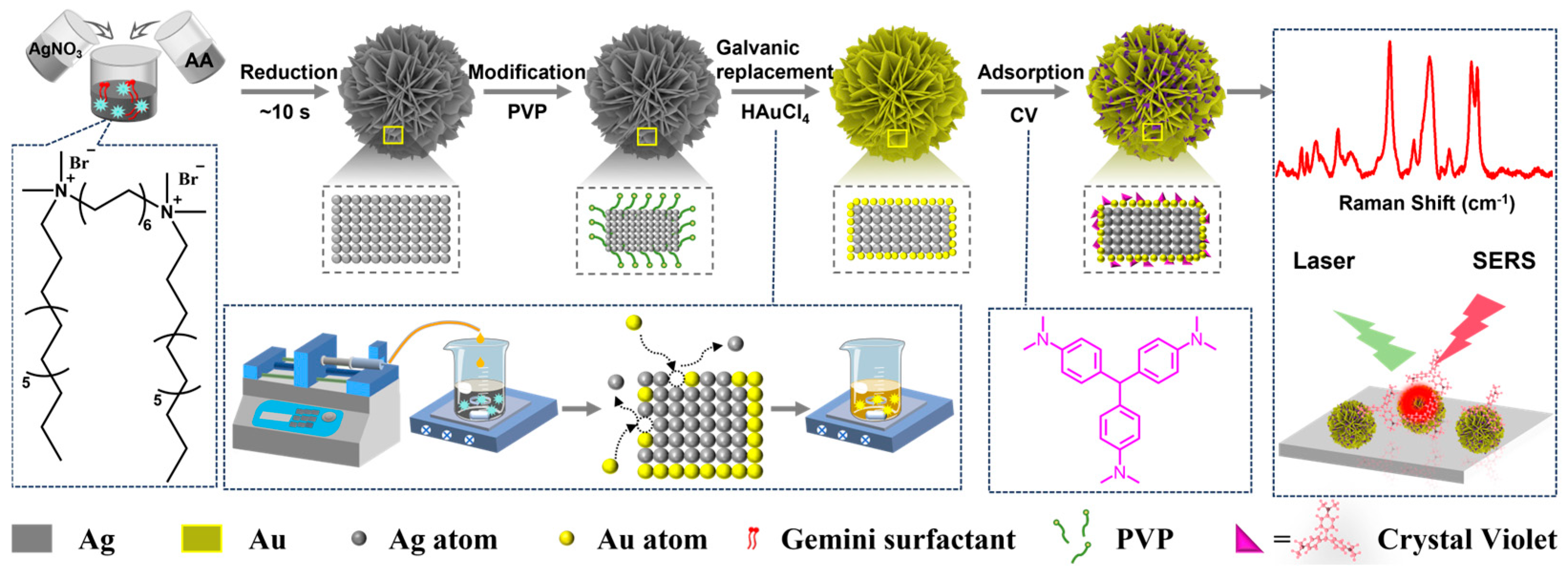
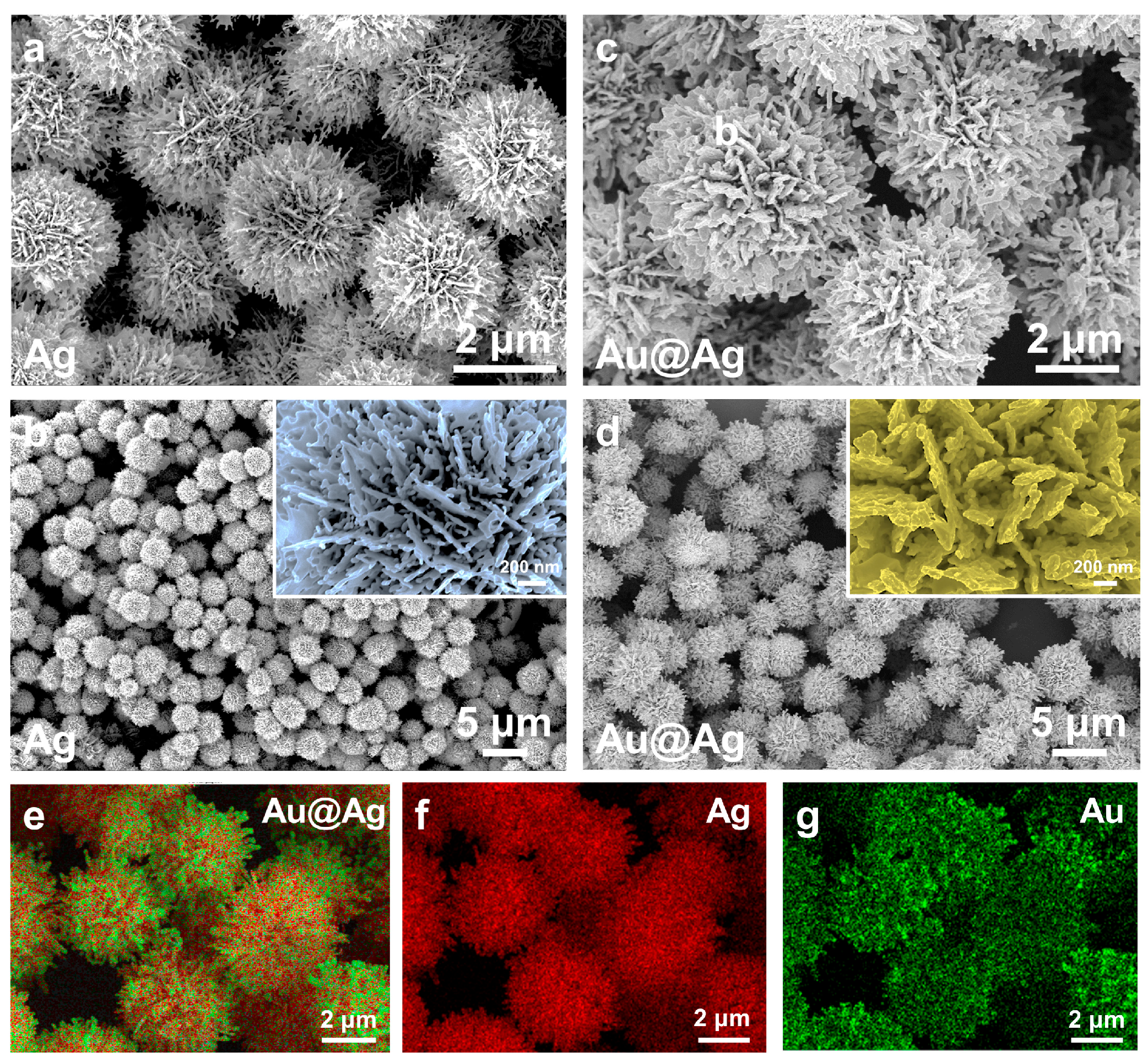
2.1. Synthesis and Characterization of Au@Ag HSs

To synthesize Au@Ag HSs, monodispersed Ag HSs were initially prepared based on our previous study [28], serving as templates for the subsequent displacement reaction. Utilizing Gemini surfactant as a directing agent and meticulously controlling the concentration of reactants and reaction temperature, we successfully synthesized structurally perfect Ag HSs within seconds (Figures S1 and S2). As depicted in Figure 1a,b and Figure S3, the Ag HSs exhibit a morphology resembling hydrangea flowers. With the exception of a few particles that exhibit considerable size deviations, the majority of Ag HSs display consistent dimensions and morphology. As shown in Figure S4, statistical analysis revealed an average particle size of 3.23 ± 0.27 μm. The insets in the upper right corners of Figure 1b clearly demonstrate that the surface features an extremely dense, flake-like petal structure. Ag HSs akin to natural blossoms typically present a sophisticated three-dimensional architecture, featuring centrally located, comparatively substantial NPs encircled by numerous smaller NPs to create a petal-like tiered arrangement. The petals on the surface of these HSs are lush, with sharp edges, and even bear numerous circular holes on their surfaces.
Using these Ag HSs as templates, which are specially protected by PVP, HAuCl4 was slowly introduced into the surface-modified Ag HSs suspension. Notably, as evidenced by prior studies, PVP plays a crucial role in our system by selectively stabilizing specific facets of Ag NPs. This facet-selective capping behavior promotes the oxidation of Ag atoms on lateral surfaces while simultaneously facilitating defect formation [29,30]. This mechanism is particularly critical as it enables precise site-selective Au deposition at predetermined locations. Building upon this principle, by precisely controlling the concentration of HAuCl4 and the injection rate, we regulated the rate of the Ag and Au3+ displacement reaction, ultimately yielding structurally perfect Au@Ag HSs. The resulting Au@Ag HSs maintain identical morphology and monodispersity to the original Ag HSs, with no significant structural alterations observed during the reaction process, as depicted in Figure 1c,d. Further, localized enlargement images reveal that in comparison to Ag HSs, the Au@Ag HSs have a noticeably increased thickness. Fortunately, the holes on the petal surfaces prior to the reaction remain largely unaffected (the insert in Figure 1d).
Further analysis of the structure and composition of the Au@Ag HSs was conducted using energy-dispersive X-ray spectroscopy (EDS) mapping. Figure 1e illustrates that Au@Ag HSs consist of two elements, with light green, indicating the presence of a small quantity of Au, which is predominantly distributed at the tips of the surface. Additionally, the uniform distribution of Ag and Au elements, as shown in Figure 1f,g, indicates that a small amount of Au is evenly dispersed across the surface of the HSs, apart from the tip regions. Additionally, the results of X-ray photoelectron spectroscopy (XPS) analysis show that the metal composition of the metal nanoclusters is Au and Ag (Figures S5–S7). It can finally be shown that the Ag HSs template can be used to synthesize a variety of bimetallic Au@Ag HSs containing both gold and silver elements. In brief, Au@Ag HSs with an almost perfect structure have been successfully fabricated, establishing a foundation for guiding related research endeavors and exploring their broad applications.

2.2. Structural Manipulation of Au@Ag HSs

To efficiently fabricate Au@Ag HSs with a flower-like appearance characterized by dense, porous petals that align with our specific requirements, we carefully optimized two key synthesis parameters: the concentration of HAuCl4 and the injection rate. Figure 2 and Figure S8 present the SEM images of Au@Ag HSs synthesized under varying injection rates and concentrations of HAuCl4. The results highlight that the synthesis of Au@Ag HSs involves a delicate balance between the concentration of HAuCl4 and its injection rate into the reaction system. Specifically, lower concentrations of HAuCl4 (1.0 mM) and slower injection rates (0.5 mL/min) favor the preservation of the Ag template’s structure and the uniform deposition of Au on the Ag surface, resulting in well-defined, flower-like structures with dense and porous petals (Figure 2a,d). However, increasing either the concentration of HAuCl4 or the injection rate leads to significant morphological changes. Higher concentrations of HAuCl4 (1.5 and 2.5 mM) and faster injection rates (1.0 and 1.5 mL/min) promote the aggregation of Au NPs, which results in structural deformations and a loss of the desired floral morphology (Figure 2b,c,e–i). Specifically, at higher concentrations and injection rates, the Au NPs tend to aggregate, leading to rougher surfaces, blunted petal edges, and, eventually, a more spheroidal overall structure (Figure 2i). That is, the optimal synthesis conditions appear to lie at the intersection of moderate HAuCl4 concentrations (1.0–1.5 mM) and relatively slow injection rates (0.5–1.0 mL/min). Under these conditions, the Au NPs can deposit uniformly on the Ag template, preserving the intricate three-dimensional morphology of the HSs while ensuring sufficient Au coverage to prevent oxidation and maintain structural stability.
Furthermore, the elemental composition of the as-prepared Au@Ag HSs was quantitatively analyzed using inductively coupled plasma optical emission spectroscopy (ICP-OES). Systematic variation in both the HAuCl4 concentration and injection rate during synthesis enabled precise control over the bimetallic composition, resulting in tunable Ag:Au mass ratios ranging from 3.17 to 28.31 (Table S1). Notably, the observed deviations between the final composition and initial feeding ratio can be attributed to the reaction kinetics. When using higher HAuCl4 concentrations or faster injection rates, more extensive structural disruption of the Ag HSs occurred. This led to increased participation of inner-layer silver atoms in the replacement reaction, consequently yielding products with lower Ag:Au mass ratios. In contrast, under milder reaction conditions, the structural integrity of the Ag HSs was better preserved, with only surface silver atoms participating in the galvanic replacement, thereby resulting in higher Ag:Au ratios. This conclusion is well supported by SEM observations, which revealed a clear correlation between structural preservation and the resulting elemental composition. The above highlights the critical role that both HAuCl4 concentration and injection rate play in determining the morphology of the Au@Ag HSs. These findings also emphasize the importance of controlling these parameters to ensure the desired structural characteristics, which are critical for applications such as SERS, where the distribution of hotspots plays a pivotal role in performance.

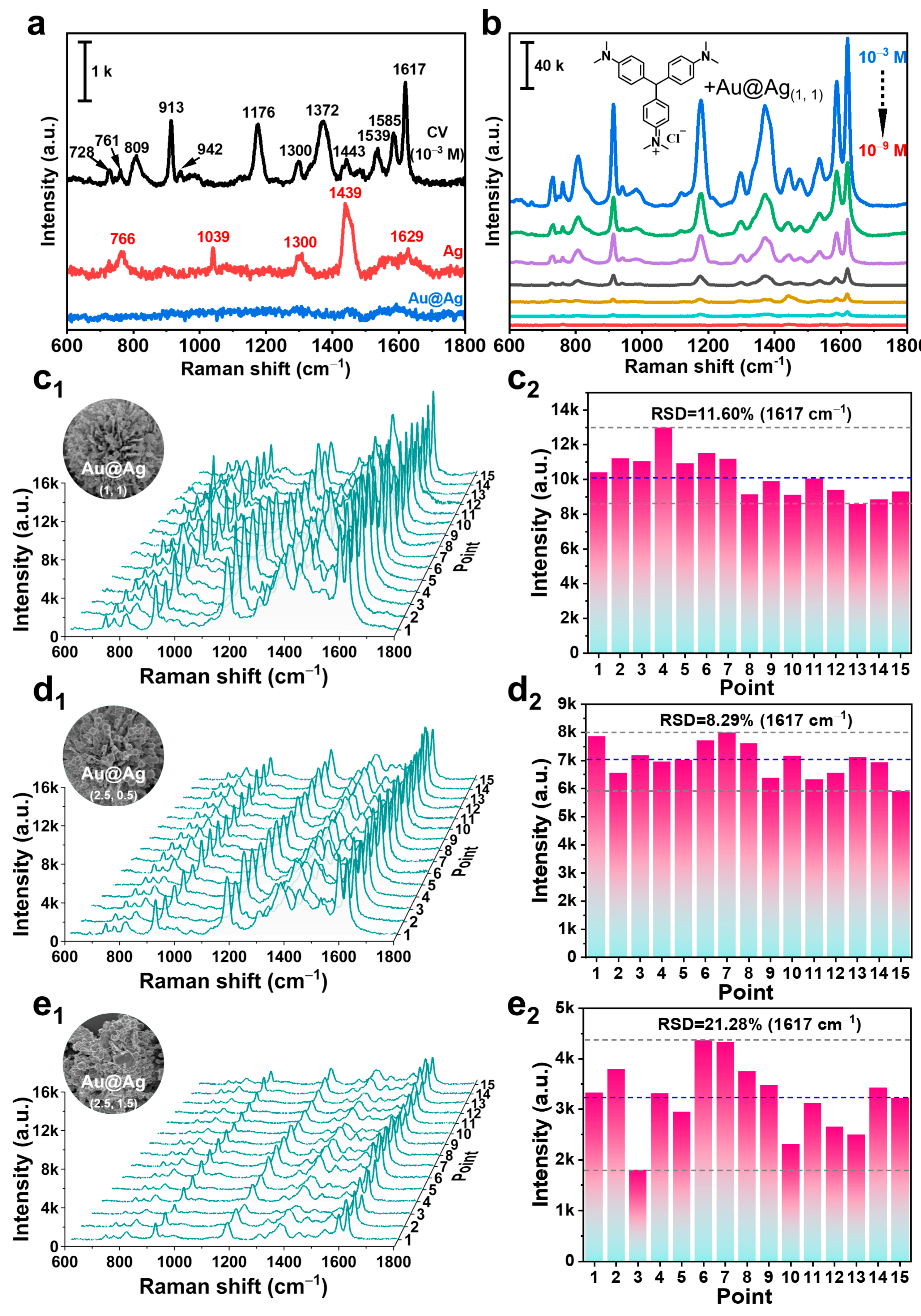
2.3. SERS Properties of Au@Ag HSs

Building on the achieved morphological control through optimization of HAuCl4 concentration and injection rate, we further explored the structure-dependent SERS performance of Au@Ag hybrid structures. For this purpose, CV was employed as a model analyte-this choice was motivated by its widespread adoption in the literature as a benchmark molecule [31], despite its known carcinogenic, mutagenic, and reproductive toxicities that have led to strict regulatory controls due to significant environmental persistence and particularly high aquatic toxicity [32]. To eliminate potential interference from surface adsorbates, Raman spectra of the bare substrates were first analyzed (Figure 3a). The Ag HSs displayed characteristic peaks associated with the Gemini surfactant coating [33], similar to those observed for CTAB [34]. In contrast, the Raman spectrum of Au@Ag HSs showed a clean baseline, confirming the effective removal of surface residues and establishing a well-defined plasmonic substrate. For CV at 10−3 M, distinct peaks were observed at 1617 cm−1 (aromatic ring vibrations) and 1585 cm−1 (C-C stretching), along with secondary peaks at 1372, 1176, 913, and 809 cm−1, corresponding to various vibrational modes within the molecule [35]. Next, we systematically assessed the SERS enhancement across a range of CV concentrations (10−3 to 10−9 M). Au@Ag HSs demonstrated significant signal amplification, with the 10−3 M CV solution showing much higher peak intensities than the unenhanced 10−3 M reference (Figure 3b). Notably, even at a concentration as low as 10−9 M, CV signals remained detectable, highlighting the exceptional sensitivity of the Au@Ag HSs. This enhancement is attributed to the hierarchical architecture and bimetallic composition of the Au@Ag HSs, which facilitate the formation of numerous electromagnetic hotspots through interparticle plasmon coupling.
To evaluate the homogeneity of the SERS signal, we conducted spatially resolved SERS mapping on four representative samples with distinct morphologies, resulting from different HAuCl4 concentrations and injection rates: 1.0 mM (1.0 mL/min), 2.5 mM (0.5 mL/min), 2.5 mM (1.5 mL/min). In addition, Ag HSs with perfect structures were used as controls for comparison. These Au@Ag HSs are denoted as Au@Ag(1, 1), Au@Ag(2.5, 0.5), and Au@Ag(2.5, 1.5), respectively (Figure 3c–e). These three morphologies were selected as the most representative among the synthesized products because they encompassed both well-defined structures and highly defective states, providing a comprehensive comparison. The pristine Au@Ag(1, 1) displayed outstanding signal uniformity, with a relative standard deviation (RSD) of 11.60% at the 1617 cm−1 peak. This is attributed to its intact flower-like structure and uniform Au encapsulation, which ensures consistent hotspot distribution across the surface [36]. In contrast, Au@Ag(2.5, 0.5) showed a slightly lower RSD (8.29%) but with reduced signal intensity. This reduction is due to partial structural collapse and a thicker Au layer, which diminishes plasmonic coupling efficiency [37]. The most degraded Au@Ag(2.5, 1.5) exhibited severe signal inconsistency (21.28% RSD) and a significant loss of signal intensity. This is a result of extensive Au aggregation and spherical deformation, disrupting the electromagnetic field confinement needed for effective SERS enhancement. These findings reveal a clear relationship between the structural integrity of Au@Ag HSs and their SERS performance. The optimized Au@Ag(1, 1) structure strikes an ideal balance between morphological preservation and Au coverage, ensuring both strong and homogeneous signal enhancement. In contrast, deviations from this optimal structure—such as incomplete Au encapsulation or excessive metal deposition—lead to a loss of hotspot density and distribution, ultimately diminishing the SERS enhancement. Significantly, when the substrate was replaced with Ag HSs featuring ideal multi-layered architecture, our experimental data demonstrated their exceptional performance in SERS enhancement while maintaining satisfactory reproducibility (RSD = 14.74%, Figures S9 and S10). These results are consistent with established literature confirming Ag as one of the most efficient SERS substrate materials [38]. The enhanced SERS activity primarily stems from Ag HSs’ well-defined hierarchical morphology and distinct petal-like edges, which effectively intensify localized surface plasmon resonance [39]. Nevertheless, it should be acknowledged that the practical implementation of Ag-based substrates may be constrained by their inherent susceptibility to oxidation under prolonged exposure [40]. To sum up, this structure-performance correlation underscores the critical importance of precise synthetic control in the development of effective plasmonic substrates for SERS applications.

3. Materials and Methods

3.1. Materials

Silver nitrate (AgNO3, ≥99.0%), ascorbic acid (AA, ≥99.0%), gold(III) chloride trihydrate (HAuCl4·3H2O, ACS, 99.99%), CV (≥99.0%), and PVP (K-30) were obtained from Sinopharm Chemical Reagent Corporation (Shanghai, China). All the chemicals were of analytical grade and were used without further purification. The cationic gemini surfactant dodecanediyl-1,12-bis(dimethylhexadecylammonium bromide) (16-12-16), used herein, was synthesized according to an earlier paper [41]. Ultrapure water (18.25 MΩ. cm) was used throughout the experiments.

3.2. Instruments and Characterization

The morphology of the products was characterized using field emission scanning electron microscopy (FE-SEM) (GeminiSEM 360, Carl Zeiss AG, Oberkochen, Germany) at 3 kV, and EDS elemental mapping analysis was performed using the same instrument operating at an accelerating voltage of 15 kV. The surface electronic properties were studied by X-ray photoelectron spectroscopy (XPS) using the Rerkin Elmerph-5000cESCA system. The Raman spectra were captured at room temperature using a Renishaw inVia Reflex Raman system, with an excitation laser wavelength of 532 nm and a recording time of 5 s. An ICP-OES (Agilent 5110 OES, Agilent, Santa Clara, CA, USA) was utilized to quantify the Au and Ag contents in the HSs.

3.3. Synthesis of Silver Hierarchical Superstructure (Ag HSs)

Typically, 15 mL of a 100 mM AgNO3 solution was introduced into a 50 mL plastic tube and placed in a 30 °C water bath to maintain a constant temperature (Note: If a glass bottle is used as a reaction vessel, there may be some product sticking to the walls) (Figures S1 and S2). Thereafter, under persistent stirring, 15 mL of a 2.5 mM 16-12-16 solution and 15 mL of a 100 mM AA solution were added in an orderly fashion. Accompanied by continuous stirring for a few seconds, resulted in a pronounced color transformation of the solution from a pale white to a deep black shade. The supernatant was carefully decanted, and the precipitate was redispersed in water and subsequently subjected to ultrasonication and centrifugation. Ultimately, the precipitate, denoted as Ag HSs, was reconstituted in ultrapure water to a concentration of approximately 5 mg/mL, rendered suitable for subsequent applications.

3.4. Synthesis of Gold-Coated Silver Hierarchical Superstructure (Au@Ag HSs)

Typically, 10 mL of a 1.0 mg/mL Ag HSs suspension was introduced into a 25 mL glass flask, where ultrasonic processing ensures a more uniform distribution. Subsequently, 0.25 mL of a 50 mM PVP solution, acting as a surface modifier, was added and thoroughly mixed. Then, 5 mL of a 1.0 mM HAuCl4·3H2O solution was incrementally introduced into the mixture using a precision syringe pump, with the flow rate set to 1.0 mL/min. After the infusion of HAuCl4·3H2O was complete, the mixture was stirred for an additional 2 h. Finally, the product was obtained by centrifugation and washing with ultrapure water, resulting in the formation of the target Au@Ag HSs. To achieve the optimal Au@Ag HSs, the injection rate and HAuCl4 concentration, as key reaction parameters, were meticulously controlled, while all other parameters were kept constant.

3.5. SERS Application

To evaluate the surface-enhanced Raman scattering (SERS) enhancement performance of different substrates, CV stock solutions with a concentration of 10−3 M were initially prepared by dissolving the powder in ethanol. Lower concentrations of CV solutions, ranging from 10−9 to 10−3 M, were subsequently obtained through serial dilutions in ethanol. For the SERS detection process, 5 mg of Au@Ag HSs powders were redispersed in 1.0 mL of ethanol using sonication to achieve a homogeneous suspension. A SERS substrate was then prepared by depositing 10 μL of this suspension onto a clean silicon wafer, followed by drying under ambient conditions. The substrate was immersed in the prepared CV ethanol solutions (10−9 to 10−3 M) for 24 h. After immersion, the substrate was allowed to air-dry naturally before being used for SERS testing.

4. Conclusions

In summary, we have successfully developed a kinetics-controlled synthetic approach for the fabrication of Au@Ag HSs with superior SERS performance. Through meticulous manipulation of the reaction kinetics by utilizing PVP as a surface modifier and precisely regulating the introduction rate of reaction precursors, we have achieved uniform gold encapsulation while preserving the structural integrity of the pre-synthesized Ag HSs templates. The resulting Au@Ag HSs exhibit remarkable structural uniformity and stability, which are crucial for their outstanding SERS performance. Our results demonstrate that the optimized Au@Ag HSs (Au@Ag(1, 1)) achieved a detection limit of 10−9 M for CV, with excellent signal reproducibility (RSD = 11.60%). This is attributed to the intact flower-like structure and uniform gold encapsulation, which ensures consistent hotspot distribution across the surface. In contrast, deviations from the optimal structure, such as incomplete gold encapsulation or excessive metal deposition, lead to a loss of hotspot density and distribution, ultimately diminishing the SERS enhancement. The kinetic-guided strategy developed here offers fundamental insights for constructing various hierarchical nanostructures (extendable to Au/Cu-based systems) while demonstrating the scalable potential for practical sensor fabrication through its robust, high-efficiency design paradigm.

Supplementary Materials

The following supporting information can be downloaded at: https://www.mdpi.com/article/10.3390/inorganics13060191/s1, Figure S1: Digital photos of different reaction times for the Ag HSs synthesis process, (a) 0 s, (b) 4 s, (c) 7 s, and (d) 9 s. Figure S2: Digital photos of different containers used for preparing Ag HSs. Figure S3: SEM images of the prepared Ag HSs. Figure S4: Size distribution histogram of the Ag HSs. Figures S5–S7: XPS spectrum of the as-prepared typical Au@Ag HSs. Figure S8: Low-magnification SEM images of Au@Ag synthesized at different injection rates and injection concentrations, (a) 1.0 mM, 0.5 mL/min, (b) 1.5 mM, 0.5 mL/min, (c) 2.5 mM, 0.5 mL/min, (d) 1.0 mM, 1.0 mL/min, (e) 1.5 mM, 1.0 mL/min, (f) 1.5 mM, 2.5 mL/min, (g) 1.0 mM, 1.5 mL/min, (h) 1.5 mM, 1.5 mL/min, (i) 2.5 mM, 1.5 mL/min. Table S1: Elemental analysis by ICP-OES was conducted on the various Au@Ag HSs. Figure S9: SERS spectra of CV molecules with the concentration of 10−6 M at 15 different positions on the Ag HSs substrate. Figure S10: Histogram of the intensity of the peak at 1617 cm−1 in the corresponding SERS spectra in Figure S9.

Author Contributions

M.L.: Methodology, Investigation, Writing—original draft. M.J.: Methodology, Investigation. H.Y.: Investigation, Validation. K.W.: Validation. P.Z.: Validation. Y.Z.: Validation. Y.X.: Methodology, Supervision, Writing—review and editing. J.L.: Methodology, Supervision, Writing—Review and editing. All authors have read and agreed to the published version of the manuscript.

Funding

This work was supported by the National Natural Science Foundation of China (No. 51973229), the Natural Science Foundation of Ningbo (No. 2024J117), the Scientific Research Foundation of NingboTech University (No. 20220920Z0221), the General Scientific Research Project of Zhejiang Education Department (No. Y202352936).

Data Availability Statement

The authors confirm that the data supporting the findings of this study are available within the article and its Supplementary Materials.

Conflicts of Interest

The authors declare no conflicts of interest.

References

  1. Itoh, T.; Procházka, M.; Dong, Z.; Ji, W.; Yamamoto, Y.S.; Zhang, Y.; Ozaki, Y. Toward a new era of SERS and TERS at the nanometer scale: From fundamentals to innovative applications. Chem. Rev. 2023, 123, 1552–1634. [Google Scholar] [CrossRef] [PubMed]
  2. Zhao, Q.; Hilal, H.; Kim, J.; Park, W.; Haddadnezhad, M.; Lee, J.; Park, W.; Lee, J.; Lee, S.; Jung, I.; et al. All-hot-spot bulk surface-enhanced raman scattering (SERS) substrates: Attomolar detection of adsorbates with designer plasmonic nanoparticles. J. Am. Chem. Soc. 2022, 144, 13285–13293. [Google Scholar] [CrossRef] [PubMed]
  3. Hilal, H.; Haddadnezhad, M.; Oh, M.J.; Jung, I.; Park, S. Plasmonic dodecahedral-walled elongated nanoframes for surface-enhanced raman spectroscopy. Small 2024, 20, 2304567. [Google Scholar] [CrossRef]
  4. Tang, X.; Kishimoto, N.; Kitahama, Y.; You, T.T.; Adachi, M.; Shigeta, Y.; Tanaka, S.; Xiao, T.H.; Goda, K. Deciphering the potential of multidimensional carbon materials for surface-enhanced raman spectroscopy through density functional theory. J. Phys. Chem. Lett. 2023, 14, 10208–10218. [Google Scholar] [CrossRef]
  5. Lin, J.; Zhang, D.; Yu, J.; Pan, T.; Wu, X.; Chen, T.; Gao, C.; Chen, C.; Wang, X.; Wu, A. Amorphous nitrogen-doped carbon nanocages with excellent SERS sensitivity and stability for accurate iden-tification of tumor cells. Anal. Chem. 2023, 95, 4671–4681. [Google Scholar] [CrossRef]
  6. Gu, C.; Li, D.; Zeng, S.; Jiang, T.; Shen, X.; Zhang, H. Synthesis and defect engineering of molybdenum oxides and their SERS applications. Nanoscale 2021, 13, 562–5651. [Google Scholar] [CrossRef] [PubMed]
  7. Qiu, J.; Chu, Y.; He, Q.; Han, Y.; Zhang, Y.; Han, L. A self-assembly hydrophobic oCDs/Ag nanoparticles SERS sensor for ultrasensitive melamine detection in milk. Food Chem. 2023, 402, 134241. [Google Scholar] [CrossRef]
  8. Hang, Y.; Wang, A.; Wu, N. Plasmonic silver and gold nanoparticles: Shape- and structure-modulated plasmonic functionality for point-of-caring sensing, bio-imaging and medical therapy. Chem. Soc. Rev. 2024, 53, 2932–2971. [Google Scholar] [CrossRef]
  9. Thakur, A.; Singh, R.; Yadav, V.; Siddhanta, S.; Jayarmaulu, K. Advancing SERS applications of 2D materials through the interplay of rational design and structure-property relationships. Small Methods 2025, e2402056. [Google Scholar] [CrossRef]
  10. Troncoso-Afonso, L.; Vinnacombe-Willson, G.A.; García-Astrain, C.; Liz-Márzan, L.M. SERS in 3D cell models: A powerful tool in cancer research. Chem. Soc. Rev. 2024, 53, 5118–5148. [Google Scholar] [CrossRef]
  11. Zhao, Y.; Kumar, A.; Yang, Y. Unveiling practical considerations for reliable and standardized SERS measurements: Lessons from a comprehensive review of oblique angle deposition-fabricated silver nanorod array substrates. Chem. Soc. Rev. 2024, 53, 14–157. [Google Scholar] [CrossRef] [PubMed]
  12. Liu, J.; Wang, Z.; Meng, Y.; Chen, C.; Chen, Q.; Wang, Y.; Dou, S.; Liu, X.; Lu, N. Increasing hotspots density for high-sensitivity SERS detection by assembling array of Ag nanocubes. Talanta 2023, 258, 124408. [Google Scholar] [CrossRef] [PubMed]
  13. Zhou, S.; Hu, Z.; Zhang, Y.; Wang, D.; Gong, Z.; Fan, M. Differentiation and identification structural similar chemicals using SERS coupled with different chemometric methods: The example of fluoroquinolones. Microchem. J. 2022, 183, 108023. [Google Scholar] [CrossRef]
  14. Fan, X.; Wu, P.; Qu, C.; Gao, Y.; Yin, P. Functionalized Ag/MOF nanocomposites based on defect engineering for highly sensitive SERS detection of organic dyes. J. Environ. Chem. Eng. 2024, 12, 112240. [Google Scholar] [CrossRef]
  15. Zhong, L.; Liu, Q.; Wu, P.; Niu, Q.; Zhang, H.; Zheng, Y. Facile on-site aqueous pollutant monitoring using a flexible, ultralight, and robust surface-enhanced raman spectroscopy substrate: Interface self-assembly of au@ag nanocubes on a polyvinyl chloride template. Environ. Sci. Technol. 2018, 52, 5812–5820. [Google Scholar] [CrossRef]
  16. Feng, Y.; Wang, G.; Chang, Y.; Cheng, Y.; Sun, B.; Wang, L.; Chen, C.; Zhang, H. Electron compensation effect suppressed silver ion release and contributed safety of Au@Ag core-shell nanoparticles. Nano Lett. 2019, 19, 4478–4489. [Google Scholar] [CrossRef]
  17. Zhang, J.; Winget, S.A.; Wu, Y.; Su, D.; Sun, X.; Xie, Z.; Qin, D. Ag@Au concave cuboctahedra: A unique probe for monitoring au-catalyzed reduction and oxidation reactions by surface-enhanced raman spectroscopy. ACS Nano 2016, 10, 2607–2616. [Google Scholar] [CrossRef]
  18. Cortie, M.B.; McDonagh, A.M. Synthesis and optical properties of hybrid and alloy plasmonic nanoparticles. Chem. Rev. 2011, 111, 3713–3735. [Google Scholar] [CrossRef]
  19. He, M.Q.; Ai, Y.; Hu, W.; Guan, L.; Ding, M.; Liang, Q. Recent advances of seed-mediated growth of metal nanoparticles: From growth to applications. Adv. Mater. 2023, 35, e2211915. [Google Scholar] [CrossRef]
  20. Yang, Y.; Liu, J.; Fu, Z.; Qin, D. Galvanic replacement-free deposition of Au on Ag for core-shell nanocubes with enhanced chemical stability and SERS activity. J. Am. Chem. Soc. 2014, 136, 8153–8156. [Google Scholar] [CrossRef]
  21. Csapo, E.; Oszko, A.; Varga, E.; Juhasz, A.; Buzas, N.; Koroesi, L.; Majzik, A.; Dekany, I. Synthesis and characterization of Ag/Au alloy and core(Ag)-shell(Au) nanoparticles. Colloids Surf. A Physicochem. Eng. Asp. 2012, 415, 281–287. [Google Scholar] [CrossRef]
  22. Zhu, Y.; Kim, S.; Ma, X.; Byrley, P.; Yu, N.; Liu, Q.; Sun, X.; Xu, D.; Peng, S.; Hartel, M.C.; et al. Ultrathin-shell epitaxial Ag@Au core-shell nanowires for high-performance and chemically-stable electronic, optical, and mechanical devices. Nano Res. 2021, 14, 4294–4303. [Google Scholar] [CrossRef]
  23. Shahjamali, M.M.; Salvador, M.; Bosman, M.; Ginger, D.S.; Xue, C. Edge-gold-coated silver nanoprisms: Enhanced stability and applications in organic photovoltaics and chemical sensing. J. Phys. Chem. C 2014, 118, 12459–12468. [Google Scholar] [CrossRef]
  24. Zaleska-Medynska, A.; Marchelek, M.; Diak, M.; Grabowska, E. Noble metal-based bimetallic nanoparticles: The effect of the structure on the optical, catalytic and photocatalytic properties. Adv. Colloid Interface Sci. 2016, 229, 80–107. [Google Scholar] [CrossRef]
  25. Lin, S.; Lin, X.; Han, S.; Zhao, H.; Hasi, W.; Wang, L. Highly monodisperse Au@Ag nanospheres: Synthesis by controlled etching route and size-dependent SERS performance of their surperlattices. Nanotechnology 2019, 30, 215601. [Google Scholar] [CrossRef]
  26. Kim, K.K.; Kim, M.; Pyun, K.; Kim, J.; Min, J.; Koh, S.; Root, S.E.; Kim, J.; Nguyen, B.T.; Nishio, Y.; et al. A substrate-less nanomesh receptor with meta-learning for rapid hand task recognition. Nat. Electron. 2023, 6, 64–75. [Google Scholar] [CrossRef]
  27. Gholami, M.; Tajabadi, F.; Taghavinia, N.; Moshfegh, A. Chemically-stable flexible transparent electrode: Gold-electrodeposited on embedded silver nanowires. Sci. Rep. 2023, 13, 17511. [Google Scholar] [CrossRef] [PubMed]
  28. Xia, Y.; Gao, Z.; Liao, X.; Pan, C.; Zhang, Y.; Feng, X. Rapid synthesis of hierarchical, flower-like Ag microstructures with a gemini surfactant as a directing agent for SERS applications. CrystEngComm 2017, 19, 6547–6555. [Google Scholar] [CrossRef]
  29. Zhang, L.; Zhang, Y.; Ahn, J.; Wang, X.; Qin, D. Defect-assisted deposition of Au on Ag for the fabrication of core-shell nanocubes with outstanding chemical and thermal stability. Chem. Mat. 2019, 31, 1057–1065. [Google Scholar] [CrossRef]
  30. Ahn, J.; Wang, D.; Ding, Y.; Zhang, J.; Qin, D. Site-selective carving and co-deposition: Transformation of Ag nanocubes into concave nanocrystals encased by Au-Ag alloy frames. ACS Nano 2018, 12, 298–307. [Google Scholar] [CrossRef]
  31. Chen, Y.; Xia, Y.; Lyu, M.; Jiang, M.; Hong, Y.; Guo, Z.; Li, J.; Fang, Z. Engineering ZIF-8@Ag core-satellite superstructures through solvent-induced tunable self-assembly for surface-enhanced Raman spectroscopy. Nanoscale 2025, 17, 4687–4694. [Google Scholar] [CrossRef] [PubMed]
  32. Li, Y.; Zhu, J.; Weng, G.; Liu, Y.; Li, J.; Zhao, J. Study on the roughen process of branches of AuAg nanostars for the improved surface-enhanced Raman scattering (SERS) to detect crystal violet in fish. Sens. Actuators B Chem. 2023, 390, 133936. [Google Scholar] [CrossRef]
  33. Sakir, M.; Akgul, E.T.; Demir, M. Highly sensitive detection of cationic pollutants on molybdenum carbide (MXene)/Fe2O3/Ag as a SERS substrate. Mater. Today Chem. 2023, 33, 101702. [Google Scholar] [CrossRef]
  34. Yu, C.; Varghese, L.; Irudayaraj, J. Surface modification of cetyltrimethylammonium bromide-capped gold nanorods to make molecular probes. Langmuir 2007, 23, 9114–9119. [Google Scholar] [CrossRef]
  35. Kitaw, S.L.; Ahmed, Y.W.; Candra, A.; Wu, T.Y.; Anley, B.E.; Chen, Y.Y.; Cheng, Y.T.; Chen, K.J.; Thammaniphit, C.; Hsu, C.C.; et al. An advanced plasmonic bimetallic nanostar composite for ultra-sensitive SERS detection of crystal violet. Nanoscale 2024, 17, 508–519. [Google Scholar] [CrossRef]
  36. Sunil, J.; Narayana, C.; Kumari, G.; Jayaramulu, K. Raman spectroscopy, an ideal tool for studying the physical properties and applications of metal-organic frameworks (MOFs). Chem. Soc. Rev. 2023, 52, 3397–3437. [Google Scholar] [CrossRef]
  37. Wang, T.; Wang, S.; Cheng, Z.; Wei, J.; Yang, L.; Zhong, Z.; Hu, H.; Wang, Y.; Zhou, B.; Li, P. Emerging core–shell nanostructures for surface-enhanced Raman scattering (SERS) detection of pesticide residues. Chem. Eng. J. 2021, 424, 130323. [Google Scholar] [CrossRef]
  38. Guselnikova, O.; Lim, H.; Kim, H.J.; Kim, S.H.; Gorbunova, A.; Eguchi, M.; Postnikov, P.; Nakanishi, T.; Asahi, T.; Na, J.; et al. New Trends in Nanoarchitectured SERS Substrates: Nanospaces, 2D Materials, and Organic Heterostructures. Small 2022, 18, e2107182. [Google Scholar] [CrossRef]
  39. Kim, J.; Lee, C.; Lee, Y.; Lee, J.; Park, S.; Park, S.; Nam, J. Synthesis, Assembly, Optical Properties, and Sensing Applications of Plasmonic Gap Nanostructures. Adv. Mater. 2021, 33, e2006966. [Google Scholar] [CrossRef]
  40. Huang, Y.; Zhang, S.; Jiang, S.; Xu, J. Improved SERS Performance on Ag-Coated Amorphous TiO2 Random Nanocavities by the Enhanced Light—Matter Coupling Effect. ACS Sustain. Chem. Eng. 2024, 12, 3234–3242. [Google Scholar] [CrossRef]
  41. Zana, R.; Benrraou, M.; Rueff, R. Alkanediyl-.alpha.,.omega.-bis(dimethylalkylammonium bromide) surfactants. 1. Effect of the spacer chain length on the critical micelle concentration and micelle ionization degree. Langmuir 1991, 7, 1072–1075. [Google Scholar] [CrossRef]
Scheme 1. Schematic illustration of the fabrication of Au@Ag HSs and its application as SERS substrate for the detection of crystal violet (CV).
Inorganics 13 00191 sch001
Figure 1. (a,b) SEM images of Ag HSs, (c,d) SEM images, and (eg) elemental mappings of Au@Ag HSs. The inset images in the top right corner of (b,d) are magnified SEM images (shown in pseudocolor).
Inorganics 13 00191 g001
Figure 2. High-magnification SEM images of Au@Ag synthesized at different injection rates and injection concentrations, (a) 1.0 mM, 0.5 mL/min, (b) 1.5 mM, 0.5 mL/min, (c) 2.5 mM, 0.5 mL/min, (d) 1.0 mM, 1.0 mL/min, (e) 1.5 mM 1.0 mL/min, (f) 2.5 mM 1.0 mL/min, (g) 1.0 mM 1.5 mL/min, (h) 1.5 mM 1.5 mL/min, (i) 2.5 mM 1.5 mL/min.
Inorganics 13 00191 g002
Figure 3. (a) Raman spectra of CV (10−3 M), Ag HSs, Au@Ag HSs. (b) SERS spectra of CV with concentrations ranging from 10−3 to 10−9 M (the concentration decreases by a 10-fold gradient). SERS spectra of CV molecules with the concentration of 10−6 M at fifteen different positions on the different substrates: (c) Au@Ag(1, 1), (d) Au@Ag(2.5, 0.5), and (e) Au@Ag(2.5, 1.5), respectively. (c1e1) Histogram of the intensity of the peak at 1617 cm−1 in the corresponding SERS spectra in (ce) (The blue and gray dashed lines respectively represent the average value and the maximum and minimum values of the SERS intensity).
Inorganics 13 00191 g003
Disclaimer/Publisher’s Note: The statements, opinions and data contained in all publications are solely those of the individual author(s) and contributor(s) and not of MDPI and/or the editor(s). MDPI and/or the editor(s) disclaim responsibility for any injury to people or property resulting from any ideas, methods, instructions or products referred to in the content.

