Article

The Growth-Inhibitory Effect of Glass Ionomer Liners Reinforced with Fluoride-Modified Nanotubes

by
Ricardo De Jesús-Pascual
1,
Elias Nahum Salmerón-Valdés
1,*,
Adriana Alejandra Morales-Valenzuela
1,
Leticia Verónica Jiménez-Rojas
2,
Rodrigo Correa-Prado
3,
Edith Lara-Carrillo
1,
Víctor Hugo Toral-Rizo
1,
Osmar Alejandro Chanes-Cuevas
4,
Ulises Velázquez-Enríquez
1,
Raúl Alberto Morales-Luckie
5 and
Javier Jaramillo-García
1
1
Center for Research and Advanced Studies in Dentistry, School of Dentistry, Autonomous University of Mexico State, Toluca 50000, Estado de Mexico, Mexico
2
Infectious Diseases Research Unit, Hospital Infantil de Mexico Federico Gómez, Mexico City 06720, Mexico
3
Tissue Bioengineering Laboratory, Postgraduate Studies and Research Division, Faculty of Dentistry, National Autonomous University of Mexico, Mexico City 04510, Mexico
4
Dental Biomaterials Laboratory, Postgraduate Division, Dental School, National Autonomous University of Mexico, Mexico City 04510, Mexico
5
Joint Research Center for Sustainable Chemistry, UAEM-UNAM, Toluca 50200, Estado de Mexico, Mexico
*
Author to whom correspondence should be addressed.
Inorganics 2025, 13(6), 190; https://doi.org/10.3390/inorganics13060190
Submission received: 7 May 2025 / Accepted: 4 June 2025 / Published: 7 June 2025
(This article belongs to the Special Issue Recent Research and Application of Amorphous Materials)

Abstract

The aim of this research was to compare the growth-inhibitory effect of halloysite-based nanotubes preloaded with sodium fluoride incorporated into two commercial glass ionomers (Vitrebond 3MTM and Ionobond VOCO) for indirect pulp capping. Methods: Sixty samples were prepared and were distributed into two control groups, two positive control groups and two experimental groups. A total of 10% of the total weight of ionomer powder required to prepare each sample was replaced with nanotubes that had been preloaded at 2000 parts per million (minimum inhibitory dose for Streptococcus mutans established in this study using the McFarland index). The growth-inhibitory effect was determined by placing the samples in Petri dishes inoculated with S. mutans for 24 h at 37 °C. Results: Regarding the control groups, only Vitrebond demonstrated a growth inhibition zone; both experimental groups showed an inhibitory effect, and statistical differences were observed when the experimental Ionobond group and control groups were compared. Conclusions: The ionomers reinforced with fluorine-modified nanotubes showed an adequate inhibitory effect on Streptococcus mutans.

Graphical Abstract

1. Introduction

The principal challenge of modern restorative dentistry is the protection and preservation of pulpal vitality with the objective to avoid a root canal treatment; in deep caries lesions, the management includes the removal of damaged tissue and the subsequent application of a liner of certain biomaterials. Liners are a relatively thin indirect pulp capping (IPC) that provides a barrier to protect dentin in deep cavity preparations with proximity to the pulp if no exposure of this tissue is detected [1,2]. They eliminate or reduce postoperative hypersensitivity in resin-based composite restorations [3]. They are widely used to provide an adequate biological seal and cariostatic action [4]. Therefore, the principal objective of any indirect pulp capping procedure is to avoid bacterial filtration to guarantee the health, vitality and protection of the pulp. Conventional glass ionomer cements (GICs) are the most used materials for IPC treatments, and they have been shown to reduce shrinkage stress and the formation of spaces in the dentin/resin adhesive interface [2,5,6]. Glass ionomer-based dental materials exhibit several advantages, the most significant of which include their fluoride-releasing capacity and biocompatibility with the dental pulp. Furthermore, these cements demonstrate low cytotoxicity, the ability to form chemical bonds with dental hard tissues, a coefficient of thermal expansion comparable to that of natural tooth structures, and bacteriostatic properties [7,8,9,10,11].
On the other hand, some disadvantages like difficult handling, low compressive and flexural strengths and low fracture resistance have been reported [12]. In recent years, RMGIC (resin-modified glass ionomer cement) has been developed to eliminate said disadvantages, and it has been used in IPC treatments [3,13,14,15].
Previous studies reported that both GICs and RMGICs could stimulate the remineralization of dentin affected by caries and contribute to the prevention of secondary caries, which is considered the main cause of failure of resin-based restorations [16,17]. The bacteriostatic function of glass ionomer has been attributed to the fluoride release ability of this material.
Fluoride is an antibacterial material that strengthens the structure of enamel, making it less susceptible to demineralization and caries formation due to the generation of fluorapatite crystals and the promotion of enamel remineralization; these mechanisms result in less soluble tissue in the acidic enamel environment that is conducive to the growth of microorganisms such as Streptococcus mutans (S. mutans) [18,19,20].
This microorganism inhabits the human oral cavity, specifically in the biofilm of dental surfaces (dental plaque), and its cariogenic potential is characterized by several key traits: rapid conversion of carbohydrates into lactic acid (acidogenicity); the ability to synthesize large amounts of glucan in the presence of sucrose; a strong affinity for colonizing the hard tissues of the tooth; the capacity to produce an extracellular polymeric matrix; and the ability to survive and proliferate in low pH environments (aciduricity).Although S. mutans is not the only microorganism that causes dental caries, it has been shown to be capable of creating a favorable environment for other aciduric and acidogenic species [21,22].
Regarding the fluoride released by GICs and RMGIs, there is controversy about the true antibacterial capacity of these cements because almost all studies have reported a fluoride release level that does not exceed 50 ppm [23,24,25,26]. Morales et al. (2020) reported that glass ionomers release approximately 17.8 ppm fluoride [27]. On the other hand, some studies mentioned that the minimum quantity required to inhibit the growth of S. mutans is between 4000 and 5000 ppm. Pradiptama et al. (2019) stated that the minimum concentration of sodium fluoride necessary to inhibit the growth of S. mutans is 4800 ppm (4.8 mg/mL) [28]. Therefore, it can be said that the antibacterial effect of fluoride-releasing materials is directly related to the quantity and duration of the fluoride release, and for this reason, the necessary quantity of fluoride to inhibit bacterial growth cannot be obtained from GICs or RMGIs.
Several studies have focused on improving the physical, mechanical and antibacterial properties of ionomers by using nanotechnology [29]. Some studies reported an improvement in the microhardness, compressive strength, shear bonding and flexural strength of GICs with the incorporation of nanoparticles into the glass ionomer powder [30,31,32]. However, there has been great interest in developing or modifying some GICs with nanostructures like nanoparticles (zinc oxide, graphene–silver and magnesium oxide nanoparticles) to enhance their antibacterial capacity [33,34,35].
Nanotubes are nanostructures with drug delivery ability; drugs can be loaded inside nanotubes, and they can improve the antibacterial properties and increase the time of action of different compounds [36,37]. There are nanotubes made of different materials, such as carbon, trititanate, graphene, boron and halloysite. Halloysite nanotubes (HNTs) are a low-cost, nanometer-sized porous tubular structure with a lumen diameter of 10–15 nm, which allows for the encapsulation of drugs or active elements, making them a nanometric container capable of releasing said drugs in a controlled manner. The outer diameter of nanotubes is approximately of 50–80 nm, and they have a length of 1 µm [38,39]. Recent research has shown that HNTs can be loaded with various antimicrobial agents [36]. Other advantages of halloysite nanotubes are their high mechanical strength, thermal stability and adequate biocompatibility [40,41].
The antibacterial properties of glass ionomer cements have been evaluated in previous studies via the agar diffusion method, which has the advantages of simplicity, low cost, easy interpretation of results and the possibility of analyzing a large number of antimicrobials and microorganisms [42,43,44,45,46]. The process consists of inoculating a microorganism onto an agar plate, and if the antimicrobial agent is effective, it will diffuse into the agar and be able to inhibit the growth of the microorganisms, and the diameter of the zone of growth inhibition around the sample is measured.
Therefore, this study aimed to load HNTs with a minimum inhibitory dose of sodium fluoride and to determine the growth-inhibitory effect of these nanotubes on S. mutans when incorporated into a GIC and an RMGIC.

2. Results

Fluoride and halloysite nanotubes were analyzed through Fourier transform infrared spectroscopy. In Figure 1a, vibrations can be observed between 1000 and 1400 cm−1, characteristic of fluorinated compounds (F), and the vibrations observed between 300 and 500 cm−1 correspond to the interaction between sodium and fluorine ions. Regarding the halloysite nanotubes (b), the flexural vibrations between 400 and 600 cm−1 correspond to the presence of silicate groups (Si-O). The vibrations between 1000 and 1200, characteristic of silicate groups, and the flexural vibrations between 600 and 700 cm−1, typical of minerals containing aluminum (Al-O) in their structure, confirmed the presence of halloysite.
As shown in Figure 2a,b, the corresponding infrared spectrum for the control groups showed functional groups belonging to OH groups between the frequencies of 3200–3600 cm−1 and the presence of aliphatic chains (C-H) between 2850 and 2960 cm−1. Bands related to silicate groups (Si-O) were observed at frequencies of 1075 for Ionobond and 1052 for Vitrebond, and the bands at frequencies of 732 for Ionobond and 723 for Vitrebond, indicative of aluminum groups (Al-O), correspond to the presence of halloysite in the positive control groups. For the experimental groups, the presence of the groups mentioned previously was observed, and the appearance of fluoride was confirmed via the band peaks at 1178 and 1456 for Ionobond and at 1143 and 1416 for Vitrebond.
Regarding the growth-inhibitory effect, the minimum and maximum inhibitory concentrations of the control group were 3000 and 4000 ppm, respectively, while for the experimental group, the concentrations were 2000 and 4000 ppm (Figure 3).
Among the control and positive control groups, only VB and VBHNT showed a bacterial growth inhibition zone of 8.3 and 8.5 mm, respectively, for S. mutans (Figure 4).
In Figure 5, it can be observed that all of the experimental groups presented an antibacterial effect on the microorganism analyzed in this study. An average bacterial growth inhibition zone of 8.60 ± 0.51 mm was observed for the IBHNT-NaF, while the VBHNT-NaF showed a greater inhibition zone than the control, with a mean of 9.10 ± 0.56 mm.
As shown in Table 1, significant differences were observed after carrying out a non-parametric Kruskal–Wallis test when the control and experimental groups were compared (p = 0.001). This test is carried out to determine if there are statistically relevant differences between two or more groups with a p value ≤ 0.05. Nevertheless, with the Kruskall–Wallis test, it was not possible to determine between which groups statistically significant differences were present. Therefore, a pairwise comparison test (Mann–Whitney U) was performed to determine between which groups these significant differences were found. The control and positive control groups for Ionobond showed no inhibitory effect, while the control and positive control groups for Vitrebond presented a similar inhibitory effect, whereby no statistically significant differences were observed between these groups. Significant differences were observed between the control groups for Ionobond and Vitrebond (p = 0.010) because Vitrebond presented an inhibitory effect of approximately 8.30 mm, whereas Ionobond did not present said inhibitory effect. When Ionobond was compared with both experimental groups, a significant difference was observed because the VBHNT-NaF and IBHNT-NaF experimental groups presented an inhibitory effect of 9.10 (p = 0.0001) and 8.60 mm (p = 0.001), respectively. Nevertheless, between the control group for Vitrebond and the VBHNT-NaF and IBHNT-NaF experimental groups, no differences were observed (Table 2).

3. Discussion

The anticariogenic effect presented by fluoride-releasing materials is directly related to the duration and amount of fluoride released [27,47]. Glass ionomers are among the dental materials that deliver the highest fluoride release. Previous studies have reported fluoride release amounts of between 17.4 and 32.6 ppm [27,28], which correspond to bacterial inhibition [48]. However, one study reported that a value of between 3040 and 5700 ppm of NaF is needed to eliminate bacteria in the oral cavity [49]; another study reported that a value of between 4000 and 6000 ppm of NaF is needed [50]. Another study reported that the minimum inhibitory amount of NaF necessary for a bactericidal effect on S. mutans is 4800 ppm [28].
Regarding the maximum inhibitory concentration of fluoride, it has been established by previous studies that excessive concentrations of fluoride can cause a regrowth because S. mutans generates a type of adaptation due to a change in the enolase enzyme inhibitor, which is irreversible and subsequently allows S. mutans to stay alive [28,49]
Several studies have tried to enhance the antibacterial characteristics of glass ionomers by incorporating different nanomaterials [30,51,52]. However, some of these studies did not report favorable results [36,53,54,55].
Nanotubes are nanostructures with the ability to encapsulate drugs; specifically, HNTs have been shown to be suitable for drug transport and prolonged release [54,56]. Massaro et al. [57]. concluded that HNTs do not present bacterial inhibition themselves, which was corroborated in this study, since the positive control groups did not show differences in their inhibitory effects from the control groups. However, HNTs are characterized by an excellent carrying capacity and ability to release and transport drugs [54]. These qualities have positively improved some characteristics of dental materials [53].
In this study, the inhibitory effect of NaF was analyzed at concentrations between 1000 and 10,000 ppm; the established maximum inhibitory concentration (4000 ppm) and minimum inhibitory concentration (3000 ppm) of NaF on S. mutans was similar to previous studies [49,50]. The positive control groups analyzed in this study did not show differences with respect to the control groups and were included only to guarantee that the inhibitory effect of the experimental groups was due to the presence of fluorine inside the nanotubes. No bacterial growth inhibition zone was observed in the Ionobond control groups analyzed in this study, so an insufficient amount of NaF was released by this glass ionomer to inhibit S. mutans. In the VB control group, a bacterial growth inhibition zone was observed. A previous study suggested that this antibacterial effect could be caused by the release of the initiator diphenyleneiodonium chloride, which can inhibit fibroblast growth and could also inhibit the growth of S. mutans [47]. Another study showed that compared to GICs, VB is strongly cytotoxic in different cell cultures [58].
In the present investigation, the antibacterial properties were improved in both experimental groups of glass ionomers modified with HNTs preloaded with NaF. In the IB control group, no antibacterial capacity was observed prior to the incorporation of HNTs, but an inhibitory effect was obtained after the incorporation of nanotubes, and the growth inhibition effect of the VBHNT-NAF experimental group increased with respect to VB because a broader bacterial growth inhibition zone was observed (1 mm approximately). However, no statistically significant differences were observed between these groups (Table 2).
With respect to the minimum and maximum concentrations of NaF required to inhibit the growth of S. mutans, HNTs promoted the antibacterial activity of NaF and expanded the bacterial inhibition interval of 3000 to 6000 ppm reported by previous studies [28,49]. However, in the experimental groups that were analyzed in our study, a growth inhibition zone for S. mutans was observed from 2000 ppm. These results demonstrate the ability of HNTs to promote NaF release and increase the antibacterial effect of glass ionomers, which is consistent with previous studies demonstrating the efficacy of HNTs loaded with other antimicrobial agents [54,56].
The ability of glass ionomer cements to inhibit bacterial growth as a consequence of their fluoride release has been critically evaluated in this study, because previous studies concluded that the inhibitory effect occurs from 5000 ppm [18]. Taking into account the amount of fluoride released by glass ionomer cements reported in previous studies, which is no higher than 50 ppm [17,18,40], these materials will release an insufficient amount of fluoride to guarantee an environment free of microorganisms like S. mutans. Considering this limitation, the clinical application of glass ionomer liners reinforced with fluoride-modified nanotubes could increase the success of indirect pulp capping therapy and decrease the probability of secondary caries, guaranteeing an aseptic environment. On the other hand, the amount of fluoride required to eliminate the S. mutans present in dental cavities will be reduced to 2000 ppm due to the capability of HNTs to improve the effect of drugs.

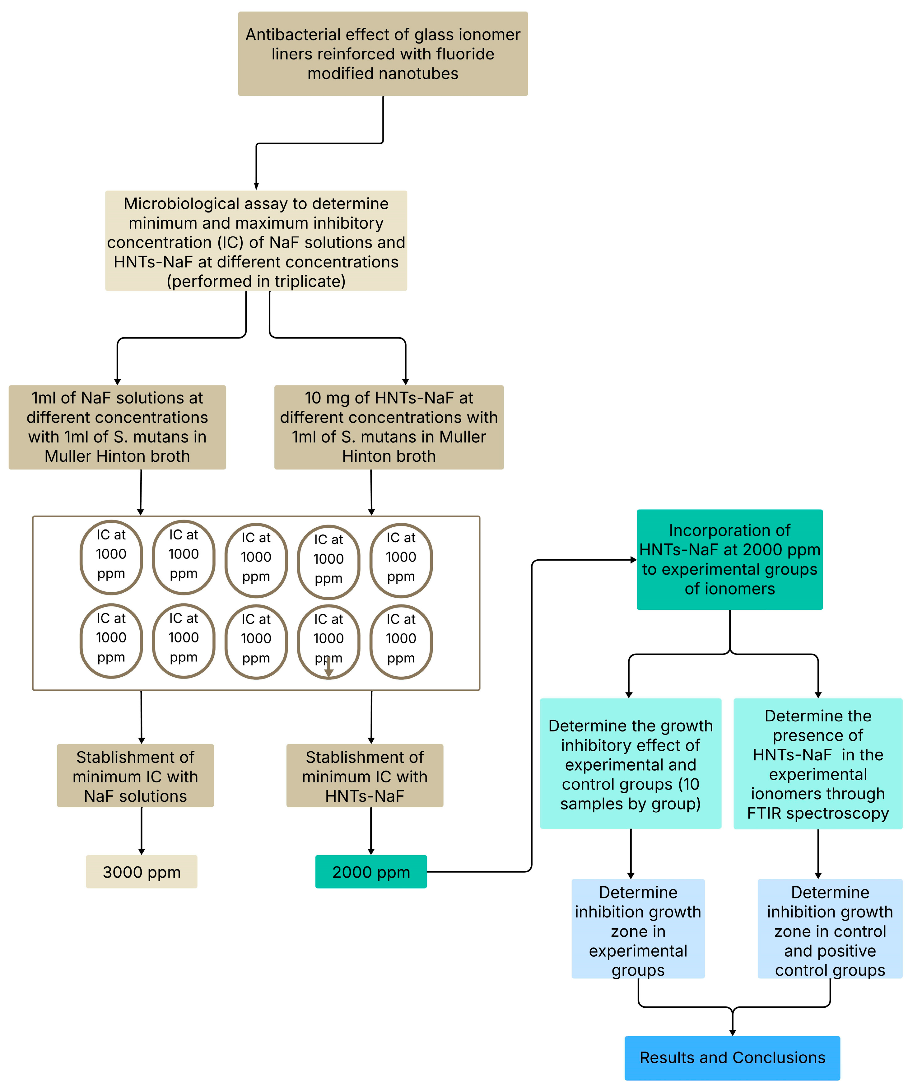
4. Materials and Methods

4.1. Determination of the Inhibitory Effect of Sodium Fluoride (NaF)

The minimum and maximum inhibitory concentrations were determined as follows. A total of 2.2106 g of 99.99% extra pure sodium fluoride (Fagalab; Mocorito, México) was weighed with an analytical balance (Shimadzu Scientific Instruments; Kyoto, Japan) and diluted in deionized water (100 mL) to obtain a solution at a concentration of 10,000 ppm [59]. From this stock solution, solutions were prepared with different concentrations with an interval of 1000 ppm (1000–10,000 ppm), and these were used as the control group. A total of 1 mL of each NaF solution was inoculated with 1 mL of S. mutans in Mueller–Hinton broth (0.5 McFarland corresponds 1.5 × 10⁸ CFU/mL) at 37 °C for 24 h in 5 mL test tubes, and this procedure was performed in triplicate (30 samples). For the experimental groups, 10 mg of HNTs preloaded with NaF was directly added to 1 mL aliquots of the prepared solutions with S. mutans in Mueller–Hinton broth with the same number of samples. Once the minimum inhibitory concentration was established, the HNTs for incorporation into the ionomers were modified with that concentration of NaF (Figure 6).

4.2. Inccorporation of Sodium Fluoride into Halloysite Nanotubes

One gram of halloysite nanotubes (Sigma–Aldrich; St. Louis, MO, USA) was weighed using the previously mentioned analytical balance. The HNTs were immersed in a prepared solution with 98% 3-(trimethoxysilyl) propyl-methacrylate (Sigma–Aldrich) diluted to 5% in 95% acetone (Sigma–Aldrich) for 24 h at a temperature of 110 °C in a drying oven (HERAtherm oven Thermo Fisher Scientific; Waltham, MA, USA).
Subsequently, these nanotubes were incorporated into 10 mL of NaF (Fagalab) at 2000 ppm (the minimum inhibitory concentration established in this study in the above-described experiments) based on the methodology of previous studies, where between 1 and 1.25 g of HNTs was mixed with 5 to 10 mL of antiseptic solution [42,54,60]. Afterwards, 10 mL of pure ethanol at 95% was added, and this solution was sonicated during 1 h. After this procedure, the solution was taken into a drying oven for 10 days at a temperature of 30 °C to remove the residual solvent and obtain the nanotubes loaded with sodium fluoride (HNT-NaF) [61].

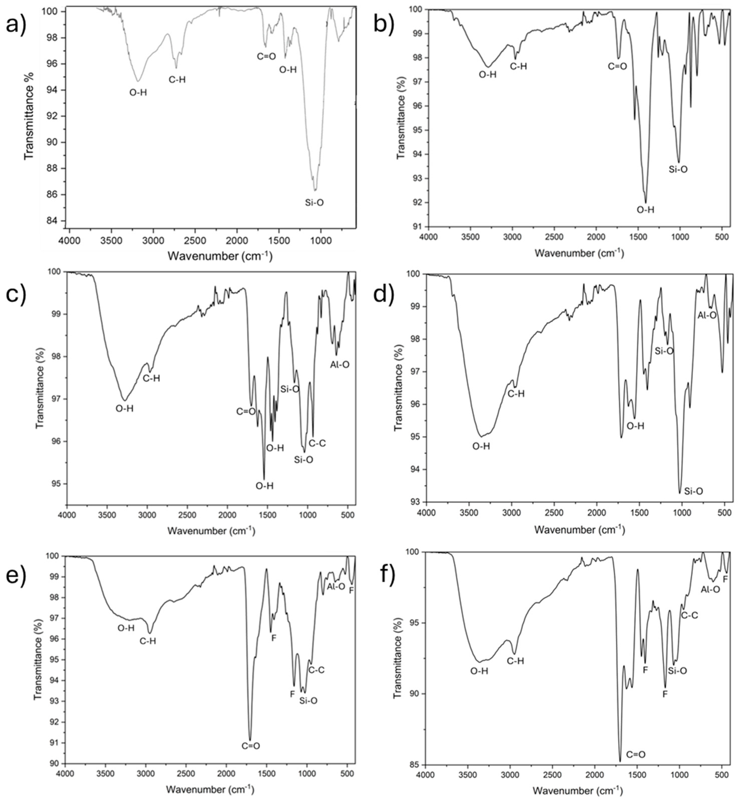
4.3. Characterization of the Samples Using FTIR (Fourier Transform Infrared Spectroscopy)

To determine the presence of fluoride in the experimental groups, Fourier transform infrared analysis was used. The glass ionomer cements with fluoride nanotubes were analyzed using a spectrometer (6700 FTIR Perkin Elmer; Waltham, MA, USA) with attenuated total reflectance, employing a diamond/zinc-selenide crystal plate. Each sample was analyzed by thirty-two scans with a 5 cm−1 spectral resolution using an infrared spectrum range between 400–4000 cm−1.

4.4. Modification of Glass Ionomers with Preloaded Fluoride Halloysite Nanotubes

For this study, 2 glass ionomers, Vitrebond (VB; 3M ESPE, St. Paul, MN, USA) and Ionobond (IB; VOCO GmbH, Cuxhaven, Germany), were used. Sixty blocks were fabricated in a Teflon matrix with a 3 mm diameter and a thickness of 1 mm. The materials were manipulated according to the instructions provided by the makers. The materials activated by light were polymerized with an LED lamp (Elipar; 3M ESPE) for 20 s; a radiometer (Demetron; Kavo Kerr, Orange, CA, USA) was necessary to confirm the correct intensity of the emitted light. This had to be greater than 400 milliwatts per square centimeter (mW/cm2).
Using the spoon provided by the manufacturer, the amount of powder indicated was retrieved from the bottle and subsequently weighed. This procedure was performed 10 times to calculate the average amount of powder corresponding to the spoon provided by the manufacturer, this procedure was repeated to elaborate each sample. The average for VB was 0.0483 mg, while the average for IB was 0.1043 mg. Then, 10% of the ionomer powder was replaced with HNTs with and without NaF to prepare the positive control and experimental groups, respectively. The total powder of these groups was mixed with the liquid following the instructions proportionated by the manufacturer.
The control group consisted of 10 blocks of each ionomer analyzed in this study. For the positive control group, 20 blocks of ionomers with HNTs were fabricated and divided into 2 groups (10 blocks per group) as follows: the VBHNT group (VB ionomer with HNTs) and the IBHNT group (IB ionomer with HNTs).
The experimental groups consisted of 10 blocks of each ionomer with HNT-NaF at 2000 ppm, which were distributed into 2 groups (VBHNT-NaF and IBHNT-NaF). The sampling distribution is presented in Figure 7.

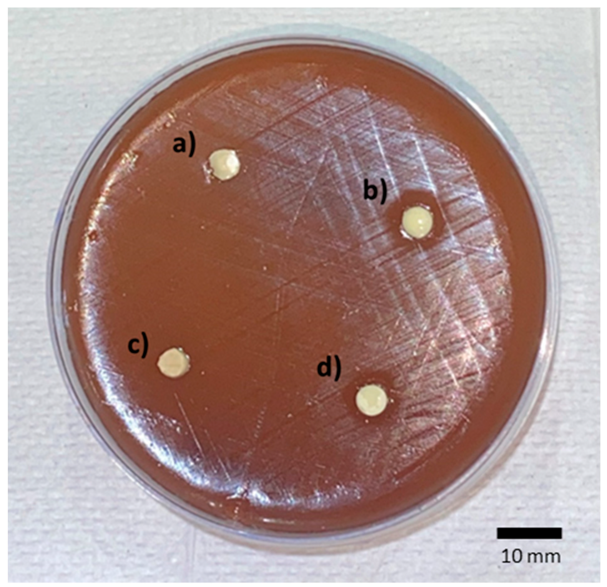
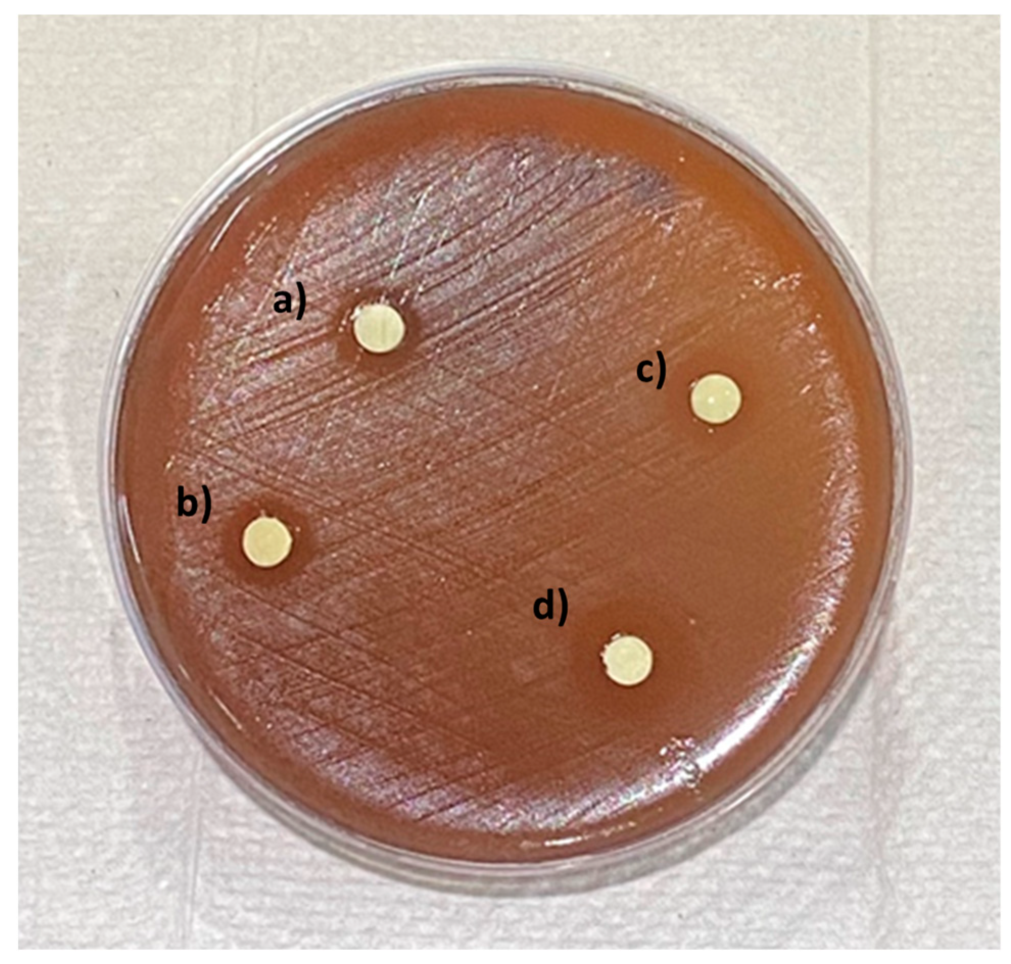
4.5. Microbiological Assay

The microbiological tests were carried out according to the CLSI-established guidelines, specifically those in the document M100 [62]. Mueller–Hinton agar was supplemented with sheep blood at a percentage of 5% (BD Columbia, Heidelberg, Germany), and this was used to seed S. mutans (ATCC) 33688 through the cross-streak method, and the samples were subsequently incubated for 24 h at 37 °C.
A bacterial suspension was prepared with a turbidity of 0.5 according to the McFarland scale (equivalent to 1.5 × 10⁸ CFU/mL) in 0.9% NaCl2 solution. The quantification of the bacteria was carried out with the plate count method. The AMH plates were fully inoculated with S. mutans without leaving any free zones, and the samples corresponding to the controls and experimental glass ionomer groups were set on the plates. The plates were taken into an incubator (Thermo Fisher Scientific, Waltham, MA, USA) with an anaerobic atmosphere with 5% CO2 for 24 h at 37 °C. This procedure was performed in triplicate. Subsequently, the bacterial growth on the plates was evaluated to determine the area of bacterial inhibition, which was measured with a Vernier caliper in millimeters.

4.6. Statistical Analysis

The SPSS 25.0 statistical program (IBM, New York, NY, USA) was used to analyze the data obtained. To perform multiple comparisons between the study groups and determine statistical differences between the study groups, the Kruskal–Wallis statistical test was necessary, and the Mann–Whitney U test was performed to determine between which study groups these statistical differences were found. In all the statistical test used in this study, a p value ≤ 0.05 was considered significant.

5. Conclusions

In this study, glass ionomers were modified by replacing 10% of the average amount of powder required with fluoride-modified halloysite nanotubes (HNT-NaF). The glass ionomer Ionobond modified with HNT-NaF obtained an efficient growth-inhibitory effect on S. mutans, which this material did not show previously. Vitrebond had a growth-inhibitory effect attributable to the methacrylates in its composition. However, this effect was increased by the incorporation of HNT-NaF, showing wider halos. Similarly, it was established that the minimum amount of sodium fluoride required to inhibit S. mutans was between 3000 and 6000 ppm, which could be improved to 2000 ppm by using HNTs.
Halloysite nanotubes are a highly promising additive for improving dental materials. Fluoride-modified halloysite nanotubes can promote the inhibitory effect of glass ionomers on S. mutans.

Limitations of the Study

In this study, only one bacterial strain was evaluated during the first 24 h. The evaluation of other bacterial strains with observations beyond this period would have been appropriate to evaluate its long-term effectiveness. It would be desirable to carry out more experiments to evaluate the materials’ antibacterial activity, such as the growth kinetics of the biofilm, the viability of bacterial cells, and the morphology of bacterial cells. The authors consider that future studies should be developed to evaluate the behavior and antibacterial capabilities of GICs and RMGIs reinforced with fluoride nanotubes under clinical conditions and analyze their physical and mechanical properties as well as their fluoride release.

Author Contributions

Investigation, R.D.J.-P.; Conceptualization, A.A.M.-V.; Software, R.A.M.-L.; Formal Analysis, U.V.-E.; Data Curation, E.L.-C.; Methodology, L.V.J.-R.; Supervision, O.A.C.-C.; Project Administration, V.H.T.-R.; Visualization, R.C.-P.; Validation, J.J.-G.; Writing—Original Draft, Writing—Review and Editing, E.N.S.-V. All authors have read and agreed to the published version of the manuscript.

Funding

This research received no external funding.

Institutional Review Board Statement

Not applicable.

Informed Consent Statement

Not applicable.

Data Availability Statement

The original contributions presented in this study are included in the article. Further inquiries can be directed to the corresponding author.

Acknowledgments

We thank the Center for Applied Physics and Advanced Technology, U.N.A.M., Campus Juriquilla, Queretaro.

Conflicts of Interest

The authors declare no conflicts of interest.

References

  1. Soubhagya, M.; Goud, K.M.; Deepak, B.S.; Thakur, S.; Nandini, T.N.; Arun, J. Comparative in Vitro Evaluation of Internal Adaptation of Resin-Modified Glass Ionomer, Flowable Composite and Bonding Agent Applied as a Liner under Composite Restoration: A Scanning Electron Microscope Study. J. Int. Oral Health 2015, 7, 27–31. [Google Scholar] [PubMed]
  2. Weiner, R. Liners and Bases in General Dentistry. Aust. Dent. J. 2011, 56, 11–22. [Google Scholar] [CrossRef] [PubMed]
  3. Strober, B.; Veitz-Keenan, A.; Barna, J.A.; Matthews, A.G.; Vena, D.; Craig, R.G.; Curro, F.A.; Thompson, V.P. Effectiveness of a Resin-Modified Glass Ionomer Liner in Reducing Hypersensitivity in Posterior Restorations: A Study from the Practitioners Engaged in Applied Research and Learning Network. J. Am. Dent. Assoc. 2013, 144, 886–897. [Google Scholar] [CrossRef] [PubMed]
  4. Mitra, S.B. Adhesion to Dentin and Physical Properties of a Light-Cured Glass-Ionomer Liner/Base. J. Dent. Res. 1991, 70, 72–74. [Google Scholar] [CrossRef]
  5. Azevedo, L.M.; Casas-Apayco, L.C.; Villavicencio Espinoza, C.A.; Wang, L.; Navarro, M.F.D.L.; Atta, M.T. Effect of Resin-Modified Glass-Ionomer Cement Lining and Composite Layering Technique on the Adhesive Interface of Lateral Wall. J. Appl. Oral Sci. 2015, 23, 315–320. [Google Scholar] [CrossRef]
  6. Rodrigues, M.d.P.; da Cunha, L.S.; Vilela, A.B.F.; Schettini, A.C.T.; de Bragança, G.F.; França, R.; Soares, C.J. Selective Carious Tissue Removal and Glass Ionomer Liner Reduction of Pulp Stress in Bulk Fill Resin Composite Restorations. Braz. Oral Res. 2021, 35, e119. [Google Scholar] [CrossRef]
  7. Kasraei, S.; Azarsina, M.; Majidi, S. In Vitro Comparison of Microleakage of Posterior Resin Composites with and without Liner Using Two-Step Etch-and-Rinse and Self-Etch Dentin Adhesive Systems. Oper. Dent. 2011, 36, 213–221. [Google Scholar] [CrossRef]
  8. Güray Efes, B.; Yaman, B.C.; Gümüştaş, B.; Tiryaki, M. The Effects of Glass Ionomer and Flowable Composite Liners on the Fracture Resistance of Open-Sandwich Class II Restorations. Dent. Mater. J. 2013, 32, 877–882. [Google Scholar] [CrossRef]
  9. Caneppele, T.; Bresciani, E.; da Silva Ávila, D.; Barcellos, D.; Pucci, C. Effect of Lining Materials on Shear Bond Strength for Composite Restorations In Vitro. Int. J. Periodontics Restor. Dent. 2017, 37, 137–143. [Google Scholar] [CrossRef]
  10. Ranjbar Omrani, L.; Moradi, Z.; Abbasi, M.; Kharazifard, M.J.; Tabatabaei, S.N. Evaluation of Compressive Strength of Several Pulp Capping Materials. J. Dent. 2021, 22, 41–47. [Google Scholar] [CrossRef]
  11. Alsunbul, H.; Khan, A.A.; Alqahtani, Y.M.; Hassan, S.A.b.; Asiri, W.; Saadaldin, S.; Alharthi, R.; Aldegheishem, A. Using Functionalized Micron-Sized Glass Fibres for the Synergistic Effect of Glass Ionomer on Luting Material. J. Funct. Biomater. 2023, 14, 550. [Google Scholar] [CrossRef]
  12. Alsunbul, H.; Khan, A.A.; De Vera, M.A.T.; Bautista, L.S.J.; Javed, R. Utilizing an Oxidized Biopolymer to Enhance the Bonding of Glass Ionomer Luting Cement Particles for Improved Physical and Mechanical Properties. Biomimetics 2023, 8, 347. [Google Scholar] [CrossRef]
  13. De Magalhães, C.S.; Hara, A.T.; Turssi, C.P.; Serra, M.C.; Giannini, M. Microhardness Evaluation around Composite Restorations Using Fluoride-Containing Adhesive Systems. J. Appl. Oral Sci. 2005, 13, 259–264. [Google Scholar] [CrossRef]
  14. Duque, C.; Negrini, T.D.C.; Sacono, N.T.; Spolidorio, D.M.P.; De Souza Costa, C.A.; Hebling, J. Clinical and Microbiological Performance of Resin-Modified Glass-Ionomer Liners after Incomplete Dentine Caries Removal. Clin. Oral Investig. 2009, 13, 465–471. [Google Scholar] [CrossRef]
  15. Deepa, V.L.; Dhamaraju, B.; Bollu, I.P.; Balaji, T.S. Shear Bond Strength Evaluation of Resin Composite Bonded to Three Different Liners: TheraCal LC, Biodentine, and Resin-Modified Glass Ionomer Cement Using Universal Adhesive: An in Vitro Study. J. Conserv. Dent. 2016, 19, 166–170. [Google Scholar] [CrossRef]
  16. Askar, H.; Krois, J.; Göstemeyer, G.; Schwendicke, F. Secondary Caries Risk of Different Adhesive Strategies and Restorative Materials in Permanent Teeth: Systematic Review and Network Meta-Analysis. J. Dent. 2021, 104, 103541. [Google Scholar] [CrossRef]
  17. Torres, C.R.G.; Mailart, M.C.; Rocha, R.S.; Sellan, P.L.B.; Contreras, S.C.M.; Di Nicoló, R.; Borges, A.B. The Influence of a Liner on Deep Bulk-Fill Restorations: Randomized Clinical Trial. J. Dent. 2020, 102, 103454. [Google Scholar] [CrossRef]
  18. Yon, M.J.Y.; Gao, S.S.; Chen, K.J.; Duangthip, D.; Lo, E.C.M.; Chu, C.H. Medical Model in Caries Management. Dent. J. 2019, 7, 37. [Google Scholar] [CrossRef]
  19. ten Cate, J.M. Current Concepts on the Theories of the Mechanism of Action of Fluoride. Acta Odontol. Scand. 1999, 57, 325–329. [Google Scholar] [CrossRef]
  20. Naik, S.V.; Attiguppe, P.; Malik, N.; Ballal, S. CPP–ACP and Fluoride: A Synergism to Combat Caries. Int. J. Clin. Pediatr. Dent. 2019, 12, 120–125. [Google Scholar] [CrossRef]
  21. Lemos, J.A.; Palmer, S.R.; Zeng, L.; Wen, Z.T.; Kajfasz, J.K.; Freires, I.A.; Abranches, J.; Brady, L.J. The Biology of Streptococcus Mutans. Microbiol. Spectr. 2019, 7, 7. [Google Scholar] [CrossRef]
  22. Kovacs, C.J.; Faustoferri, R.C.; Bischer, A.P.; Quivey, R.G. Streptococcus Mutans Requires Mature Rhamnose-Glucose Polysaccharides for Proper Pathophysiology, Morphogenesis and Cellular Division. Mol. Microbiol. 2019, 112, 944–959. [Google Scholar] [CrossRef]
  23. Kucukyilmaz, E.; Savas, S.; Kavrik, F.; Yasa, B.; Botsali, M. Fluoride Release/Recharging Ability and Bond Strength of Glass Ionomer Cements to Sound and Caries-Affected Dentin. Niger. J. Clin. Pract. 2017, 20, 226–234. [Google Scholar] [CrossRef]
  24. Hironaka, N.G.L.; Trizzi, J.Q.; Yoshida, N.M.; Cury, J.A.; Tabchoury, C.P.M.; Botelho, J.N.; Pini, N.I.P.; Pascotto, R.C. Use of Sonic Waves in Bubble Formation, Microhardness and Fluoride Release of a High-Viscosity Glass-Ionomer Cement. J. Investig. Clin. Dent. 2019, 10, e12456. [Google Scholar] [CrossRef]
  25. Bamoussa, A.; Assery, M.; Pani, S. Fluoride Release and Recharge Abilities of Zinc-Reinforced Glass Ionomer Cement in Comparison to Traditional High Strength Glass Ionomers. Saudi J. Oral Sci. 2015, 2, 69. [Google Scholar] [CrossRef]
  26. Cibim, D.D.; Saito, M.T.; Giovani, P.A.; Borges, A.F.S.; Pecorari, V.G.A.; Gomes, O.P.; Lisboa-Filho, P.N.; Nociti-Junior, F.H.; Puppin-Rontani, R.M.; Kantovitz, K.R. Novel Nanotechnology of TiO2 Improves Physical-Chemical and Biological Properties of Glass Ionomer Cement. Int. J. Biomater. 2017, 2017, 7123919. [Google Scholar] [CrossRef]
  27. Morales-Valenzuela, A.A.; Scougall-Vilchis, R.J.; Lara-Carrillo, E.; Garcia-Contreras, R.; Salmeron-Valdes, E.N.; Aguillón-Sol, L. Comparison of Fluoride Release in Conventional Glass-Ionomer Cements with a New Mechanical Mixing Cement. Oral Health Prev. Dent. 2020, 18, 319–324. [Google Scholar] [CrossRef]
  28. Pradiptama, Y.; Purwanta, M.; Notopuro, H. Antibacterial Effects of Fluoride in Streptococcus Mutans Growth in Vitro. Biomol. Health Sci. J. 2019, 2, 1. [Google Scholar] [CrossRef]
  29. Najeeb, S.; Khurshid, Z.; Zafar, M.; Khan, A.; Zohaib, S.; Martí, J.; Sauro, S.; Matinlinna, J.; Rehman, I. Modifications in Glass Ionomer Cements: Nano-Sized Fillers and Bioactive Nanoceramics. Int. J. Mol. Sci. 2016, 17, 1134. [Google Scholar] [CrossRef]
  30. Ching, H.S.; Luddin, N.; Kannan, T.P.; Ab Rahman, I.; Abdul Ghani, N.R.N. Modification of Glass Ionomer Cements on Their Physical-Mechanical and Antimicrobial Properties. J. Esthet. Restor. Dent. 2018, 30, 557–571. [Google Scholar] [CrossRef] [PubMed]
  31. Kheur, M.; Kantharia, N.; Iakha, T.; Kheur, S.; Husain, N.A.H.; Özcan, M. Evaluation of Mechanical and Adhesion Properties of Glass Ionomer Cement Incorporating Nano-Sized Hydroxyapatite Particles. Odontology 2020, 108, 66–73. [Google Scholar] [CrossRef] [PubMed]
  32. Noori, A.J.; Kareem, F.A. Setting Time, Mechanical and Adhesive Properties of Magnesium Oxide Nanoparticles Modified Glass-Ionomer Cement. J. Mater. Res. Technol. 2020, 9, 1809–1818. [Google Scholar] [CrossRef]
  33. Malekhoseini, Z.; Rezvani, M.B.; Niakan, M.; Atai, M.; Bassir, M.M.; Alizade, H.S.; Siabani, S. Effect of Zinc Oxide Nanoparticles on Physical and Antimicrobial Properties of Resin-Modified Glass Ionomer Cement. Dent. Res. J. 2021, 18, 73. [Google Scholar] [CrossRef]
  34. Noori, A.J.; Kareem, F.A. The Effect of Magnesium Oxide Nanoparticles on the Antibacterial and Antibiofilm Properties of Glass-Ionomer Cement. Heliyon 2019, 5, e02568. [Google Scholar] [CrossRef]
  35. Chen, J.; Zhao, Q.; Peng, J.; Yang, X.; Yu, D.; Zhao, W. Antibacterial and Mechanical Properties of Reduced Graphene-Silver Nanoparticle Nanocomposite Modified Glass Ionomer Cements. J. Dent. 2020, 96, 103332. [Google Scholar] [CrossRef]
  36. Barman, M.; Mahmood, S.; Augustine, R.; Hasan, A.; Thomas, S.; Ghosal, K. Natural Halloysite Nanotubes/Chitosan Based Bio-Nanocomposite for Delivering Norfloxacin, an Anti-Microbial Agent in Sustained Release Manner. Int. J. Biol. Macromol. 2020, 162, 1849–1861. [Google Scholar] [CrossRef]
  37. Ostadhossein, F.; Moitra, P.; Altun, E.; Dutta, D.; Sar, D.; Tripathi, I.; Hsiao, S.-H.; Kravchuk, V.; Nie, S.; Pan, D. Function-Adaptive Clustered Nanoparticles Reverse Streptococcus Mutans Dental Biofilm and Maintain Microbiota Balance. Commun. Biol. 2021, 4, 846. [Google Scholar] [CrossRef]
  38. Fakhruddin, K.; Hassan, R.; Khan, M.U.A.; Allisha, S.N.; Razak, S.I.A.; Zreaqat, M.H.; Latip, H.F.M.; Jamaludin, M.N.; Hassan, A. Halloysite Nanotubes and Halloysite-Based Composites for Biomedical Applications. Arab. J. Chem. 2021, 14, 103294. [Google Scholar] [CrossRef]
  39. Yuan, P.; Tan, D.; Annabi-Bergaya, F. Properties and Applications of Halloysite Nanotubes: Recent Research Advances and Future Prospects. Appl. Clay Sci. 2015, 112–113, 75–93. [Google Scholar] [CrossRef]
  40. Carazo, E.; Borrego-Sánchez, A.; García-Villén, F.; Sánchez-Espejo, R.; Aguzzi, C.; Viseras, C.; Sainz-Díaz, C.I.; Cerezo, P. Assessment of Halloysite Nanotubes as Vehicles of Isoniazid. Colloids Surf. B Biointerfaces 2017, 160, 337–344. [Google Scholar] [CrossRef]
  41. Alkatheeri, M.S.; Palasuk, J.; Eckert, G.J.; Platt, J.A.; Bottino, M.C. Halloysite Nanotube Incorporation into Adhesive Systems—Effect on Bond Strength to Human Dentin. Clin. Oral Investig. 2015, 19, 1905–1912. [Google Scholar] [CrossRef]
  42. Salmerón-Valdés, E.N.; Cruz-Mondragón, A.C.; Toral-Rizo, V.H.; Jiménez-Rojas, L.V.; Correa-Prado, R.; Lara-Carrillo, E.; Morales-Valenzuela, A.A.; Scougall-Vilchis, R.J.; López-Flores, A.I.; Hoz-Rodriguez, L.; et al. Mechanical Properties and Antibacterial Effect on Mono-Strain of Streptococcus Mutans of Orthodontic Cements Reinforced with Chlorhexidine-Modified Nanotubes. Nanomaterials 2022, 12, 2891. [Google Scholar] [CrossRef]
  43. Takahashi, Y.; Imazato, S.; Kaneshiro, A.V.; Ebisu, S.; Frencken, J.E.; Tay, F.R. Antibacterial Effects and Physical Properties of Glass-Ionomer Cements Containing Chlorhexidine for the ART Approach. Dent. Mater. 2006, 22, 647–652. [Google Scholar] [CrossRef]
  44. Hoszek, A.; Ericson, D. In Vitro Fluoride Release and the Antibacterial Effect of Glass Lonomers Containing Chlorhexidine Gluconate. Oper. Dent. 2008, 33, 696–701. [Google Scholar] [CrossRef]
  45. Türkün, L.Ş.; Türkün, M.; Ertuǧrul, F.; Ateş, M.; Brugger, S. Long-Term Antibacterial Effects and Physical Properties of a Chlorhexidine-Containing Glass Ionomer Cement. J. Esthet. Restor. Dent. 2008, 20, 29–44. [Google Scholar] [CrossRef]
  46. Balouiri, M.; Sadiki, M.; Ibnsouda, S.K. Methods for in Vitro Evaluating Antimicrobial Activity: A Review. J. Pharm. Anal. 2016, 6, 71–79. [Google Scholar] [CrossRef]
  47. Vermeersch, G.; Leloup, G.; Delmée, M.; Vreven, J. Antibacterial Activity of Glass-Ionomer Cements, Compomers and Resin Composites: Relationship between Acidity and Material Setting Phase. J. Oral Rehabil. 2005, 32, 368–374. [Google Scholar] [CrossRef]
  48. Palenik, C.J.; Behnen, M.J.; Setcos, J.C.; Miller, C.H. Inhibition of Microbial Adherence and Growth by Various Glass Ionomers in Vitro. Dent. Mater. 1992, 8, 16–20. [Google Scholar] [CrossRef]
  49. Bowden, G.H.W. Effects of Fluoride on the Microbial Ecology of Dental Plaque. J. Dent. Res. 1990, 69, 653–659. [Google Scholar] [CrossRef] [PubMed]
  50. Perales Zamora, S.; Guillen Bordaz, C.; Loayza De La Cruz, R.; Alvarado Menacho, S.; Torres Ramos, G.; Guillén Astete, A.; Anticona Huaynate, C. El Flúor En La Prevención de Caries En La Dentición Temporal. Barnices Fluorados. Odontol. Sanmarquina 2014, 9, 33. [Google Scholar] [CrossRef]
  51. Vamsi, K.; Siddiqui, F. Antimicrobial Effect of an Experimental Glass Ionomer Cement against Pathogens Associated with Deep Carious Lesions. J. Contemp. Dent. Pract. 2018, 19, 824–829. [Google Scholar] [CrossRef] [PubMed]
  52. Heidarinasab, A.; Ahmad Panahi, H.; Faramarzi, M.; Farjadian, F. Synthesis of Thermosensitive Magnetic Nanocarrier for Controlled Sorafenib Delivery. Mater. Sci. Eng. C 2016, 67, 42–50. [Google Scholar] [CrossRef]
  53. Renné, W.G.; Lindner, A.; Mennito, A.S.; Agee, K.A.; Pashley, D.H.; Willett, D.; Sentelle, D.; Defee, M.; Schmidt, M.; Sabatini, C. Antibacterial Properties of Copper Iodide-Doped Glass Ionomer-Based Materials and Effect of Copper Iodide Nanoparticles on Collagen Degradation. Clin. Oral Investig. 2017, 21, 369–379. [Google Scholar] [CrossRef] [PubMed]
  54. Feitosa, S.A.; Palasuk, J.; Geraldeli, S.; Windsor, L.J.; Bottino, M.C. Physicochemical and Biological Properties of Novel Chlorhexidine-Loaded Nanotube-Modified Dentin Adhesive. J. Biomed. Mater. Res. B Appl. Biomater. 2019, 107, 868–875. [Google Scholar] [CrossRef]
  55. Huang, Q.; Wang, S.; Sun, Y.; Shi, C.; Yang, H.; Lu, Z. Effects of Ag/ZnO Nanocomposite at Sub-Minimum Inhibitory Concentrations on Virulence Factors of Streptococcus Mutans. Arch. Oral Biol. 2020, 111, 104640. [Google Scholar] [CrossRef]
  56. Feitosa, S.A.; Palasuk, J.; Kamocki, K.; Geraldeli, S.; Gregory, R.L.; Platt, J.A.; Windsor, L.J.; Bottino, M.C. Doxycycline-Encapsulated Nanotube-Modified Dentin Adhesives. J. Dent. Res. 2014, 93, 1270–1276. [Google Scholar] [CrossRef]
  57. Massaro, M.; Lazzara, G.; Milioto, S.; Noto, R.; Riela, S. Correction: Covalently Modified Halloysite Clay Nanotubes: Synthesis, Properties, Biological and Medical Applications. J. Mater. Chem. B 2017, 5, 4246. [Google Scholar] [CrossRef]
  58. Leyhausen, G.; Abtahi, M.; Karbakhsch, M.; Sapotnick, A.; Geurtsen, W. Biocompatibility of Various Light-Curing and One Conventional Glass-Ionomer Cement. Biomaterials 1998, 19, 559–564. [Google Scholar] [CrossRef] [PubMed]
  59. Salmerón-Valdés, E.; Scougall-Vilchis, R.; Alanis-Tavira, J.; Morales-Luckie, R. Comparative Study of Fluoride Released and Recharged from Conventional Pit and Fissure Sealants versus Surface Prereacted Glass Ionomer Technology. J. Conserv. Dent. 2016, 19, 41–45. [Google Scholar] [CrossRef]
  60. Kalagi, S.; Feitosa, S.A.; Münchow, E.A.; Martins, V.M.; Karczewski, A.E.; Cook, N.B.; Diefenderfer, K.; Eckert, G.J.; Geraldeli, S.; Bottino, M.C. Chlorhexidine-Modified Nanotubes and Their Effects on the Polymerization and Bonding Performance of a Dental Adhesive. Dent. Mater. 2020, 36, 687–697. [Google Scholar] [CrossRef]
  61. Degrazia, F.W.; Leitune, V.C.B.; Takimi, A.S.; Collares, F.M.; Sauro, S. Physicochemical and Bioactive Properties of Innovative Resin-Based Materials Containing Functional Halloysite-Nanotubes Fillers. Dent. Mater. 2016, 32, 1133–1143. [Google Scholar] [CrossRef] [PubMed]
  62. CLSI M100; Performance Standards for Antimicrobial Susceptibility Testing. Clinical and Laboratory Standards Institute CLSI: Berwyn, PA, USA, 2020.
Figure 1. Fourier transform infrared spectroscopy of fluoride (a) and halloysite nanotubes (b).
Inorganics 13 00190 g001
Figure 2. Fourier transform infrared spectroscopy of study groups. The control groups of (a) Ionobond (IB) and (b) Vitrebond (VB) and the positive control groups. (c) Ionobond with nanotubes (IBHNT), (d) Vitrebond with nanotubes (VBHNT), (e) Ionobond with fluoride-modified nanotubes (IBHNT-NaF) and (f) Vitrebond with fluoride-modified nanotubes (VBHNT-NaF).
Inorganics 13 00190 g002
Figure 3. Inhibitory concentrations of control (NaF) and experimental (HNTs-NaF) groups, shown in ppm (parts per million).
Inorganics 13 00190 g003
Figure 4. Growth inhibition zone of control and positive control groups. (a) Ionobond (IB) and (b) Vitrebond (VB), which formed the control group; (c) Ionobond with halloysite nanotubes (IBHNT) and (d) Vitrebond with halloysite nanotubes (VBHNT), which formed the positive control group.
Inorganics 13 00190 g004
Figure 5. Growth inhibition zone of experimental groups: (a,b) IB with HNT-NaF at 2000 ppm, (c,d) VB with HNT-NaF at 2000 ppm.
Inorganics 13 00190 g005
Figure 6. Sketch of experimental procedures.
Inorganics 13 00190 g006
Figure 7. Sample distribution diagram, (n) sample size.
Inorganics 13 00190 g007
Table 1. Kruskal–Wallis’ test comparing the antibacterial effect between the experimental and control groups analyzed.
GroupsMean (SD)
Control group: Vitrebond (VB)8.30 (0.483)
Experimental group: (VBHNT-NaF)9.10 (0.568)
Positive control group Vitrebond: (VBHNT)8.50 (0.527)
Control group: Ionobond (IB)0.00 (0.00)
Positive control group Ionobond: (IBHNT)0.00 (0.00)
Experimental group: IBHNT-NaF8.60 (0.516)
Kruskal–Wallis‘ testp = 0.001 *
Mean: average of growth inhibition zone in millimeters; SD: standard deviation; *: p ≤ 0.05.
Table 2. Mann–Whitney U test.
GroupsStatisticsStatistics Deviationp Value
IB–VB25.2003.4020.010 *
IB–IBHNT0.0000.0001.000
IB–VBHNT−27.100−3.6580.004 *
IB–IBHNT-NaF−29.000−3.9150.001 *
IB–VBHNT-NaF−38.700−5.2240.0001 *
IBHNT–VBHNT−27.100−3.6580.004 *
IBHNT–IBHNT-NaF−29.000−3.9150.001 *
IBHNT–VBHNT-NaF−38.700−5.2240.0001 *
VB–IBHNT25.2003.4020.010 *
VB–VBHNT−1.900−0.2561.000
VB–IBHNT-NaF−3.800−0.5131.000
VB–VBHNT-NaF−13.500−1.8221.000
VBHNT–IBHNT-NaF−1.900−0.2561.000
VBHNT–VBHNT-NaF−11.600−1.5661.000
IBHNT-NaF–VBHNT-NaF9.7001.3091.000
IB, VB: control groups; IBHNT-NaF, VBHNT-NaF: experimental groups; *: p ≤ 0.05.
Disclaimer/Publisher’s Note: The statements, opinions and data contained in all publications are solely those of the individual author(s) and contributor(s) and not of MDPI and/or the editor(s). MDPI and/or the editor(s) disclaim responsibility for any injury to people or property resulting from any ideas, methods, instructions or products referred to in the content.

