Article

Exploring the Anti-Corrosion, Photocatalytic, and Adsorptive Functionalities of Biogenically Synthesized Zinc Oxide Nanoparticles

by
Syed Najmul Hejaz Azmi
1 and
Mahboob Alam
2,*
1
Applied Sciences Department, College of Applied Sciences and Pharmacy, University of Technology and Applied Sciences-Muscat, P.O. Box 74, Al Khuwair 133, Oman
2
Division of Smart Safety Engineering, Dongguk University Wise, 123 Dongdae-ro, Gyeongju-si 780714, Republic of Korea
*
Author to whom correspondence should be addressed.
Inorganics 2024, 12(7), 199; https://doi.org/10.3390/inorganics12070199
Submission received: 1 July 2024 / Revised: 14 July 2024 / Accepted: 15 July 2024 / Published: 22 July 2024

Abstract

:
This study reported the synthesis of ZnO nanoparticles (ZnO NPs) using Cucurbita pepo L. seed extract and explored their multifunctional properties such as anti-corrosion, photocatalytic, and adsorption capabilities. The synthesized ZnO NPs were characterized by Fourier-transform infrared spectroscopy (FTIR) to identify their functional groups, thermogravimetric analysis (TGA) to assess their thermal stability, transmission electron microscopy (TEM), and scanning electron microscopy with energy-dispersive X-ray spectroscopy (SEM-EDX) to determine their size, morphology, and elemental composition. The characterization of biofabricated ZnO NPs revealed an average particle size of 32.88 nm; however, SEM displayed a tendency for the particles to agglomerate. Furthermore, the X-ray diffraction (XRD) and EDX analysis confirmed the NPs as ZnO, matching patterns reported in the literature. In this study, the potential of the biogenic ZnO NPs was explored for multifunctional applications. Zinc oxide nanoparticles exhibited a higher capacity for adsorbing hydrogen sulfide (H2S) compared to bulk zinc oxide, mostly because of their larger surface area. In addition, electrochemical studies demonstrated a substantial enhancement in the corrosion resistance of mild steel in a 1.0 M HCl solution. ZnO NPs also demonstrated remarkable photodegradation effectiveness, reducing 75% of methyl orange in 60 min under sun-light irradiation. This implies that they could be used to remediate organic pollutants (organic dyes) from wastewater.

1. Introduction

Nanoparticles, with their remarkable properties stemming from their high surface-to-volume ratio, represent the cornerstone of nanotechnology, a multidisciplinary field revolutionizing various aspects of human life [1,2,3]. Among its branches, green nanotechnology stands out for its cost-effective and predictable synthesis methods, promising a sustainable future [4,5,6,7]. The surge in nanomaterial production and consumption underscores their growing significance. Zinc oxide nanoparticles (ZnO NPs) are emerging as a particularly versatile candidate, especially in agriculture and environmental remediation [8,9]. ZnO NPs have garnered substantial attention owing to their unique properties and eco-friendly synthesis approaches, such as utilizing extracts from various sections of plants [10,11,12,13,14]. These nanoparticles hold promise for a wide range of applications, from antimicrobial coatings to water treatment. However, alongside the enthusiasm for their applications, concerns about the safety and toxicity of nanomaterials persist. This has urged researchers to explore biocompatible synthesis methods and assess their environmental impact rigorously [15]. In this context, the biogenic synthesis of ZnO NPs presents a promising avenue, leveraging the reducing and capping properties of microbial enzymes and biomolecules [16]. Such approaches offer a sustainable alternative to conventional synthesis methods. They also hold potential for diverse applications, including wastewater treatment and agricultural practices [17]. The rise of nanoscience has revolutionized various scientific disciplines, particularly in the field of environmental remediation. Zinc oxide nanoparticles are one of the most interesting and promising nanomaterials. They have special properties, such as a wide band gap (3.37 eV), the ability to oxidize strongly, and excellent photocatalytic performance [18,19]. These characteristics make ZnO NPs ideal candidates for the environmental and medical sectors [20]. As a result, ZnO NPs synthesis has shifted toward environmentally friendly methods using biological substrates such as plant extracts, bacteria, and fungi. These methods offer cost-effectiveness, simplicity, and reduced environmental risks, making them suitable for biomedicine and environmental remediation. Traditional methods often involve toxic solvents, high energy consumption, and hazardous by-products. Green synthesis methods eliminate these risks by using natural reducing and stabilizing agents found in biological materials. ZnO NPs have shown promise in photocatalysis for pollutant removal, offering a sustainable solution for water purification and pollution control. Further research is needed to optimize the environmental impact of green synthesis methods and explore different biological substrates [13,17]. Bio-extracts in aqueous medium can create a protective layer for ZnO NPs, preventing corrosive agents from reaching the metal surface. The combination of organic compounds, such as saponins, quinones, and alkaloids, increases corrosion inhibition. These compounds also enhance stability, as reported in the literature on Cucurbita pepo L. seeds [21,22,23]. This green technology reduces reliance on toxic chemical inhibitors, making anti-corrosion treatments more sustainable and environmentally friendly. The use of bio-extracts in aqueous solutions increases efficiency.
In addressing the pressing issue of water pollution, the efficiency of nanomaterials in adsorbing and degrading contaminants, particularly dyes and heavy metals, has been a focal point [24]. Traditional wastewater treatment methods often fall short in addressing the escalating pollution levels, necessitating innovative approaches like nanotechnology [25]. By harnessing the superior properties of ZnO NPs, researchers aim to develop efficient solutions for wastewater treatment. These properties include high reactivity and stability, making the solutions eco-friendly. These solutions can mitigate environmental hazards while conserving energy. Moreover, the potential of ZnO NPs extends beyond wastewater treatment, encompassing diverse fields such as medicine, electronics, and catalysis. Their biocompatibility and antimicrobial properties make them promising candidates for biomedical applications, ranging from drug delivery systems to wound healing. As studies advance, it is crucial to investigate the synthesis mechanisms, enhance the performance, and overcome any constraints connected with ZnO NPs. Green synthesis refers to the biosynthesis of nanoparticles from natural sources. This method has become increasingly popular because it is both eco-friendly and cost-effective. Plant extracts, including those derived from roots to seeds, have been explored as potential sources for synthesizing nanoparticles with desirable properties [26,27,28,29]. These biogenic nanoparticles offer several advantages over chemically synthesized counterparts, including reduced toxicity and increased biocompatibility [30].
This study explores the utilization of biogenic ZnO NPs synthesized using an extract derived from Cucurbita pepo L. seeds. This environmentally sustainable process includes numerous benefits compared to conventional chemical-based approaches. The process reduces its adverse impact on the environment and exploits the beneficial components in the seed extract to potentially modify the characteristics of the resultant ZnO NPs.
The goal of this study is to examine the biogenic synthesis, photocatalytic activity, anti-corrosion capabilities, and adsorption capacity of these naturally derived ZnO NPs. Furthermore, the nanoparticles’ durability upon adsorption is assessed to determine their potential for reuse and long-term effectiveness. The objective of this research is to develop biofabricated ZnO NPs with adjustable characteristics for various uses, including anti-corrosion coatings, photocatalysis, and adsorption studies. An overview of the biosynthesis process for ZnO NPs employing various section plant extracts is provided in Table 1. These extracts, rich in bioorganic compounds, serve as reducing and stabilizing agents in the synthesis process, leading to the formation of ZnO NPs with different morphologies and sizes. Table 1 also represents characterization techniques important for analyzing structural, morphological, and chemical properties of the synthesized ZnO NPs. Additionally, it also demonstrates the practical applications of these ZnO NPs in various fields.

2. Results and Discussion

2.1. Characterization of Seed Extract-Mediated Biofabricated ZnO Nanoparticles

2.1.1. SEM, EDX, and TEM

Scanning electron microscopy (SEM) and energy-dispersive X-ray spectroscopy (EDX) were used to analyze the size and morphology of the biofabricated ZnO NPs. The SEM-EDX analysis confirmed the presence of ZnO NPs and their elemental composition. As shown in Figure 1a, agglomerates of ZnO NPs were observed, ranging in size from 1 to 5 µm, with irregular and non-uniform shapes. Aggregation of nanoparticles is a common phenomenon. It is possible to avoid this by using certain agents, altering drying techniques, enhancing their dispersion, and managing concentration and storage. These results, however, supported the earlier studies and pointed out the reproducibility of the biosynthesis process [29].
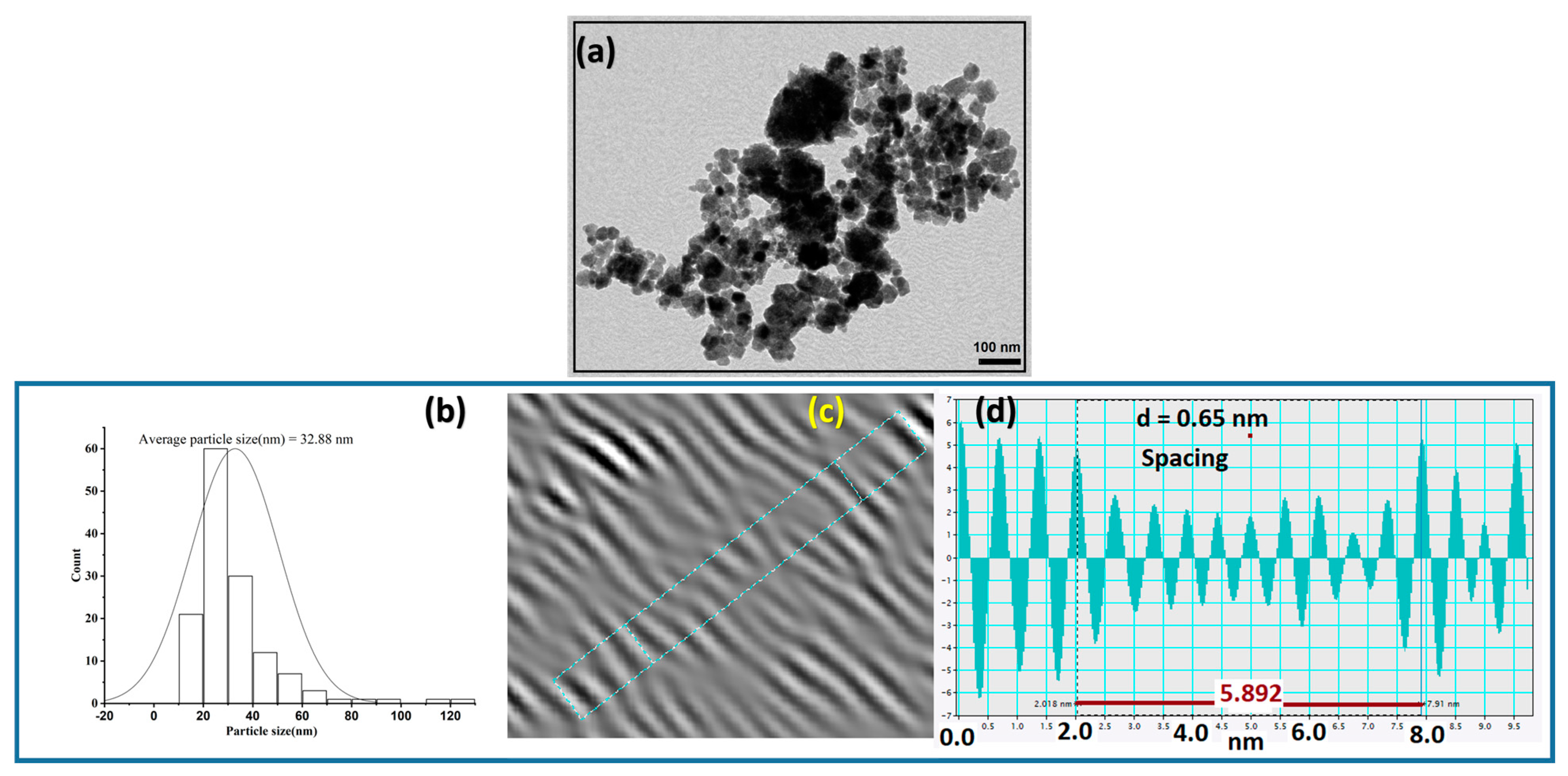
The EDX test revealed strong signals for zinc (71.85%) and oxygen (28.15%), which showed that ZnO NPs are existed (Figure 1b). These NPs likely formed due to the reduction in zinc ions in the seed extract. Furthermore, as shown in Figure 1b, EDX analysis of the ZnO sample indicated that it was of high purity. The EDX spectrum shows peaks for zinc (Zn) and oxygen (O), with atomic percentages of 38.39% Zn and 61.61% O, respectively. This suggests that the biogenic ZnO NPs have very little impurity content. The observed strong signal energy peaks for Zn-O in the 0–2 keV range further support the presence of ZnO NPs. The presence of a peak at around 2.1 keV [50] in the EDX pattern profile (Figure 1b) is attributed to the application of gold coating. This is a conventional method employed to improve the resolution of the SEM image. Transmission electron microscopy (TEM) analysis provides the dimensions and morphology of the zinc oxide nanoparticles that were produced by biosynthesis. Referring to the TEM images, the majority of the nanoparticles had a range of morphologies with thickness variations, including spheres, hexagons, triangles, and irregular forms (Figure 2a). The nanoparticles’ diameter measurements varied from 10 to 120 nanometers (nm). However, the histogram (Figure 2b) shows that the average particle size was 32.88 nm. Using DigitalMicrograph (DM) [51,52], the d-spacing of the lattice fringes was determined to be 0.65 nm (Figure 2c,d). The analysis showed that some nanoparticles aggregated together to form agglomerates (Figure 2a). Due to their high surface energy and large specific surface area, these nanoparticles are prone to aggregation. The observed agglomeration is likely a result of the drying process.

2.1.2. FT-IR, XRD, and UV Analysis

Fourier transform infrared (FTIR) spectroscopy is used to identify the functional groups present in biogenic nanoparticles produced from metallic precursors utilizing seed extract. The presence of ZnO NPs in the sample is further confirmed by FTIR analysis, as seen in Figure 3a, which supports SEM-EDX’s findings (Figure 1a). A distinctive peak in the spectra, located at 513 cm−1, is indicative of the Zn-O stretching vibration. This observation is consistent with earlier reports [53], according to which ZnO usually exhibits 400–600 cm−3 vibrational bands. Further analysis of the FTIR spectrum (Figure 3a) shows a broad peak at 3411 cm−1. This peak can be attributed to the O–H stretching vibration of the hydroxyl group, possibly originating from the alcohol functional group [29]. The presence of this peak indicates that biomolecules containing hydroxyl (OH) groups are involved in the formation process of ZnO NPs. These biomolecules are expected to play a dual role: (1) reducing Zn2+ ions to Zn0 and (2) stabilizing the bioreduced ZnO nanoparticles synthesized using the extract. The FTIR spectrum also revealed other informative peaks at 3165 cm−1 and 2981 cm−1. In addition, the alcoholic extract from Cucurbita pepo L. seeds has been analyzed using GC-MS, which identified a wide range of phytoconstituents, as per our earlier research [54]. The infrared (IR) data also confirms the presence of functional groups in bioactive compounds, suggesting the occurrence of reducing and capping agents in the extract. The biomolecules that have been identified are hexadecanoic acid ethyl ester, oleic acid, 9-octadecenoic acid (cis-vaccenic acid), 9,12-octadecadienoic acid, 1-chloroheptadecene, and 6,11-dimethyl-2,6,10-dodecatrien-1-ol in alcoholic seed extracts.
These peaks can be attributed to stretching vibrations of C-H bonds in organic molecules, possibly originating from various functional groups [55]. The presence of these peaks indicates the adsorption of biomolecules on the surface of nanoparticles [56]. Moreover, the C=C stretching vibration of an adsorbed molecule may be responsible for the peak at 1678 cm−1. It is expected that bio-nanoparticle FTIR analysis will provide insight into the potential uses of phytochemicals in extracts. The aqueous-based extract of seeds contains phytochemicals like phenols, terpenes, and flavonoids that may help turn metal ions into metals during the nanoparticle formation process.
The synthesis of biogenic ZnO NPs obtained from seed extract of Cucurbita pepo L. is confirmed by the PXRD graph (Figure 3b). The presence of sharp diffraction peaks at 2θ values of 31.74°, 34.40°, 36.22°, 47.39°, and 56.35° corresponds to Miller indices (100), (002), (101), (102), and (110), respectively. The reference patterns for the spherical and hexagonal wurtzite crystal phases of ZnO can be found on JCPDS cards (No. 36-1451, 87-0720, 89-7102, and 01-079-0206). These peaks match them. In line with earlier studies [57,58], this PXRD pattern shows that the biogenic ZnO NPs have a well-crystallized structure. ZnO NPs size was estimated in this work using X-ray diffraction (XRD). Through the application of the Scherrer equation, the average nanoparticle diameter was determined using the full width at half maximum (FWHM) of the intense peak in the XRD pattern. The average crystallite size (D) can be estimated using the Scherrer equation: D = Kλ/(β cos θ). In this equation, θ is the Bragg angle, K is a shape factor (usually 0.9), λ is the X-ray wavelength (0.15418), and β is the FWHM of the diffraction peak in radians after instrumental broadening adjustment. Regarding crystallite size, 13.72 nm was the estimated size. The size of a single, uniformly diffracting region within a material is referred to as the crystallite size in XRD analysis. However, this crystallite size may or may not coincide with the total particle size. SEM analysis revealed a larger particle, raising the possibility that it was a single nanoparticle composed of several smaller nanocrystals. Moreover, documents suggest that the broadening of XRD peaks can be attributed to flaws and internal stress [59].
UV-Vis spectroscopy is utilized to study, characterize, and identify metal nanoparticles, particularly within the 200–800 nm light wavelength range. By looking at a surface plasmon resonance (SPR) at 361.7 nm (Figure 3c), this method proves that ZnO NPs are synthesized from seed extract. The reaction mixture’s color change from light cream to brown indicates nanoparticle synthesis. Cucurbita pepo L. seed extract, rich in various biomolecules, serves as a valuable source for zinc nanoparticle synthesis; it also provides stabilization for the NPs. The observed absorption peaks correlate with nanoparticle size, with larger diameters exhibiting absorption at longer wavelengths. Higher bioactive component concentrations in the extract are responsible for the increase in SPR band intensity over time [60]. A tauc plot using the direct method [(αhν)^1/2 vs. eV] was used to find the band gap energy (Eg) of ZnO NPs (Figure 3d). The measurement revealed an Eg of 3.29 eV, indicating that these nanoparticles have potentially excellent optical properties. The size of ZnO NPs was further estimated to be in the range of 7.42–8.0 nm. This estimate is derived from the band gap energy (Eg) value and the absorption maximum using a simplified form of the Brus equation [61] given below.
diameter = hc/(λ_max × Eg_J) × 2
where
h is Planck’s constant (6.626 × 10⁻³⁴ J s), c is the speed of light (3 × 10⁸ m/s), λmax is the absorption maximum wavelength of the nanoparticles (in meters), Eg_J is the band gap energy of the bulk material in Joules (convert electron volts (eV) to Joules by multiplying by 1.602 × 10⁻¹⁹ J/eV. The Bruce equation is one technique for determining the diameter of spherical semiconductor nanocrystals. It works with the material’s known bulk band gap energy (Eg) and maximum absorption wavelength (λmax) from UV-visible spectroscopy. The Brus equation gives an estimate of the particle size. It assumes spherical nanoparticles; however, the real size may differ based on the shape of the nanoparticle and other factors.

2.2. Thermogravimetric Analysis (TGA)

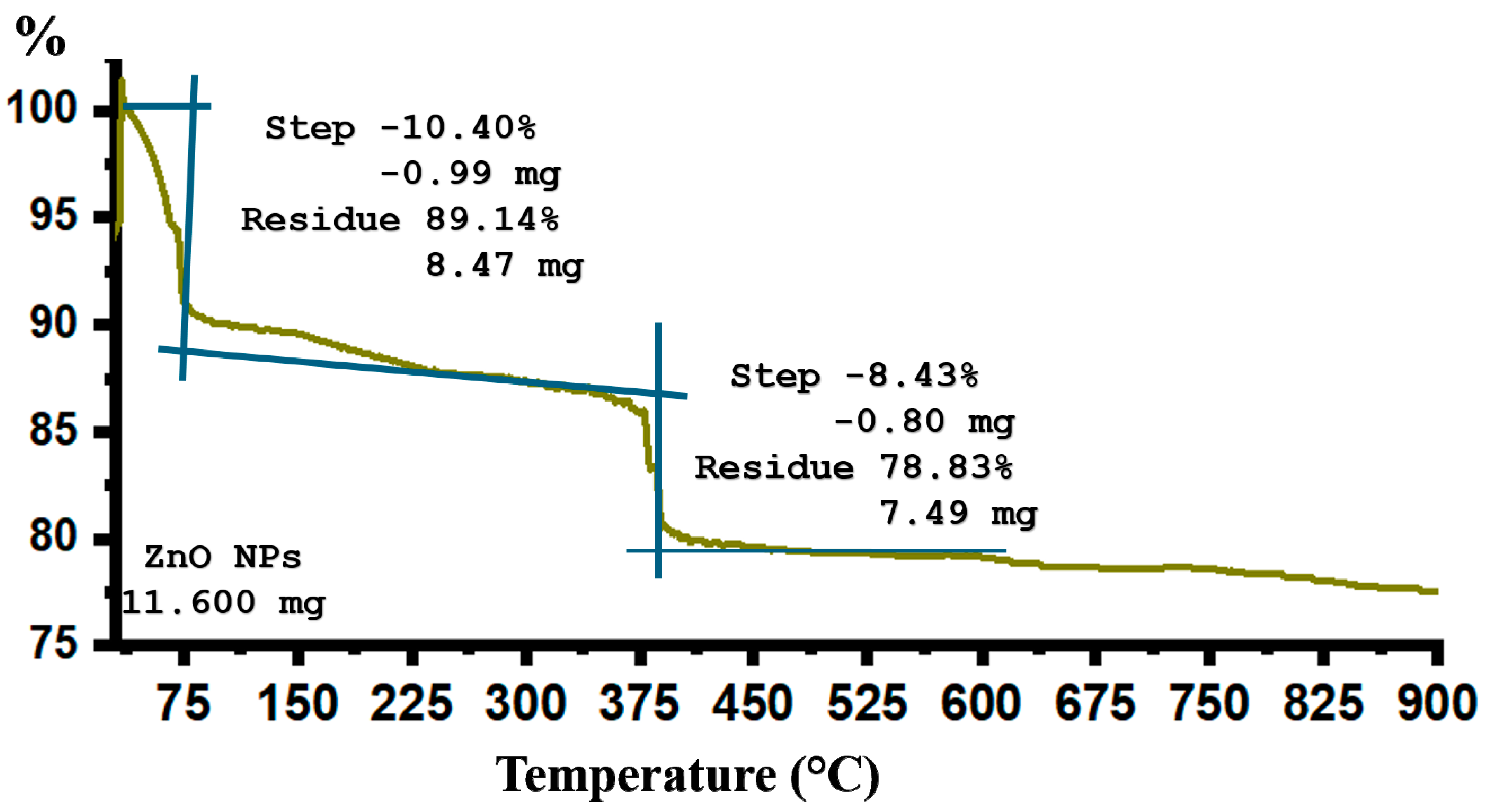
Thermogravimetric analysis (TGA) was performed to evaluate the thermal stability and organic content of seed-derived ZnO NPs (Figure 4). The weight loss of ZnO NPs powder was measured when it was heated from room temperature to 900 °C. The TGA curve (Figure 4) reveals two distinct phases of weight loss. The initial decrease below 100 °C corresponds to the evaporation of water from the sample surface. Initially, water evaporation led to a decrease in weight below 100 °C. At higher temperatures, we observed a two-stage weight decrease. The first stage, occurring around 68 °C, signified the breakdown of organic compounds linked with the nanoparticles. Additionally, a moderate weight loss of about 10.40% was observed in the sample in the temperature range of 20 °C to 80 °C. This weight loss may be attributed to the evaporation of water absorbed on the surface of the sample. This phenomenon may also denote a reduction in moisture content and organic materials within the ZnO NPs. TGA analysis yielded similar results [62], aligning with previous research. Another significant weight loss (8.41%) occurred in the temperature range of 380 °C to 500 °C. It is likely due to the breakdown of plant bioorganic compounds present on the ZnO NPs’ surface [63]. Significantly, the TGA curve showed no weight loss over 600 °C, demonstrating the stability of ZnO NPs in this temperature range.

2.3. Analysis of Photocatalytic Activity

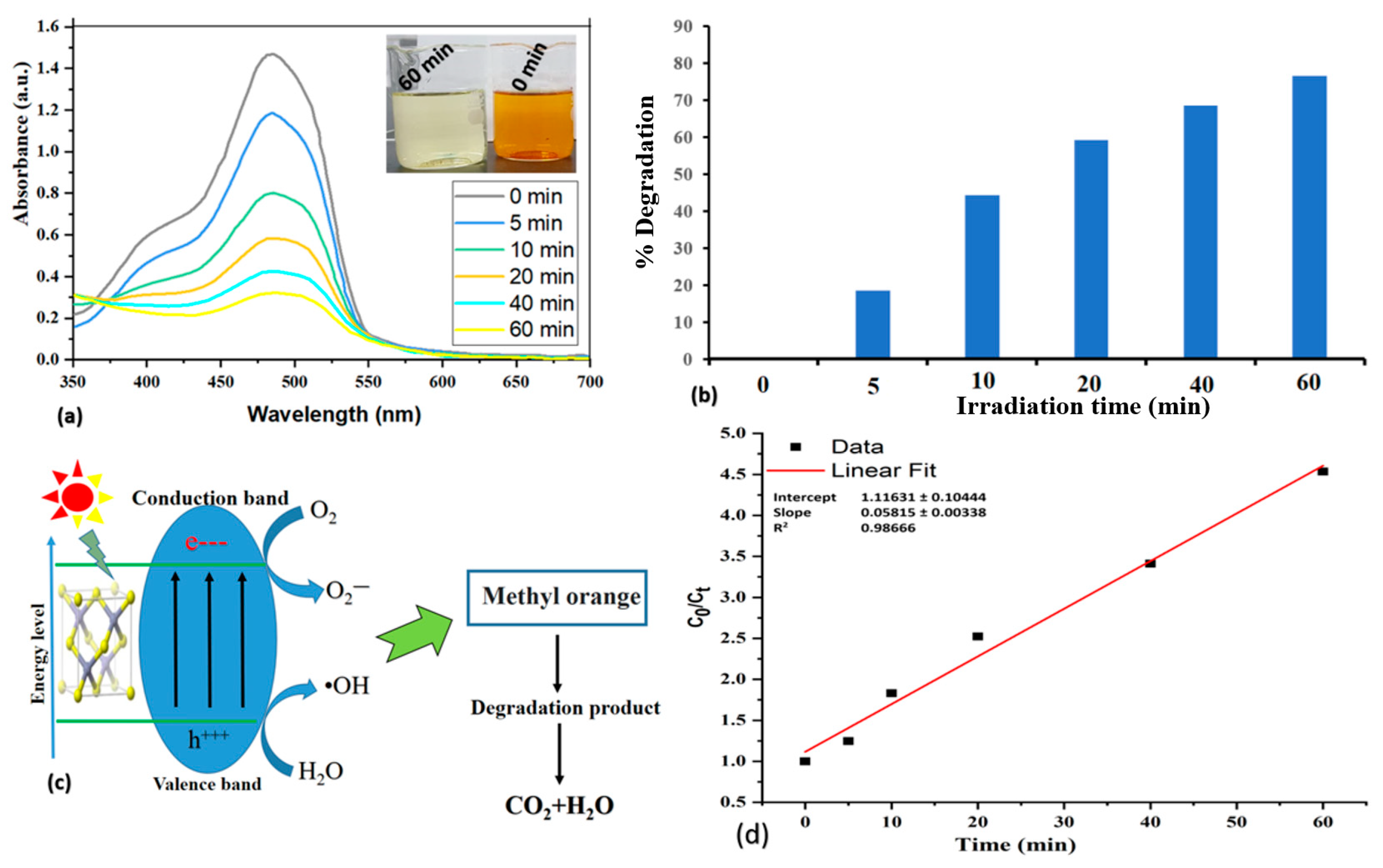
An experiment was conducted to examine the degradation potential of methyl orange (MO) dye. The experiment involved exposing the dye to sunlight while incorporating ZnO NPs as photocatalysts. The investigation focused on observing the degradation process of methyl orange at various time intervals within the visible region, utilizing ZnO NPs. Analysis of the absorption spectrum revealed noticeable reductions in the peaks associated with methyl orange, demonstrating its photo degradability over time (refer to Figure 5). This suggests that ZnO NPs can be used as an effective photocatalyst for the degradation of MO dye in wastewater. The degradation of the methyl orange dye through the photocatalytic reaction was observed by monitoring the absorption peak at 484 nm. These peaks gradually decreased with increasing exposure time, indicating that a photocatalytic degradation process occurred. Figure 5 shows the analytical results for the decomposition of methyl orange (MO). The peak intensity of the dye gradually decreases over time, indicating that the dye is degrading. Careful inspection of the diagram (Figure 5a,b) reveals clear evidence of dye degradation. This degradation can be attributed to the generation of reactive oxygen species promoted by ZnO NPs under light irradiation (Figure 5c). The main mechanism of dye degradation under sunlight is considered to be the photocatalytic generation of electron–hole pairs within the ZnO NPs. H2O and O2 molecules are oxidized by the free holes and electrons moving in the valence band and conduction band to generate OH radicals and O2 ions, respectively [64].
Studies have shown that the interaction of sunlight with ZnO nanoparticles can enhance the photocatalytic activity of various materials [65,66]. This enhanced light absorption is anticipated to improve the capacity of the nanoparticles to absorb light. This improved light absorption will facilitate the production of electron–hole pairs that can interact with the adsorbed dye molecules. This contact is believed to facilitate the degradation process, as seen in our investigation with MO dye. Figure 5a shows a decrease in adsorption intensity with increasing exposure time. This indicates that the ZnO nanoparticles effectively interact with the dye molecules. This effective interaction between ZnO nanoparticles and dye molecules provides evidence for the observed degradation phenomenon. Smaller nanoparticles or surface imperfections in semiconductors like ZnO are commonly associated with blue shifts, which refer to shorter wavelength absorption, and wider band gaps. According to the reported findings [67,68,69], these phenomena might explain the enhanced photocatalytic activity observed in our system. This degradation is accompanied by significant decolorization of the dye. Notably, more than 60% of the dyes were degraded in just 30 min in the presence of ZnO NPs. On the other hand, in the absence of ZnO NPs, no dye degradation was noticed. This emphasizes the critical role played by these nanoparticles in promoting the degradation reaction. ZnO NPs are effective photocatalysts for the degradation of methyl orange dye. The excellent degradation observed in these nanoparticles can be attributed to their larger surface area, which can enhance adsorption. This enhanced adsorption capacity is a key prerequisite for efficient photodegradation. Preliminary results from experimental studies demonstrate the great potential of ZnO NPs as efficient photocatalysts for methyl orange dye decomposition. Consequently, this increased migration leads to a higher degradation efficiency, as illustrated in a schematic diagram (Figure 5a–c).
Figure 5d depicts the biosynthesized ZnO NP-facilitated photocatalytic degradation of methyl orange (MO). Figure 5d shows the variation of the C0/Ct ratio over time, where C0 and Ct represent the dye concentrations at time 0 and time t, respectively. Here, C0 is equivalent to the initial absorbance A0, and Ct is the absorbance at a specific time t. Analysis of the graphs (Figure 5a) shows a gradual decrease in absorbance, indicating that the MO concentration decreases with increasing irradiation time. This means that ZnO NPs can effectively degrade the dye, with a maximum degradation of 75–80% within 60 min. Control experiments were also set up and confirmed that this degradation was attributed to the photocatalytic activity of ZnO NPs rather than solvent effects. Figure 5d displays a linear fit with an intercept of 1.11631 ± 0.10444, slope of 0.05815 ± 0.00338, and R2 value of 0.98666. ZnO NPs exhibit a consistent degradation pattern for methyl orange, indicating their reliable performance and effectiveness in this application. A pseudo-first-order kinetic plot of ln(C/Cₜ; concentration) versus irradiation time (min) was constructed. The results confirmed that the photocatalytic degradation of MO followed pseudo-first-order kinetics. The high correlation coefficient (R2 > 0.90714) indicated a satisfactory linear fit between the experimental data and the pseudo-first-order kinetic model [70]. The rate constant of the photocatalytic degradation of MO using the prepared ZnO NPs was 0.02398 min−1, as shown in Figure 6a. In summary, a noteworthy coefficient of correlation (R2 = 0.90714) enhances the level of confidence in the proposed kinetic model’s precision and dependability. The estimated half-life of 28.90 min (t1/2 ≈ 28.90 min) provides an important information on the dynamics of degradation noticed in the experimental setup.
Furthermore, various methods for the synthesis of ZnO NPs were analyzed based on the literature and this study. Their effectiveness in removing methyl orange (MO) dye from water was also examined. The results are summarized in Table 2. The analysis in Table 2 demonstrates various factors influencing the effectiveness of ZnO NPs for methyl orange (MO) dye removal. These factors include the synthesis method (e.g., sol–gel, sonochemical, solution combustion, hydrothermal, chemical precipitation, laser-generated, green synthesis), MO concentration, type of light source, amount of ZnO NPs used, exposure time, and percentage of MO dye removed. ZnO nanoparticles were produced using an eco-friendly method, degrading methyl orange color in sunlight, offering advantages over traditional methods involving harsh chemicals or excessive energy. The primary findings indicate that ZnO nanoparticles manufactured using green methods exhibit significant efficacy in eliminating 10 mg/L of methyl orange dye. This remarkable removal capacity is achieved with a relatively modest dosage of 50 mg/L, following a 60-min exposure to sunshine. The dye removal efficacy of 75–80% achieved with this process is comparable to other reported methods. This is especially noteworthy considering the shorter exposure period and the environmentally benign aspect of the green synthesis methodology. In summary, the findings indicate that the ZnO nanoparticles manufactured using green methods have the potential to effectively, rapidly, and persistently eliminate dyes in aqueous solutions.

2.4. Stability and Reusability of ZnONPs

Assessing the stability and cost of industrial feasibility requires examining the long-term effectiveness of photocatalysis and adsorption. The capacity of ZnO NPs (40 mg) for repeated use in degrading the MO solution (10 mg/L) was evaluated through five cycles of exposure to sun radiation. Following each photodegradation cycle, the catalyst underwent centrifugation and drying and was then collected for another degradation run. Following five successive rounds of photodecomposition, there was no discernible alteration in the catalytic efficacy of the biogenic ZnO NPs, indicating the good stability of the biogenic photocatalyst. Over the course of five repetitions, the efficiency of ZnO nanoparticles in the degradation of MO dye exhibited a slight decrease, dropping from 98% to 93%. These findings demonstrate the high durability and recyclability of the biosynthesized ZnO NPs, as depicted in Figure 6b. These findings match with previously documented [79,80,81,82,83] outcomes regarding the degradation of MO. The negligible decrease in activity indicates a high level of resistance to photo corrosion, which is a crucial aspect for practical use. Additionally, the adsorption of hydrogen sulfide (H2S) was investigated in five consecutive trials. The recovery of ZnO NPs in each cycle was achieved using centrifugation, washing, and drying. After undergoing five cycles, as depicted in Figure 6b, the effectiveness of adsorption fell from 95% to 65%. Following five H2S adsorption cycles, ZnO NPs exhibited a 30% reduction in adsorption capacity. The gradual accumulation of chemical intermediates on the catalyst surface may be the cause of the efficiency decrease. After multiple cycles, it may also be due to structural alterations in the ZnO NPs. After each exposure cycle, the nanocatalysts were collected by centrifugation, washed with deionized water, and dried in an oven at 70 °C overnight. These nanoparticles obtained from different photodegradation and H2S adsorption cycles were used to run UV-Vis spectroscopy to check the stability. The UV-Vis spectra of the nanoparticles from photodegradation cycles (Figure 6c) clearly show that there is no significant change in the UV spectrum after 5 reaction cycles, demonstrating the stability of the biosynthesized ZnO NPs [84]. However, as shown in Figure 6d, the UV spectra of the nanoparticles obtained from different H2S adsorption cycles show significant changes. Changes in the UV-visible spectrum may indicate nanoparticle aggregation. Aggregation can cause changes in particle size and surface area, which can affect their effectiveness and stability [85]. The broad peak at 361.7 nm continues to flatten, and the shape of this peak changes, indicating alterations in the nanoparticles’ properties and suggesting instability. In conclusion, unlike the H2S adsorption cycles, the nanoparticles display greater stability during the photodegradation cycles. The UV-Vis spectra show negligible changes after the photodegradation cycles, indicating that the nanoparticles are able to better maintain their shape and properties under these conditions. In contrast, the significant spectrum shifts observed during the H2S adsorption cycles indicate instability and modification of the nanoparticles.

2.5. Analysis of Corrosion Studies

Electrochemical polarization measurements are a technique used to study the behavior of electrochemical systems, such as corrosion processes or electrochemical reactions. It involves applying a potential (voltage) to an electrode immersed in an electrolyte solution and measuring the resulting current response. Three-electrode cells provide a more accurate configuration for studying corrosion than a two-electrode setup. This configuration consists of three key components: the working electrode (WE) (usually a metal subject to corrosion), the counter electrode (CE) (usually composed of an inert platinum wire), and the reference electrode (RE), such as silver/silver chloride (Ag/silver chloride). Furthermore, the potentiostat allows precise control of the potential applied to the WE. The onset and end potentials measured at WE were −700 mV and +250 mV, respectively. The Tafel polarization curve shown in Figure 7a is used to characterize the corrosion behavior of metal samples. The linear Tafel slopes of the anodic (βa) and cathodic (βc) reactions were extracted. Additionally, the corrosion current density (Icorr) was measured. The corrosion potential (Ecorr) was calculated by extrapolating the linear segments of the curves. Table 3 lists the data and related information, such as inhibition efficiency. Figure 7a presents polarization curves describing applied potential versus current density for mild steel corrosion in the presence and absence of ZnO NPs. The curve can be divided into two distinct regions: the anode part and the cathode part. In the anode region, metal oxidation occurs. Instead, metal reduction occurs in the cathode region. The corrosion potential, in the context of corrosion studies, denotes the electrical potential of a metal surface in the absence of any external current. It represents the point at which the anodic and cathodic currents are in equilibrium. This potential is measured relative to a reference electrode in the corrosive electrolyte. Figure 7a illustrates the polarization curve for corrosion of mild steel, plotting the applied potential versus current density. The curve shows the effect of ZnO NPs on the corrosion process. In the presence of ZnO NPs, the cathodic curve shows a more pronounced shift toward lower current densities compared to the anodic curve. This observation suggests that the inhibitor reduces the rate of hydrogen evolution reaction on the mild steel surface. The adsorption of corrosion inhibitor molecules onto the metal surface is responsible for this effect. Change in corrosion potential (Ecorr) is a reported metric used to classify a substance as an anodic or cathodic inhibitor. An inhibitor was categorized as either an anodic or cathodic. This categorization took place when the Ecorr value deviated by more than 85 mV in either the anodic or cathodic direction, as compared to the uninhibited system, respectively. If the shift is less than 85 mV, the inhibitor is considered mixed.
This study investigates the effect of inhibitors on metal corrosion behavior. The maximum change in corrosion potential (ΔEcorr) is approximately 16 mV, which is calculated as [(ΔEcorr = Ecorr(inhibitor) − Ecorr(blank)]. This value suggests that the mixed inhibitor does not alter the basic reaction mechanism. The inhibitor likely works by physically adhering to the metal surface and blocking the active site. This leaves less space for corrosion reactions to occur. Inhibitors at a concentration of 100 µg/mL in 1.0 M hydrochloric acid (HCl) solution provide the greatest protection against mild steel corrosion. This effect is caused by the formation of a protective inhibitor film on the metal surface, which blocks the interaction between the metal and the corrosive acid. These observations are consistent with previously reported studies [35,37,86].
The findings shown in Table 3 demonstrate a significant reduction in the corrosion rate (Icorr) as a result of the inhibitors. This indicates their influence on both the anodic and cathodic reactions. These findings suggest that ZnO NPs have a function in restricting the release of hydrogen and the dissolution of metals in a solution. In order to determine the efficacy of ZnO NPs as a preservative, different levels were introduced. Inhibitors like ZnO NPs were added, and the corrosion current density decreased. This is because the cathode potential goes up, which means the corrosion rate goes down. The inhibition efficiency is directly related to the ZnO NPs concentration, reaching a maximum value of 83.66% at 100 μg/mL (100 ppm). The findings indicate that higher concentrations of ZnO NPs suppressed both anodic and cathodic processes, as seen by a reduction in Tafel slopes. This observation indicates an obstacle in the overall kinetics of corrosion. Moreover, the observed reduction in corrosion current density (Icorr) at elevated concentrations can be ascribed to the adsorption of inhibitor molecules onto the surface of the mild steel. These findings demonstrate the potential of biogenic ZnO NPs as effective corrosion inhibitors for mild steel in 1.0 M HCl solutions. The present results highlight the potential of biologically derived nanoparticles as corrosion inhibitors, especially in corrosive chemical environments. Increasing inhibitor concentration results in an increase in surface coverage (θ) of the metal surface. An increase in θ is directly related to an increase in inhibition efficiency. Essentially, the greater the number of adsorbed inhibitor molecules, the more effective the corrosion protection. In addition, Table 3 employs the descriptor “mmpy” to represent the material loss rate (MLR) or corrosion rate (CR). The measure, denoted in millimeters per year (mm/yr), provides a direct representation of the thickness of the corroded material. The CR values presented in Table 3 illustrate the extent to which corrosion has diminished with increasing concentrations of the ZnO NPs inhibitor. Moreover, the mechanism of mild steel deterioration in a hydrochloric acid solution with a concentration of 1 M was examined. This experiment utilized electrochemical impedance spectroscopy (EIS) to examine samples, comparing those with and without nanoparticles. EIS is a commonly used technique for studying corrosion processes, lending credibility to the method. In Figure 7b, the Nyquist plots show a depressed capacitive loop and an inductive loop. As the concentration of ZnO NPs increases, the diameter of the capacitive loop gets larger. This suggests that adding these nanoparticles hinders charge transfer at the interface between the mild steel and the solution. In the presence of 100 ppm NPs, the highest impedance modulus value reached approximately 285 Ω cm2, a tenfold increase compared to the blank solution. These results demonstrate the strong adsorption of nanoparticles on mild steel surfaces, resulting in enhanced corrosion inhibition at higher nanoparticle concentrations.

2.6. Evaluation of H2S Adsorption Capacity

The adsorption capacity of synthesized ZnO NPs and bulk ZnO was evaluated through the experimental setup [87] shown in Figure 8a. Freshly prepared H2S was passed through equal amounts of bulk ZnO and ZnO NPs (2 g each) for different times (0 to 10 min), with the gas flow appropriately adjusted. The degree of absorption was assessed by periodically drawing aliquots of lead acetate using a syringe and measuring the intensity of absorption using a UV-Vis spectrophotometer (Figure 8b). The lead acetate solution was then shown to turn from clear to black in hue. The absorption intensity of bulk ZnO increased over time. This was accompanied by a darkening of the solution, attributed to lead sulfide formation, within 10 min. The absorption intensity of ZnO NPs followed a similar pattern over 20 min when the test went beyond the set period (0 to 10 min). Initially, there was no appreciable color change in the presence of ZnO NPs. After 7 min, the lead acetate underwent a progressive color change, as indicated by the increasing degree of absorption in the solution. Nevertheless, the change was not as pronounced as that observed in the presence of bulk ZnO. This is evidenced in the graphs showing the absorption intensity over time. The experiment was conducted in triplicate, yielding approximately consistent results. The large surface-to-volume ratio of nanoparticles, which results in heightened surface activity, is responsible for this phenomenon.
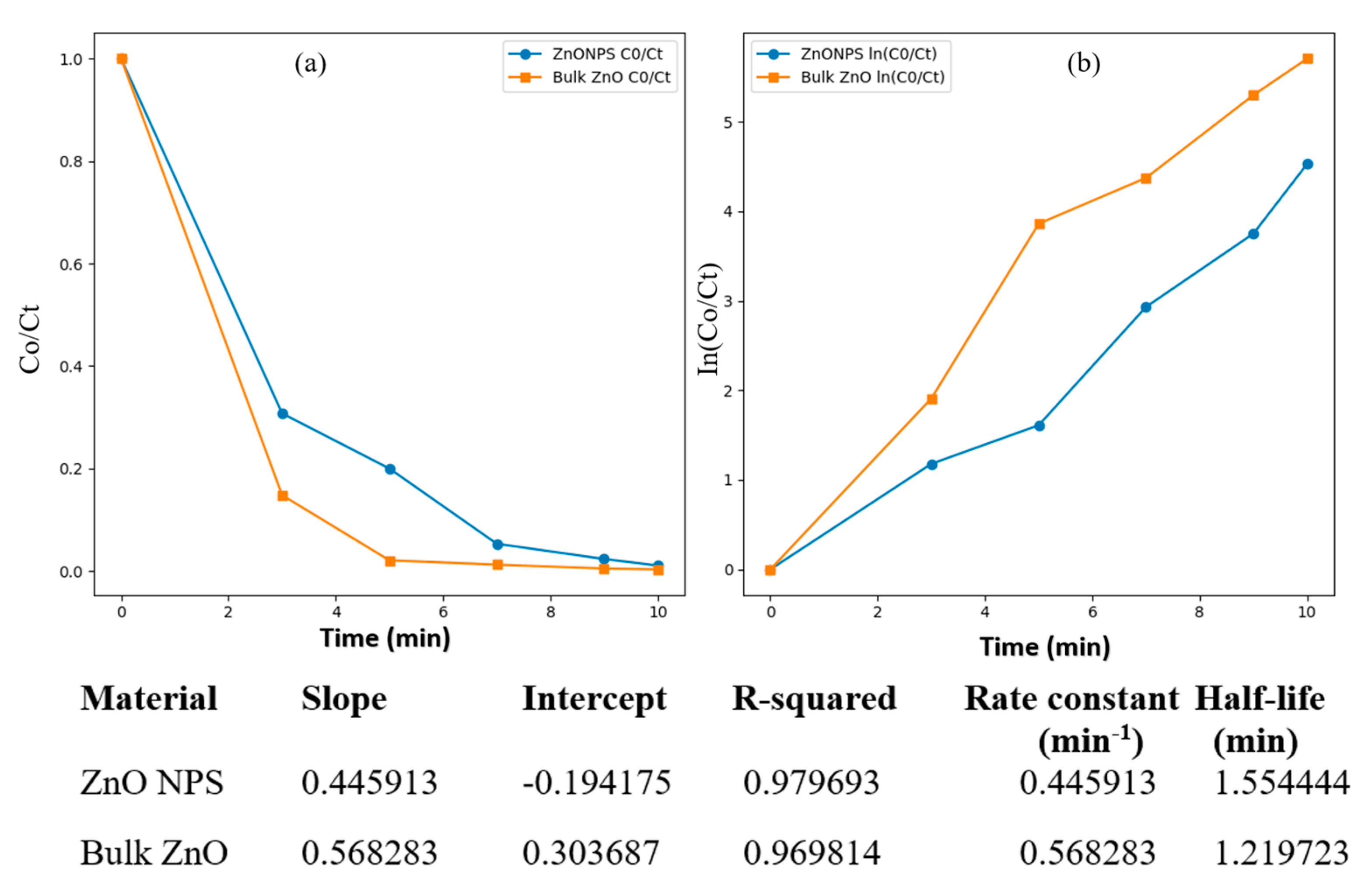
Consequently, these characteristics may have contributed to a more pronounced adsorption of H2S compared to its bulk counterpart. In the process of ZnO adsorbing H2S, a reaction occurs in which ZnO reacts with H2S to form ZnS and H2O. Thus, inert zinc sulfide is produced, and unadsorbed H2S passes into the lead acetate solution, forming lead sulfide, which changes color from white to black. Our findings from the kinetic investigation comparing ZnO NPs and bulk ZnO’s H2S gas adsorption reveal important differences and follow a pseudo-first-order kinetic model (In(Ct/C0) = k1t). Due to their higher surface area, ZnO NPs adsorb more H2S than bulk ZnO. Due to their higher surface activity, ZnO NPs absorb more H2S molecules, delaying the lead acetate solution’s color change from clear to black. The plot of normalized concentration (C0/Ct) against time for bulk ZnO and ZnO NPs is shown in Figure 9a. In this, H2S gas concentration changes over time as it interacts with different materials. The normalized concentration of ZnO NPs declines slower than bulk ZnO. This indicates that they adsorb H2S more efficiently and release it more slowly into the environment. This experiment shows that materials’ surface characteristics and active sites affect adsorption kinetics. Kinetic parameters in experiments provide quantitative support for these observations (Figure 9b). The rate constant for ZnO NPs was lower (0.445913 min−1) than for bulk ZnO (0.568283 min−1), which means that the reaction with H2S started more slowly. At the same time, ZnO NPs had a longer half-life (1.554444 min) than bulk ZnO (1.219723 min), which means that the adsorption process continued over time. Experimental results match our observations. ZnO NPs absorb more H2S than bulk ZnO and release less into the lead acetate chamber. ZnO NPs’ more active sites and surface area make adsorption stronger. This delays the lead acetate solution’s color shift. Bulk ZnO adsorbs less H2S but desorbs it faster, causing the lead acetate solution to change color early.
This shows faster response kinetics and higher initial H2S release than ZnO NPs. These studies demonstrate ZnO-based materials’ unique adsorption and desorption behaviours, which have environmental remediation and gas purification consequences.

3. Materials and Methods

3.1. Chemicals

Cucurbita pepo L. seeds were acquired from the market. Zinc acetate GR, iron sulfide, NaOH, HCl, and methyl orange (MO) were obtained from Merck and Sigma, respectively.

3.2. Preparation of Plant Extract and Utilization in Biosynthesis of Zinc Oxide Nanoparticles

Seeds of Cucurbita pepo L. were washed and cleaned thoroughly with double-distilled water and then crushed into fine powder. Subsequently, the powdered seeds were air-dried at room temperature. In a 250 mL round-bottomed flask containing 100 mL of double-distilled water, add 2 g of powdered seeds [29]. The flask was then refluxed for 1 h, followed by cooling to room temperature and filtering through Whatman No. 1 filter paper to obtain the plant extract. The filtrate obtained was utilized for the synthesis of ZnO nanoparticles, employing soluble compounds derived from Cucurbita pepo L. seeds. For the synthesis process, 20 mL of the seed extract was heated for 15 min at 50 °C in a 250 mL conical flask, in accordance with our previous research [14,29]. Subsequently, a solution comprising 50 mL of 91 mM zinc acetate was added to the flask. Upon addition of the zinc acetate solution, the reaction mixture exhibited a yellowish coloration, followed by the precipitation of cream-colored zinc hydroxide precipitates. The reduction process to zinc hydroxide was facilitated by allowing the reaction mixture to stand undisturbed for 30 min. Following this, centrifugation at 15,000 rpm for 10 min was performed to separate the reaction mixture from the solid precipitate. For additional examination and use, the precipitate was carefully gathered and vacuum-dried at 40 to 50 °C. The formation of ZnO NPs was primarily confirmed using UV-visible spectroscopy. Biogenic ZnO nanoparticles were extensively characterized using various techniques (FTIR, XRD, EDS, TEM, SEM) to determine their crystal structure, composition, size, and morphology.

3.3. Characterization of Biofabricated ZnO NPs

To fully characterize the bio-fabricated ZnO NPs, various techniques were employed to validate their synthesis, assess stability, evaluate chemical and physical properties, and compare them with previously reported studies. These techniques include scanning electron microscopy (SEM) and energy-dispersive X-ray spectroscopy (EDX), Fourier transform infrared spectroscopy (FTIR), ultraviolet-visible spectroscopy (UV-Vis), X-ray diffraction (XRD), and thermogravimetric analysis (thermal analysis). SEM and EDX analyses were conducted by placing a small portion of bio-derived ZnO NPs on a carbon stud pre-coated with conductive double-sided carbon tabs. The sample’s ZnO NPs surface was coated with a fine layer of gold using an auto coater before imaging with SEM under high vacuum conditions and at a 15 kV accelerating voltage. SEM imaging was performed at various magnifications, including X1000 and X5000. For EDX analysis, the mode was switched from SEM to EDX, and the resulting EDX data appeared as a graph showing weight percentage or atomic percentage. The bio-produced ZnO NPs were analyzed for size, shape, and morphology using transmission electron microscopy (TEM) (Hitachi, Japan). FTIR analysis involved placing the seed-derived ZnO NPs sample on the diamond stage of an FTIR machine, pressing it with a pressure clamp, and embedding it with KBr. The OPUS software (v.5.5) was then used to run the sample, and the resulting data appeared as a graph displaying bands ranging from 4000 to 500 cm−1. XRD analysis was carried out using a Pan Analytical X-pert Pro instrument equipped with a Cu-Kα radiation source, operating at 45 kV and 40 mA. The XRD scans covered the 2θ range of 20–80°. UV-Vis spectroscopy of the synthesized ZnO NPs was performed using a UV-Vis spectrophotometer (UV-2450, Shimadzu, Kyoto, Japan) with a resolution of 1 nm. The measurement spanned from 200 to 800 nm to observe the conversion of metal oxide ions into metal oxide nanoparticles. A blank reference of double-distilled water was used to account for background effects. TGA analysis was utilized to assess the thermal stability and weight loss of the ZnO NPs with increasing temperature. The Mettler Toledo TGA instrument was employed for this analysis, with approximately 10 mg of ZnO NPs placed in the instrument’s pan under a regulated atmosphere using oxygen gas. The resulting data were displayed as thermogravimetric or TGA curves, showing the mass change of ZnO NPs versus temperature or time.

3.4. Photocatalytic Experiment

In this study, the photo-degradation of organic dyes was investigated using methyl orange (MO) as one of the dyes. MO is a toxic azo dye commonly found in the textile, leather, and paper industries. The photocatalytic degradation performance of bio-ZnO nanoparticles was evaluated using MO in an aqueous solution (10 mg/L) under direct sunlight irradiation. First, the calculated amount (50 mg) of ZnO nanoparticles was added to the aqueous solution of MO. The solution was then stirred for one hour in the dark to achieve equilibrium. The solution mixture was then exposed to direct sunlight for 60 min on a sunny day. A control solution was prepared and maintained in the same manner as the experimental solution to check for any color changes in the MO dye solution. Five milliliters of suspension from the experimental and control solutions were taken at predetermined intervals. The samples were centrifuged at 7000 rpm for 10 min. Each sample’s supernatant was taken for UV-Vis spectrophotometer absorption analysis. The photocatalytic degradation of MO is accompanied by a gradual decrease in the absorption intensity at λmax = 484 nm. The wavelength scan range is 350 to 700 nm. This indicates that the MO dye degrades over time. The % degradation of the dyes was determined by the equation [30,88].
%   D e g r a d a t i o n = A o A / A o × 100  
The initial and final absorbance of the dye solution were denoted as Ao and A, respectively. The control experiments were performed in the dark to determine the absorption loss. To further analyze the results, the kinetic equation associated with the initial dye concentration (C₀), concentration after irradiation (Cₜ), rate constant (k), and reaction time (t) was used: ln(C₀/Cₜ) = kt. After the reaction completion, the catalyst was separated from the mixture by using centrifugation. It was then cleaned with doubly distilled water and dried in order to be ready for the next cycle. The stability of the catalyst was evaluated by examining the alterations in the X-ray diffraction (XRD) pattern of the isolated ZnO nanoparticles following the last stage of the cycling process.

3.5. Corrosion Studies

3.5.1. Samples Compositions and Preparation

The mild steel samples examined were primarily composed of iron (Fe), with particular quantities of additional components incorporated. In terms of weight percentage, the composition of the substance except iron is as follows: 0.21% carbon (C), 0.38% silicon (Si), 0.05% manganese (Mn), 0.05% sulfur (S), 0.09% phosphorus (P), and 0.01% aluminum (Al). In the corrosion experiments, mild steel samples were prepared by cutting into 10 mm × 10 mm squares. To ensure accurate surface uniformity for corrosion testing, samples were mechanically polished before experiments. Before corrosion testing, the samples were mechanically polished with a series of 800, 1000, and 1500 grit emery paper, a fine-grained abrasive, to ensure a smooth surface. A common practice in materials science is to use progressively finer grit sizes, using sandpaper (e.g., emery) for mechanical polishing. This method helps achieve a smooth surface finish, which is critical for reliable corrosion test results [27]. Before experiments, the samples were degreased with denatured ethanol, rinsed with acetone, and cleaned with distilled water to remove contaminants and avoid initial corrosion. The polished and cleaned samples were then dried and stored in a desiccator to prevent moisture accumulation.

3.5.2. Preparation for Corrosive Environments

An acidic environment simulating corrosive conditions was prepared by mixing biosynthesized ZnONPs solutions with 1.0 M hydrochloric acid (HCl). To study the effect of zinc oxide nanoparticles (ZnO NPs) on corrosion behavior, solutions with different ZnO NPs concentrations were prepared. These solutions included a control solution without ZnO NPs (0 µg/mL) and four experimental solutions with increasing concentrations of ZnO NPs (10, 20, 40, and 100 µg/mL or ppm). ZnO NPs solutions were prepared by diluting the 1 mg/mL stock solution with distilled water.

3.5.3. Electrochemical Polarization Measurement

Electrochemical studies were performed using an AUTOLAB potentiostat at room temperature. Data acquisition and analysis were facilitated by GPES 4.9 (General Electrochemistry Software), EC-Lab 10.4 software (trial version), and Origin2024 (trial version). The study utilized a typical three-electrode cell setup. The working electrode was comprised of a conductive substrate, such as glassy carbon or FTO-coated glass, onto which varying concentrations of ZnO NPs were adsorbed. The typical surface area of the working electrode is 10 mm2. A platinum electrode was used as the counter electrode, while a saturated silver/silver chloride electrode was used as the reference electrode. The electrolyte solution contained 1.0 M hydrochloric acid (HCl) and different concentrations of ZnO NPs. The effect of inhibitors on corrosion was obtained through polarization experiments. The potential of the working electrode (mild steel) was varied, and the resultant current was observed. The Tafel behavior was investigated within the suitable anodic potential range, typically ranging from −700 mV to +250 mV, employing standard scan speeds of 1.0 mV/s. To ascertain the corrosion current density, the linear segment of the Tafel plot was extrapolated to the corrosion potential. Following the acquisition of the current density, the corrosion rate and inhibition efficiency (%IE) were then computed. Furthermore, the evaluation of surface coverage (θ) and inhibitory efficiency (%IE) might be conducted by employing well-established Equations (1) and (2).
Surface coverage (θ) = I0corr − Icorr/I0corr
Inhibitor efficiency (IE%) = I0corr − Icorr/I0corr × 100

3.6. Adsorption Study

Caution! H2S is poisonous and fatal if inhaled. This experiment requires a well-ventilated fume hood, gloves, safety glasses, and an H2S-approved respirator.
Purified biosynthesized ZnO NPs were dried, and 2 g was introduced into the reaction vessel. A 1% (w/v) lead acetate solution was carefully prepared by dissolving 1 g of lead acetate in 100 mL of deionized water in an Erlenmeyer flask and placed on a magnetic stirrer for gentle mixing. In a separate round-bottomed flask, iron sulfide (FeS; 0.1 g) was mixed with 15 mL of 1 M dilute sulfuric acid solution (approximately 1% v/v) and gently heated. Before starting the experiment, make sure that all connections are sealed to prevent gas leakage before reaching the flask containing the prepared lead acetate solution. Subsequently, aliquots of the lead acetate solution were drawn using a syringe at intervals of 0, 3, 5, 7, 9, and 10 min from the reaction vessel. These aliquots were then analyzed by UV-visible spectrophotometry at a fixed wavelength of 450 nm to monitor the adsorption of H2S generated during the reaction of iron sulfide and sulfuric acid, observing any corresponding color changes. A similar set of reactions was also performed in the presence of bulk ZnO to compare the obtained results with ZnO NPs. As the H2S concentration in the lead acetate solution increases, the color becomes darker due to the formation of lead sulfide, increasing the absorption intensity of the lead solution.

4. Conclusions

In conclusion, this study successfully employed Cucurbita pepo L. seeds for the synthesis of ZnO nanoparticles. Different methods of characterization, such as TEM, SEM, EDX, UV-vis, XRD, and FTIR, confirmed the seed-derived NPs’ nanostructure, morphology, elemental composition, purity, and ZnO presence. Additionally, TGA analysis revealed the good thermal stability of the ZnO NPs at high temperatures, suggesting their potential for various applications. Furthermore, the study revealed enhanced H2S adsorption capacity of ZnO NPs compared to bulk ZnO, which is due to their higher surface area. The study also investigated the effect of these biogenic ZnO NPs on the corrosion resistance of mild steel. Electrochemical measurements revealed that ZnO NPs significantly improved the steel’s corrosion resistance. In a separate study, biogenic ZnO nanoparticles achieved an excellent photodegradation efficiency of 75% for methyl orange under sunlight within 60 min. This suggests that biogenic ZnO NPs could be used to treat water pollution caused by organic dyes. This work highlights the effective environmentally-friendly production of ZnO NPs, which have potential uses in gas adsorption, corrosion prevention, and the breakdown of organic contaminants through photocatalysis. Subsequent investigations will prioritize evaluating their effectiveness in real-world settings to determine their practical utility. While our current study provides valuable insights under controlled laboratory conditions, it has limitations in representing real-world scenarios. To definitively assess their practical value, future studies will focus on evaluating the effectiveness of these nanoparticles in complex natural environments (in situ). The goal of this ongoing research is to bridge the differences between laboratory findings and real environmental issues. Furthermore, it seeks to ensure the effectiveness and long-term viability of biogenic ZnO NPs in practical pollution reduction methods.

Author Contributions

Methodology, S.N.H.A. and M.A.; Software, M.A.; Formal analysis, S.N.H.A.; Investigation, S.N.H.A.; Resources, S.N.H.A. and M.A.; Data curation, M.A.; Writing—original draft, S.N.H.A.; Writing—review & editing, M.A.; Supervision, M.A. All authors have read and agreed to the published version of the manuscript.

Funding

This research received no external funding.

Data Availability Statement

The data that support the findings of this study are available within the article.

Conflicts of Interest

The authors declare no conflicts of interest.

References

  1. Ying, S.; Guan, Z.; Ofoegbu, P.C.; Clubb, P.; Rico, C.; He, F.; Hong, J.J.E.T. Innovation. Green synthesis of nanoparticles: Current developments and limitations. Environ. Technol. Innov. 2022, 26, 102336. [Google Scholar] [CrossRef]
  2. Barabadi, H.; Ovais, M.; Shinwari, Z.K.; Saravanan, M. Anti-cancer green bionanomaterials: Present status and future prospects. Green Chem. Lett. Rev. 2017, 10, 285–314. [Google Scholar] [CrossRef]
  3. Samuel, M.S.; Ravikumar, M.; John, J.A.; Selvarajan, E.; Patel, H.; Chander, P.S.; Soundarya, J.; Vuppala, S.; Balaji, R.; Chandrasekar, N. A review on green synthesis of nanoparticles and their diverse biomedical and environmental applications. Catalysts 2022, 12, 459. [Google Scholar] [CrossRef]
  4. Nasrollahzadeh, M.; Sajjadi, M.; Sajadi, S.M.; Issaabadi, Z. Green nanotechnology. In Interface Science and Technology; Elsevier: Amsterdam, The Netherlands, 2019; Volume 28, pp. 145–198. [Google Scholar]
  5. Khan, S.H. Green nanotechnology for the environment and sustainable development. In Green Materials for Wastewater Treatment; Springer: Berlin/Heidelberg, Germany, 2020; pp. 13–46. [Google Scholar]
  6. Sukul, P.K.; Kar, C. Green Conversion Methods to Prepare Nanoparticle. In Bioinspired and Green Synthesis of Nanostructures: A Sustainable Approach; Wiley Online Library: Hoboken, NJ, USA, 2023; pp. 115–139. [Google Scholar]
  7. Bharadwaj, K.K.; Rabha, B.; Pati, S.; Sarkar, T.; Choudhury, B.K.; Barman, A.; Bhattacharjya, D.; Srivastava, A.; Baishya, D.; Edinur, H.A. Green synthesis of gold nanoparticles using plant extracts as beneficial prospect for cancer theranostics. Molecules 2021, 26, 6389. [Google Scholar] [CrossRef]
  8. Zhou, X.-Q.; Hayat, Z.; Zhang, D.-D.; Li, M.-Y.; Hu, S.; Wu, Q.; Cao, Y.-F.; Yuan, Y.J.P. Zinc oxide nanoparticles: Synthesis, characterization, modification, and applications in food and agriculture. Processes 2023, 11, 1193. [Google Scholar] [CrossRef]
  9. Soltani, S.; Gacem, A.; Choudhary, N.; Yadav, V.K.; Alsaeedi, H.; Modi, S.; Patel, A.; Khan, S.H.; Cabral-Pinto, M.M.; Yadav, K.K. Scallion peel mediated synthesis of zinc oxide nanoparticles and their applications as nano fertilizer and photocatalyst for removal of organic pollutants from wastewater. Water 2023, 15, 1672. [Google Scholar] [CrossRef]
  10. Adraoui, I.; Mamouni, R.; Saffaj, N.; Achemchem, F. Eco-friendly synthesis of zinc oxide nanoparticles using saffron extract and their photocatalytic and antibacterial activities. J. Mater. Res. 2023, 38, 2874–2884. [Google Scholar] [CrossRef]
  11. Singh, J.; Dutta, T.; Kim, K.-H.; Rawat, M.; Samddar, P.; Kumar, P. ‘Green’synthesis of metals and their oxide nanoparticles: Applications for environmental remediation. J. Nanobiotechnol. 2018, 16, 84. [Google Scholar] [CrossRef]
  12. Shnawa, B.H.; Jalil, P.J.; Al-Ezzi, A.; Mhamedsharif, R.M.; Mohammed, D.A.; Biro, D.M.; Ahmed, M.H. Evaluation of antimicrobial and antioxidant activity of zinc oxide nanoparticles biosynthesized with Ziziphus spina-christi leaf extracts. J. Environ. Sci. Health Part C 2024, 42, 93–108. [Google Scholar] [CrossRef]
  13. Sutharappa Kaliyamoorthy, T.; Subramaniyan, V.; Renganathan, S.; Elavarasan, V.; Ravi, J.; Prabhakaran Kala, P.; Subramaniyan, P.; Vijayakumar, S. Sustainable environmental-based ZnO nanoparticles derived from Pisonia grandis for future biological and environmental applications. Sustainability 2022, 14, 17009. [Google Scholar] [CrossRef]
  14. Tabrez, S.; Khan, A.U.; Hoque, M.; Suhail, M.; Khan, M.I.; Zughaibi, T.A. Biosynthesis of ZnO NPs from pumpkin seeds’ extract and elucidation of its anticancer potential against breast cancer. Nanotechnol. Rev. 2022, 11, 2714–2725. [Google Scholar] [CrossRef]
  15. Ranjha, M.M.A.N.; Shafique, B.; Rehman, A.; Mehmood, A.; Ali, A.; Zahra, S.M.; Roobab, U.; Singh, A.; Ibrahim, S.A.; Siddiqui, S.A. Biocompatible nanomaterials in food science, technology, and nutrient drug delivery: Recent developments and applications. Front. Nutr. 2022, 8, 778155. [Google Scholar] [CrossRef] [PubMed]
  16. Huq, M.A.; Apu, M.A.I.; Ashrafudoulla, M.; Rahman, M.M.; Parvez, M.A.K.; Balusamy, S.R.; Akter, S.; Rahman, M.S. Bioactive ZnO Nanoparticles: Biosynthesis, Characterization and Potential Antimicrobial Applications. Pharmaceutics 2023, 15, 2634. [Google Scholar] [CrossRef] [PubMed]
  17. Despotović, V.; Finčur, N.; Bognar, S.; Šojić Merkulov, D.; Putnik, P.; Abramović, B.; Panić, S. Characterization and Photocatalytic Performance of Newly Synthesized ZnO Nanoparticles for Environmental Organic Pollutants Removal from Water System. Separations 2023, 10, 258. [Google Scholar] [CrossRef]
  18. Chemingui, H.; Missaoui, T.; Mzali, J.C.; Yildiz, T.; Konyar, M.; Smiri, M.; Saidi, N.; Hafiane, A.; Yatmaz, H. Facile green synthesis of zinc oxide nanoparticles (ZnO NPs): Antibacterial and photocatalytic activities. Mater. Res. Express 2019, 6, 1050b4. [Google Scholar] [CrossRef]
  19. Chemingui, H.; Lafi, R.; Missaoui, T.; Montasser, I.; Hafiane, A.; Kamoun, M. Eco-friendly approach for the synthesis of zinc oxide nanoparticles (ZnO NPs) using Verbena officinalis L: Enhancement of photocatalytic activity under UV light and application of response surface methodology. In Biomass Conversion and Biorefinery; Springer: Berlin/Heidelberg, Germany, 2024; pp. 1–19. [Google Scholar]
  20. Chemingui, H.; Moulahi, A.; Missaoui, T.; Al-Marri, A.H.; Hafiane, A. A novel green preparation of zinc oxide nanoparticles with Hibiscus sabdariffa L.: Photocatalytic performance, evaluation of antioxidant and antibacterial activity. Environ. Technol. 2024, 45, 926–944. [Google Scholar] [CrossRef]
  21. Wal, A.; Singh, M.R.; Gupta, A.; Rathore, S.; Rout, R.R.; Wal, P. Pumpkin Seeds (Cucurbita spp.) as a Nutraceutical Used in Various Lifestyle Disorders. Nat. Prod. J. 2024, 14, 118–137. [Google Scholar] [CrossRef]
  22. Ethiraj, S.; Balasundaram, J. Phytochemical and biological activity of Cucurbita seed extract. J. Adv. Biotechnol. 2016, 6, 813–821. [Google Scholar] [CrossRef]
  23. Muchirah, P.N.; Waihenya, R.; Muya, S.; Abubakar, L.; Ozwara, H.; Makokha, A. Characterization and anti-oxidant activity of Cucurbita maxima Duchesne pulp and seed extracts. J. Phytopharm. 2018, 7, 134–140. [Google Scholar] [CrossRef]
  24. Zhao, X.; Baharinikoo, L.; Farahani, M.D.; Mahdizadeh, B.; Farizhandi, A.A. Experimental modelling studies on the removal of dyes and heavy metal ions using ZnFe2O4 nanoparticles. Sci. Rep. 2022, 12, 5987. [Google Scholar] [CrossRef]
  25. Goutam, S.P.; Saxena, G.; Roy, D.; Yadav, A.K.; Bharagava, R.N. Green synthesis of nanoparticles and their applications in water and wastewater treatment. In Bioremediation of Industrial Waste for Environmental Safety: Volume I: Industrial Waste and Its Management; Springer: Berlin/Heidelberg, Germany, 2020; pp. 349–379. [Google Scholar]
  26. Alam, M. Analyses of biosynthesized silver nanoparticles produced from strawberry fruit pomace extracts in terms of biocompatibility, cytotoxicity, antioxidant ability, photodegradation, and in-silico studies. J. King Saud Univ.-Sci. 2022, 34, 102327. [Google Scholar] [CrossRef]
  27. Huda Abd Kadir, N.; Ali Khan, A.; Kumaresan, T.; Khan, A.U.; Alam, M. The impact of pumpkin seed-derived silver nanoparticles on corrosion and cytotoxicity: A molecular docking study of the simulated AgNPs. Green Chem. Lett. Rev. 2024, 17, 2319246. [Google Scholar] [CrossRef]
  28. Jose, L.M.; Raj, R.A.; Sajan, D.; Aravind, A. Adsorption and photocatalytic activity of biosynthesised ZnO nanoparticles using Aloe Vera leaf extract. Nano Express 2021, 2, 010039. [Google Scholar] [CrossRef]
  29. Kadir, N.H.A.; Murugan, N.; Khan, A.A.; Sandrasegaran, A.; Khan, A.U.; Alam, M. Evaluation of the cytotoxicity, antioxidant activity, and molecular docking of biogenic zinc oxide nanoparticles derived from pumpkin seeds. Microsc. Res. Tech. 2024, 87, 602–615. [Google Scholar] [CrossRef] [PubMed]
  30. Alam, M. Photocatalytic activity of biogenic zinc oxide nanoparticles: In vitro antimicrobial, biocompatibility, and molecular docking studies. Nanotechnol. Rev. 2021, 10, 1079–1091. [Google Scholar] [CrossRef]
  31. Al-Askar, A.A.; Hashem, A.H.; Elhussieny, N.I.; Saied, E. Green biosynthesis of zinc oxide nanoparticles using Pluchea indica leaf extract: Antimicrobial and photocatalytic activities. Molecules 2023, 28, 4679. [Google Scholar] [CrossRef] [PubMed]
  32. Al Rahbi, A.S.; Al Mawali, A.H.; Al Rawahi, S.S.; Al Dighishi, R.K.; Al Abri, F.A.; Ahmed, A.; Rahman, S. Green synthesis of zinc oxide nanoparticles from Salvadora persica leaf extract: Characterization and studying methyl orange removal by adsorption. Water Pract. Technol. 2024, 19, 1219–1231. [Google Scholar] [CrossRef]
  33. Sharma, D.; Sabela, M.I.; Kanchi, S.; Mdluli, P.S.; Singh, G.; Stenström, T.A.; Bisetty, K. Biosynthesis of ZnO nanoparticles using Jacaranda mimosifolia flowers extract: Synergistic antibacterial activity and molecular simulated facet specific adsorption studies. J. Photochem. Photobiol. B Biol. 2016, 162, 199–207. [Google Scholar] [CrossRef] [PubMed]
  34. Kahsay, M.H. Synthesis and characterization of ZnO nanoparticles using aqueous extract of Becium grandiflorum for antimicrobial activity and adsorption of methylene blue. Appl. Water Sci. 2021, 11, 45. [Google Scholar] [CrossRef]
  35. Akhir, R.M.; Norashikin, M.H.; Mahat, M.M.; Bonnia, N.N. Biosynthesis of Zinc Oxide Nanoparticles for Corrosion Protection Application. Int. J. Eng. Technol 2018, 4, 488–492. [Google Scholar]
  36. Beddiar, H.; Boudiba, S.; Benahmed, M.; Hanini, K.; Tamfu, A.N.; Boudiba, L.; Soltani, H.; Laouer, H.; Akkal, S. Evaluation of the Potential of Daucus crinitus Extracts and Their Synthesized ZnO Nanoparticles in Inhibiting the Corrosion of Carbon Steel. J. Chem. 2024, 2024, 7703829. [Google Scholar] [CrossRef]
  37. Al-Senani, G.M. Synthesis of ZnO-NPs using a Convolvulus arvensis leaf extract and proving its efficiency as an inhibitor of carbon steel corrosion. Materials 2020, 13, 890. [Google Scholar] [CrossRef] [PubMed]
  38. Faisal, S.; Jan, H.; Shah, S.A.; Shah, S.; Khan, A.; Akbar, M.T.; Rizwan, M.; Jan, F.; Wajidullah; Akhtar, N. Green synthesis of zinc oxide (ZnO) nanoparticles using aqueous fruit extracts of Myristica fragrans: Their characterizations and biological and environmental applications. ACS Omega 2021, 6, 9709–9722. [Google Scholar] [CrossRef] [PubMed]
  39. Vasiljevic, Z.; Vunduk, J.; Bartolic, D.; Miskovic, G.; Ognjanovic, M.; Tadic, N.B.; Nikolic, M.V. An Eco-friendly Approach to ZnO NP Synthesis Using Citrus reticulata Blanco Peel/Extract: Characterization and Antibacterial and Photocatalytic Activity. ACS Appl. Bio Mater. 2024, 7, 3014–3032. [Google Scholar] [CrossRef] [PubMed]
  40. Gamedze, N.P.; Mthiyane, D.M.; Mavengahama, S.; Singh, M.; Onwudiwe, D.C. Biosynthesis of ZnO nanoparticles using the aqueous extract of Mucuna pruriens (utilis): Structural characterization, and the anticancer and antioxidant activities. Chem. Afr. 2024, 7, 219–228. [Google Scholar] [CrossRef]
  41. Vishnupriya, B.; Nandhini, G.E.; Anbarasi, G. Biosynthesis of zinc oxide nanoparticles using Hylocereus undatus fruit peel extract against clinical pathogens. Mater. Today Proc. 2022, 48, 164–168. [Google Scholar] [CrossRef]
  42. Hayat, K.; Din, I.U.; Alam, K.; Khan, F.U.; Khan, M.; Mohamed, H.I. Green synthesis of zinc oxide nanoparticles using plant extracts of Fumaria officinalis and Peganum harmala and their antioxidant and antibacterial activities. In Biomass Conversion and Biorefinery; Springer: Berlin/Heidelberg, Germany, 2024; pp. 1–15. [Google Scholar]
  43. Dappula, S.S.; Kandrakonda, Y.R.; Shaik, J.B.; Mothukuru, S.L.; Lebaka, V.R.; Mannarapu, M.; Amooru, G.D. Biosynthesis of zinc oxide nanoparticles using aqueous extract of Andrographis alata: Characterization, optimization and assessment of their antibacterial, antioxidant, antidiabetic and anti-Alzheimer’s properties. J. Mol. Struct. 2023, 1273, 134264. [Google Scholar] [CrossRef]
  44. Naiel, B.; Fawzy, M.; Halmy, M.W.A.; Mahmoud, A.E.D. Green synthesis of zinc oxide nanoparticles using Sea Lavender (Limonium pruinosum L. Chaz.) extract: Characterization, evaluation of anti-skin cancer, antimicrobial and antioxidant potentials. Sci. Rep. 2022, 12, 20370. [Google Scholar] [CrossRef] [PubMed]
  45. Tilahun, E.; Adimasu, Y.; Dessie, Y. Biosynthesis and optimization of ZnO nanoparticles using Ocimum lamifolium leaf extract for electrochemical sensor and antibacterial activity. ACS Omega 2023, 8, 27344–27354. [Google Scholar] [CrossRef]
  46. Mishra, D.; Chitara, M.K.; Negi, S.; Pal Singh, J.; Kumar, R.; Chaturvedi, P. Biosynthesis of zinc oxide nanoparticles via leaf extracts of Catharanthus roseus (L.) G. Don and their application in improving seed germination potential and seedling vigor of Eleusine coracana (L.) Gaertn. Adv. Agric. 2023, 2023, 7412714. [Google Scholar] [CrossRef]
  47. Thi, T.U.D.; Nguyen, T.T.; Thi, Y.D.; Thi, K.H.T.; Phan, B.T.; Pham, K.N. Green synthesis of ZnO nanoparticles using orange fruit peel extract for antibacterial activities. RSC Adv. 2020, 10, 23899–23907. [Google Scholar]
  48. Ramamoorthy, S.; Surendhiran, S.; Senthil Kumar, D.; Murugesan, G.; Kalaiselvi, M.; Kavisree, S.; Muthulingam, S.; Murugesan, S. Evaluation of photocatalytic and corrosion properties of green synthesized zinc oxide nanoparticles. J. Mater. Sci. Mater. Electron. 2022, 33, 9722–9731. [Google Scholar] [CrossRef]
  49. Shaghaghi, Z.; Mollaei, S.; Amani-Ghadim, A.R.; Abedini, Z. Green synthesis of ZnO nanoparticles using the aqueous extract of Platanus orientalis: Structural characterization and photocatalytic activity. Mater. Chem. Phys. 2023, 305, 127900. [Google Scholar] [CrossRef]
  50. Boora, A.; Duhan, S.; Rohilla, B.; Malik, P.; Sehrawat, S.; Goyat, M.; Mishra, Y.K.; Kumar, V. A three-dimensional ZnO/TUD-1 nanocomposite-based multifunctional sensor for humidity detection and wastewater remediation. Mater. Adv. 2024, 5, 4467–4479. [Google Scholar] [CrossRef]
  51. Plana-Ruiz, S.; Krysiak, Y.; Portillo, J.; Alig, E.; Estradé, S.; Peiró, F.; Kolb, U. Fast-ADT: A fast and automated electron diffraction tomography setup for structure determination and refinement. Ultramicroscopy 2020, 211, 112951. [Google Scholar] [CrossRef] [PubMed]
  52. Roslova, M.; Smeets, S.; Wang, B.; Thersleff, T.; Xu, H.; Zou, X. InsteaDMatic: Towards cross-platform automated continuous rotation electron diffraction. J. Appl. Crystallogr. 2020, 53, 1217–1224. [Google Scholar] [CrossRef] [PubMed]
  53. Ramesh, P.; Saravanan, K.; Manogar, P.; Johnson, J.; Vinoth, E.; Mayakannan, M. Green synthesis and characterization of biocompatible zinc oxide nanoparticles and evaluation of its antibacterial potential. Sens. Bio-Sens. Res. 2021, 31, 100399. [Google Scholar] [CrossRef]
  54. Dwivedi, P.; Malik, A.; Hussain, H.Z.F.; Jatrana, I.; Imtiyaz, K.; Rizvi, M.A.; Mushtaque, M.; Khan, A.U.; Alam, M.; Rafatullah, M. Eco-Friendly CuO/Fe3O4 Nanocomposite synthesis, characterization, and cytotoxicity study. Heliyon 2024, 10, e27787. [Google Scholar] [CrossRef]
  55. Coates, J. Interpretation of infrared spectra, a practical approach. Encycl. Anal. Chem. 2000, 12, 10815–10837. [Google Scholar]
  56. Bhuyan, T.; Mishra, K.; Khanuja, M.; Prasad, R.; Varma, A. Biosynthesis of zinc oxide nanoparticles from Azadirachta indica for antibacterial and photocatalytic applications. Mater. Sci. Semicond. Process. 2015, 32, 55–61. [Google Scholar] [CrossRef]
  57. Del Buono, D.; Di Michele, A.; Costantino, F.; Trevisan, M.; Lucini, L. Biogenic ZnO nanoparticles synthesized using a novel plant extract: Application to enhance physiological and biochemical traits in maize. Nanomaterials 2021, 11, 1270. [Google Scholar] [CrossRef]
  58. Rajashekara, S.; Shrivastava, A.; Sumhitha, S.; Kumari, S. Biomedical Applications of Biogenic Zinc Oxide Nanoparticles Manufactured from Leaf Extracts of Calotropis gigantea (L.) Dryand. BioNanoScience 2020, 10, 654–671. [Google Scholar] [CrossRef]
  59. Othman, A.; Ali, M.A.; Ibrahim, E.; Osman, M. Influence of Cu doping on structural, morphological, photoluminescence, and electrical properties of ZnO nanostructures synthesized by ice-bath assisted sonochemical method. J. Alloys Compd. 2016, 683, 399–411. [Google Scholar] [CrossRef]
  60. Gupta, M.; Tomar, R.S.; Kaushik, S.; Mishra, R.K.; Sharma, D. Effective antimicrobial activity of green ZnO nano particles of Catharanthus roseus. Front. Microbiol. 2018, 9, 2030. [Google Scholar] [CrossRef] [PubMed]
  61. Brus, L.E. Electron–electron and electron-hole interactions in small semiconductor crystallites: The size dependence of the lowest excited electronic state. J. Chem. Phys. 1984, 80, 4403–4409. [Google Scholar] [CrossRef]
  62. Moharram, A.; Mansour, S.; Hussein, M.; Rashad, M. Direct precipitation and characterization of ZnO nanoparticles. J. Nanomater. 2014, 2014, 20. [Google Scholar] [CrossRef]
  63. Padalia, H.; Chanda, S. Characterization, antifungal and cytotoxic evaluation of green synthesized zinc oxide nanoparticles using Ziziphus nummularia leaf extract. Artif. Cells Nanomed. Biotechnol. 2017, 45, 1751–1761. [Google Scholar] [CrossRef] [PubMed]
  64. Chen, X.; Wu, Z.; Liu, D.; Gao, Z. Preparation of ZnO photocatalyst for the efficient and rapid photocatalytic degradation of azo dyes. Nanoscale Res. Lett. 2017, 12, 143. [Google Scholar] [CrossRef] [PubMed]
  65. Kamarulzaman, N.; Kasim, M.F.; Rusdi, R. Band gap narrowing and widening of ZnO nanostructures and doped materials. Nanoscale Res. Lett. 2015, 10, 346. [Google Scholar] [CrossRef]
  66. Venkatesan, S.; Suresh, S.; Arumugam, J.; Ramu, P.; Pugazhenthiran, N.; Jothilakshmi, R.; Prabu, K. Sunlight assisted degradation of methylene blue dye by zinc oxide nanoparticles green synthesized using Vitex negundo plant leaf extract. Results Chem. 2024, 7, 101315. [Google Scholar] [CrossRef]
  67. Samavati, A.; Awang, A.; Samavati, Z.; Ismail, A.F.; Othman, M.; Velashjerdi, M.; Rostami, A. Influence of ZnO nanostructure configuration on tailoring the optical bandgap: Theory and experiment. Mater. Sci. Eng. B 2021, 263, 114811. [Google Scholar] [CrossRef]
  68. Pal, D.; Chattopadhyay, S. Surface and interface effects: Properties of nanostructured ZnO. In Nanostructured Zinc Oxide; Elsevier: Amsterdam, The Netherlands, 2021; pp. 253–287. [Google Scholar]
  69. Gaur, J.; Kumar, S.; Pal, M.; Kaur, H.; Batoo, K.M.; Momoh, J.O. Current trends: Zinc oxide nanoparticles preparation via chemical and green method for the photocatalytic degradation of various organic dyes. Hybrid Adv. 2023, 5, 100128. [Google Scholar] [CrossRef]
  70. Bharadwaj, K.K.; Rabha, B.; Pati, S.; Choudhury, B.K.; Sarkar, T.; Gogoi, S.K.; Kakati, N.; Baishya, D.; Kari, Z.A.; Edinur, H.A. Green synthesis of silver nanoparticles using Diospyros malabarica fruit extract and assessments of their antimicrobial, anticancer and catalytic reduction of 4-nitrophenol (4-NP). Nanomaterials 2021, 11, 1999. [Google Scholar] [CrossRef]
  71. Arroyo-Ortega, G.; Paz, J.; Armendariz, I.O.; Camacho-Montes, H.; León, C.; Reyes-Blas, H.; Rodríguez-González, C. Photocatalytical degradation of methyl orange (MO) using ZnO nanoparticles from alkaline wasted batteries. The effect of the MO, catalyst, and organic loads. Dig. J. Nanomater. Biostruct. (DJNB) 2022, 17, 1241–1248. [Google Scholar] [CrossRef]
  72. Raliya, R.; Avery, C.; Chakrabarti, S.; Biswas, P. Photocatalytic degradation of methyl orange dye by pristine titanium dioxide, zinc oxide, and graphene oxide nanostructures and their composites under visible light irradiation. Appl. Nanosci. 2017, 7, 253–259. [Google Scholar] [CrossRef]
  73. Souza, D.A.R.; Gusatti, M.; Sanches, C.; Moser, M.; Kuhnen, N.; Riella, H. Initial studies of photocatalytic discolouration of methyl orange by using ZnO nanostructures. Chem. Eng. Trans. 2013, 32, 2275–2280. [Google Scholar]
  74. Vasei, H.V.; Masoudpanah, S.; Adeli, M.; Aboutalebi, M.; Habibollahzadeh, M. Mesoporous honeycomb-like ZnO as ultraviolet photocatalyst synthesized via solution combustion method. Mater. Res. Bull. 2019, 117, 72–77. [Google Scholar] [CrossRef]
  75. Rahimi, R.; Yaghoubi-Berijani, M.; Zargari, S.; Rabbani, M.; Shariatinia, S. SnTCPP-modified ZnO nanorods prepared via a simple co-precipitation method: Application as a new photocatalyst for photodegradation and photoreduction processes. Res. Chem. Intermed. 2016, 42, 4697–4714. [Google Scholar] [CrossRef]
  76. Peerakiatkhajohn, P.; Butburee, T.; Sul, J.-H.; Thaweesak, S.; Yun, J.-H. Efficient and rapid photocatalytic degradation of methyl orange dye using Al/ZnO nanoparticles. Nanomaterials 2021, 11, 1059. [Google Scholar] [CrossRef]
  77. Adeel, M.; Saeed, M.; Khan, I.; Muneer, M.; Akram, N. Synthesis and characterization of Co–ZnO and evaluation of its photocatalytic activity for photodegradation of methyl orange. ACS Omega 2021, 6, 1426–1435. [Google Scholar] [CrossRef]
  78. Ajil, A.H.; Ahmed, N.M.; Yam, F.; Zango, Z.U.; Wadi, I.A.; Binzowaimil, A.M.; Aldaghri, O.; Ibnaouf, K.H.; Cabrera, H. Enhancing methyl orange degradation with laser-generated ZnO and Ce-doped ZnO nanoparticles. Appl. Sci. 2023, 13, 11857. [Google Scholar] [CrossRef]
  79. Sultana, K.A.; Hernandez Ortega, J.; Islam, M.T.; Dorado, Z.N.; Alvarado-Tenorio, B.; Galindo-Esquivel, I.R.; Noveron, J.C. Saccharide-Derived Zinc Oxide Nanoparticles with High Photocatalytic Activity for Water Decontamination and Sanitation. Sustain. Chem. 2023, 4, 321–338. [Google Scholar] [CrossRef]
  80. Algarni, T.S.; Abduh, N.A.; Aouissi, A.; Al Kahtani, A. Photodegradation of methyl orange under solar irradiation on Fe-doped ZnO nanoparticles synthesized using wild olive leaf extract. Green Process. Synth. 2022, 11, 895–906. [Google Scholar] [CrossRef]
  81. Ali, J.; Irshad, R.; Li, B.; Tahir, K.; Ahmad, A.; Shakeel, M.; Khan, N.U.; Khan, Z.U.H. Synthesis and characterization of phytochemical fabricated zinc oxide nanoparticles with enhanced antibacterial and catalytic applications. J. Photochem. Photobiol. B Biol. 2018, 183, 349–356. [Google Scholar] [CrossRef] [PubMed]
  82. Alharthi, M.N.; Ismail, I.; Bellucci, S.; Salam, M.A. Green synthesis of zinc oxide nanoparticles by Ziziphus jujuba leaves extract: Environmental application, kinetic and thermodynamic studies. J. Phys. Chem. Solids 2021, 158, 110237. [Google Scholar] [CrossRef]
  83. Baruah, R.; Goswami, M.; Das, A.M.; Nath, D.; Talukdar, K. Multifunctional ZnO bionanocomposites in the treatment of polluted water and controlling of multi-drug resistant bacteria. J. Mol. Struct. 2023, 1283, 135251. [Google Scholar] [CrossRef]
  84. El Faroudi, L.; Saadi, L.; Barakat, A.; Mansori, M.; Abdelouahdi, K.; Solhy, A. Facile and Sustainable Synthesis of ZnO Nanoparticles: Effect of Gelling Agents on ZnO Shapes and Their Photocatalytic Performance. ACS Omega 2023, 8, 24952–24963. [Google Scholar] [CrossRef] [PubMed]
  85. Moja, M.M.; Mapossa, A.B.; Chirwa, E.M.N.; Tichapondwa, S. Photocatalytic degradation of 2, 4-dichlorophenol using nanomaterials silver halide catalysts. Environ. Sci. Pollut. Res. 2024, 31, 11857–11872. [Google Scholar] [CrossRef] [PubMed]
  86. Sarah, M.; Sihem, A.; Nabila, B.; Asma, Y.; Rafik, C.; Kamilia, M.; Karima, A. Corrosion inhibiting effects of biosynthesized ZnO nanoparticles by the extract of Plectranthus amboinicus leaves. Inorg. Chem. Commun. 2024, 112836, in press. [Google Scholar] [CrossRef]
  87. Prasad, K.; Jha, A.K. ZnO nanoparticles: Synthesis and adsorption study. Nat. Sci. 2009, 1, 129. [Google Scholar] [CrossRef]
  88. Zewde, D.; Geremew, B. Biosynthesis of ZnO nanoparticles using Hagenia abyssinica leaf extracts; their photocatalytic and antibacterial activities. Environ. Pollut. Bioavailab. 2022, 34, 224–235. [Google Scholar] [CrossRef]
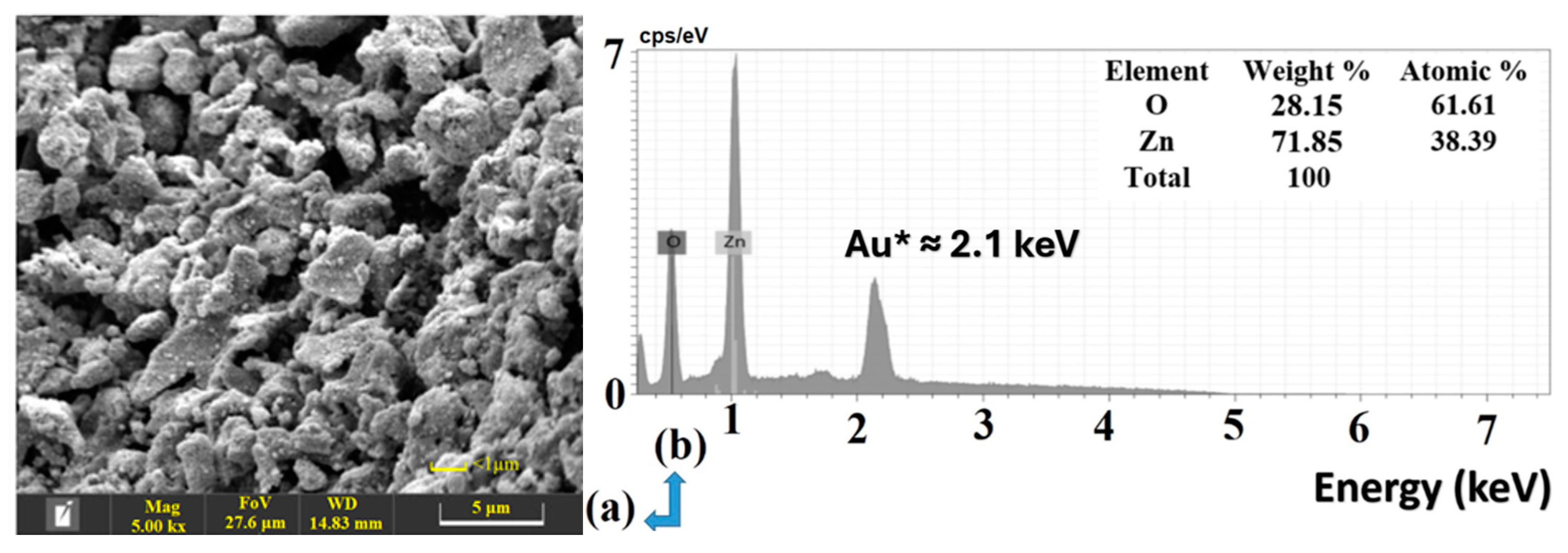
Figure 1. (a) shows the observed agglomerates of biofabricated ZnO NPs, while (b) shows the ZnO NPs EDX pattern profile.
Inorganics 12 00199 g001
Figure 2. (a) TEM micrographs of biogenic nanoparticles obtained from Cucurbita pepo L. seed extract, (b) A particle size distribution histogram determined from the TEM image, (c) TEM image showing lattice fringes, and (d) Spacing measurement with Digital Micrograph indicating a d-spacing of 0.65 nm.
Inorganics 12 00199 g002
Figure 3. Characterization of biosynthesized ZnO nanoparticles using seed extract: (a) FTIR spectrum, (b) XRD pattern, (c) UV-vis spectrum, and (d) Tauc’s plot for bandgap energy (Eg) determination.
Inorganics 12 00199 g003
Figure 4. TGA curve of biogenic ZnO NPs.
Inorganics 12 00199 g004
Figure 5. (a) Photocatalytic degradation of dye using biogenic ZnO NPs, (b) Bar diagram degradation efficiency showing % degradation versus exposer time, (c) Photo trigger mechanism of ZnO NPs in dye degradation in direct sunlight, and (d) Photodegradation plot of Co/Ct vs. Time.
Inorganics 12 00199 g005
Figure 6. (a) Degradation kinetic plot of In(C0/Ct) vs. Time. (b) Reusing as-prepared ZnO NPs in various cycles for degradation and adsorption efficiencies. (c) UV-Vis spectra of ZnO NPs obtained from various photodegradation cycles compared with the initial UV spectrum of as-prepared ZnO NPs. (d) UV-Vis spectra of ZnO NPs obtained from various H2S adsorption cycles compared with the initial UV spectrum of as-prepared ZnO NPs.
Inorganics 12 00199 g006
Figure 7. (a) Potentiodynamic polarization curves of mild steel immersed in 1.0 M hydrochloric acid (HCl) solution in the absence and presence of inhibitor, and (b) Electrochemical impedance spectra (EIS) Nyquist plots for corrosion inhibition of steel in 1.0 M HCl containing various concentrations of ZnO NPs as a green inhibitor.
Inorganics 12 00199 g007
Figure 8. (a) The setup for the experiment to study how ZnO nanoparticles adsorb H2S and (b) the adsorption curve, which shows the change in absorbance over time in minutes.
Inorganics 12 00199 g008
Figure 9. Kinetic analysis of H2S adsorption onto ZnO NPs and bulk ZnO. (a) Plot of normalized concentration (C0/Ct)) vs. time for both materials and (b) Kinetic plot of ln(C0/Ct)) vs. time for ZnO NPs and bulk ZnO.
Inorganics 12 00199 g009
Table 1. Biosynthesis of ZnO NPs using various sections of aqueous extracts and their characterization, as well as applications in photocatalytic degradation, adsorption, and corrosion inhibition activities.
S.No.Plant SectionCharacterizationApplications *Ref
Biological AssayPDA/E/ASCIA/Ad
1Verbena offcinalis leaf extractXRD, FTIR, SEM, TEM, DLS, UV, 19.44 nm, Eg = 3.3 eV, spherical Congo Red/97% [19]
2Laurus nobilis plant extractPhotoluminescence, XRD, FTIR, SEM, TEM, UV, 20–30 nm, Eg = 3.30 eV. BET/BJHAntibacterial (E. coli)Remazol Brilliant Red F3B/99%/60 min [18]
3Hibiscus sabdariffa L. flower extractFTIR, XRD, 15 nm, SEM/EDX, DLS, BET, TEM, ZP, wurtzite, Eg = 3.21 eVAntibacterial (E. coli, S. aureus) DPPH (IC50 value of 38 μg/mL)MO under UV irradiation/99%/60 min [20]
4Pluchea indica leaf extractFTIR, UV, XRD, TEM, SAED, SEM/EDX, DLS, 21.9 nmAntimicrobial (P. aeruginosa; E. faecalis, B. subtilis; S. aureus; C. albicans; C. neoformansMB/95%/150 min [31]
5Salvadora persica leaf extractSEM-EDX, FTIR, XRD, UV-Vis, 32–68 nm, Langmuir isotherm model, hexagonal/rod-shaped MO/68%/100 min [32]
6Jacaranda mimosifolia flowers extractMicrowave-assisted synthesis, XRD, HRTEM, 2–4 nm GC–MS, Eg = 4.03 eVAntibacterial, E. coli, E. faecium Adsorption by molecular modeling[33]
7Becium grandiflorumUV–Vis, FTIR, XRD and SEM–EDSAntimicrobial, S. epidermidis, S. aureus, K. pneumonia, P. aeruginosa, E. coliMB under UV irradiation/69%/200 min [34]
8Pandanus amaryllifolius leaves extractXRD, wurtzite, FESEM, EDX, 4.15 nm, 27.19 nm, 35.69 nm Corrosion inhibition, 1.0 M HCl, 79.43%[35]
9Daucus crinitus extracts (DCE)UV-vis, SEM-EDS Corrosion inhibitors for carbon steel (CS) in HCl, inhibitory efficiencies; 80.20, 91.20%[36]
10Convolvulus arvensis leaf extractFTIR, UV-vis Inhibitor of carbon steel corrosion, 1 M HCl solution, 91%[37]
11Pinecone extract (PCE)UV-Vis, FTIR, XRD, HRTEM, and SAED, 40 to 60 nm, sphere-like morphologyAntimicrobial, E. coli, B. subtilis, H. insolens, M. indicus, biocompatibility, cytotoxicityPhotocatalytic activity, MB, 60%. 30 min [30]
12Fruit extracts of Myristica fragransXRD (41.23 nm), FTIR, SEM (43.3 to 83.1 nm), UV (Eg = 2.57 eV), semispherical shape, TEM (35.5 nm), DLS, Zeta (66 nm and −22.1 mV), TGAProtein kinase inhibition assay. Antidiabetic, antioxidant, antilarvicidal
K. pneumoniae, E. coli, P. aeruginosa, S. aureus
Photocatalytic, MB, 88% [38]
13Citrus reticulata BlancoFTIR, UV–vis, hexagonal wurtzite, Eg = 2.84–3.14 eV, 7 and 26 nm via XRD, ZP = −20 mV, * PLAntimicrobial; E. coli, S. enteritidis, S. aureus, antioxidant; CUPRAC assay
ABTS
Photocatalytic, acid green dye, 94%/90 min [39]
14Pumpkin seed extractFTIR, XRD, FESEM/, TEM, 48–50 nmAnticancer
breast cancer
[14]
15Pumpkin seed extractFTIR, UV, XRD, SEM–EDX, TEM/SAED, 50 to 100 nm, TGAAnticancer
HCT 116, DPPH (IC50 of 142.857 μg/mL)
[29]
16Aqueous extract of Mucuna pruriensFTIR, UV, XRD, SEM/EDX, Spherical, TEM/SAED, 30.50 nm, Eg = 3.75 eVAnticancer
HeLa, HEK 293, antioxidant, DPPH (IC50 = 4.10 µg mL−1)
[40]
17Hylocereus undatus fruit peel extractFTIR, UV, XRD, SEM/EDX, spherical shape, 10–100 nm;Antimicrobial activity; E. coli; K. Pneumoniae; P. aeruginosa; B. subtilis; C. albicans [41]
18Fumaria officinalis and Peganum harmalaFTIR, UV, XRD, 25.10 nm, irregular rods, sphericalAntioxidant
ABTS, (41.67 & 39.79%)
antibacterial (S. aureus; C. michiganensis
[42]
19Andrographis alataUV–Vis, FT-IR, XRD, SEM, EDAX, HR-TEM, DLS, 35–53 nmAntibacterial, antioxidant (DPPH, ABTS), antidiabetic, anti-Alzheimer [43]
20Sea lavenderUV–Vis, FT-IR, XRD, ~ 41 nm, hexagonal/cubic crystalline. SEM, EDAX, TEM, GC–MS, TGA, 41 nmAnti-skin cancer IC50 = 409.7 µg/mL/cytotoxicity/antimicrobial activity E. coli; C. albicans/DPPH IC50 =  95.80 μg/mL [44]
21Ocimum lamifolium leaf extractUV–vis, TGA/DTA, FTIR, XRD, SEM-EDX, TEM, HRTEM, SAED, 6.5–22.8 nm, Eg~3.2 eVAntimicrobial, E. coli, S. aureus, P. aeruginosa, S. pyogenElectrocatalytic activity [45]
22Leaf extracts of Catharanthus roseus (L.) G. DonUV-Vis FTIR, FE-SEM, EDX, and TEM, 44.5 nm, nonspherical, ZP (−18.8 mV)Seed germination [46]
23Orange fruit peel extractXRD, FTIR, TGA, TEM, 10–20 nmAntibacterial, E. coli, S. aureus [47]
24Neem plant extractsSEM/EDX, 23–40 nm, spherical-shaped, DLS (27.81 nm), Eg= 3.24 eV Photocatalytic
MO, 95%, 120 min; Rhodamine-B, 90%, 120 min
CIS
Nyquist, Tafel, uncoated and ZnO NPs coated Zn metal plates (3.5% NaCl)
[48]
25Platanus orientalisFT-IR, PXRD 23.48 nm, UV-Vis, PL, FESEM-EDX, TEM-SAED, spherical, BET Photocatalytic acid red 14/85%/45 min [49]
26Cucurbita pepo L. seed extractSEM, EDX, TEM 32.88 nm, FT-IR, PXRD 13.72, UV-Vis, TGA, Eg of 3.29 eV, d-spacing of 0.65 nm MO dye, 75–80%/60 minCSI for mild steel (MS) in 1.0 HCl, inhibitory efficiencies 83.66%; Nyquist plots; H2S AD capacity with ZnO NPs and bulk ZnO.Present work
* PDA/E stands for Photo Degradation Activity/Efficiency; AS stands for Adsorption Study; CIS stands for Corrosion Inhibition Study, and AD for Adsorption; PL for Photoluminescence.
Table 2. Comparison of ZnO nanoparticles synthesized by different methods and their efficiency in removing methyl orange dye under different light sources.
Synthesis MethodDye Used/Conc./Nature of RadiationZnO NPs
Dosage
Exposure
Time (min.)
% RemovalRef.
Sol–gelMO/40 mg·L−1/UV200 mg/L12065[71]
Sol–gelMO/100 ppm/UV1000 ppm12042[72]
SolochemicalMO/0.02 g·L−1/UV0.1 g·L−112 h78–80[73]
Solution combustionMO/15 mg·L−1/UV0.1 g18052[74]
Hydrothermal synthesisMO/10 mg·L−1/UV0.6 g L−124040[75]
Sol–gelMO/50 mg·L−1/UV30 mg24080[76]
Chemical PrecipitationMO/100 mg·L−1/UV0.05 g12050[77]
Laser-GeneratedMO/20 ppm/Sunlight0.05 g12089[78]
GreenMO/10 mg·L−1/Sunlight50 mg6075–80Present
Table 3. Shows the corrosion parameters for samples with and without inhibitors derived from polarization measurements.
InhibitorConc. (ppm)
Inhibitor
Ecorr (mV)icorr
(μA/cm2)
βa
(mV/dec)
βc
(mV/dec)
θCR
mmpy
ηPDP (%) or
% I.Ep
blank0−440.52211.588110.2202.4-2.47636-
ZnO NPs10−416.2059.41279.6159.90.720.6953471.92
20−415.9256.56178.9143.60.730.6619773.26
40−422.8440.05676.1144.20.810.4688081.06
100−446.0334.56082.7110.10.840.4044883.66
Disclaimer/Publisher’s Note: The statements, opinions and data contained in all publications are solely those of the individual author(s) and contributor(s) and not of MDPI and/or the editor(s). MDPI and/or the editor(s) disclaim responsibility for any injury to people or property resulting from any ideas, methods, instructions or products referred to in the content.